INVESTIGATINGTHEROLEOF N-DEACETYLASE/N-SULFOTRANSFERASE2IN
HEPARINBIOSYNTHESIS
Ryan Matthew Bullis
A dissertation submitted to the faculty of the University of North Carolina at Chapel Hill in partial fulfillment of the requirements for the degree of Doctor of Philosophy in Pharmaceutical
Sciences (Chemical Biology and Medicinal Chemistry)
Chapel Hill 2013
Approved by:
Michael Jarstfer, Ph.D.
Jian Liu, Ph.D.
Robert Linhardt, Ph.D.
Andrew Lee, Ph.D.
ii
iii
ABSTRACT
RYAN BULLIS: Investigating the Role of N-deacetylase/N-sulfotransferase 2 in Heparin Biosynthesis
(Under the direction of Jian Liu, Ph.D.)
Heparan sulfate (HS) is a highly sulfated polysaccharide, produced ubiquitously in the
human body, which plays a key role in signaling and regulatory events. Heparin (HP), a
structural analog of HS produced in mast cells, maintains a higher density of sulfation. HP is
widely regarded for its anticoagulant properties. However, its mode of production, by extraction
from animal tissues, has been recently compromised. This presents an opportunity to develop a
new approach to synthesize HP and improve its safety and efficacy. The overall goal of our
research is to develop an enzymatic-based approach to the design of HP-like compounds. A key
component of this goal is to understand the biosynthetic pathway of HP.
The biosynthesis of HS has long been thought of as a stepwise process, consisting of:
initiation of polysaccharide synthesis on a core protein, elongation, and modification. Recent
research has shown that this process is a more dynamic event involving cooperation between the
glycosyltransferase enzymes, epimerase, and sulfotransferases. Because the process is
nontemplate driven, interactions among enzymes are key components. N-deacetylase/N
-sulfotransferase (NDST) is the initial -sulfotransferase to modify the polysaccharide. Most of the
subsequent reactions rely on the GlcNS residue for substrate binding. NDST-2 is responsible for
iv
the substrate specificity and modification patterns of NDST-2 to gain a thorough understanding
of HP biosynthesis.
We present a study of NDST-2 modification using a structurally-defined oligosaccharide
library. We identified a pentasaccharide as the smallest oligosaccharide modified by NDST-2.
We determined that NDST-2 does not have a directional mode of action, and is unaffected by
pre-existing N-sulfation on the oligosaccharide. We demonstrated by one pot reaction that NDST-2 can cooperate with C5-epimerase, and 2-O-sulfotransferase to form an IdoUA2S-GlcNS domain in vitro. The domain was formed in both short and long oligosaccharides already
carrying GlcNS and IdoUA2S. This was a key discovery in uncovering the biosynthetic pathway
of HP, as this repeating domain comprises up to 90% of the overall structure of HP. The result
v
To my sweet baby boy, Noah M. Bullis, whose smile and laughter always provides escape when
times are tough and constantly reminds me of the bigger picture in life.
To my fiancé, Maria C. Cruz, whose love and encouragement has lit the path through my
vi
ACKNOWLEDGEMENTS
First I would like to extend my sincerest thanks to my advisor, Dr. Jian Liu. His
expertise in the field of glycobiology is unmatched and I cannot express how much I have
learned from him. He has guided me through my toughest of times in the lab, when both of us
wanted to throw in the towel. Now, coming out the other end of the gauntlet, he has truly helped
to transform me in so many ways. I came to UNC as an inexperienced student and with his
extended support I will leave UNC as a seasoned scientist ready to make a difference in the
world. He tells me often how he has never seen such a sudden transformation in any student he
has advised, but I have also never heard of any advisor as patient, forgiving, and understanding
as Dr. Liu.
Next, I would like to acknowledge Dr. Linhardt for his assistance in supporting my
graduate education through a joint supplemental grant between our laboratories. I would also
like to extend my appreciation for the incredible hospitalities upon my visits to RPI. I have
learned a great deal of the business side of drug development through my involvement in the
BRB startup meetings. In addition, I was able to attend the glycosaminoglycan summer
conference at RPI which truly broadened the scope of my knowledge and understanding in the
field of glycobiology. I would also like to send my wholehearted gratitude to my other
vii
spent providing intelligent suggestions and guidance through the development of my research
project.
Finally, I would like to extend my unfeigned thanks to the members of my lab, both
former and current. I would like to thank Dr. Juzheng Sheng for his mentorship with the design
of the oligosaccharide substrate library and his guidance in the development of the NDST-2
substrate specificity study. I also would like to show my great appreciation and admiration for
our lab ‘Momma’ Yongmei Xu. Yongmei always has helpful advice and suggestions and is truly
a wonderful scientist and person. I would like to acknowledge the help that she has given to my
project by providing starter oligosaccharides, which were only minimally modified for use in the
second section of my project. I would also like to thank the former members of my lab for their
support, Dr. Renpeng Liu, Dr. Liz Chappell, Dr. Kai Li, Dr. Xianxuan Zhou, and Justin Roberts,
as well as current labmates, Truong Pham, Kasemsiri Chandarajoti, Tim O’Leary, Po-Hung
Hsieh, Dr. Wen Zhou, and Susan Woody. Finally, I would like to send a special thank you to the
“Liu ladies”, Dr. Heather Bethea, Dr. Courtney Law, and Dr. Sherket Peterson. These three
former labmates were always there for me when I needed them most to discuss science or life in
general. Without the help of everyone involved, this dissertation and the scientific knowledge I
viii
TABLE OF CONTENTS
LIST OF TABLES ... XIII
LIST OF FIGURES ... XIV
LIST OF ABBREVIATIONS ... XVIII
INTRODUCTION...1
HEPARAN SULFATE PROTEOGLYCANS ...1
Chemical Structure of Heparan Sulfate ...2
Structural Analysis of Heparan Sulfate ...6
BIOSYNTHESIS OF HEPARAN SULFATE ...10
Initiation ...10
Polymerization ...12
Modification ...15
BIOSYNTHESIS OF HEPARIN ...34
GAGosome model ...38
BIOLOGICAL FUNCTIONS OF HEPARAN SULFATE ...40
Anticoagulation ...40
Cell Proliferation and Differentiation ...45
ix
Inflammation ...49
Tumor Progression ...53
STATEMENT OF PROBLEM ...56
MATERIALS AND METHODS ...57
CULTURING INSECT CELLS ...57
GENERATING THE RECOMBINANT BACULOVIRUS EXPRESSION VECTOR ...59
Cloning into pFastBac-Mel-HT ...59
Transformation and Analysis of Recombinant Expression Vector ...60
GENERATING THE RECOMBINANT BACMID ...61
Transforming DH10BacTME. coli ...61
Isolating Recombinant Bacmid DNA ...61
Analyzing Recombinant Bacmid DNA by PCR ...62
PRODUCING RECOMBINANT BACULOVIRUS ...62
Transfecting SF9 Insect Cells ...62
Isolating P1 Viral Stock ...63
Amplifying Baculoviral Stock ...63
Expressing NDST-2 ...64
IDENTIFICATION OF NDST-2ACTIVITY ...65
NDST-2 Activity Assay ...65
Polysaccharide Purification by DEAE-Sephacel and Radioisotope Quantification ...66
x
Heparin Lyase Degradation ...66
LARGE SCALE EXPRESSION OF NDST-2 ...67
Culturing SF9 Insect Cells in Serum Free Media ...67
Expression of NDST-2 in Large Scale Shaker Flasks ...68
PURIFICATION OF NDST-2COUPLED TO THE FPLCSYSTEM ...68
Toyopearl Heparin AF HC-650M Chromatography ...68
Nickel Sepharose 6 Fast FlowTMAffinity Chromatography for His 6-Tagged NDST-2 ...69
CHEMOENZYMATIC SYNTHESIS OF STRUCTURALLY DEFINED OLIGOSACCHARIDES ...70
KfiA and pmHS2 Guided Oligosaccharide Backbone Elongation ...70
GlcNTFA Deacetylation and Subsequent Sulfation with NST-1 ...71
C5-Epi and 2OST Modification ...73
NDST-2MODIFICATION OF STRUCTURALLY DEFINED OLIGOSACCHARIDES ...73
ANALYSIS OF NDST-2MODIFIED STRUCTURALLY DEFINED OLIGOSACCHARIDES ...74
Q Sepharose Fast FlowTM-HPLC Purification and Analysis ...74
PAMN-HPLC Analysis...74
DEAE-HPLC Analysis ...74
Mass Spectrometry Analysis of NDST-2 Modified Oligosaccharides ...75
Tandem Mass Spectrometry Analysis of NDST-2 Modified Oligosaccharides ...75
IDENTIFYING THE COOPERATIVE ROLES OF NDST-2,C5-EPI, AND 2OST USING STRUCTURALLY DEFINED IDEAL SUBSTRATES IN A ONE POT REACTION ...75
xi
DETERMINATION OF THE SUBSTRATE SPECIFICITY OF
N-DEACETYLASE/N-SULFOTRANSFERASE ISOFORM 2 ...78
EXPRESSION OF NDST-2,COLUMN SELECTION AND PURITY...80
SYNTHESIS OF STRUCTURALLY DEFINED OLIGOSACCHARIDE LIBRARY ...87
DETERMINATION OF MINIMAL SIZE SUBSTRATE TO BE MODIFIED BY NDST-2 ...94
VISUALIZING THE NDST-2MODE OF ACTION ...97
UNDERSTANDING THE EFFECT OF PRE-EXISTING OLIGOSACCHARIDE SUBSTRATE N-SULFATION ON THE MODIFICATION OF NDST-2 ...123
CONCLUSIONS ...131
ESTABLISHING THE COOPERATIVE ROLES OF NDST-2, 2-O-SULFOTRANSFERASE, AND C5-EPIMERASE IN HEPARIN SYNTHESIS ...135
UNDERSTANDING THE INTERDEPENDENCY OF NDST,2OST, AND C5-EPI ...138
EXAMINING THE INFLUENCE OF GLUCOSAMINE SULFATION STATE ON THE REVERSIBILITY OF C5EPI ...139
MACROMOLECULAR COMPLEX THEORY OF HEPARIN BIOSYNTHESIS ...140
C5-Epi/2OST Modification of Fully N-sulfated Octasaccharide ...142
ANALYZING NDST-2MODIFICATION OF AN IDEAL OLIGOSACCHARIDE SUBSTRATE CONTAINING 2-O-SULFATED IDURONIC ACID ...145
ENZYMATIC SYNTHESIS OF IDOUA2S-GLCNSDOMAIN IN IDEAL OLIGOSACCHARIDE SUBSTRATES USING A ONE POT APPROACH WITH NDST-2,C5-EPI, AND 2OST ...148
LOW PHNO2DEGRADATION OF I2S-OCTA PRODUCT ...152
xii
CONCLUSIONS ...157
APPENDIX I. CURRICULUM VITAE ...164
xiii
LIST OF TABLES
Table 1 - NDST-2 purification analysis ...86
Table 2 - Library of structurally defined oligosaccharide substrates ...88
xiv
LIST OF FIGURES
Figure 1 - Disaccharide repeating units of HS ...3
Figure 2 - Domain structures of HS ...4
Figure 3 - Chemical structure of AT-binding pentasaccharide ...5
Figure 4 - Substrate specificity among various heparin lyase isoforms ...8
Figure 5 - Nitrous acid degradation of heparan sulfate...9
Figure 6 - Chemical structure of HS linkage region and corresponding biosynthetic enzymes ....11
Figure 7 - Elongation of the HS polysaccharide ...14
Figure 8 - Enzymatic modifications of HS ...16
Figure 9 - NDST reaction ...17
Figure 10 - Crystal structure of NST in complex with PAP ...18
Figure 11 - C5-epimerase mechanism ...24
Figure 12 - C5-Epi mode of irreversibility ...25
Figure 13 - 2OST reaction ...26
Figure 14 - 6OST reaction ...28
Figure 15 - 3OST reaction ...30
Figure 16 - GAGosome biosynthetic model ...39
Figure 17 - Blood coagulation cascade ...42
Figure 18 - Ternary complex of antithrombin, thrombin, and a heparin mimetic ...44
xv
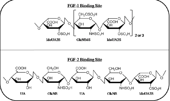
Figure 20 - Chemical structure of heparan sulfate FGF-1, and FGF-2 binding sites ...48
Figure 21 - Chemical structure of gD-binding octasaccharide required for viral entry ...49
Figure 22 - Roles of HS and HP in mediating the inflammatory response ...51
Figure 23 - Counting cells using a hemacytometer ...58
Figure 24 - Baculovirus expression system ...82
Figure 25 - FPLC chromatogram of the purification of NDST-2 by AF Heparin 650M affinity chromatography ...85
Figure 26 - FPLC chromatogram of the purification of NDST-2 by Nickel Sepharose 6 Fast Flow chromatography ...85
Figure 27 - Analysis of purified NDST-2 ...86
Figure 28 - Chemoenzymatic synthesis of structurally defined oligosaccharide library ...89
Figure 29 - Purity and structural analysis of Tetra-1 ...89
Figure 30 - Purity and structural analysis of Penta-1 ...90
Figure 31 - Purity and structural analysis of Hexa-1 ...90
Figure 32 - Purity and structural analysis of Hepta-1 ...91
Figure 33 - Purity and structural analysis of Octa-1 ...91
Figure 34 - Purity and structural analysis of Nona-1 ...92
Figure 35 - Purity and structural analysis of Deca-1 ...92
Figure 36 - Purity and structural analysis of Nona-2 ...93
Figure 37 - Purity and structural analysis of Deca-2 ...93
Figure 38 - Tetra-1 + NDST-2 reaction analysis ...95
Figure 39 - Penta-1 + NDST-2 reaction analysis ...96
xvi
Figure 41 - Hexa-1 + NDST-2 reaction analysis ...100
Figure 42 - N-sulfo hexasaccharide MS/MS analysis ...101
Figure 43 - Hepta-1 + NDST-2 reaction analysis ...103
Figure 44 - N-sulfo heptasaccharide MS/MS analysis ...104
Figure 45 - Octa-1 + NDST-2 HPLC reaction analysis ...107
Figure 46 - Octa-1 + NDST-2 ESI-MS reaction analysis ...108
Figure 47 - N-sulfo octasaccharide 1 MS/MS analysis ...109
Figure 48 - N-sulfo octasaccharide 2 MS/MS analysis ...109
Figure 49 - N-sulfo octasaccharide 3 MS/MS analysis ...110
Figure 50 - Nona-1 + NDST-2 HPLC reaction analysis ...113
Figure 51 - Nona-1 + NDST-2 ESI-MS reaction analysis ...114
Figure 52 - N-sulfo nonasaccharide 1 MS/MS analysis...115
Figure 53 - N-sulfo nonasaccharide 2 MS/MS analysis...115
Figure 54 - N-sulfo nonasaccharide 3 MS/MS analysis...116
Figure 55 - Deca-1 + NDST-2 HPLC reaction analysis ...119
Figure 56 - Deca-1 + NDST-2 ESI-MS reaction analysis ...120
Figure 57 - N-sulfo decasaccharide 1 MS/MS analysis ...121
Figure 58 - N-sulfo decasaccharide 2 MS/MS analysis ...122
Figure 59 - N-sulfo decasaccharide 3 MS/MS analysis ...122
Figure 60 - Nona-2 + NDST-2 reaction analysis ...125
xvii
Figure 62 - N-sulfo nonasaccharide 5 MS/MS analysis...127
Figure 63 - Deca-2 + NDST-2 reaction analysis ...129
Figure 64 - N-sulfo decasaccharide MS/MS analysis ...130
Figure 65 - Demonstration of C5-epimerase irreversibility with N-sulfo octasaccharide 2 ...140
Figure 66 - Proposed model for synthesis of IdoUA2S-GlcNS domain of heparin ...142
Figure 67 - Analysis of fully N-sulfated octasaccharide + C5-Epi/2OST ...144
Figure 68 - Analysis of NDST-2 modification of I2S-octa ...147
Figure 69 - Analysis of I2S-octa + NDST-2/C5-Epi/2OST one pot reaction ...150
Figure 70 - PAMN-HPLC analysis of I2S-octa + C5-Epi/2OST negative control ...151
Figure 71 - Analysis of I2S-tetradeca + NDST-2/C5-Epi/2OST reaction...153
xviii
LIST OF ABBREVIATIONS
∆UA ∆4,5
-unsaturated uronic acid
∆UA2S ∆4,5
-unsaturated 2-O-sulfated uronic acid
2OST 2-O-sulfotransferase
3OST 3-O-sulfotransferase
6OST 6-O-sulfotransferase
AnMan 2,5-anhydromannitol
AT Antithrombin
C5-Epi C5-epimerase
CHO Chinese hamster ovary
CS Chondroitin sulfate
DEAE Diethyl aminoethyl
DMSO Dimethyl sulfoxide
DS Dermatan sulfate
EPS Epimerization site
ESI-MS Electrospray ionization mass spectrometry
EXT Exostosin
EXTL Exostosin like
FBS Fetal bovine serum
xix
GAG Glycosaminoglycan
Gal Galactose
GalT-I/II Galactosyltransferase I/II
GlcNAc N-acetyl glucosamine
GlcNAc6S N-acetylated, 6-O-sulfated glucosamine
GlcNAcT-I/II Glucosaminyltransferase I/II
GlcNH2 Unsubstituted glucosamine
GlcNS N-sulfated glucosamine
GlcNTFA N-trifluoracetylated glucosamine
GlcUA Glucuronic acid
GlcUAT-I/II Glucuronyltransferase I/II
GlmU Glucosamine-1-phosphate acetyltransferase/N-acetylglucosamine-
1-phosphate uridyl transferase
GPI Glycosylphosphatidylinositol
HI-FBS Heat inactivated fetal bovine serum
HP Heparin
HPPG Heparin proteoglycan
HPLC High performance liquid chromatography
HS Heparan sulfate
HSPG Heparan sulfate proteoglycan
HSV-I Herpes simplex virus-1
xx
IdoUA2S 2-O-sulfated iduronic acid
LMW HP Low molecular weight heparin
MOI Multiplicity of infection
MRRS Mode of reaction recognition site
MS Mass spectrometry
MS/MS Tandem mass spectrometry
MWCO Molecular weight cut-off
NDST N-deacetylase/N-sulfotransferase
NST N-sulfotransferase
PAMN-HPLC Polyamine high performance liquid chromatography
PAP 3’-phosphoadenosine 5’-phosphate
PAPS 3’-phosphoadenosine 5’-phosphosulfate
PFU Plaque forming unit
PMSF Phenylmethanesulfonylfluoride
RPIP-HPLC Reverse phase ion-pairing high performance liquid chromatography
SFM Serum free media
TFA Trifluoroacetic acid
UDP Uridine diphosphate
VEGF Vascular endothelial growth factor
Xyl Xylose
Chapter I
Introduction
Heparan Sulfate Proteoglycans
Heparan sulfate (HS) is a highly sulfated, linear polysaccharide that resides in
nearly every tissue at the cell surface and within the extracellular matrix. It is classified
as a glycosaminoglycan (GAG) (1). GAGs are negatively charged heteropolysaccharides
consisting of repeating disaccharide units of uronic acid and an amino sugar usually
ranging between 10-100kDa in molecular weight (1). The major categories of GAGs are
keratan sulfate, chondroitin sulfate (2) and dermatan sulfate (DS), heparin (HP) and HS,
and hyaluronic acid (1). The distinguishing features among each category of GAG are
attributed to differences in monosaccharide identities that comprise the repeating
disaccharide units and glycosidic linkages between each monosaccharide (3). For
example, HP and HS contain the amino sugar glucosamine, while CS and DS contain
galactosamine. They can further be divided into two classifications: sulfated and
non-sulfated. Hyaluronic acid is the only GAG to be non-sulfated (3). All other GAGs are
highly sulfated at various positions and deprotonated at physiological pH giving rise to a
2
GAGs are normally covalently linked as side chains to a core protein and exist as
a proteoglycan (PG). Only HA exists as a free polysaccharide not linked to any core
protein (3). In the case of HS there are several associated core proteins that give rise to
the HSPG, such as serglycin, syndecans, glypicans, perlecans and agrin (3). Perlecans
and agrin are found in the extracellular matrix, while syndecans and glypicans remain
bound to the cell surface. Glypicans are linked to the cell surface through a
glycosylphosphatidylinositol motif, while syndecans normally possess a transmembrane
domain (4-8). Serglycin is the only known intracellular core protein, and is the only PG
to carry HP as its GAG side chains. This HPPG is found in the secretory granule of
hematopoietic cell types, such as connective tissue mast cells (9). The core protein is
mostly responsible for the distribution of HS among various tissue types, while the HS
GAG is responsible for protein signaling leading to an array of physiological events (3).
Chemical Structure of Heparan Sulfate
Heparan sulfate naturally exists as a heterogeneous molecule containing various
modifications throughout. The placement of these modifications throughout the
polysaccharide backbone is maintained by a wide array of biosynthetic enzymes present
in the lumen of the golgi apparatus (10). In addition, there are multiple isoforms for
many of these various enzymes that maintain unique substrate binding capacity and
unique modification patterns. On the whole, these modifications represent unique
binding sites for various biologically relevant proteins, implicating the role of HS across
3
The basic building block of HS structure is a repeating disaccharide backbone of
D-glucuronic acid (GlcUA) covalently linked to D-N-acetyl glucosamine (GlcNAc)
through β-1,4 and α-1,4 alternating glycosidic linkages (Figure 1) (12). The glucosamine
residue may undergo several modifications on its way to becoming fully functional HS.
It can be sulfated at the 3-OH and 6-OH positions, and the N-position can either be acetylated, sulfated, or, to a lesser extent, be presented as a free amine. Only 1-7% of the
glucosamine residues remain as a free amine (13). The GlcUA residue can become L
-iduronic acid (IdoUA) by interconversion at the C5-position. While GlcUA is only found
in the 4C1 chair conformation, IdoUA can reside in either the 1C4 chair or 2S0 skew boat,
thus allowing for much more conformational flexibility (14, 15). In addition, GlcUA and
IdoUA can be sulfated at the 2-OH position. These modifications do not run to
completion and are governed by the presence or absence of pre-existing modifications.
The ability of these molecules to possess such large combinations of modifications gives
rise to heterogeneous macromolecules that are capable of binding and signaling many
various proteins and controlling a multitude of biological events.
Figure 1. Disaccharide repeating units of HS. The uronic acid monosaccharide units can exist as either GlcUA or IdoUA. Sulfation (R=-SO3) at
the 2-O position of IdoUA is common, sulfation at the 2-O position of GlcUA is less common. Sulfation at the 6-O position of glucosamine is common. Sulfation at the 3-O position of glucosamine is rare. Both N-acetyl (R’=-Ac, GlcNAc) and N-sulfo (R’=-SO3, GlcNS)
4
HS may be further characterized by the domain structures that define its overall
topography. HS is known to possess stretches of highly sulfated domains which are
flanked by domain structures carrying very low sulfation density (Figure 2). The highly
sulfated domains carry out much of the protein signaling due to their negative charge
density (16). These domains are often referred to as NS domains due to the presence of
repeating GlcNS residues. Many of the HS biosynthetic enzymes rely on the presence of
GlcNS residues in order to carry out their reactions, thus adding sulfation density to these
domains. The domains carrying low sulfation density are known as NAc domains due to
the presence of repeating GlcNAc residues (17). These two domain structures are
separated by segments of repeating disaccharides carrying both the GlcNAc and GlcNS
residues (18)
5
HP is a structural analogue of HS. It has been widely exploited therapeutically
for its anticoagulant properties for over half a century (19). It maintains the same
repeating disaccharide structure (UA-GlcN) but is different from HS mainly due to its
high density of sulfation. On average, HP maintains 2.6 sulfo groups per disaccharide
unit, while HS disaccharides carry 0.6 sulfo groups (3). HP is the most highly sulfated
GAG that is currently known (20). Between 70-90% of its overall structure consists of
the repeating disaccharide IdoUA2S-GlcNS6S, while only 30-60% of HS N-sulfated glucosamine residues (21). The remainder of HP structure is made up of other
combinations of modification and the unique AT binding pentasaccharide
(GlcNAc6S-GlcUA-GlcNS3S6S-IdoUA2S-GlcNS6S) that confers its anticoagulant activity (Figure 3)
(22). In addition to degree of sulfation, fully elongated HP polysaccharide chains are
generally longer (Mr = 60,000-100,000 Da) than HS (Mr = 22,000-45,000) (23, 24).
6
Structural Analysis of Heparan Sulfate
The heterogeneous nature of HS polysaccharides has always presented a
formidable challenge for researchers in attempting to decipher the specific
monosaccharide sequence that constitutes each molecule. The sheer length, 100-400
monosaccharide units, in addition to the extensive array of potential modifications
present, makes purification of a structurally defined HS nearly impossible (7). This is a
critical barrier to the understanding the structure/function relationship between HS and
their protein signaling partners. As a result, glycobiologists have determined that the best
method for elucidating structures of polysaccharides is to use various methods of
degradation into much shorter oligosaccharide or disaccharide fragments. Recent
research has also developed dependable chemoenzymatic methods for designing
structurally defined oligosaccharides which can also be utilized for probing
structure/function relationships of resultant HS fragments (25). The oligosaccharides can
then be analyzed by a variety of laboratory methods such as High Performance Liquid
Chromatography (HPLC), and capillary electrophoresis. Electrospray ionization mass
spectrometry (ESI-MS), and tandem mass spectrometry (MS/MS) are also useful
methods for determining oligosaccharide chain length and modifications (26). Using
these techniques in a combinatorial approach has allowed researchers to study HS
structure, function, and synthesis like never before.
Depolymerization of HS polysaccharides into disaccharides for compositional
analysis has been reported using both enzymatic and chemical approaches. Following
depolymerization, these disaccharides are then able to be analyzed by anion-exchange
7
analysis (27, 28). These techniques will reveal the relative composition of the HS
polysaccharides. Using these methods, there have been 22 various disaccharides reported
from naturally occurring HS and HP (7).
Enzymatic degradation of HS polysaccharides is achieved using the family of
heparin lyase enzymes originally derived from Flavobacterium heparium. There are 3 known isoforms of heparin lyase, each expressing unique substrate modification sites
based on the identities of the surrounding monosaccharide residues (Figure 4) (7).
Heparin lyase I has shown the capability to cleave the glycosidic bond between
GlcNS-IdoUA2S. Heparin lyase III has less selectivity as it cleaves the bond between
GlcNAc/NS-GlcUA. Finally heparin lyase II has the least selectivity as it will cleave the
glycosidic linkage between GlcNAc/NS-GlcUA/IdoUA (29). The disaccharide products
of these enzymatic reactions can then be readily analyzed by RPIP-HPLC against
appropriate standards to determine the modifications within (30, 31). This is made
possible by the creation of a ∆4,5
-unsaturated uronic acid residue during the reaction,
which maintains a UV absorbance at 232nm (7). However, the creation of this uronic
acid species results in an inability to determine the original identity of the uronic acid in
the original polysaccharide sample. Using this enzymatic method has aided researchers
in elucidating the identities and relative abundance of individual modified disaccharides
at the polysaccharide or oligosaccharide levels. These discoveries have led to a more
8
Another method for depolymerization of polysaccharides is nitrous acid
degradation. Nitrous acid reacts with GlcNS or GlcNH2 residues to form
2,5-anhydromannose residues, which are then reduced by sodium borohydride to form
anhydromannitol (Figure 5) (35). At pH 1.5, nitrous acid will react with GlcNS residues.
While at a higher ph (4.5-5.5) nitrous acid will exhibit selectivity for the unsubstituted
free amine on the GlcNH2 residue. In the case of GlcNAc residues, the polysaccharide
must undergo deacetylation by treatment with hydrazine before going through the high
9
low and elevated pH will lead to nearly complete digestion of the polysaccharide or
oligosaccharide to be analyzed. The resultant disaccharide products can then be analyzed
by RPIP-HPLC by coelution with appropriate standards (36). The key advantage of this
method over lyase degradation lies in its ability to determine the configuration of the
uronic acid residue in from the parent molecule (7). However, the main drawback to this
method is the need for radioisotope or fluorescent labeling of the compound prior to
digestion in order for HPLC detection (35, 37).
10
On the whole, a combinatorial approach of chemical and enzymatic
depolymerization techniques is a useful tool for researchers to identify structural motifs
within HS polysaccharides and oligosaccharides that result in biological function. This
knowledge may then be exploited for use in therapeutics that may one day be used to
treat a wide range of health problems.
Biosynthesis of Heparan Sulfate
Heparan sulfate biosynthesis can be divided into three separate phases before a
fully functional HS proteoglycan is rendered. These phases are 1) Initiation, 2)
Polymerization, and 3) Modification. The biosynthetic process mostly takes place in the
Golgi apparatus, where the biosynthetic enzymes line the lumen. The initiation phase
consists of attachment of a tetrasaccharide linkage region to the core protein. Next, the
polymerization phase consists of repeated attachment of monosaccharides to form the
polysaccharide backbone structure. Finally, the modification phase consists of an array
of various deacetylation, sulfation, and epimerization provided by many various enzymes
to produce biologically active HS (38).
Initiation
The initiation phase of proteoglycan formation is identical across each GAG
classification, from chondroitin sulfate and dermatan sulfate to heparan sulfate and
heparan. Each GAG chain is attached to the core protein through a common linkage
region of –GlcUAβ3Galβ3Galβ3Xylβ3-L-[ser] (Figure 6). The transition step from initiation to elongation will determine the identity of the GAG to be synthesized (39).
11
UDP-xylose to specific serine residues on the protein by xylosyltransferase (XT).
Xylosyltransferase is a golgi associated type II transmembrane domain protein that
performs the initial xylosylation in the early cisternae of the Golgi apparatus before
moving into the lumen for subsequent modification (40, 41).The GAG attachment sites
on the core protein contain the consensus sequence Ser-Gly/Ala-X-Gly (X stands for any
amino acid) (42). There are two vertebrate β4-xylosysltransferases, XylT-I and XylT-II.
These enzymes have 55% homology and both recognize the same consensus sequence for
GAG attachment (40, 43). Elimination of XylT in CHO cells resulted in an inability for
these cells to produce HS or CS, thus showing their necessity in GAG biosynthetic
initiation (44).
The next step in the initiation pathway of GAG biosynthesis relies on the addition
of two D-galactose residues from their UDP-galactose donors by the enzymes galactosyltransferase I and II (GalT-I & GalT-II). GalT-I is a member of the β4
galactosyltransferase family while GalT-II is a member of the β3 galactosyltransferase
family (45, 46). Both of these galactosyltransferase enzymes are single isoforms
12
localized in the medial Golgi (47, 48). CHO cell mutants lacking GalT-I have been
shown to be deficient in HS and CS (49). While small interfering RNA has been used to
impede GalT-I, also resulting in a deficiency of HS and CS (47). These results show the
requirement of both of these enzymes in the biosynthesis of GAGs.
Finally, the linkage region is completed by the addition of GlcUA by
glucuronyltransferase 1 (GlcAT-I). GlcAT-I is a member of a family of β-1,3-glucuronyl
transferases that exhibit action of glycoproteins and glycolipids (50, 51). CHO cells
lacking GlcAT-I have been shown to be deficient in HS and CS, thus exhibiting its
necessity for GAG chain synthesis (52).
Formation of the linkage region can be selectively regulated through a series of
sulfations and phosphorylations. For example, the Gal residues in CS linkage regions
may be selectively sulfated at the 4-O and 6-O positions, while they are not in HS (53). These sulfations may serve to either enhance formation of CS or block the synthesis of
HS (39). Studies have shown that the presence of these sulfations on Galβ1-3Gal
enhances the activity of GlcAT-I to complete the formation of the linkage region (53-55).
Phosphorylation at the 2-O position of the xylose residue has also been shown to inhibit the action of GalT-I. This has been postulated as a rate limiting step in the formation of
GAG chains (56).
Polymerization
Once the linkage region is in place, the repeating disaccharide structure of
alternating GlcUA and GlcNAc is ready to be assembled. The polymerization of HS is
13
family consists of 5 currently characterized glycosyltransferase enzymes: EXTL1,
EXTL2, EXTL3, EXT1, and EXT2 (57-59). As mentioned previously, the initial transfer
of α-GlcNAc to the non-reducing end of the tetrasaccharide linkage region is the
divergent point between synthesis of HS and CS. When CS is to be made, a β-N -acetylgalactosamine residue is added to the tetrasaccharide linkage region by a CS
GalNAcT enzyme (42). The transfer of the initial α-GlcNAc monosaccharide to the
linkage region relies on a transferase with GlcNAcT-I activity, wheras polymerization
relies on GlcNAcT-II activity. Exostosin-like 2 (EXTL2) and EXTL3 have both
demonstrated GlcNAcT-I activity (57, 58). While EXTL2 only possesses GlcNAcT-I
activity, EXTL3 also has demonstrated GlcNAcT-II activity and most likely plays a role
in HS polymerization as well. EXTL1 has only GlcNAcT-II activity and is most likely
involved in polymerization of the growing polysaccharide chain (57). Each of the EXTL
isozymes has the ability to recognize the amino acid sequence on the core protein that is
proximal to the GAG attachment site. For the initial α-GlcNAc transfer, they prefer
attachment sites that are flanked by acidic and hydrophobic amino acids with repeating
serine-glycine units (28,42,60-63). This means that the EXTL enzyme is also able to
interact with the core protein through a separate domain, in addition to binding the
tetrasaccharide linkage region, in order to selectively polymerize a HS polysaccharide
14
After the initial α-GlcNAc is added to the tetrasaccharide linkage region,
polymerization of the repeating disaccharide backbone of GlcUA and GlcNAc is
mediated by a HS-polymerase enzymatic complex of EXT1 and EXT2, with most likely
some influence by EXTL1 and EXTL3 (59). The activities of these glycosyltransferases
were demonstrated in mice models. EXT1 knockout mice exhibited developmental
defects and absence of HS, while EXT2 knockout mice exhibited the same
developmental defects (10, 64). EXT1 and EXT2 were also expressed separately in
yeast, which does not naturally produce HS. Both enzymes displayed GlcNAc
transferase and GlcUA transferase activities, but at a lower level than when they were
expressed together (65). This suggests that the biologically relevant form of these
enzymes is a complex of EXT1 and EXT2 which are responsible for HS chain
polymerization. In fact, it has been reported that these enzymes form a hetero-oligomeric
complex in vivo that can be localized to the golgi apparatus (66).
15
The biosynthesis of heparan sulfate and heparin is most often presented as a linear
process of initiation, polymerization, and modification of the polysaccharide backbone.
However, this model may only be telling a one dimensional story of a much more highly
dynamic and cooperative nature of biosynthetic machinery used to create fully functional
heparan sulfate and heparin. The initial evidence pointing to this cooperative mode of
heparan sulfate biosynthesis was reported by Lidholt, Kjellen, and Lindahl in 1989. They
found that elongating polysaccharide chains in mouse mastocytoma microsomal fractions
were synthesized to be larger when incubated in the presence of the sulfo donor
3’-phosphoadenosine 5’-phosphosulfate (PAPS) than those incubated without the donor.
The sulfated polysaccharide chains were nearly tenfold larger by average molecular
weight than their non-sulfated counterparts (67). In addition, they found that GlcUA
transferase ability of EXT1 greatly favored a previously N-sulfated substrate versus a substrate with N-acetylated or N-unsubstituted glucosamine residues (68). Taken together, the evidence has existed for over two decades that N-sulfation and
polysaccharide elongation have significant influence over one another and most likely
occur simultaneously. Given that N-deacetylase/N-sulfotransferase (NDST) is responsible for the deacetylation and sulfation of the GlcNAc residues, it has been
postulated that it may form an enzyme complex with the HS polymerase enzymes during
elongation (69). There are also numerous other examples of coordination between HS
biosynthetic modification enzymes that will be discussed further below.
Modification
As the polysaccharide backbone is built, a series of modification reactions take
16
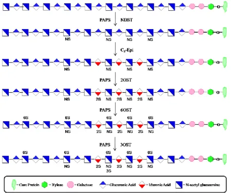
(Figure 8). The first modification to take place is a deacetylation reaction at the N -position of GlcNAc residues. Subsequently, most of these glucosamine residues are
transferred a sulfo group from the sulfo donor PAPS. Both reactions are catalyzed by the
bi-functional enzyme N-deacetylase/N-sulfotransferase. Next, the enzyme C5-epimerase (C5-epi) converts some of the GlcUA residues into IdoUA by interconversion at the C5
position of the hexuronic ring. This reaction is followed by sulfation at the 2-O-position of some of the uronic acid residues by 2-O-sulfotransferase (2OST). Finally, the
glucosamine residues can be sulfated at the 6-O and 3-O positions by 6-
O-sulfotransferase (6OST) and 3-O-sulfotransferase (3OST) respectively (70). Each
enzyme possesses unique substrate modification capabilities across each of their isoforms
leading to final polysaccharide structures with unique properties.
Figure 8. Enzymatic modifications of HS. The first modification to take place is the deacetylation and sulfation at the N-position of GlcNAc, facilitated by NDST. Next, C5-Epi converts some of the
17 Glucosaminyl N-Deacetylase/N-Sulfotransferase
The first enzyme to modify the growing polysaccharide chain is N-deacetylase/N -sulfotransferase (NDST). It is the most essential of the modifications to the
polysaccharide backbone as nearly every other HS biosynthetic enzyme, 6-O -sulfotransferase notwithstanding, relies on N-sulfation for substrate recognition and modification, including the HS polymerases as stated previously (71). This notion arises
from the fact that in both HS and HP, the regions with large sulfation density always have
long stretches of GlcNS residues. As mentioned previously, it is a bifunctional enzyme
with two separate domains responsible for carrying out both deacetylation at the N -position of GlcNAc and addition of a sulfo group to the N-position, thus creating GlcNS (Figure 9) (72). There are 4 known isoforms of NDST (1-4) that possess the same overall
structure and 65-80% nucleotide sequence identity (73). Sequence alignment and
molecular modeling have revealed that these enzymes are type II transmembrane domain
proteins with a 12-13 amino acid cytoplasmic tail, an approximately 20 amino acid
transmembrane domain, a short domain that varies among each isoform, and a globular
domain that houses the catalytic sites. They also contain a PAPS binding domain that is
highly conserved across many various sulfotransferase enzymes (73). Other analyses
have revealed that the sulfotransferase domains of the NDST enzymes are located near
the carboxyl end of the protein with a deacetylase domain located more towards the
amino terminus (74-77).
18
Although none of the x-ray crystal structures of the full length proteins have been
solved, Kakuta et al has published an x-ray crystal structure of the sulfotransferase
domain of NDST-1 (NST) in complex with PAP (Figure 10) (76). They found that NST
consisted of a single α/β fold with a highly conserved five strand β-sheet that is
characteristic of the PAPS binding site across most sulfotransferases. The PAP molecule
is also further stabilized by a three strand β-sheet that runs anti-parallel and contains a
disulfide bond. The PAPS binding site is sandwiched between the structural regions α1,
α6, β1, β7, the 5’-phosphate binding loop between β1 and α1, and a random coil between
β8 and α13. There is also a substrate binding cleft between α6 and a random coil that
runs between β2 and α2. In addition, lysine 614 is able to form a hydrogen bond with the
5’-phosphate of PAPS (76, 78). The random coil that runs along the bottom of the
substrate binding cleft is reported to differ in charge and composition among each
isoform and has been postulated to be a major source of substrate preference among the
NDSTs (73).
19
Each of the four isoforms has unique capabilities to show preference towards
certain substrates and modify them in specific reproducible patterns. Based on overall
tissue expression patterns the NDST isoforms can be divided into two groups:
NDST1/NDST-2 and NDST-3/NDST-4 (73, 79, 80). The mRNAs for NDST-1 and
NDST-2 are expressed in every tissue in the body and are therefore considered to be the
most essential in the HS and HP biosynthesis pathways. It is important to note that these
mRNAs are translationally regulated (81). NDST-1 is the protein that becomes translated
in most cells, while NDST-2 is highly translated in connective tissue type mast cells,
whereas NDST-1 is barely detected (79). HP naturally resides in mast cells, thus
implicating the role of NDST-2 in HP biosynthesis. NDST-3 and NDST-4 have only
been found in adult brain and fetal tissues and seem to have a very narrow functionality
(73, 79, 80). The biological significance of each NDST isoform has been studied in
mouse models and is discussed below.
NDST-1 is widely believed to be the most influential isoform in the biosynthetic
pathway of HS. NDST-1studies in mice have shown that this enzyme is absolutely
essential for synthesizing HS in vivo. Mice lacking NDST-1 were arrested in
development where they underwent neonatal respiratory distress and death. This was
marked by a reduction in lamellar bodies and microvilli versus the control group, in
addition to higher glycogen content. The lungs of the NDST-1-/- mice also had a lower
concentration of phospholipids and disaturated phosphatidylcholine (82). This evidence
shows that NDST-1 is absolutely essential for early lung development and sustaining life.
Additional knockout experiments have revealed the role of NDST-1 in many various
20
clearance of lipoproteins, and development of lobuloalveolar in the mammary gland
(83-86).
In addition to studies on the biological function of NDST-1, Sheng et al
performed multiple experiments using structurally defined oligosaccharide substrates to
prove the much anticipated role of NDST-1 in HS biosynthesis (26). NDST-1 was seen
to have no preference for its initial sulfation of the substrate, leading to multiple products
for a single substrate. This initial observation shows that NDST-1 is a key player in
producing the structural heterogeneity that defines HS. They also were able to
demonstrate the role that this isoform plays in forming the domain structures prevalent in
the overall structure of HS. It was reported that the enzyme moves in a processive
fashion while sulfating the substrate consecutively in a direction from the nonreducing
end towards the reducing end of the polysaccharide, stopping when the enzyme reaches
the residue that is four units from the reducing end. This data provides a platform for the
formation of the NS domain within HS (26). They also found that in the presence of a
pre-existing N-sulfated substrate NDST-1 would allow for a gap of at least 5 sugar residues before initiating the subsequent N-sulfation. This data provides evidence that NDST-1 also has the substrate recognition capability to identify a pre-existing GlcNS
causing the enzyme to form a NAc domain (26). With all of these recently discovered
data, one can infer that NDST-1 is perhaps the most influential of the NDST isoforms
when it comes to HS biosynthesis. In fact, studies have shown that the levels of HS
polymerase expression (EXT 1 and EXT2) can have a significant impact on NDST-1
expression and affect the overall structure of HS that is produced. Overexpression of
-21
sulfation, while overexpression of EXT1 resulted in decreased expression. It has been
postulated that NDST-1 may compete with EXT1 for binding to EXT2 (69). This data
provides yet another potential example of HS biosynthetic enzyme cooperation that could
be an essential element of HS biosynthesis.
If NDST-1 is essential to HS biosynthesis, but causes the formation of
heterogeneous products with an appreciable number of NAc domains, there has to be
another isoform that is responsible for the nearly 90% of NS domains that are prevalent
in the structure of HP. That isoform is believed to be NDST-2. HP resides solely in the
intracellular vesicles of connective tissue-type mast cells (87). In these specific cell
types, NDST-2 is widely expressed, while NDST-1 is barely detectable (79). So it can be
reasonably assumed that this isoform plays a key role in the biosynthesis of HP. The
function of NDST-2 in vivo was studied using gene knockout experiments in mice. Researchers found that NDST-2-/- mice were unable to produce fully sulfated HP. As a
result, the morphology of the mast cells was altered as they contained lower levels of
histamine and mast cell proteases. Otherwise, these mice were viable and fertile. This
study also led to the understanding that perhaps the key physiological role of HP is to
sequester positively charged proteases within mast cells (87, 88). NDST-2 also possesses
a greater ratio of deacetylase/sulfotransferase activity, also implicating its role in creating
the highly sulfated domains of HP (73). There is also some evidence in microsomes
suggesting that NDST-2 prefers to act on substrate locations containing pre-existing
GlcNS residues (89, 90). This could help explain the much longer NS domains in HP
22
Significantly less is known about the isoforms NDST-3 and NDST-4. These
isoforms have very limited tissue distribution patterns and it is not clear as to the extent
of their roles in HS biosynthesis. A murine knockout of NDST-3 revealed its tissue
specific role in HS biosynthesis. NDST-3-/- mice were able to develop normally and were
fertile, exhibiting only minor hematological and behavioral phenotypes. However, there
was a detectable change in the sulfation level of HS located in the brain regions (80).
NDST-3 has been shown to possess a significantly larger deacetylase/sulfotransferase
activity than the other isoforms. NDST-4 displays the opposite activity with much more
sulfotransferase activity (73).
In addition to the interaction between the HS polymerase enzymes, there also
appears to be cooperation between NDSTs and 2-OST/C5-epi which will be discussed
below.
Glucuronosyl C5-Epimerase
The structural heterogeneity of HS is not only determined by the array of sulfation
on the polysaccharide backbone, but also by configuration of the uronic acid residues in
the repeating disaccharide. Initially, a GlcUA monosaccharide will be added to the
elongating chain by the HS polymerase enzymes, EXT1/EXT2. The pyranose ring of
GlcUA can only exist in the chair conformation, thus limiting the substrate flexibility in
binding to proteins (14). This potential limitation is erased by the enzyme C5-epimerase.
This enzyme has the ability to catalyze the reaction from D-glucuronic acid to L-iduronic acid by interconversion at the C5-position (91). The reaction proceeds through a putative
23
to the ring resulting in potential ring interconversion (Figure 11) (92, 93). IdoUA has the
ability to take a chair or skew-boat conformation as it resides in the HS polysaccharide
(14). Only one known isoform of C5-Epi exists in the human genome. It is predicted to
be a type II transmembrane domain protein with a 17 amino acid transmembrane domain
and an 11 amino acid cytoplasmic tail. Overall it maintains an approximate molecular
weight of 70kDa consisting of 618 amino acids (94). A murine gene knockout study
revealed that a lack of this enzyme proved to be neo-natal lethal with mice displaying
developmental defects of the lungs and kidneys (70). Thus, the conversion of the uronic
acid residue into a more flexible conformation is absolutely essential for binding of HS to
a wide array of proteins and for maintaining life. The substrate binding site for C5-Epi
requires a GlcNS residue to be located on the reducing end adjacent to the uronic acid
residue to be modified. However, if the adjacent GlcNS residue or uronic acid residue is
24
The most prevalent problem for researchers attempting to characterize C5-Epi has
always been the “reversibility” of the enzyme, or its capability to convert back to GlcUA
(92). Recent research has uncovered a mode of irreversibility for C5-Epi that relies solely
on a code of N-sulfation. Irreversibility of C5-Epi depends on the sulfation state of the glucosamine residue that sits three residues to the non-reducing end of the GlcUA to be
modified (100). This glucosamine at this site is termed the mode of reaction recognition
site (MRRS), while the GlcUA is termed the epimerization product site (EPS) (Figure
12). When the MRRS is N-sulfated or N-unsubstituted, the EPS will remain reversible and can interconvert between GlcUA and IdoUA. However, when the MRRS is N -acetylated the EPS will become locked into an iduronic acid conformation (100). This
data proves the hypothesis that the state of the glucosamine residues, either N-acetylated,
N-sulfated or N-unsubstituted, are critically important for subsequent epimerization reactions to occur.
Figure 11. C5-Epimerase mechanism. C5-Epi abstracts a proton from carbon 5 of GlcUA forming a
25 Uronosyl 2-O-Sulfotransferase
Uronosyl-2-O-sulfotransferase is closely related to C5-Epi because it has the ability to catalyze the transfer of a sulfo group to the 2-O position of either IdoUA or GlcUA (Figure 13) (101). It is the only sulfotransferase to modify the uronic acid residue
in the HS biosynthetic pathway. The relationship of C5-Epi and 2-OST has been
demonstrated in mutant CHO cells defective for 2-OST. C5-Epi was localized to the
endoplasmic reticulum without 2-OST expression, but upon reintroduction of 2-OST,
these proteins seemed to form a complex and move to the golgi apparatus. In addition,
there was no epimerase activity in the mutant cells until the introduction of 2-OST,
suggesting that these enzymes depend on one another for stability, localization, and
function (48). 2-OST has a preference for IdoUA and is also associated with N-sulfation as it only modifies uronic acid residues with N-sulfation present at the adjacent GlcNS residue linked at the nonreducing end. Like C5-Epi, there is only one isoform of this
sulfotransferase in the human genome. It has a molecular weight of 43 kDa, consisting of
356 amino acids (102). Studies have confirmed that this gene is essential for
development and sustaining life. 2-OST -/- mice displayed flaws in development of the Figure 12. C5-Epi mode of irreversibility. C5-Epi is able to act in an irreversible mode when a
26
eyes and kidneys resulting in death during the neonatal period (103). Researchers have
also demonstrated that 2-OST is critical for cell and axon migration in C. elegans (104). 2-OST has also shown the capability to decrease NDST activity, as CHO cells containing
a 2-OST deficient mutation were shown to have more GlcNS residues than wild-type
(105). Taken together, all of the data so far points to regulation and interaction between
various HS biosynthetic enzymes. The HS polymerase enzymes, NDSTs, C5-Epi, and
2-OST all have influence on one another and most likely these reactions are all occurring
simultaneously as the polysaccharide is being constructed.
27 Glucosaminyl 6-O-Sulfotransferase
Beyond the 2-OST modification is where the true HS protein binding assembly
takes place. O-sulfation at the glucosamine unit has long been understood as a pre-requisite for protein binding and downstream signaling events to occur (106). Seeing as
how there is only a single isoform of 2-OST, it remains unable to produce a distinct
binding code for proteins to read and interact. The key players in creating distinct HS
sequences are 3-O-sulfotransferase and 6-O-sulfotransferase. Between these two enzymes there are ten isoforms, each with its own substrate binding capabilities and
modification profiles (11). These are the steps that truly define the specific patterns that
make up the heterogeneous HS polysaccharides, resulting in such a wide array of
biological events.
6-O-sulfotransferase catalyzes the addition of a sulfo group from the sulfo donor PAPS to the 6-O-position of glucosamine (Figure 14). There are three isoforms of 6OST-(1-3), and each isoform is predicted to maintain type II transmembrane topology
consisting of 401, 506, and 470 amino acids respectively. They display between 50-57%
homology across each isoform (106). Regulation of 6-O-sulfation has been implicated as a major factor in HS binding to and signaling FGF2 and FGF1, resulting in cell
differentiation (107). Sulfation at the 6-O-position has been observed in 2 of the
characteristic HS domain structures, the NS and NS/NAc domains. Considering that the
28
context of a GlcUA or IdoUA at the nonreducing end of the GlcNS residue, IdoUA was
the favored substrate. Moreover 6-O-sulfation occurred more favorably at sites that were linked to a 2-O-sulfate uronic acid residue (109). The critical differences among these isozymes are their target substrate preferences.
Each isoform differs in its preference of uronic acid at the nonreducing end of the
glucosamine residue to be modified. 6-OST-1 seems to prefer IdoUA-GlcNS, but has
also been shown to modify GlcNAc. 6-OST-2 can modify both IdoUA-GlcNS and
GlcUA-GlcNS, but is very dependent on substrate concentration. 6-OST-3 has the ability
to act on both substrates with no dependency on concentration (106, 110). These
substrate specificities seem to imply that 6-OST-1 performs most of the modification in
the NS domain, where IdoUA is most prevalent, while 6-OST-2 and 6-OST-3 most likely
modify the glucosamine residues within the NS/NAc domain of HS. The expression of
each isoform also seems to be tissue specific and may imply that each isoform is
responsible for creating structures that will be used in signaling proteins within each
specific physiological location. Northern blot analysis on murine tissue has shown that
the expression of 6-OST-1 is greatest in the liver, where there is a large abundance of the
disaccharide 2SIdoUA-GlcNS6S. 6-OST-2 is mainly expressed in the brain,
corresponding to the elevated level of GlcUA-GlcNS6S disaccharide (106).
29 Glucosaminyl 3-O-sulfotransferase
Glucosaminyl 3-O-sulfotransferase is arguably the most crucial modification for protein binding and signaling. 3-OST transfers a sulfo group from the donor PAPS to the
3-O-position of glucosamine (Figure 15). Unlike the other HS biosynthetic enzymes, 3-OST-1 is not a transmembrane protein. Instead, it resides inside the lumen of the golgi
apparatus and lacks a cytoplasmic domain (111). 3-OST-1 does, however, maintain
approximately 50% homology to the NDSTs, mostly in the C-terminal sulfotransferase
domain (112). 3-O-sulfation is present in binding sequences of antithrombin and the viral gD-envelope protein of HSV-1. It is a relatively rare modification, accounting for
approximately 0.5% of HS sulfation, and its presence creates finite structures for HS
ligand binding (30, 36). The ability of 3-OST to provide such an array of unique
structures that various proteins can recognize and bind most likely comes from its full
arsenal of seven distinct isoforms in the human genome, 3-OST-1, 3-OST-2, 3-OST-3A,
3-OST-3B, 3-OST-4, 3-OST-5, and 3-OST-6 (111, 113-115). Each isoform has the
ability to transfer a sulfo group to the 3-O-position of glucosamine, but the modification depends on the conformation and sulfation state of the uronic acid linked to the
nonreducing end (116a). Studies in zebrafish have revealed that some of the isoforms are expressed ubiquitously, while others are localized to very specific tissues (117). This
data provides the notion that certain isoforms provide certain tissue specific
modifications to finite HS polysaccharide structures that are needed for specific protein
30
Perhaps the most extensively studied protein binding site within HS structures is
the antithrombin-binding pentasaccharide. This structure contains a critical 3-O-sulfation that is required for binding to antithrombin, creating a conformational change in the
protein that enhances its affinity for thrombin resulting in inhibition of blood coagulation
(118). In addition, 3-O-sulfation has been implicated in the binding of herpes simplex virus type 1 to the cell membrane through interaction with the viral glycoprotein gD,
resulting in the initiation of viral entry (32). Interestingly, a very similar modification has
also been implicated in the binding of cyclophilin B, a cyclosporine A-binding protein,
which signals for migration and integrin-mediated adhesion of peripheral blood T
lymphocytes (119). As one can see, the 3-O-sulfo group is very important for protein signaling and based on the variations provided by the different isoforms, researchers have
only scratched the surface in terms of proteins that bind these sites in HS
polysaccharides.
Human 3-OST-1 consists of 307 amino acid residues and has a molecular weight
of approximately 36kDa. It has a 93% homology to mouse 3-OST-1. 3-OST-1 is believe
to be a key player and provide the final modification in the development of the
AT-binding site (111). This isoform is capable of recognizing and sulfating the disaccharides
GlcUA-GlcNS and GlcUA-GlcNS6S, as well as IdoUA-GlcNS and IdoUA-GlcNS6S,
31
presence of 2-O-sulfation at the nonreducing end of the GlcNS residue to be modified will inhibit catalysis by 3-OST-1. Mice knockout studies have shown that it is not the
only enzyme capable of creating the AT-binding domains and that there may be some
level of cooperation between the various isoforms (121). They also showed lethality of
this knockout in a specific genetic background, in addition to intrauterine growth
retardation.
A crystal structure of 3-OST-1 has been solved at 2.5Å resolution bound to PAP
(116a). This structure displayed striking similarities to the structure of NST-1. It displays the common α/β fold that is consistent with structures of most other
sulfotransferase enzymes. Based on site-directed mutagenesis studies it was determined
that the residues Arg-67, Lys-68, Arg-72, Glu-90, His-92, Asp-95, Lys-123, and Arg-276
are all necessary for the enzyme to maintain activity. When compared to the structure of
NST-1, it was determined that Arg-67, Arg-72, His-92, and Asp-95 are conserved among
each 3-OST isoform. These residues are not conserved in the structure of NST-1, thus
may play a role in substrate recognition of 3OST-1 (116a).
Recently, two additional crystal structures have been solved which shed light on
the substrate binding specificities of 3-OST-1 and 3-OST-3 (116b). The first structure is
a ternary complex between 3-OST-1 and a heptasaccharide substrate. The second
structure is a ternary complex between 3-OST-3 and a tetrasaccharide substrate. Both
substrates maintain a common trisaccharide motif of GlcNS6S-IdoUA2S-GlcNS6S. It is
evident that the substrates bind 3-OST-1 and 3-OST-3 in a different orientation through a
change in the configuration of the IdoUA2S residue in the trisaccharide motif. This
32
while adopting the 1C4 conformation when binding to 3-OST-1. This conformational
difference results in a change in the positioning of the reducing end of the substrate
causing OST-3 interaction with the substrate to be mediated by a metal ion, while
3-OST-1 maintains a more direct interaction. In addition to differences in substrate binding,
the substrate specificity between the two isoforms is clearly mediated by the 2-O -sulfation at the uronic acid residue to the nonreducing end of the 3-O-sulfation site. For 3-OST-1, the GlcUA cannot be 2-O-sulfated due to the placement of this residue ~2.8Å from the carboxylate on the reducing end IdoUA2S, resulting in charge repulsion due to
the close proximity of these residues after binding the enzyme. But after binding to
3-OST-3 the charge repulsion is less because the negatively charged groups are now 3.2Å
away, and there is an additional hydrogen bonding interaction with Lys259. Thus, the
specificities between 3-OST-1 and 3-OST-3 are clearly due to a difference in substrate
binding.
3-OST-2 maintains a substrate preference of GlcUA2S-GlcNS and
IdoUA2S-GlcNS (31, 122). Studies have shown that 3-OST-2 function is activated in the pineal
glands of rats only during the daytime (123, 124). The physiological significance of this
observation remains to be fully understood. 3-OST-3A and 3-OST-3B catalyze the
addition of a 3-O-sulfation to the disaccharides containing the sequences IdoUA2S-GlcNS and IdoUA2S-GlcNH2. The 3-OST-3 isoforms are responsible for the 3-O -sulfation that is seen in the gD-binding sequence that binds herpes simplex virus type 1,
allowing for viral entry into the cell (32). 3-OST-3B has been found to be an important
player in the notch signaling pathway in drosophila. Interfering RNA was used to disrupt
33
Notch protein (125). 3-OST-2 and 3-OST-3A/B are believed to not be involved in the
formation of the AT-binding domain. Cell extracts that were transfected with these
enzymes showed 300-fold lower activity towards antithrombin versus cell extracts
transfected with 3-OST-1 (31). 3-OST-4 and 3-OST-6 have also demonstrated the
capability of creating a binding site to facilitate HSV-1 fusion in CHO cells (115, 126).
3-OST-5 has demonstrated the unique capability of sulfating both substrates that will lead
to AT-binding sites, as well as, those that will lead to HSV-1 entry (114).
Researchers have attempted to decipher the mechanisms by which the isoforms, in
particular how 3-OST-1, 3-OST-3, and 3-OST-5, recognize their various preferred
substrates to carry out their modification. The substrate recognition mechanism is a
result of amino acid residues at two sites: the catalytic site and the gate (127). Residues
at the catalytic site can recognize monosaccharide units adjacent to the acceptor
glucosamine, but these residues are all conserved among the 3-OST isoforms. Thus, it
was determined that the bulk of substrate specificity was most likely coming from the
gate residues of each isoform. These amino acids interact with the monosaccharide unit
that is three sugar units to the nonreducing end of the acceptor monosaccharide (127).
Point-mutations at the gate amino acids were introduced into both 3-OST-1 and 3-OST-3
isoforms. 3-OST-1 mutations consisted of E88G and H271G, essentially decreasing the
size of these gate amino acids. The resultant mutant displayed more 3-OST-5 like
activity by modifying polysaccharides to have reduced activity towards antithrombin and
greater interaction with the gD viral envelope protein (127). In contrast, they installed
point mutations in the gate amino acids of OST-5 in an attempt to make it have more
34
larger ones, consisting of S120E and A306H. These mutants displayed the ability to
produce polysaccharides with more AT-binding ability and less interaction with the gD
viral protein (127). These site-directed mutagenesis studies did not completely shift the
activity one way or the other, thus there seems to be more unknown factors playing a role
in the substrate recognition mechanisms of the 3-OST isoforms. These postulated factors
could be charge/charge interactions or conformational flexibility resulting from the
incorporation of the IdoUA residue (127).
BIOSYNTHESIS OF HEPARIN
Considering that most of the biosynthetic enzymes which define heparan sulfate
structure are also involved in the biosynthesis of the structural analog heparin, a key
question to ask is what sets heparin apart? Given that heparin is only produced in mast
cells and that heparan sulfate is produced nearly everywhere else in the body, there must
be something special about the mast cell.
The structure of HS is governed by the presence of three basic domain regions.
The NAc domains consist of mostly GlcUA-GlcNAc repeating disaccharide units. The
NAc/NS domains consist of random intermittent GlcNS residues mostly linked to
GlcUA. Finally, the NS domains consist of long stretches of GlcNS residues (128).
These stretches, however, are not fully sulfated at all positions and seem to vary
depending on tissue type and location (129, 130). The structure of HP is essentially one
long mostly homogeneous NS domain, as it contains upwards of 90%