THE ROLE OF THE INFLAMMASOME IN THE RESPIRATORY INNATE IMMUNE RESPONSE TO VIRUSES AND POLLUTANTS: INSIGHTS FOR ASTHMA
PATHOGENESIS
Rebecca N. Bauer
A dissertation submitted to the faculty at the University of North Carolina at Chapel Hill in partial fulfillment of the requirements for the degree of Doctor of Philosophy in the Curriculum
of Toxicology within the School of Medicine.
Chapel Hill 2014
iii ABSTRACT
Rebecca N. Bauer: The role of the inflammasome in the respiratory innate immune response to viruses and pollutants: insights for asthma pathogenesis
(Under the direction of Ilona Jaspers)
iv
v
vi
ACKNOWLEDGEMENTS
It would take many pages to truly acknowledge of all the people who helped me get here. I would like to thank my mentor, Dr. Ilona Jaspers, for the privilege of working in her laboratory and teaching me the ins and outs of research. I could not ask for a more supportive mentor, and I truly have her to thank for being where I am today. Thank you to my committee members for their guidance and preparing me for this day. I would like to thank all of the members of the Jaspers lab and co-workers at the CEMALB who along the way helped me with all of my research endeavors, motivated me when times were tough, and made coming to work fun. I of course have to give a special shout out to my “partner-in-crime,” Megan, who sat beside me during most of graduate school, made me laugh, accepted my tears, and in the process has
become a true friend. Thank you to all of the friends I have made at UNC who have helped make graduate school much more enjoyable. To my fellow members of the “pharm club,” little did we know how much more than a study group this would become. Thanks for laughs and the
kvetching. To my roommates of apartment H201, Liz and Becca, thanks for making Chapel Hill feel like home. To Glenn, thank you for keeping me sane, providing a sounding board (until you ask the really tough questions), and most importantly, for love. I look forward to our new
vii PREFACE
Explanation of my contribution to each chapter and acknowledgement of other contributors: Chapter1: Parts of the “Initiation of the innate immune response by pattern recognition receptors
(PRRs)” section were adapted the review article Bauer, RN; Diaz-Sanchez, D; Jaspers I. Effects of air pollutants on innate immunity: the role of toll-like receptors and nucleotide-binding oligomerization domain–like receptors. J Allergy Clin Immunol. 2012 Jan; 129(1):14-24 with permission from Elsevier and are marked accordingly throughout the chapter. David Diaz-Sanchez wrote the introduction, Ilona Jaspers wrote the conclusion, and I wrote the main text that was reviewed and revised by David Diaz-Sanchez and Ilona Jaspers. Only sections from the main text of this article that were authored by me are included in this dissertation. Additionally, Figures 1.3 and 1.4 are adapted from the same review article.
viii
Rebecca Fry performed the statistical analysis for the real-time PCR array and reviewed the manuscript. David Peden provided insight on the conception of the study and
revisions of the manuscript. Ilona Jaspers was the senior author on the study and oversaw the conception, design, data analysis and interpretation, and manuscript preparations. Chapter 3: The findings in this chapter are unpublished. I conceived of the hypothesis, performed
all of experiments, analyzed all of the data, and wrote the chapter. Corey Jania and Stephen Tilleyprovided the mice for this study, and performed the house-dust mite sensitization protocol in their laboratory. I assisted Corey Jania with the collection of samples, preformed all subsequent sample analysis, and summarized the findings for this chapter. Luisa Brighton processed the fixed lungs and preformed the
immunohistochemistry. Ellen Glista-Baker was a second scorer of the lung pathology and performed the goblet cell hyperplasia analysis. Ilona Jaspers was the senior author on the study and oversaw the conception, design, data analysis and interpretation, and
manuscript preparations.
Chapter 4: The findings in this chapter are unpublished. I conceived of the hypothesis, performed all of experiments, analyzed all of the data, and wrote the chapter. The samples for this study were collected from a human exposure study performed in collaboration with Dr. Kelly Duncan and the U.S. EPA. Charles Esther performed the glutathione and purine metabolite mass spectrometry. Hye-Young Kim and Ned Porter performed the oxysterol analysis. Ilona Jaspers was the senior author on the study and oversaw the conception, design, data analysis and interpretation, and manuscript preparations.
ix
performed the caspase-3 immunohistochemistry. Loretta Mueller helped with the design and execution of the 16HBE14o- and pulmonary macrophage co-culture model and provided technical assistance with the flow cytometry and confocal microscopy. Ilona Jaspers was the senior author on the study and oversaw the conception, design, data analysis and interpretation, and manuscript preparations.
x
TABLE OF CONTENTS
LIST OF FIGURES ... xiii
LIST OF TABLES ... xvi
LIST OF ABBREVIATIONS AND SYMBOLS ... xvii
Chapter 1: Introduction ... 21
1.1 Respiratory mucosal host defense against pathogens and pollutants ... 21
1.1.1 Cells of the respiratory innate immune system ... 22
1.1.2 Adaptive immunity in the lung ... 29
1.1.3 Cell-cell communication underlies immunologic homeostasis ... 31
1.2 Initiation of the innate immune response by pattern recognition receptors1 ... 32
1.2.1 Toll-like receptors (TLRs) ... 33
1.2.2 RIG-I like Receptors (RLRs) ... 35
1.2.3 Nod-like Receptors (NLRs) ... 36
1.2.4 Inflammasome signaling ... 37
1.2.5 PRRs in the response to pathogens versus pollutants ... 38
1.3 Asthma overview... 40
1.3.1 Innate immunity in asthma development ... 41
1.3.2 Innate immunity in asthma exacerbation: ... 43
1.4 Summary ... 48
1.5 REFERENCES ... 54
Chapter 2: Influenza enhances caspase-1 in bronchial epithelial cells from asthmatics and is associated with pathogenesis ... 69
xi
2.2 Materials and Methods ... 71
2.3 Results ... 76
2.4 Discussion ... 79
2.5 REFERENCES ... 109
Chapter 3: Caspase-1 deficiency modifies in vivo development of house dust mite-induced allergic airway disease ... 114
3.1 Introduction ... 114
3.2 Materials and Methods ... 118
3.3 Results ... 122
3.4 Discussion ... 126
3.5 REFERENCES ... 143
Chapter 4: Investigation of damage associated molecular patterns and activation of the inflammasome in the airway of healthy volunteers after in vivo ozone exposure ... 150
4.1 Introduction ... 150
4.2 Materials and methods ... 153
4.3 Results ... 158
4.4 Discussion ... 161
4.5 REFERENCES ... 175
Chapter 5: Airway Macrophage and Epithelial Cell Interaction Modifies the Innate Immune Response to Ozone ... 181
5.1 Introduction ... 181
5.2 Materials and Methods ... 184
5.3 Results ... 187
5.4 Discussion ... 192
5.5 REFERENCES ... 206
Chapter 6: Discussion ... 211
xii
6.2 The inflammasome in response to ozone ... 217
6.3 The inflammasome and asthma ... 222
6.4 Epithelial cells as orchestrators of innate immunity and asthma ... 228
6.5 Final Insights ... 232
xiii
LIST OF FIGURES
Figure 1.1 Respiratory epithelium at homeostasis ...50
Figure 1.2 Respiratory mucosal host defense to an antigen ...51
Figure 1.3 Toll-like receptor (TLR) signaling ...52
Figure 1.4 Activation of the NLRP3 inflammasome ...53
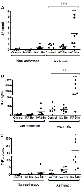
Figure 2.1 HBEC from asthmatics have enhanced production of pro-inflammatory cytokines in response to IAV infection ...97
Figure 2.2 Gene- specific qRT-PCR confirmation of inflammasome- related gene expression ...98
Figure 2.3 Caspase-1 and PYCARD co-localize with IAV-infection ...99
Figure 2.4 Additional immunofluorescence images of caspase-1 and PYCARD localization ...100
Figure 2.5 3-dimensional and side view of caspase-1 and PYCARD localization ...101
Figure 2.6 HBEC from asthmatics and non-asthmatics have similar levels of cytotoxicity. ...102
Figure 2.7 HBEC from asthmatics have enhanced expression of antiviral genes. ...103
Figure 2.8 Viral replication is not significantly increased in HBEC from asthmatics. ...104
Figure 2.9 Correlation between expression of innate immune genes and quantity of influenza HA transcripts ...105
Figure 2.10 Casp1-/- MTEC have diminished antiviral gene expression in response to IAV infection. ...106
Figure 2.11 Casp1-/-MTEC have diminished viral replication ...107
Figure 2.12 Viral replication is necessary for the reduced innate immune response in Casp1-/- MTEC ...108
Figure 3.1 Schematic of house dust mite (HDM) sensitization protocol ...134
xiv
Figure 3.3 Casp1-/- mice have enhanced neutrophil influx with HDM sensitization ...136
Figure 3.4 Histopathology assessment of lung inflammation and damage ...137
Figure 3.5 Lungs from HDM-sensitized Casp1-/- mice have enhanced inflammation ...138
Figure 3.6 Assessment of goblet cell metaplasia ...139
Figure 3.7 HDM-sensitization and caspase-1 deficiency do not affect Il1b gene expression or pro-IL-1Β cleavage ...140
Figure 3.8 WT and Casp1-/- have similar expression of Neutrophil elastase and Cathepsin G in lung tissue ...141
Figure 3.9 HDM-sensitized Casp1-/- mice have reduced airway resistance with methacholine challenge ...142
Figure 4.1 Oxysterols analyzed by LC-MRM analysis ...168
Figure 4.2 Trend for increased gutathione (GSH) oxidation and lactate dehyrodrogenase (LDH) the airway at 1 hour post-O3 exposure. ...169
Figure 4.3 O3 exposure enhances hyaluronic acid and uric acid in the airway ...170
Figure 4.4 O3 exposure enhances levels of oxysterols in the airway ...171
Figure 4.5 O3 enhances IL-8 in the bronchoalveolar lavage fluid ...172
Figure 4.6 O3 exposure did not modify surface expression of CD14, TLR4, and CD44 by Mac at 1 hour post-exposure ...173
Figure 4.7 O3 exposure primes Mac for an enhanced inflammatory response to ATP ...174
Figure 5.1 Schematic of the human bronchial epithelial cell (16HBE) and airway macrophage (Mac) co-culture model ...198
Figure 5.2 Characterization of the human bronchial epithelial cell (16HBE) and airway macrophage (Mac) co-culture model ...199
Figure 5.3 Co-culture of Mac with 16HBE modifies Mac immunophenotype in response to O3 exposure. ...200
Figure 5.4 Exposure to O3 reduces Mac phagocytosis of S. Aureus bioparticles ...201
xv
Figure 5.6 No effect of O3 or co-culture on cleaved caspase-3 ...203 Figure 5.7 Exposure of Mac to conditioned media from 16HBE exposed to Air
xvi
LIST OF TABLES
Table 2.1 Subject Characteristics ...85
Table 2.2 Summary of subject characteristics ...86
Table 2.3 Antigens for allergy skin test ...87
Table 2.4 Cytokine levels and Influenza HA mRNA 6 hours post-IAV infection ...88
Table 2.5 Cytokine levels and Influenza HA mRNA 24 hours post-IAV infection ...89
Table 2.6 Genes with significantly different expression in HBEC from asthmatics vs. non-asthmatics at baseline (control treatment) and 24 hours post-IAV infection ...90
Table 2.7 Full inflammasome array results at baseline (A) and 24 hours after IAV infection (B)...92
Table 3.1 Summary of cytokine levels in bronchoalveolar lavage fluid (BAL) fluid, lung homogenate, and serum ...133
xvii
LIST OF ABBREVIATIONS AND SYMBOLS 16HBE 16HBE14o- (human bronchial epithelial) cell line
a_SecoB alkynyl secosterol B
AHR airway hyperresponsiveness AIM2 absent in melanoma 2 ALR AIM2-like receptor ALI air-liquid interface
AMP adenosine monophosphate AP-1 activating protein-1 ASM airway smooth muscle ATP adenosine triphosphate BAL bronchoalveolar lavage CLR C-lectin type receptor Casp1-/- caspase-1 deficient mice CholEPα cholesterol epoxide α CholEPβ cholesterol epoxide β
COPD chronic obstructive pulmonary disease Ctsg cathepsin G
DAMP damage associated molecular pattern DC dendritic cell
xviii FLICA measure of caspase-1 activity FSC forward scatter
G alveolar tissue dampening
GM-CSF granulocyte-macrophage colony stimulating factor GSH glutathione
GSSG glutathione disulfide H&E hematoxylin and eosin HA hyaluronic acid
HBEC human bronchial epithelial cells HDM house dust mite
Hr hour
IAV influenza virus IFN interferon
Ig immunoglobulin
IL interleukin
Influenza HA hemagglutinin
KC keratinocyte chemoattractant
LC-MRM liquid chromatography-multiple-reaction monitoring LDH lactate dehydrogenase
LGP2 laboratory of genetics and physiology 2 and a homolog of mouse D11lgp2) LLOD lower limit of detection
xix
MARCO macrophage receptor with collagenous structure MAVS mitochondrial antiviral signaling protein
MDA5 melanoma differentiation associated factor 5 mDC myeloid dendritic cell
MFI mean fluorescence intensity MHC major histocompatibility complex MS mass spectrometry
MTEC murine tracheal epithelial cells
MYD88 Myeloid differentiation primary response gene 88 ND non-detectable
NF-kB nuclear factor kappa-light-chain-enhancer of activated B cells NK cell natural killer cell
NLR nucleotide-binding oligomerization domain receptors NLRC NLR family CARD domain-containing protein NLRP NACHT, LRR and PYD domains-containing protein
NLRX1 nucleotide-binding oligomerization domain, leucine rich repeat containing X1 NOD nucleotide-binding oligomerization domain
O3 ozone
OVA ovalbumin
xx PI propidium iodide
PMN polymorphonuclear cell Ppm parts per million
PRR pattern recognition receptor
PYCARD PYD and CARD domain containing qRT-PCR quantitative real time PCR
Raw central airway resistance RIG-I retinoic acid-inducible gene 1
RIPK2 Receptor-interacting serine/threonine-protein kinase 2 RL total resistance of the lung
RLR RIG-I-like receptor RSV respiratory syncytial virus
RV rhinovirus
S. aureus Staphylococcus aureus Seco A secosterol A
Seco B secosterol B SSC side scatter
Th1 type 1 helper T lymphocyte Th17 type 17 helper T lymphocyte Th2 type 2 helper T lymphocyte TLR toll-like receptor
TSLP thymic stromal lymphoprotein
21
Chapter 1:Introduction1
1.1 Respiratory mucosal host defense against pathogens and pollutants
The respiratory mucosal surface is the first line of defense against the inhaled environment, including pathogens and pollutants. A tightly regulated immune system is necessary to effectively maintain immunologic homeostasis while permitting the appropriate response to injury (1). The immune system is composed of both an innate and adaptive component. Innate immunity is the non-specific first response to infection or injury and is composed of both structural cells, such as epithelial cells, and resident and circulating innate immune cells, such as macrophages and neutrophils (2). The crosstalk between these cell types is necessary to determine the appropriate inflammatory response to a stimulus and to inform the adaptive immune system of injury. The adaptive immunity is an antigen specific, cell- and antibody-mediated immune response to eliminate specific invading pathogens (3). The adaptive immune response is mediated by B and T lymphocytes and typically results in immunological memory of the insult (4, 5). Effective interaction between the innate and adaptive immune systems is crucial not only to clear the insult, but also to protect the body from unnecessary inflammation and self-destruction. Diseases such as asthma occur when the immune system inappropriately responds to normally harmless stimuli (6). The chronic inflammation associated with asthma causes airway remodeling and bronchoconstriction that is exacerbated by a normally innocuous stimulus, leading to difficulties breathing (7).
1Sections of this introduction were adapted from Bauer, RN; Diaz-Sanchez, D; Jaspers I. Effects of air pollutants on
22
The work presented in this dissertation is largely centered on the role of the innate immune system in the respiratory mucosal host response to pathogens and pollutants and how these pathways may contribute to asthma pathogenesis. A brief overview of the innate immune cells in the airway and their contribution to allergy and asthma will be covered below. Given the important interplay between innate and adaptive immunity and the crucial role that lymphocytes play in asthma development and pathogenesis, adaptive immune cells will also be briefly
discussed.
1.1.1 Cells of the respiratory innate immune system
Airway epithelial cells (EC). The respiratory epithelium represents the main barrier
23
Beyond acting as a physical barrier, the epithelium is an active participant in host defense. EC produce an epithelial lining fluid composed of mucins and surfactants that moisten the airway and trap inhaled toxins or pathogens (16, 17). The mucins produced by EC together with movement of cilia traps foreign stimuli and moves them up the airway through a
mechanism termed the “mucociliary escalator” (17, 18). The epithelial lining fluid also contains antimicrobial mediators (e.g. β-defensins and lactoferrin) and antioxidants (e.g. glutathione) that protect the airway (17). Additionally, EC play a major role in the secretion of immunoglobulin A (IgA). IgA produced by plasma cells in the lamina propria binds to the polymeric
immunoglobulin receptor on the basolateral side of EC, and is transported to the luminal side before secretion as secretory IgA, which neutralizes bacteria and toxins and prevents microbe adhesion to EC (19). EC produce a number of inflammatory and chemotactic mediators that alert nearby cells of damage or infection and are thus crucial to the initiation of innate immune
responses in the airway (20).
Alterations in EC function are associated with a number of airway diseases, including asthma, chronic obstructive pulmonary disease, and cystic fibrosis (8). Disruption of EC barrier function, increased goblet cell numbers, and remodeling of the airway epithelium are key characteristics of asthma, suggesting that alterations in the airway epithelium may significantly contribute the asthma pathogenesis (22). EC produce a number of DAMPs and
cytokines/chemokines that attract and activate other immune cells, such as dendritic cells and eosinophils, which are major contributors to both the initiation and progression of asthma and will be described below (21).
Pulmonary macrophages (Mac). Pulmonary Mac, which are derived from monocytic
24
Mac are among the first cells to contact inhaled antigens, and thus serve a critical role in the initiation of innate immune responses (24). The primary functions of pulmonary Mac include phagocytosis of cellular/tissue debris and inhaled particulates, destruction of inhaled pathogens and infected cells by release of reactive oxidants through a mechanism termed respiratory burst, antigen presentation to T lymphocytes, and production of inflammatory and chemotactic
mediators to regulate inflammation in the airway (23, 25). Adoptive transfer studies have shown that lung microenvironment plays a key role in determining Mac phenotype and function (26). Pulmonary Mac more resemble dendritic cells and have lower phagocytic and antigen
presentation function compared to other tissue resident or infiltrating Mac (23, 26, 27). This subdued Mac phenotype is important to prevent inflammatory responses to the many harmless stimuli in the inhaled environment (1). Additionally, like EC, Mac phenotype and function may vary based on anatomical location within the lung, and previous studies have shown that
bronchial Mac, interstitial Mac, and alveolar Mac differ in phenotype and function (28). Recently, Mac phenotypic characterization has received increased attention and Mac have been broadly subdivided into “classically activated” and “alternatively activated” subtypes (29). Classically activated Mac are induced by proinflammatory type 1 helper T lymphocyte (Th1) cytokines, such as IFNγ and LPS, and are characterized by production of proinflammatory cytokines and microbicidal reactive oxygen and nitrogen species. Conversely, alternatively activated Mac are characterized by production of anti-inflammatory and tissue repair mediators, such IL-10 and TGFβ, and expression of scavenger receptors, such as CD206 (macrophage mannose receptor) (25). However, these categories are primarily derived from in vitro
25
Moreover, these categories were primarily derived in mice or from peripheral blood/ bone marrow-derived Mac. The application of alternative and classical characterization has not been well verified in the lung or using human Mac (31, 32).
The pro- and anti-inflammatory functions of Mac may play dual roles in the pathogenesis of asthma and allergy (33). In a mouse model of adoptive transfer of Mac from unsensitized mice to allergen-sensitized mice ameliorated airway inflammation and airway hyperresponsiveness, highlighting that the anti-inflammatory functions of Mac may suppress asthma pathology (34). However, the pro-inflammatory functions of Mac can contribute to asthma pathogenesis by enhancing inflammatory cell migration to the airway. Mac from an ovalbumin mouse model of allergic asthma have been shown to produce more IL-17 that recruits neutrophils to airway, a characteristic of exacerbation and severe asthma (35). These diverging findings suggest that Mac function must be exteremely fine-tuned to mainatain a homeostatic state in the lung, and that more studies are needed to explore the anti-inflammatory versus pro-inflammatory functions of Mac in asthma pathogenesis.
Dendritic cells (DC). DC are the major antigen presenting cell in the lung, and thus serve
as important linkers of the innate and adaptive immune responses. Two primary populations of DC exist in the lung, myeloid DCs (mDC) and plasmacystoid DCs (pDC), and the populations differ in recruitment and function. pDC are recruited to the lung upon viral infection,
26
process it into short fragments that are presented on major histocompatibility complex molecules to T cells (21). DC express co-stimulatory molecules such as CD40 that are required for T cell activation, and lack of co-stimulatory receptors can lead to T lymphocyte anergy, a mechanism of inactivation and tolerance (37). In addition, upon recognition of an antigen or danger signal DC produce cytokines and chemokines to attract other immune cells and promote T cell differentiation (38). As presentation of self-antigens or normally harmless allergens to T lymphocytes and differentiation of T lymphocytes to type 2 helper cells is considered an early step in the development of allergic asthma, DC have been shown to play major role in the development of asthma (39, 40).
Neutrophils. Neutrophils, along with eosinophils and basophils, are members of the
granulocyte family. Neutrophils are the first immune cells to be recruited to the site of
short-27
lived. Though some neutrophils are removed by apoptosis, others may undergo necrosis leading to the release of granules that may damage the surrounding tissue (41). Neutrophils, via their high reactivity, have been shown to contribute to a number of airway diseases, including severe asthma, chronic obstructive pulmonary disease, and acute respiratory distress syndrome (42, 46).
Eosinophils. Eosinophils develop and mature in the bone marrow in the presence of IL-5,
IL-3, and GM-CSF and are recruited to the airway upon an inflammatory insult by Th2 cytokines such as IL-4, IL-5, and IL-13 and chemokines such as CCL5 and the eotaxins (47). Eosinophils play a major role in antimicrobial response to parasitic helminths and RNA viruses and are also a hallmark cell of allergy (47, 48). Eosinophils produce a number of proinflammatory cytokines, chemokines, and lipid mediators (such as leukotrienes) and contain granules that harbor
cytotoxic cationic proteins such as major basic protein (MBP), eosinophil cationic protein (ECP), eosinophil peroxidase (EPO), and eosinophil-derived neurotoxin (EDN) that can damage both pathogens and tissue (47). Eosinophils are hallmark of allergic diseases and contribute to asthma by inducing mast cell and basophil degranulation, smooth muscle contraction that contributes to airway hyperresponsiveness (AHR), and mucus production (49, 50).
Basophils. Basophils account for less than 1% of granulocytes in the spleen and blood
(51, 52). Like eosinophils, basophils are thought to be involved in the response to parasitic helminthes and promote allergy (52, 53). As indicated by their name, basophils contain
28
cytokines promote Th2 lymphocyte differentiation and responses (51). Due to their
responsiveness to IgE and migration to sites of ongoing allergic inflammation, basophils are considered late phase effector cells of allergic diseases, including asthma (53). Additionally, basophils may also play a role in the induction of allergic inflammation, as they have been shown to migrate to lymphoid tissue and produce mediators such as TSLP and IL-4 that promote CD4+ T cell differentiation (54, 55).
Mast cells. Mast cells are long-lived cells that reside near epithelial tissue, airway smooth
muscle, blood vessels, nerves, and in mucus-producing glands (56). Like eosinophils and basophils, mast cells harbor granules that contain cytokines, histamine, proteoglycans, leukotrienes, and proteases such as chymase and β-tryptase (56). Mast cell degranulation is activated by FcεRI receptor binding of antigen-specific IgE, as well as activation of innate immune pattern recognition receptors such as the TLRs (57). The release of these mediators have important effects on the lung: mediators such as histamine and leukotrienes act on airway smooth muscle cells to induce bronchoconstriction whereas proteases play a key role in host defense by cleaving proinflammatory mediators, extracellular matrix proteins, and cell surface proteins (53, 57, 58). Since mast cells are resident in the lung, they are important mediators of the early phase of allergic responses (53).
Natural killer (NK) cells. NK cells compose approximately 10% of resident lymphocytes
29
inflammatory mediators, such as IFNγ, Th2 cytokines (IL-5 and IL-13), or regulatory cytokines such as IL-10 (61). Activation of NK cells is based both on interaction with receptors and cytokines. NK cell activating receptors include the natural cytotoxicity receptor NKp46, the Fc receptor CD16, and NKG2D, which recognizes ligands on “stressed” cells, such as UL16 Binding Protein 3 (ULBP3) (62). NK cells are also activated by cytokines, in particular type 1 interferons, IL-12, and IL-18 (60). In addition to antiviral and anti-tumor activity, NK cells are involved in allergy and asthma pathogenesis in the lung. NK cells can be activated by IgE and may release of a number of cytokines that contribute to the allergen sensitization, such as IL-4, IL-5 and IL-13 (63).
1.1.2 Adaptive immunity in the lung
Adaptive immunity is a specific, cell- and antibody-mediated immune response to an antigen that is mediated by T and B lymphocytes and typically results in memory (64). T lymphocytes mediate cellular immunity, whereas B lymphocytes recognize antigens and
differentiate into antibody producing cells called plasma cells (64). T lymphocytes can be further broken down into CD4+ helper, CD4+ regulatory, and CD8+ cytotoxic lymphocytes, all of which express the αβ antigen receptor (65). Another type of T lymphocyte, called γδ T
lymphocytes based on the antigen receptor, are also found in the airway and are considered to be more part of the innate immune response, but these cells will not be discussed here (66).
30
conversely quiescent and may exist for years after encountering an antigen. Upon a second stimulus, memory cells quickly proliferate and mount strong immune responses (67).
In the absence of airway inflammation, the number of T and B lymphocytes in the airway is relatively low (1). Resident T lymphocytes may lie under the epithelium in the lamina propria or within the epithelial cell layer (1, 5, 68). Also within the lamina propria are plasma cells that produce mainly mucosal IgA antibodies (1, 69, 70). The adaptive immune response is closely tied to innate immune responses in the lung, thus with activation of innate immunity, an increased number of T and B lymphocytes migrate to the airway (3, 21). Innate antigen
presentation cells, particularly DC, are important linkers of the two systems. DC present
processed antigen to naïve T lymphocytes via a major histocompatibility (MHC) complexes (6, 38). Based on the type of MHC complex, the naïve T lymphocyte will differentiate to a CD4+ (MHC class II) or CD8+ (MHC Class I) T lymphocyte (64, 65). MHC Class I stimuli represent active intracellular infections, which require destruction of the cell by a cytotoxic T lymphocyte, whereas MHC class II mediators represent extracellular mediators that may cleared by immune cells. Based on the microenvironment, CD4+ helper T lymphocytes differentiate into subtypes such as type 1, 2, and 17 helper cells (6, 65). Type 1 helper (Th1) cells are induced by cytokines such as IFNγ and IL-12, Th2 by IL-4, and Th17 by TGFβ with IL-6 or IL-1 (6, 71, 72). Each helper cell subtype produces different sets of cytokines: Th1 produce IFNγ and IL-12, Th2 produce IL-4, IL-5, and IL-13, and Th17 produce IL-17, and these mediators then attract appropriate immune cells to clear the stimuli (65, 71). B cells differentiate inlymphoid tissue upon recognition of an antigen and, in some cases, a second stimulus from a helper T
31
such as IgG, IgE, or IgA isotypes (4). For example, IL-4, which is secreted by Th2 helper cells, induces a switch to the IgE isotype, which is an important mediator of allergy and asthma (6).
Increased numbers Th2 and Th17 cells, as well as Th2- or Th17-skewed cytokines produced by innate cells have been implicated in the development and pathogenesis of allergy and asthma (6). Th2 cytokines are associated with B lymphocyte isotype switching to IgE and mast cell, eosinophil, and basophil recruitment and degranulation (72). Th17 cytokines are associated with neutrophilic asthma (71).
1.1.3 Cell-cell communication underlies immunologic homeostasis
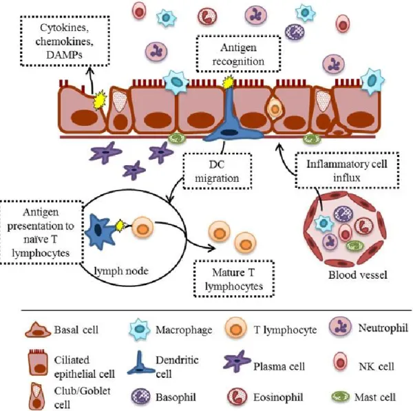
Immunologic homeostasis requires a complex interaction between cells of the innate and adaptive immune systems. Resident immune cells in the lung are constantly interacting with inhaled stimuli and must distinguish between innocuous and pathogenic agents. At baseline conditions, Mac and EC are two of the most populous cells in the airway, suggesting that they coordinate the immune response in tandem (1) (Figure 1.1). The lung lining fluid secreted by EC provides a number of regulatory molecules that are vital to host defense, and the mucociliary escalator provides a mechanism to remove pathogens and particles from the airway (17). Mac phagocytose antigens to prevent the development of specific immune responses and exist in a subdued phenotype with poor antigen presentation capabilities to prevent acquired immune responses unless truly necessary (23, 26, 27). The interaction between Mac and EC at baseline, and how signals from EC may modify Mac phenotype will be explored later in this dissertation (Chapter 5). Additionally, underlying the epithelium is a population of DC that constantly
32
Upon activation of innate immune receptors, resident immune cells produce a number of mediators to recruit inflammatory cells in the airway and activate other resident cells in the lung (Figure 1.2). Resident DC travel to the draining lymph node to present antigens to T and B cells and initiate adaptive immune responses (38). The onslaught of cytokines and chemokines produced by resident cells in the lung recruit circulating immune cells from the blood to the airway, including neutrophils, eosinophils, and basophils (6). The molecular and cellular events that follow limit the infection or injury, but in the process can damage the surrounding tissue. Resolution is another highly coordinated process that involves clearance of damaged tissue and reactive inflammatory cells by apoptosis and phagocytosis, the release of pre-resolving mediators (e.g. lipoxins, resolvins, protectins, and maresins), and reconstitution of the epithelial barrier (41, 73). Failure to resolve inflammation in the airway can lead chronic inflammatory diseases such as asthma, which will be discussed in more detail below (section 1.3) (7, 42, 73, 74). In the next section, the molecular mechanisms that initiate the innate immune response will be explored.
1.2 Initiation of the innate immune response by pattern recognition receptors1
33
by inhalation of oxidant gases such as ozone (O3) (77). Stimulation of PRRs by PAMPs and DAMPs activates downstream signaling pathways that culminate in the production of cytokines and chemokines to attract leukocytes and antigen presenting cells (78). In the case of antigen presentation cells such as DCs, activation of PRRs may provide a necessary maturation signal that leads to activation of naïve T lymphocytes in the draining lymph nodes (38).
There are several classes of PRRs, including the Toll-like receptors (TLRs), C-type lectin receptors (CLRs), Retinoic acid-inducible gene (RIGI)-I-like receptors (RLRs), AIM2-like receptors (ALRs) and NOD-like receptors (NLRs) (79, 80). TLRs and CLRs are transmembrane associated receptors, whereas RLRs, ALRs, NLRs are cytoplasmic proteins. A number of studies have demonstrated the role of TLR signaling in both pathogen- and pollutant-induced
inflammation. More recently, NLRs and the subset that assemble and oligomerize to form the complex known as the inflammasome have been implicated in both pathogenic and sterile inflammatory responses (81). RLRs play an important role in the innate immune response to RNA viruses, such as influenza, and can also form inflammasome complexes (82). CLRs, such as the Dectins and Mannose Receptor (CD206), though important for the recognition of
carbohydrates on microorganisms, will not be discussed here (83). [Adapted from (84)]
1.2.1 Toll-like receptors (TLRs)
34
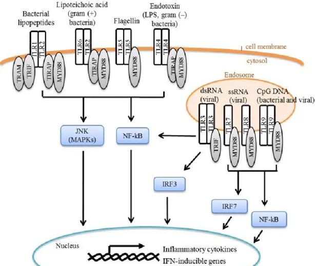
well as EC (86). Each TLR is associated with specific recognition patterns: extracellular TLRs 1, 2, 4, and 5 sense bacterial components such as lipoproteins and the bacterial wall component LPS, whereas endosomal TLRs 3, 7, 8, and 9 recognize nucleic acids (Figure 1.3). (80) Notably, TLRs have been implicated in the response to a variety of pathogens, pollutants, and DAMPs, indicating that these receptors are not limited to one specific ligand. For example, TLR4 plays a key role in the innate immune response to the air pollutant O3, an oxidant gas that cannot be directly bound by a receptor, suggesting that DAMPs from O3-inudced damage of the airway mediate TLR4 activation (87).
Interaction of the TLR with its specific ligand results in the activation of a signaling cascade leading to the production of innate effector molecules and the initiation of the adaptive immune response (88, 89) (Figure1.3). TLRs signal to the cytoplasm through adaptor proteins, such as myeloid differentiation primary response gene (MyD88), toll-interleukin 1 receptor (TIR) domain containing adaptor protein (TIRAP), TIR-domain-containing adapter-inducing interferon-β (TRIF), and translocation associated membrane protein (TRAM), all of which harbor a Toll-Interleukin-1 Receptor (TIR) domain for the recruitment of the adaptor protein to the TLR cytoplasmic domain via a TIR-TIR interaction (85). MyD88 is an adaptor protein shared by all TLRs except TLR3, which instead utilizes TRIF to signal to the cytoplasm. The TLR signaling cascades result in the activation of transcription factors in the cytoplasm, such as nuclear factor kappa-light-chain-enhancer of activated B cells (NF-κB), interferon regulatory Factors (IRFs), and activation protein 1 (AP-1), which then induce or inhibit the transcription of genes involved in inflammatory and immune responses (78, 90).
35
(ARDS), asthma, and chronic obstructive pulmonary disease (COPD) (78). For example, ARDS may be triggered by bacterial or viral infections and non-infectious insults such as environmental exposures or trauma, which may stimulate TLR signaling and initiate an inflammatory response and tissue damage (78, 91, 92). Additionally, TLR4, the endotoxin receptor, has been shown to play a role in the induction of Th2 biased immune response in the lung and the development of asthma (81). Consequently, mutations in several TLRs, including TLR4, have been associated with asthma (78, 93, 94). Clearly, the TLR signaling pathways play an important role in
initiating the immune response to PAMPs and DAMPs which, when left unchecked, can lead to tissue injury and airway disease. [Adapted from (84)]
1.2.2 RIG-I like Receptors (RLRs)
36
domain and activate processing of IL-1β and IL-18 (96). Inflammasome signaling will be discussed in detail below (section 1.2.4).
1.2.3 Nod-like Receptors (NLRs)
Nucleotide-binding oligomerization domain receptors (NLRs) are a family of
cytoplasmic PRRs that that are characterized by the presence of three domains: a C-terminal leucine-rich repeat domain that binds ligands, a central NATCH nucleotide binding domain that is important for oligomerization, and N-terminal effector domain that may be a pyrin, CARD, or BIR domain (97). The N-terminal effector domain determines the subfamily of the NLR: NLRB has a BIR domain and contains one member NAIP; the NLRC family has a CARD domain and contains NOD1, NOD2, NLRC3, NLRC4, NLRC5, and NLRX1; and the NLRP family has a pyrin domain and includes NLR1 through NLRP14 (98). Notably, the members of these
subfamilies may differ in mice (99). The ligands recognized by these receptors are highly varied. For example, NLRC4 recognizes microbial bacterial flagellin and type III secretion machinery, NLRP1 recognizes muramyl dipeptide (a bacterial peptidoglycan), and NLRP3 recognizes a wide range of stimuli from influenza to uric acid crystals (100). Engagement of these receptors results in a cascade of intracellular signaling leading in the production of innate immune
37
1.2.4 Inflammasome signaling
Upon recognition of a PAMP or DAMP, several NLRs, RIG-I, and the DNA-binding IFI200 family member absent in melanoma 2 (AIM2) may form a multi-protein complex termed the inflammasome. The inflammasome is composed of a PRR, pro-caspase-1, and the adaptor protein Pyd and card domain containing (PYCARD) (103). This complex has been best studied in myeloid cells, particularly Mac and monocytes, though recently there has been increased interest in inflammasome signaling in non-myeloid cells (104).
Upon formation of the inflammasome complex, caspase-1 is autoactivated from the pro- form to the cleaved active form, which can then catalyze the proteolytic processing and release of IL-1β and IL-18. Caspase-1 mediated processing and the release of mature cytokines requires two distinct stimuli (100, 105). First, an inflammatory stimulus activates the transcription of pro-cytokines, such as pro-IL1β and pro-IL18. This signal may be triggered by TLR activation and downstream signaling through the NF-κB or MAPK pathways. The second signal induces formation of the inflammasome complex, cleavage of caspase-1, and then pro-cytokine
maturation and release (Figure 1.4). Active caspase-1 may also participate in one of several other effector mechanisms, including induction of a caspase-1 dependent form of programmed cell death termed pyroptosis, activation of NF-kB, and cleavage of proteins involved in glycolysis, apoptosis, and cytoskeleton functions (106, 107).
38
generally thought to sense general changes in cellular homeostasis rather than directly binding to a ligand (Figure1.4) (100, 110). Activation of the NLRP3-mediated IL-1β/IL-18 processing actually requires three steps: pro-IL-1β and pro-IL-18 transcription, up-regulation of NLRP3 transcription, and NLRP3-PYCARD-caspase-1 association to form the inflammasome complex (105). Mitochondria are thought to play a key role in NLRP3 inflammasome assembly and activation. Mitochondria may serve as an activation platform for the NLRP3 inflammasome and a source of reactive oxygen species and DNA, both of which have been shown to either directly activate NLRP3 or up-regulate its expression (111-114). Alterations in ion concentrations in the cell have also been associated with NLRP3 inflammasome activation, as low levels of
intracellular potassium (K+) and high levels of intracellular calcium (Ca+) can activate inflammasome formation (110, 115, 116). Finally, the NLRP3 inflammasome may also be activated by lysosamal damage, as caused by phagocytosis of large crystals or fibers (e.g. asbesotos or silica). Rupture and release of lysosomal contents, such as cathepsin B, may trigger NLRP3 inflammasome activation (117). Thus, a number of questions remain regarding
inflammasome activation, and inflammasome activation likely involves a combination of mechanisms that is tailored for specific PRRs and stimuli. [Adapted from (84)]
1.2.5 PRRs in the response to pathogens versus pollutants
Pattern recognition is typically associated with the response to microbial stimuli that may be directly recognized by PRRs via a PAMP, yet PRRs are also implicated in the inflammatory response to a number of pollutants. Neutrophilic influx is a shared characteristic of exposure to both pollutants and the bacterial cell component LPS, leading to the hypothesis that the
39
important for the inflammatory response to air pollutants (75, 87). Exactly how pollutants activate PRRs such as TLR4 is not as clear. For particulates such as particulate matter or biomass, endotoxin is a common contaminate that can activate TLR4 signaling (118, 119). Yet, other pollutants, such as O3, induce sterile inflammation and cannot directly bind a PRR, suggesting that pollutant-induced injury may generate DAMPs that activate PRRs (87). For example, O3 is associated with fragmentation of the extracellular matrix component hyaluronic acid (HA), and these fragments can be recognized by PRRs including TLR2 and TLR4 to induce innate immune responses (91, 120, 121).
Inflammasome signaling has received increased attention as an innate immune pathway induced by a wide array of stimuli, including pathogens, DAMPs, and pollutants. Whereas infection with a virus or bacteria may directly activate AIM2, RIG-I, and other receptors in the cytoplasm and induce inflammasome signaling, the mechanism by which pollutants activate inflammasome signaling is less clear. Some pollutants, such as asbestos fibers and silica crystals, have been shown to induce phagosomal destabilization, lysosomal damage, and the generation of reactive oxygen species in Mac that induce NLRP3 inflammasome formation (117). Other pollutants, such as O3, more likely activate inflammasome signaling through a secondary mediator or DAMP. Indeed, in murine studies the NLRP3 inflammasome be activated by HA fragments, which may contribute to O3-induced airway hyperresponsiveness in mice (122, 123). Thus, the inflammasome may be a common pathway by which pollutants and pathogens
stimulate similar inflammatory responses.
40
mechanism contributing to the pathogenesis of immune diseases, such as allergy and asthma (6). Indeed, viral infection and inhalation of air pollutants are known causes of asthma exacerbation, suggesting innate immunity contributes to asthma pathogenesis. The role of innate immunity in asthma will be discussed below.
1.3 Asthma overview
Over 20 million Americans suffer from asthma, a chronic lung disease characterized by airway inflammation and variable and reversible obstruction of the airway (7).The rates of asthma incidence have continued to increase over the past 50 years, which is thought to be due to a combination of changes in lifestyle and urbanization (124). Asthma is an extremely
heterogeneous disease with a diverse etiology and pathophysiology (7). Asthma is thought to be caused by a combination of genetic and environmental factors. A number of susceptibility genes for asthma have been identified that are involved in pattern recognition, T lymphocyte
differentiation and function, epithelial cell function, and lung function (125). Yet, not all
individuals with familial history or susceptibility genes have asthma, suggesting that interaction with additional environmental factors is necessary (125, 126). Environmental factors that are associated with asthma susceptibility include prenatal maternal smoking, early life viral infection, lack of exposure to infectious agents (known as the “hygiene hypothesis”), and inhalation of air pollutants (124, 127).
The most common form of asthma is allergic asthma, which is induced by allergy to an antigen such as house dust mite (HDM), pollen, or mold and is dependent on the presence of IgE antibodies specific for the allergen. Non-allergic asthma is exacerbated by stimuli such as
exercise, cold air, medications (e.g. aspirin), and stress (128). Both forms of asthma are
41
IL-13. These cytokines induce B cell isotype switching to IgE synthesis, the recruitment of eosinophils, basophils, and mast cells to airway, goblet cell metaplasia, and bronchial reactivity (21). Notably, other helper T lymphocytes can also contribute the pathogenesis of asthma, such as Th17 lymphocytes, which produce IL-17 and attract neutrophils to the airway (71). Chronic inflammation of the airways leads to airway remodeling, obstruction, and airway
hyperresponsiveness (AHR) that mediate the asthma symptoms of wheezing and difficulty breathing (7).
Though adaptive immunity, mediated by B and T lymphocytes, is clearly central to asthma pathophysiology, the innate immune system is essential for the initiation and propagation of these adaptive responses and of particular significance during asthma exacerbation (3, 6, 129). The role of innate immunity in asthma development and asthma exacerbation will be briefly introduced below. Primarily allergic asthma development will be discussed here as it is the most common type of asthma.
1.3.1 Innate immunity in asthma development
Both structural airway epithelial cells and innate immune cells, such a DC, are important contributors to the induction and maintenance of Th2-mediated immune responses (21).
Increases in airway permeability caused by damage to tight junctions as caused by inhalation of viruses, pollutants, or allergens that contain proteases (e.g. certain HDM species) can lead to increased access of antigen to the underlying tissue or intraepithelial DCs (21, 74). DC that have recognized an antigen then migrate to the draining lymph node, where they present the processed antigen to naïve T lymphocytes (21).
42
presentation of a harmless antigen leads to the induction of tolerance, or unresponsiveness of the adaptive immune system to an antigen (6, 21). In order to be fully active, DCs must be
stimulated by PAMP or DAMP activation of innate pattern recognition receptors (TLRs and CLRs) or through proteolytic activity of the allergen (e.g. proteases found in some HDM) that can induce protease activated receptors (PARs) (21, 130). Activation of PRRs induces the production of pro-Th2 cytokines and the expression of co-stimulatory molecules by DCs. Additionally, activation of PRRs on EC induces the release of cytokines, including granulocyte-macrophage colony stimulating factor (GM-CSF), IL-33, and thymic stromal lymphoprotein (TLSP), that prime DC and promote Th2 cell differentiation (131). For example, TSLP produced by EC induces expression of the co-stimulatory molecule OX40L by DCs (132). Thus, there is a clear role for innate immune receptors in the development of asthma, and previous studies have indicated that mice deficient in PRRs such as TLR4 are protected against the development of allergic inflammation (133).
With appropriate activation of DC and the presence of Th2-polarized cytokines in the draining lymph node, particularly IL-4, naïve T cells will differentiate to Th2 lymphocytes (6). Th2 lymphocytes produce cytokines such as IL-4, IL-5, IL-13 that drive allergic inflammation, including B cell isotype switching to IgE synthesis and recruitment or activation of mast cells, eosinophils, and basophils to the airway (6). Degranulation of mast cells, eosinophils, and basophils induces airway smooth muscle contraction and goblet cell metaplasia that are
43
Despite the important link between PRRs and asthma development, the contribution of the inflammasome signaling pathway to asthma remains unclear. Due to the involvement of NLRP3 inflammasome in the response to many PAMPs and DAMPs associated with allergic diseases, such as alum and several bacterial and viral species, the NLRP3 inflammasome has been best investigated for a a role in asthma development (134-138). Results from these studies are conflicting and have not identified a clear role for NLRP3 or caspase-1 in the development of allergic inflammation (131, 135,136). Likewise, studies investigating the role of IL-1 receptor (IL-1R) and IL-1 axis signaling in murine models of allergic inflammation are also unclear (131, 133, 134, 137). These studies suggest contribution of inflammasome signaling to asthma
development is complex, and may differ depending on the iniator of asthma pathogenesis.
1.3.2 Innate immunity in asthma exacerbation:
Exacerbations of asthma symptoms can lead to emergency department visits, hospitalization, and even fatality in very severe cases (129, 139). There are many causes of asthma exacerbation, including viral infections, allergens, occupational exposures, exercise, stress, and air pollutants (129). In response to these stimuli, structural cells of the airway, such as EC and airway smooth muscle, as well as inflammatory cells, including macrophages, mast cells, eosinophils, and neutrophils initiate an innate immune response, including production of
proinflammatory and cytotoxic mediators that lead to acute asthma symptoms (140). Here we will explore two common causes of exacerbation,respiratory viral infections and air pollution, and the innate immune responses that mediate these effects.
Virus-induced asthma exacerbation. The leading cause of an asthma exacerbation is
44
Rhinovirus (RV) is the most frequent cause of virus-induced asthma exacerbation, though other viruses including influenza A virus (IAV) and respiratory syncytial virus (RSV) are also
common (142). IAV infection frequently reaches seasonal epidemic and, at times, pandemic proportions and thus is a serious concern for patients with asthma (144).
Airway ECs are the primary target for respiratory viruses such as RV and IAV. In vitro and in vivo studies have found that the barrier function of EC in asthmatics is impaired due to disruptions in tight junctions (22). Additionally, EC repair and proliferation of basal cells is reduced in the airways of asthmatics (74, 145). Finally, genome-wide expression comparisons of ECs from asthmatics to non-asthmatics found that a number of genes involved in repair and immune responses are differentially expressed by EC from asthmatics (146). Thus, it is not surprising that EC from asthmatics respond differently to viral infection. Upon infection with a virus, pattern recognition receptors such as TLR3 and RIG-I recognize the virus and induce downstream cytokine, chemokine, and antiviral mediators (20). Previous studies have found that EC from asthmatics have deficient interferon (IFN) production after infection and increased production of the inflammatory cytokines IL-1, IL-6, IL-8, and GM-CSF with RV or RSV infection, correlating with the increased pro-inflammatory response to viral infections observed in vivo (146-150). Virus-induced damage of EC may increase airway permeability to allergens. Indeed, previous studies have shown a synergistic effect of virus infection and allergen exposure on hospital admissions for asthma (151).
45
or during an exacerbation (49). Neutrophils are very reactive and secrete a variety of cytokines and proteases such as neutrophil derived elastase and matrix metalloproteinase 9 that damage the airway and contribute to AHR and increased mucus production (154, 155, 156). Virus-induced asthma exacerbations may also be related to an imbalance of Th1/Th2 responses to the virus. In patients with RV infection, reduced CD4+ T cell production of IFNγ and increased IL-4, IL-5, and IL-13 were associated with more severe RV-induced asthma symptoms (152).
In summary, alterations in EC response, increased airway inflammation, and altered Th1/Th2 response to virus infection are key mediators of virus-induced asthma exacerbation. The specific molecular signaling pathways contribute to altered response to virus must be further investigated.Specifically, the function of inflammasome signaling in virus-induced asthma exacerbations has not been studied despite clear involvement of the inflammasome in the innate immune response to several respiratory infections. such as IAV (157). Moreover, epithelial cells from asthmatics have been shown to have enhanced IL-1 expression after RV infection,
suggesting the inflammasome signaling may be altered in airway epithelial cells from asthmatics and thereby contribute to asthma pathogenesis (146). Results shown in Chapter 2 will elucidate the role of inflammasome signaling in the airway epithelial cell innate immune to IAV infection, and how asthma may modified this signaling pathway.
Pollutant-induced asthma exacerbation. Exposure to air pollutants such as particulate
matter (PM), diesel exhaust (DE), and ozone (O3) are associated with increased rates of hospitalization for asthma exacerbation (158-160). Pollutants induce oxidative stress that
46
pollutants may also serve as carriers for allergens and endotoxin, and thus induce synergistic inflammatory responses (118, 119, 140). Finally, air pollutants may directly affect lung function by activating nocicepetive receptors (162). Though air pollutants may have direct effects on the airway, air pollutants have an even greater adjuvant effect, whereby the inflammation and damage caused by air pollutants may prime for more severe response to a subsequent environmental allergen (163-165).
The effects of DE on asthma and allergy have been best characterized. DE particles (DEP) consist of a carbon core with a large surface area to absorb heavy metals and organic compounds, including allergens (140, 166). DEP has effects on both innate and adaptive immune responses that may contribute to asthma exacerbation. Both in vitro and in vivo studies suggest that DEP simulates B lymphocyte production of IgE, which then bind to receptors on mast cells and basophils to induce degranulation (167, 168). DEP also enhances Th2-cytokine production, particularly IL-4 and IL-5, as well as induction of IL-8 (a potent neutrophil chemoattractant) and GM-CSF among other mediators (169, 170). The effects of DEP on airway inflammation are even greater in combination with exposure to an allergen, suggesting that DEP may serve as an adjuvant for allergic inflammation and Th2-skewed responses (163, 171) .
Epidemiological studies suggest a clear association between exposure to O3 and asthma exacerbations (81, 160). O3 induces both a nociceptive response that affects lung function and an inflammatory response in the airway, and these effects may be more severe in individuals with asthma (81). The effects of O3 on lung function are observed immediately after exposure and are likely mediated by stimulation of nociceptive receptors and/or by stimulation of C fibers in the lung that induce smooth muscle contraction and increased airway resistance (172, 173).
47
suggesting separate mechanisms mediate these effects (81, 174). O3-induced airway
inflammation is thought to be mediated by oxidative stress and damage of the airway epithelium and resident cells. O3 may directly damage cells of the airway, but due to its high reactivity O3 likely does not penetrate beyond the epithelial layer or deep into the lung (77). O3 also reacts with lipid or protein components of the lung lining fluid and induces reactive intermediates such as oxidized lipids and proteins that can travel further into the lung (175-177). Oxidized lipids as well as damaged tissue and cellular debris can serve as DAMPs for PRRs (178, 179). For example, fragments of the extracellular matrix component hyaluronic acid (HA) found in the airway after exposure to O3 activate TLR4 and the NLRP3 inflammasome to induce
inflammation and AHR (121, 123, 179, 180). O3-induced airway damage is greater in asthmatics compared to healthy volunteers and asthmatics have enhanced O3-induced inflammatory
cytokine levels in the airway, such as IL-1β and IL-6 that correlates with increased neutrophilia in the airway with O3 (181-183). Additionally, an increase in eosinophils has been observed in the airway of allergic asthmatics after O3 exposure (182, 184).
Epidemiological studies indicate that there may be a 24-48 hour lag period between O3 exposure and asthma-related exacerbation, suggesting that increased airway sensitivity and inflammation may prime individuals for enhanced response to an inhaled allergen (81, 185). Indeed, previous controlled exposure studies have found that allergic asthmatics have heightened airway reactivity to allergens after exposure (165, 185, 186). These effects may be due to
alterations in inflammatory cell phenotype. Previous studies have shown that Mac and
48
that O3 served as an adjuvant for OVA-induced airway inflammation and activated expression of the co-stimulatory molecule CD86 on DCs (188).
In summary, inhalation of O3 or other pollutants induce inflammation that may
exacerbate the chronic airway inflammation associated with asthma and prime the airway for an enhanced response to a subsequent inhaled stimulus, such as an allergen. As air pollutants are associated with oxidative damage in the airway, DAMPs likely play a particularly important role in the initiation of inflammation in the airway and the chronically inflammed asthmatic airway may be more susceptible to this damage. Many DAMPs found in the airway after exposure to air pollutants, such as HA, may activate inflammasome signaling. Interestingly, asthmatics subjects exposed to O3 in vivo have been shown to have enhanced HA in the airway after exposure to O3, which was correlated with elevated IL-1β (181, 182). Thus inflammasomem signaling and DAMPs may be important contributors to pollutant-induced asthma exacerbation. In chapters 4 and 5, the contribution of DAMPs to the innate immune response to O3 will be investigated.
1.4 Summary
The respiratory mucosal host defense system is composed of a complex network of innate and adaptive cells that together must distinguish between innocuous and injurious inhaled
stimuli. Resident immune cells, such as EC and Mac, are crucial for determining the very initial responses to inhaled stimuli and recruiting the appropriate inflammatory cells to the airway. The innate immune response informs the development of adaptive immunity. Failure to respond to a pathogenic stimulus may result in susceptibility to infection, whereas excessive response to a harmless stimulus may lead to diseases such asthma.
49
pathogenesis. Given that the inflammasome may be a common signaling pathway shared by pathogens and pollutants, we chose to investigate this pathway in the context of two known causes of asthma exacerbation: viral infection and inhalation of the oxidant air pollutant O3. Viral components serve as PAMPs and can be directly recognized by PRRs, whereas O3 is a model of sterile inflammation that activates PRRs via DAMPs. In chapters 2 and 3, the role of the inflammasome in virus-induced asthma exacerbation and the development of allergic
inflammation will be discussed. In chapters 4 and 5, we investigated the role of DAMPs and the inflammasome in the innate immune response to O3, and how the interaction between resident EC and Mac influence the response to O3. Our results uncover new insights on the innate immune response to viruses and pollutants in humans that may contribute to asthma
50
Figure 1.1 Respiratory epithelium at homeostasis. The respiratory epithelium of the bronchial mucosa is composed of four main types of airway epithelial cells: 1) ciliated epithelial cells; 2) goblet (mucus-producing) cells; 3) club (secretory) cells; and 4) basal (precursor) cells.
Epithelial cells secrete a number of mediators that make up the lung lining fluid, including mucins, antioxidants, proteases, and anti-proteases, and transport secretory immunoglobulin A (sIgA) from plasma cells to the lumen. Macrophages patrol the airway and clear debris,
51
Figure 1.2. Respiratory mucosal host defense to an antigen. Damage to epithelial cells by an injurious stimulus causes the release of damage associated molecular patterns (DAMPs),
52
Figure 1.3. Toll-like receptor (TLR) signaling. TLRs are membrane-associated pattern
53
54
1.5 REFERENCES
1. Holt PG, Strickland DH, Wikstrom ME, Jahnsen FL. Regulation of immunological homeostasis in the respiratory tract. Nat Rev Immunol. 2008 Feb; 8(2): 142-152.
2. Janeway CA,Jr, Medzhitov R. Innate immune recognition. Annu Rev Immunol. 2002; 20: 197-216.
3. Iwasaki A, Medzhitov R. Regulation of adaptive immunity by the innate immune system. Science. 2010 Jan 15; 327(5963): 291-295. PMCID: PMC3645875.
4. Pieper K, Grimbacher B, Eibel H. B-cell biology and development. J Allergy Clin Immunol. 2013 Apr; 131(4): 959-971.
5. Lefrancois L, Puddington L. Intestinal and pulmonary mucosal T cells: Local heroes fight to maintain the status quo. Annu Rev Immunol. 2006; 24: 681-704.
6. Holgate ST. Innate and adaptive immune responses in asthma. Nat Med. 2012 May 4; 18(5): 673-683.
7. Kudo M, Ishigatsubo Y, Aoki I. Pathology of asthma. Front Microbiol. 2013 Sep 10; 4: 263. PMCID: PMC3768124.
8. Proud D, Leigh R. Epithelial cells and airway diseases. Immunol Rev. 2011 Jul; 242(1): 186-204.
9. Kojima T, Go M, Takano K, Kurose M, Ohkuni T, Koizumi J, Kamekura R, Ogasawara N, Masaki T, Fuchimoto J, Obata K, Hirakawa S, Nomura K, Keira T, Miyata R, Fujii N, Tsutsumi H, Himi T, Sawada N. Regulation of tight junctions in upper airway epithelium. Biomed Res Int. 2013; 2013: 947072. PMCID: PMC3591135.
10. Winkelmann A, Noack T. The clara cell: A "third reich eponym"? Eur Respir J. 2010 Oct; 36(4): 722-727.
11. Irwin RS, Augustyn N, French CT, Rice J, Tedeschi V, Welch SJ, Editorial Leadership Team. Spread the word about the journal in 2013: From citation manipulation to invalidation of patient-reported outcomes measures to renaming the clara cell to new journal features. Chest. 2013 Jan; 143(1): 1-4.
12. Davis CW, Dickey BF. Regulated airway goblet cell mucin secretion. Annu Rev Physiol. 2008; 70: 487-512.
55
14. Rackley CR, Stripp BR. Building and maintaining the epithelium of the lung. J Clin Invest. 2012 Aug 1; 122(8): 2724-2730. PMCID: PMC3408736.
15. Williams MC. Alveolar type I cells: Molecular phenotype and development. Annu Rev Physiol. 2003; 65: 669-695.
16. Lillehoj EP, Kato K, Lu W, Kim KC. Cellular and molecular biology of airway mucins. Int Rev Cell Mol Biol. 2013; 303: 139-202.
17. Parker D, Prince A. Innate immunity in the respiratory epithelium. Am J Respir Cell Mol Biol. 2011 Aug; 45(2): 189-201. PMCID: PMC3175551.
18. Diamond G, Legarda D, Ryan LK. The innate immune response of the respiratory epithelium. Immunol Rev. 2000 Feb; 173: 27-38.
19. Gloudemans AK, Lambrecht BN, Smits HH. Potential of immunoglobulin A to prevent allergic asthma. Clin Dev Immunol. 2013; 2013: 542091. PMCID: PMC3649226.
20. Vareille M, Kieninger E, Edwards MR, Regamey N. The airway epithelium: Soldier in the fight against respiratory viruses. Clin Microbiol Rev. 2011 Jan; 24(1): 210-229. PMCID: PMC3021210.
21. Hammad H, Lambrecht BN. Dendritic cells and airway epithelial cells at the interface between innate and adaptive immune responses. Allergy. 2011 May; 66(5): 579-587.
22. Swindle EJ, Collins JE, Davies DE. Breakdown in epithelial barrier function in patients with asthma: Identification of novel therapeutic approaches. J Allergy Clin Immunol. 2009 Jul; 124(1): 23-34; quiz 35-6.
23. Laskin DL, Weinberger B, Laskin JD. Functional heterogeneity in liver and lung macrophages. J Leukoc Biol. 2001 Aug; 70(2): 163-170.
24. Harbeck RJ. Immunophenotyping of bronchoalveolar lavage lymphocytes. Clin Diagn Lab Immunol. 1998 May; 5(3): 271-277. PMCID: PMC104508.
25. Laskin DL, Sunil VR, Gardner CR, Laskin JD. Macrophages and tissue injury: Agents of defense or destruction? Annu Rev Pharmacol Toxicol. 2011; 51: 267-288. PMCID:
PMC3670679.
26. Guth AM, Janssen WJ, Bosio CM, Crouch EC, Henson PM, Dow SW. Lung environment determines unique phenotype of alveolar macrophages. Am J Physiol Lung Cell Mol Physiol. 2009 Jun; 296(6): L936-46. PMCID: PMC2692811.
27. Bilyk N, Holt PG. Cytokine modulation of the immunosuppressive phenotype of pulmonary alveolar macrophage populations. Immunology. 1995 Oct; 86(2): 231-237. PMCID:
56
28. Franke-Ullmann G, Pfortner C, Walter P, Steinmuller C, Lohmann-Matthes ML, Kobzik L. Characterization of murine lung interstitial macrophages in comparison with alveolar
macrophages in vitro. J Immunol. 1996 Oct 1; 157(7): 3097-3104.
29. Mosser DM, Edwards JP. Exploring the full spectrum of macrophage activation. Nat Rev Immunol. 2008 Dec; 8(12): 958-969. PMCID: PMC2724991.
30. Porcheray F, Viaud S, Rimaniol AC, Leone C, Samah B, Dereuddre-Bosquet N, Dormont D, Gras G. Macrophage activation switching: An asset for the resolution of inflammation. Clin Exp Immunol. 2005 Dec; 142(3): 481-489. PMCID: PMC1809537.
31. Murray PJ, Wynn TA. Obstacles and opportunities for understanding macrophage polarization. J Leukoc Biol. 2011 Apr; 89(4): 557-563. PMCID: PMC3058818.
32. Shaykhiev R, Krause A, Salit J, Strulovici-Barel Y, Harvey BG, O'Connor TP, Crystal RG. Smoking-dependent reprogramming of alveolar macrophage polarization: Implication for pathogenesis of chronic obstructive pulmonary disease. J Immunol. 2009 Aug 15; 183(4): 2867-2883. PMCID: PMC2873685.
33. Balhara J, Gounni AS. The alveolar macrophages in asthma: A double-edged sword. Mucosal Immunol. 2012 Nov; 5(6): 605-609.
34. Bang BR, Chun E, Shim EJ, Lee HS, Lee SY, Cho SH, Min KU, Kim YY, Park HW. Alveolar macrophages modulate allergic inflammation in a murine model of asthma. Exp Mol Med. 2011 May 31; 43(5): 275-280. PMCID: PMC3104249.
35. Song C, Luo L, Lei Z, Li B, Liang Z, Liu G, Li D, Zhang G, Huang B, Feng ZH. IL-17-producing alveolar macrophages mediate allergic lung inflammation related to asthma. J Immunol. 2008 Nov 1; 181(9): 6117-6124.
36. Masten BJ, Olson GK, Tarleton CA, Rund C, Schuyler M, Mehran R, Archibeque T, Lipscomb MF. Characterization of myeloid and plasmacytoid dendritic cells in human lung. J Immunol. 2006 Dec 1; 177(11): 7784-7793.
37. Kapsenberg ML. Dendritic-cell control of pathogen-driven T-cell polarization. Nat Rev Immunol. 2003 Dec; 3(12): 984-993.
38. Vermaelen K, Pauwels R. Pulmonary dendritic cells. Am J Respir Crit Care Med. 2005 Sep 1; 172(5): 530-551.
39. Gill MA. The role of dendritic cells in asthma. J Allergy Clin Immunol. 2012 Apr; 129(4): 889-901.
57
41. Bratton DL, Henson PM. Neutrophil clearance: When the party is over, clean-up begins. Trends Immunol. 2011 Aug; 32(8): 350-357. PMCID: PMC3151332.
42. Grommes J, Soehnlein O. Contribution of neutrophils to acute lung injury. Mol Med. 2011 Mar-Apr; 17(3-4): 293-307. PMCID: PMC3060975.
43. Segal AW. How neutrophils kill microbes. Annu Rev Immunol. 2005; 23: 197-223. PMCID: PMC2092448.
44. Faurschou M, Borregaard N. Neutrophil granules and secretory vesicles in inflammation. Microbes Infect. 2003 Nov; 5(14): 1317-1327.
45. Brinkmann V, Reichard U, Goosmann C, Fauler B, Uhlemann Y, Weiss DS, Weinrauch Y, Zychlinsky A. Neutrophil extracellular traps kill bacteria. Science. 2004 Mar 5; 303(5663): 1532-1535.
46. Gernez Y, Tirouvanziam R, Chanez P. Neutrophils in chronic inflammatory airway diseases: Can we target them and how? Eur Respir J. 2010 Mar; 35(3): 467-469.
47. Rosenberg HF, Dyer KD, Foster PS. Eosinophils: Changing perspectives in health and disease. Nat Rev Immunol. 2013 Jan; 13(1): 9-22.
48. Fahy JV. Eosinophilic and neutrophilic inflammation in asthma: Insights from clinical studies. Proc Am Thorac Soc. 2009 May 1; 6(3): 256-259.
49. Fahy JV. Eosinophilic and neutrophilic inflammation in asthma: Insights from clinical studies. Proc Am Thorac Soc. 2009 May 1; 6(3): 256-259.
50. Rothenberg ME, Hogan SP. The eosinophil. Annu Rev Immunol. 2006; 24: 147-174. 51. Siracusa MC, Comeau MR, Artis D. New insights into basophil biology: Initiators, regulators, and effectors of type 2 inflammation. Ann N Y Acad Sci. 2011 Jan; 1217: 166-177. PMCID: PMC3076807.
52. Karasuyama H, Mukai K, Tsujimura Y, Obata K. Newly discovered roles for basophils: A neglected minority gains new respect. Nat Rev Immunol. 2009 Jan; 9(1): 9-13.
53. Sawaguchi M, Tanaka S, Nakatani Y, Harada Y, Mukai K, Matsunaga Y, Ishiwata K, Oboki K, Kambayashi T, Watanabe N, Karasuyama H, Nakae S, Inoue H, Kubo M. Role of mast cells and basophils in IgE responses and in allergic airway hyperresponsiveness. J Immunol. 2012 Feb 15; 188(4): 1809-1818.