Characterizing the Evolution of the Macrophage-Tropic Entry Phenotype in SIV Samuel Raines
Honors Thesis Swanstrom Lab
Readers: Dr. David Margolis, Dr. Helen Lazear Department of Biology
ABSTRACT:
INTRODUCTION:
Human immunodeficiency virus type 1 (HIV-1) is a progressive disease characterized by the slow depletion of CD4+ T cells and fatal immune dysfunction if left untreated (1, 2). HIV-1 enters host cells by the binding of the glycoprotein Envelope (Env) to the CD4 receptor and then either the CCR5 or CXCR4 coreceptor to mediate fusion of the virus and host cell membrane (1, 2). Most transmitted HIV-1 viruses are adapted to entering cells expressing the CCR5 coreceptor and a high density of CD4, similar to the levels expressed on CD4+ T cells, and much higher than the levels expressed on monocytes or macrophages (3, 4). As pathogenesis progresses, decreasing levels of T cells, expressing high densities of CD4, may select for viruses capable of infecting cells expressing a low density of CD4 such as macrophages; which may explain why macrophage-tropic HIV-1 is often found in the central nervous system (CNS) late in disease (3-7).
with compartmentalization and macrophage-tropic evolution comes with barriers such as invasive sampling techniques. Thus, a reproducible model system capable of generating meaningful data is scientifically important.
Currently, research shows many of simian immunodeficiency virus’ (SIV) pathogenetic characteristics closely parallel that of HIV-1 (8-10). For instance, some samples of SIV in rhesus macaques can utilize CD4+ T cells to infect and proliferate (8). Furthermore, SIV has been shown to use CCR5 and various CD4 densities depending on cell and tissue type (8, 9). Additionally, previous studies have found evidence of compartmentalization via CNS isolates containing distinct evolving viral lineages capable of infecting macrophages (10-14). Thus, SIV infection of rhesus macaques represents a possible model system for studying compartmentalization and macrophage-tropic evolution.
multiple time points throughout the course of infection showed macrophage-tropic phenotypes, indicating no evidence for evolution of macrophage tropism in SIV and the possibility of persistent macrophage tropic viruses throughout pathogenesis.
Along with this study, we endeavored to generate an inducible cell line better equipped to phenotype SIV. Currently, Affinofile cells allow researchers to phenotype HIV-1’s ability to enter cells expressing a low density of CD4 (i.e. macrophage tropism) or those with a high density of CD4 for entry (i.e. T cell tropic); however, a similar model system for assessing the cellular tropism of SIV does not exist (15-17). Thus, we attempted to generate a similar cell line that would express inducible rhesus CD4 and constitutively express rhesus CCR5 to assess the ability of our cloned SIV envs to efficiently enter cells expressing a low density of rhesus CD4.
MATERIALS AND METHODS
Rhesus macaque infection and collection of virus
SIV Amplicon Cloning
Previously isolated amplicons of the full SIV env gene were sequenced by Dr. Christa Sturdevant from four different SIV-infected rhesus macaques (monkey ID: 21783, 21787, 21775, and 21602), and were cloned using our previously described method (22). First, Phusion Hot-Start High-Fidelity DNA Polymerase PCR was used to amplify the DNA. Second, the Phusion PCR products were gel extracted from a 1% agarose gel. The desired 3kb bands were visualized with a blue box reader using the QIAquick Gel Extraction Kit (Qiagen) with the following modifications: wash buffer was allowed to incubate for 5 minutes, and during the elution step, 50 µL of dH2O was applied, incubated, eluted, and then the steps were repeated. Final, purified DNA was then stored at -20 °C (22).
Following this, a TOPO cloning/ligation reaction with a pcDNA 3.1 Directional TOPO Expression Kit (Invitrogen) was used to clone the DNA of interest. Envelope DNA, salt, and TOPO vector were mixed and incubated at room temperature for 30 minutes. Following this, the reaction mixture was added to 50 µL of Stbl2 cells (ThermoFisher), incubated on ice for 30 minutes, heat shocked at 42 °C for 25 seconds, and then incubated on ice for 2 minutes. Then, 450 µL of SOC media was added to the resulting mixture and incubated at 30 °C for 90 minutes. The bacteria was spun down to maximize concentration and then plated on carbenicillin treated agar plates at 50
PCR were 5’-CACCTGGAGACACCCTTGAAGGAGC3’ and 5’ GAGACATGTCTATTGCCAATTTG-3’ to amplify env. After 40 cycles, the PCR product was visualized on a gel and the band of interest was identified. The bacteria in the matching well of the 24 well plate was then grown in LB-Carb media overnight, and a Qiagen Miniprep kit was used for DNA extraction.
Cell Culture
293T cells were cultured in Dulbecco’s Modified Eagle Medium (DMEM) with 4.5 g/liter glucose (Cellgro) and 10% fetal bovine serum (FBS), 100 U/mL penicillin, and 100 µg/mL streptomycin (Sigma) (20). Affinofile cells were cultured in DMEM with 10% dialyzed Fetal Bovine Serum (FBS) (12–14 kD dialyzed; Atlanta Biologicals) and Blasticidin 50 µg/ml (Invitrogen) referenced as Affinofile media. Cells were maintained at 37 °C and passaged every 2-3 days (21).
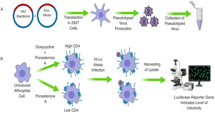
Pseudotyping Virus
controls with known HIV env sequences and entry phenotypes were tested as well as two negative controls lacking an env sequence. Following two days of incubation, the resultant virus was sterile filtered and stored at -80 °C.
Titering Virus
later, infectivity was measured. First, media was removed from the wells and washed twice with PBS. Then, 50 µL of lysis buffer was added and incubated at room temperature for 15 minutes. The solution was frozen overnight at -80 °C (5, 6).
Data Collection
The thawed 96 well plates were examined using a firefly luciferase assay under a luminometer (Promega) to discern the level of infectivity (5, 6). Readings in RLUs were quantified and plotted to determine an infectivity curve. Virus was diluted in Affinofile media so that 1 mL = 30 million RLUs when titered on Affinofiles (5, 6).
High Low Assay
Calculating High Low Results
Data corresponding to infectivity from the luciferase assay was gathered and used to calculate percent infectivity for both low and high CD4 wells. This data was then compared to assess the viruses’ ability to infect at low vs. high CD4 and plotted on a bar graph (5).
293T Co-Transfection with Puromycin selectable marker and SIV CCR5 Protocol
Transfection with Inducible rh-CD4 Vector and Tet-On Repressor
Previously transfected cells with CCR5 were transfected using the same method above except with a Tet-On repressor and PO/6 vector containing a sequenced rhesus CD4 gene isolated from restriction digests (Thermo-Fisher K102001). Single cell colonies were selected following selection with Blasticidin and Zeocin and are awaiting flow cytometry confirmation (Thermo-Fisher K102001).
RESULTS:
Pseudotyped virus with SIV env in HIV-1 subtype B backbone shows M-tropic entry phenotype for both plasma and CSF.
To assess if viral tropism shifts over time and in different compartments within SIV as a model system, viral entry phenotyping assays of pseudotyped virus with SIV Envelope proteins were conducted. We selected env sequences from macaques 21783, 21787, 21775, and 21602 to create a representative population for assessing change in tropism over time and compartment.
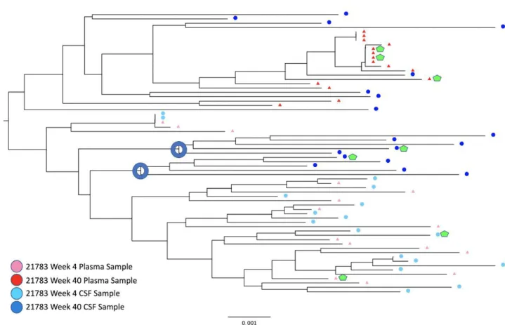
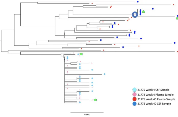
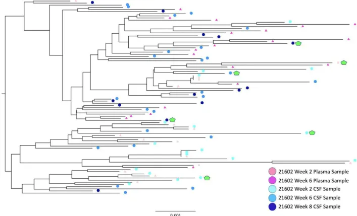
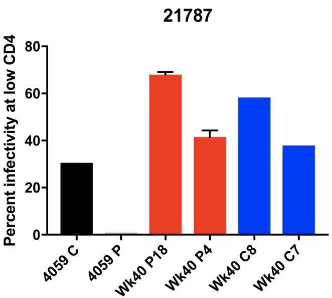
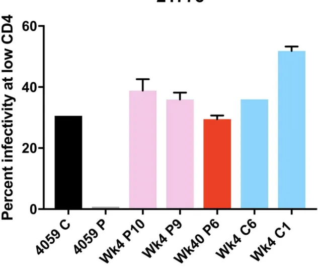
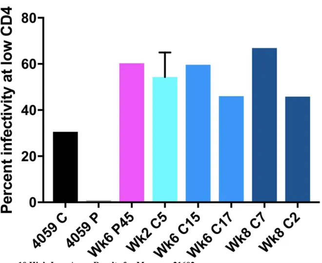
For the viruses derived from the monkey 21783, samples from both the CSF and plasma showed an M-tropic phenotype (Figure 7). Furthermore, no significant increase in macrophage tropism was found from viruses expressing Envelope proteins isolated from later time points compared to earlier time points. Viruses isolated from the monkey 21787, sampled from both the CSF and plasma, also showed an M-tropic phenotype (Figure 8). Viruses sampled from the monkey 21775 from both the CSF and plasma showed an M-tropic phenotype (Figure 9). Similarly, viruses derived from the monkey 21602, from both the CSF and plasma showed an M-tropic phenotype (Figure 10). Along with this, no significant increase in macrophage tropism was found in Envs isolated from later time points for any sampled virus. Together these results indicate that HIV-1 subtype B pseudotyped viruses with an SIV derived env vector show M-tropic entry phenotypes regardless of the compartment or time point in all tested samples.
New cell line expresses constitutive rhesus CCR5
both experimental conditions, CCR5 was not made inducible. A rhesus CD4 gene was transfected utilizing a Tet-On system, and cells are currently being selected for single cell cloning and expansion.
DISCUSSION:
Overview
SIV is one of the closest genetic relatives of HIV-1, and consequently, shares numerous similarities in structure and pathogenesis (15). In order to assess the validity of SIV as a possible model system for the evolution of macrophage-tropic HIV-1, 21 envelope genes were successfully cloned, and used to generate pseudovirus. These were tested for macrophage tropism based on their ability to infect Affinofile cells expressing a low density of CD4 and a high density of CCR5. The env genes were cloned from CSF and plasma isolates longitudinally sampled from four SIV-infected rhesus macaques.
Phylogenetic Analysis
Cloning and High Low Assay Analysis
To analyze this question, SIV envelope sequences were selected from Dr. Sturdevant’s phylogenetic trees to assess the changes in entry phenotype over time and in each compartment. Following cloning and transfection of env vectors with an HIV-1 subtype B backbone, a viral entry phenotyping assay was conducted on Affinofile cells (Figures 5, 6). Results indicated that
Factors influencing SIV as a model system.
1. Affinofile cells express human CD4 receptors and human CCR5 coreceptors on the surface of the cell. CD4 and CCR5 molecules in rhesus macaques differ from human CD4 and CCR5 at multiple residues affecting conformation (8-10). Therefore, Affinofile cells may not be able to accurately measure the phenotype of SIV Envelopes to recapitulate interactions with rhesus cells in vivo. This could occur if SIV uses human CD4 efficiently and therefore infects Affinofile cells well regardless of CD4 density.
4. Finally, the density of CD4 on rhesus macaque cells is not known. If rhesus macaque cells express lower densities of CD4, this could help to explain the result of consistent macrophage-tropic phenotypes. The definition of a cell with low density CD4 for a human could be different, and therefore the assay would not have detected a marked difference if the Affinofiles with low density CD4 were higher than CD4+ cells in rhesus macaques. The net result would be the ability to infect Affinofiles at both high and low CD4 densities, because both conditions were sufficiently high density when compared to rhesus macaque cells.
Future directions of a rhesus macaque cell line and SIV backbone.
In order to address these caveats, our lab has been working on generating a cell line capable of better reproducing in vivo interactions of SIV within rhesus macaques through the use of a cell line featuring inducible rhesus CD4 and constitutive rhesus CCR5. Along with this, we are working with collaborators to obtain an SIV backbone for pseudotyping to control for possible effects due to differences between HIV-1 and SIV backbones throughout viral entry phenotyping assays. Finally, we are also working with collaborators to determine the density of CD4 on representative CD4+ cells within the plasma and CSF of rhesus macaques to address possible constraints to using phenotyping assays with Affinofiles.
FIGURES
Figure 1. Rhesus Macaque Viral Phylogeny 21783
Figure 2. Rhesus Macaque Viral Phylogeny 21787
Figure 3. Rhesus Macaque Viral Phylogeny 21775
Figure 4. Rhesus Macaque Viral Phylogeny 21602
Figure 5. CD4 Density on cells susceptible to HIV entry and infectivity curves vs. CD4 density
Figure 6. Diagram of Pseudotyping Virus and High Low Assay
Figure 7. High Low Assay Percent Infectivity for 21783
Figure 8.High-Low Assay Results for Macaque 21787
Figure 9. High-Low Assay Results for Macaque 21775
Figure 10.High-Low Assay Results for Macaque 21602
Figure 11. Flow Cytometry Results for 293T Cells Transfected with rhCCR5
ACKNOWLEDGMENTS
I would like to thank the entire Swanstrom Lab and especially Dr. Ronald Swanstrom for allowing me to work, learn, and grow throughout an incredible undergraduate research experience. I would also like to thank Laura Kincer supervising me in numerous ways, helping throughout all parts of the project, teaching me methods, and for generally being amazing. Dr. Sarah Joseph, thank you for your help with flow cytometry, aiding my understanding of viral tropisms, and for being so friendly. Thank you to Christa Sturdevant for providing envelope amplicons and phylogenies from each monkey. Thank you Ean Spielvogel for helping me with a variety of questions I had throughout the project. Thank you to Dr. Adrienne Swanstrom for helping me find reagents for the cell line and answering many questions.
REFERENCES
1. Chan DC, Kim PS. 1998. HIV entry and its inhibition. Cell 93:681– 684. http://dx.doi.org/10.1016/S0092-8674(00)81430-0.
2. Wyatt R, Sodroski J. 1998. The HIV-1 envelope glycoproteins: fusogens, antigens, and immunogens. Science 280:1884 –1888. http://dx.doi.org/10 .1126/science.280.5371.1884. 3. Joseph SB, Swanstrom R. The evolution of HIV-1 entry phenotypes as a guide to changing target cells. J Leukoc Biol. 2018;103:421–431.https://doi.org/10.1002/JLB.2RI0517-200R 4. Schnell G, Joseph S, Spudich S, Price RW, Swanstrom R. 2011. HIV-1 replication in the central nervous system occurs in two distinct cell types. PLoS
Pathog.7:e1002286.http://dx.doi.org/10.1371/journal.ppat.1002286
6. Joseph SB, Arrildt KT, Swanstrom AE, et al.Quantification of entry phenotypes of
macrophage-tropic HIV-1 across a wide range of CD4 densities. J Virol.2014;88:1858–1869. 7.Blackard JTHIV compartmentalization: a review on a clinically important phenomenon. Curr HIV Res.
2012 Mar;10(2):133-42.
8. Chen Z, Zhou P, Ho DD, Landau NR, Marx PA. Genetically divergent strains of simian immunodeficiency virus use CCR5 as a coreceptor for entry. Journal of Virology.
1997;71(4):2705-2714.
9. Del Prete et al. Derivation and Characterization of a Simian Immunodeficiency Virus SIVmac239 Variant with Tropism for CXCR4. J. Virol. October 2009 vol. 83 no. 19 9911-9922.
10. Matsuda et al. Characterization of Simian Immunodeficiency Virus (SIV) That Induces SIV Encephalitis in Rhesus Macaques with High Frequency: Role of TRIM5 and Major Histocompatibility Complex Genotypes and Early Entry to the Brain J. Virol. November 2014 vol. 88 no. 22 13201-13211
11. Chen, M.F., Westmoreland, S., Ryzhova, E.V. et al. Journal of NeuroVirology (2006) 12: 73. https://doi.org/10.1080/13550280600654565
12. Holmes EC, Zhang LQ, Simmonds P, Ludlam CA, Brown AJ. 1992. Convergent and
divergent sequence evolution in the surface envelope glycoprotein of human immunodeficiency virus type 1 within a single infected patient. Proc Natl Acad Sci U S A 89:4835– 4839.
http://dx.doi.org /10.1073/pnas.89.11.4835.
13. Huang CC, Tang M, Zhang MY, Majeed S, Montabana E, Stanfield RL, Dimitrov DS, Korber B, Sodroski J, Wilson IA, Wyatt R, Kwong PD. 2005. Structure of a V3-containing HIV-1 gp120 core. Science 310:1025– 1028. http://dx.doi.org/10.1126/science.1118398.
14. De Jong JJ, De Ronde A, Keulen W, Tersmette M, Goudsmit J. 1992. Minimal requirements for the human immunodeficiency virus type 1 V3 domain to support the syncytium-inducing phenotype: analysis by single amino acid substitution. J Virol 66:6777– 6780.
15. Pohlmann S. et al. Amino acid 324 in the simian immunodeficiency virus SIVmac V3 loop can confer CD4 independence and modulate the interaction with CCR5 and alternative
coreceptors.J Virol.2004 Apr;78(7):3223-32.
16. Johnston, Samantha et al. A Quantitative Affinity-Profiling System That Reveals Distinct CD4/CCR5 Usage Patterns among Human Immunodeficiency Virus Type 1 and Simian Immunodeficiency Virus Strains. JOURNAL OF VIROLOGY Nov. 2009, p. 11016–11026
18. Chikere, Kelechi et al. Affinofile profiling: How efficiency of CD4/CCR5 usage impacts the biological and pathogenic phenotype of HIV. Virology 435 (2013) 81–91
19. Schnell G, Spudich S, Harrington P, Price RW, Swanstrom R.2009. Compartmentalized human immunodeficiency virus type 1 originates from long-lived cells in some subjects with HIV-1-associated dementia. PLoS Pathog.5: e1000395
20. Wei X, Decker JM, Liu H, Zhang Z, Arani RB, Kilby JM, Saag MS, Wu X, Shaw GM, Kappes JC. 2002. Emergence of resistant human immunodeficiency virus type 1 in patients receiving fusion inhibitor (T-20) monotherapy. Antimicrob Agents Chemother 46:1896–1905. doi:10.1128/AAC.46.6.1896-1905.2002.
21. Johnston SH, Lobritz MA, Nguyen S, Lassen K, Delair S, Posta F, Bryson YJ, Arts EJ, Chou T, Lee B. 2009. A quantitative affinity-profiling system that reveals distinct CD4/CCR5 usage patterns among human immunodeficiency virus type 1 and simian immunodeficiency virus strains. J Virol 83:11016–11026. doi:10.1128/JVI.01242-09.
22. Sturdevant CB, Dow A, Jabara CB, SB, Schnell G, Takamune N, Mallewa M, Heyderman RS, Van Rie A, Swanstrom R. 2012. Central nervous system compartmentalization of HIV-1 subtype C variants early and late in infection in young children. PLoS Pathog 8:e1003094. doi:10.1371/journal.ppat.1003094.
23. Sturdevant CB, (2013) Viral Characteristics associated with human immunodeficiency virus type 1 infection of the central nervous system early following transmission. 17-158.
24. Hahn BH, Shaw GM, De Cock KM, Sharp PM (2000) AIDS as a zoonosis: scientific and public health implications. Science 287: 607–614.
25. Pandrea I, Sodora DL, Silvestri G, Apetrei C (2008) Into the wild: simian
immunodeficiency virus (SIV) infection in natural hosts. Trends in Immunology 29: 419– 428.
26. Rey-Cuillé MA, Berthier JL, Bomsel-Demontoy MC, Chaduc Y, Montagnier L, et al. (1998) Simian immunodeficiency virus replicates to high levels in sooty mangabeys without inducing disease. Journal of Virology 72: 3872–3886.
27. Silvestri G, Sodora D, Koup RA, Paiardini M, O’Neil SP, et al. (2003) Nonpathogenic SIV infection of sooty mangabeys is characterized by limited bystander immunopathology despite chronic high-level viremia. Immunity 18: 441–452.
28. Stremlau M, Owens CM, Perron MJ, Kiessling M, Autissier P, Sodroski J. The
29. Williams, K. C., & Burdo, T. H. (2009). HIV and SIV infection: the role of cellular restriction and immune responses in viral replication and pathogenesis. APMIS : acta pathologica, microbiologica, et immunologica Scandinavica, 117(5-6), 400-12.