RELIABLE CHANGE INDICES OF VISUAL AND SENSORY PERFORMANCE MEASURES
Taryn Elizabeth Gilrein
A thesis submitted to the faculty of the University of North Carolina at Chapel Hill in partial fulfillment of the requirements for the degree of Master of Arts in the Department
of Exercise and Sports Science at The University of North Carolina at Chapel Hill. (Athletic Training)
Chapel Hill 2014
ii © 2014
iii ABSTRACT
TARYN ELIZABETH GILREIN: Reliable change indices of visual and sensory performance measures.
(Under the direction of Jason P. Mihalik)
The purpose was to determine the test-retest reliability and establish reliable
change indices for measures of visual and sensory performance in healthy college
students. Participants were administered several clinical and research tests of static and
dynamic visual acuity, gaze stability, and visual-motor sensory performance 14 days
apart. The test-retest ranged from 0.08 to 0.81 across all measures, and some
demonstrated significant practice effects. Clinicians should recognize employing reliable
change indices is but one method to manage concussed patients, and should consider
employing other tools to assess those tests demonstrating the lowest reliability. A
secondary purpose was to explore if visual deficits exist in college athletes who have
been cleared to return to play following concussion. We were unable to sufficiently
power these exploratory analyses. Therefore, subsequent studies should evaluate the
sensitivity, specificity, and predictive values of visual performance testing in the context
iv
TABLE OF CONTENTS
ABSTRACT ... iii!
LIST OF TABLES ... vii!
LIST OF FIGURES ... viii!
CHAPTER I ... 1!
Research Questions ... 3!
Research Hypotheses ... 4!
Variables ... 4!
Independent variables ... 4!
Dependent variables ... 5!
Operational Definitions ... 6!
Delimitations ... 6!
Limitations ... 6!
Assumptions ... 6!
CHAPTER II ... 8!
Introduction ... 8!
Concussion Epidemiology ... 9!
Current assessments/management of concussion ... 12!
Baseline Measures ... 13!
Symptoms ... 14!
v
Postural Control ... 17!
Multifaceted Approach ... 18!
Return to Play Decision ... 19!
Importance of Sports Vision for Athletes ... 20!
Visual Deficits after Concussion ... 21!
Visual Assessments ... 23!
King-Devick Test ... 26!
Why Test Vision? ... 26!
CHAPTER III ... 28!
Participants ... 28!
Instrumentation ... 28!
Dynamic Visual Acuity Test ... 28!
Gaze Stability Test ... 29!
King-Devick Test ... 31!
Procedure ... 32!
Data Reduction ... 32!
Data Analysis ... 33!
CHAPTER IV ... 41!
Introduction ... 41!
Methods ... 43!
Participants ... 43!
Instrumentation ... 43!
vi
Gaze Stability Test ... 45!
King-Devick Test ... 46!
Procedure ... 47!
Data Reduction ... 47!
Data Analysis ... 48!
Results ... 49!
Discussion ... 50!
Limitations ... 58!
Conclusions ... 59!
APPENDIX 1. PARTICIPANT DEMOGRAPHIC FORM ... 62!
APPENDIX 2. ADDENDUM TO SPARQ DESCRIPTIONS ... 64!
vii
LIST OF TABLES
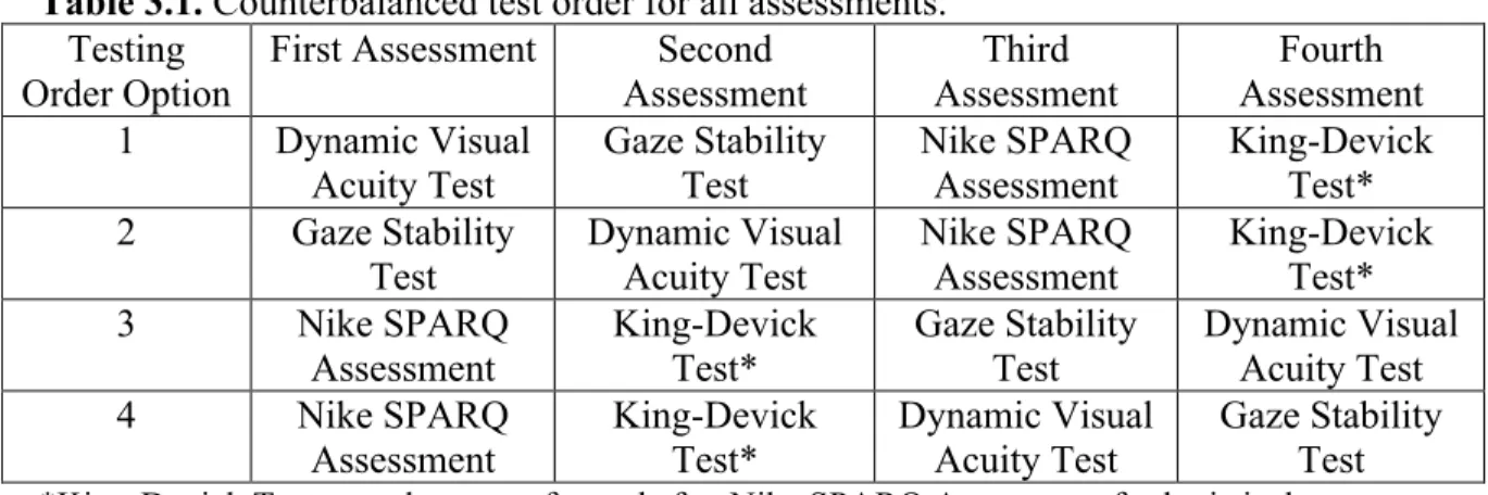
Table 3.1. Counterbalanced test order for all assessments. ... 35!
Table 3.2. Data analysis summary table. ... 36!
Table 3.3. Nike SPARQ Sensory Station module descriptions... 37!
Table 4.1. Healthy sample descriptive statistics used to compute predictive values ... 60!
viii
LIST OF FIGURES
Figure 3.1. NeuroCom test setup ... 38!
Figure 3.2. Nike SPARQ Sensory Station test constructs ... 39!
1
CHAPTER I INTRODUCTION
Concussions have recently become a spotlight health concern in today’s society.
The number of reported concussions continues to rise with approximately 1.6 to 3.8
million reported each year2 and many other suspected head injuries that go unreported.3
Concussion, or mild traumatic brain injury, has been defined as a complex
pathophysiologic process affecting the brain and its functioning capacities following an
injury to the head.4 Concussions typically results from either a direct blow to the head or
a traumatic force to the body that transmits an impulsive force to the head.5 The trauma to
the head leads to an energy crisis in the brain that results in decreased oxygen delivery
and functioning.6 As a result, concussions can result in multiple debilitating symptoms
including cognitive, balance, and visual deficits. These deficits typically resolve in 7-10
days, but may last several months in a subset of the population.4 Currently, concussion
evaluation typically includes reliable and sensitive7-9 measures of symptoms,
neurocognition, and postural control, but often does not address aspects, such as dynamic
or static vision or gaze stability, which may be associated with concussion.
Ideally, the results of the current post-concussion assessments are compared to the
individual’s baseline measures. Not all sports teams have the benefit of baseline testing
every athlete or have the medical staff to assess concussed patients. In the absence of
pre-injury scores for an athlete, the post-pre-injury measures can be compared to normative data
2
cognitive, memory, and balance measures should be utilized when comparing subjects to
a ‘norm’ for return to play decisions in addition to the individual baseline comparison.10
The computerized neuropsychological tests provide more sensitive and objective
measures when compared to the subjective pencil and paper battery tests. The
computerized battery also demonstrated a longer duration of symptoms in patients
post-concussion when compared to paper and pencil assessments.11
An important performance measure that is not considered in current
post-concussion return to participation is visual performance. Vision is critical to athletes of
all expertise levels and every sport.12,13 The visual demands and subsequent skills needed
for hitting a baseball, catching a football, and spotting a 4-inch balance beam are vastly
different. In order to objectively determine the demands necessary of the specific athlete,
a task analysis is performed considering the environment, opponents, targets and other
external factors involved in the particular sport. Athletes are required to take visual
perception and interpret the information to create a motor response dependent on the
stimuli. If an athlete is not receiving visual feedback fast enough to assess the situation
and act on the information, performance will suffer.14 Both static and dynamic visual
acuity measures should be taken and analyzed to expose deficits that could potentially
affect an athlete’s functional ability during dynamic sport. Visual performance measures
should be incorporated into the baseline testing of athletes to compare and observe
progress or deficit throughout the season, regardless of injury.15 These measures should
include assessments that address visual skills such as depth perception, reaction time, and
3
objective measure of a subject’s vision should be recorded and evaluated, similar to a
subject’s cognitive processing and postural stability.
Concussion evaluation paradigms typically use a pre-injury (baseline) to
post-injury comparison to identify deficits caused by concussion. In some cases, baseline
measures are not available, so it is important to have a ‘normal’ value with which to
compare an athlete’s results. The change from an individual’s pre-injury to post-injury
score can be compared to a reliable change index (RCI). Comparison to an RCI creates a
more sensitive conclusion as to if the athlete has returned to a normal measure prior to
resuming participation after injury, specifically concussion. Visual deficits have been
demonstrated following concussion,16 but there is a lack of data supporting the validity
and reliability of visual assessments that might aid in concussion evaluation and
management. To accurately assess the visual system in order to manage patients with
visual deficits, a clinical measure must be reliable, sensitive, and clinically applicable.
The primary purpose of this study was to determine the test-retest reliability and reliable
change indices for measures of visual performance in college athletes. A secondary and
exploratory purpose was to determine if visual deficits exist in college athletes who
report being asymptomatic following concussion.
Research Questions
1. What is the reliability of visual and sensory performance measures (Nike SPARQ
Sensory Station, NeuroCom Gaze Stability Test, NeuroCom Dynamic Visual
4
2. What are the reliable change indices for visual and sensory performance measures
(Nike SPARQ Sensory Station, NeuroCom Gaze Stability Test, NeuroCom
Dynamic Visual Acuity Test, and King-Devick Test) in healthy college students?
3. Exploratory: Is there a significant difference in visual and sensory performance
between concussed patients compared to match healthy controls?
Research Hypotheses
1. There will be moderate reliability across serial visual and sensory performance
assessments (Nike SPARQ Sensory Station, NeuroCom Gaze Stability Test,
NeuroCom Dynamic Visual Acuity Test, and King-Devick Test) in healthy
college students.
2. Reliable change indices will be computed and yield clinically reasonable
confidence intervals.
3. Exploratory: Concussed individuals will perform worse on visual and sensory
performance measures compared to match healthy controls.
Variables
Independent variables
1. Time
a. Testing Session One
b. Testing Session Two
2. Group
a. Healthy College Students
5 Dependent variables
1. Nike SPARQ Sensory Station
a. Visual Clarity (Static visual acuity)
b. Contrast Sensitivity
c. Depth Perception
d. Near-Far Quickness
e. Target Capture (Dynamic visual acuity)
f. Perception Span
g. Eye-Hand Coordination
h. Go/No-Go
i. Reaction Time
j. Response Time
k. Motor Movement Time
2. Gaze Stability Test (NeuroCom)
a. Maximum head speed (degrees/second) achieved while correctly identifying
orientation of visual stimulus; measured in yaw, pitch, and roll directions
3. Dynamic Visual Acuity (NeuroCom)
a. Dynamic visual acuity loss (dynamic visual acuity minus static visual acuity);
measured in the Logarithm of the Minimum Angle of Resolution (logMAR)
4. King-Devick Test
a. Completion time
b. Number of errors committed
6 Operational Definitions
1. Healthy participants: athletic individual participating in physical activity three to
four times per week.
2. Concussed participant: varsity or club athlete ages 18-25 who underwent a direct
blow to the head, neck, or elsewhere on the body with an impulsive force
transmitted to the head that resulted in concussive symptoms and were diagnosed
by the respective doctor with a concussion.
3. Matched control: uninjured individual matched to the concussed patients based on
age, gender, sport, and position on team. (Exploratory Research Question 3)
Delimitations
1. Data limited to college athletes at the University of North Carolina at Chapel Hill.
2. There will only be two data collection time intervals.
3. The SPARQ Sensory Station, Neurocom GST and DVAT, and the King-Devick
Test are the only visual measures that were used.
Limitations
1. Attrition rate leading to small sample size.
2. Low concussion rate leading to small sample size.
3. Forced to cease testing if symptoms return during assessments.
Assumptions
1. Nike SPARQ and NeuroCom SOT will accurately record data.
2. All participants will provide full effort during testing.
7
4. All participants will be truthful and honest about concussion symptoms and
history.
5. The ability of the examiner will not interfere with testing results.
6. Effects of mental and physical fatigue will not significantly alter participant’s
visual performance.
7. A convenient sample of athletes chosen based on proximity and availability will
accurately represent the population.
8. Data will be properly interpreted by examiner.
9. Team physicians and athletic trainers are properly evaluating and diagnosing
8
CHAPTER II
REVIEW OF LITERATURE
Introduction
Concussions are a serious health concern in today’s society and have led to a heightened sense of awareness in all factions of sports and athletics. It has been estimated that well over 1 million people in the United States sustain a concussion annually, leading to a critical movement for prevention, education, and research surrounding
concussion.2,17 The main focus of current sports-related concussion research is the identification of cognitive and vestibular deficits post-injury. Due to the complexity of the injury, a comprehensive paradigm of assessments examining cognitive function, postural-stability, and neurological symptoms should be used to assess an individual who has sustained a suspected concussion.4 The more assessments utilized by a clinician for evaluation of a potential concussed individual, the more sensitive the tests become to identifying deficits due to a concussion.18 A multifaceted approach is recommended, because a combination of measures increases the sensitivity to greater than 90 percent, compared to the sensitivities of one single assessment, which range from 43 to 80 percent.19,20
9
recovery. This leads us to ask the question: what deficits are we missing in our current
assessment of a concussed individual in the absence of symptoms? One potential deficit
we may be missing is visual disturbances. The visual system accounts for 80 percent of
an individual’s sensory input and 50 percent of the brain’s pathways are devoted to
vision.21 Visual deficits and symptoms related to vision have been identified in concussed
individuals upon sideline evaluation post-concussion but visual testing is still not always
recognized as part of the recommended evaluation.16 Visual performance measures
should be considered in evaluation of a concussion, because vision is important to all
individuals, but particularly athletes.
Concussion Epidemiology
Annually, there are approximately 44 million children and young adults
participating in organized sports, and approximately 170 million adults participating in
some type of physical activity in the United States.22,23 The large number of children and
adults participating in physical activity and sports further confirms the need for
educational programs that promote awareness of high risk situations and demonstrate
preventative measures. With the implementation of educational programs in the past few
years, there has been a significant decrease in the number of catastrophic injuries from
head injuries.24
Each year, there are an estimated 1.6 to 3.8 million people who report sustaining a
sports concussion,17,25,26 making it the most common traumatic brain injury in athletic
young adults.27 The number is only an approximation because many of these head
injuries may go unreported.3,28 The culture of the sport, attitude of the athlete, and
10
subjective statements and athletes are now admitting to lying about symptoms in order to
continue playing.3,29
There is a higher incidence of concussion in adolescents that is speculated to be
the result of younger, more susceptible brains. Traumatic brain injury in children and
adolescents can lead to persistent cognitive dysfunction, even when no initial effects are
observed.6 Increased susceptibility to concussion in children and adolescents, as
compared to adults, has been attributed to decreased myelination, a greater head-to-body
ratio, and thinner cranial bones, all which provide less protection to the developing
cortex.30-32 Females are also thought to be at a higher risk for sustaining a concussion,
both at the high school and collegiate level.33 Barnes et al. suggested that female soccer
players are more susceptible due to the biomechanical factors such as smaller head to ball
ratios and weaker musculature.34 It is speculated that the higher frequency of concussion
in male sports compared to female sports may be attributed to the different styles of play
including lacrosse, basketball, and softball.33 Concussions after getting hit by a pitch are
more likely in baseball than in softball.33 Other studies have suggested that females are
more likely to report symptoms after a potential concussion when compared to males
who may try to play through the pain.3
There is no single agreed upon definition of concussion. In 1996, the Congress of
Neurological Surgeons in America agreed on the following definition: a concussion is
“...a clinical syndrome characterized by immediate and transient post-traumatic
impairment of neural functions, such as alteration of consciousness, disturbance of vision
11
of vision comprised in the definition of concussion, as it is part of some clinician’s
post-injury evaluation, but is not always highlighted.
The most recent National Athletic Training Association Consensus Statement
released included the following definition: “Concussion is defined as a complex
pathophysiological process affecting the brain, induced by traumatic biomechanical
forces.”5 The statement goes on to describe common features that are typically, but not
always seen in those individuals who sustain a concussion. The most common
mechanism includes either a direct blow to the head, or indirect blow to anywhere on the
body transmitting “impulsive” forces to the head that lead to the rapid onset of temporary
neurologic function impairment.5 Recently, it has been determined that a concussion is a
functional injury rather than a structural injury and typically there is no abnormalities
found on standard neuroimaging that identify a concussion.5
The etiology of concussion is largely dependent on the sport;36 the majority of
these injuries occurring in contact sports such as football, boxing, hockey, in addition to
soccer and basketball.32,36-38 Concussions occur during a direct blow or indirect impact to
the head, face, or neck and lead to a rapid acceleration and subsequent deceleration of the
brain.5 The biomechanical forces lead to linear and rotational accelerations in the brain
causing injury to delicate white matter and brain tissue, ultimately leading to the
biochemical response that results in the presence of the acute symptoms of a
concussion.6,39
The rotational and linear acceleration and deceleration of the head are the most
common mechanisms of injury and result in shearing, compressive, and tensile forces to
12
the presentation of temporary clinical signs and symptoms that should be evaluated by a
healthcare professional to determine the status and further management of the
individual.6,40 Physical signs that present after a concussion include loss of
consciousness, amnesia, behavioral changes including irritability, cognitive impairment,
slowed reaction time, sleep disturbance, headaches, blurred or double vision, feeling in a
‘fog’, or increased emotional sensitivity.5,41
After the physical trauma of a concussion, a metabolic cascade ensues at the
cellular level in the pathways of the brain.6 There is a release of potassium as well as an
influx of calcium in the neurons that ultimately reduces the cell’s ability to generate
oxygen.6 Essentially, the high energy demand of brain cells, restricted blood flow, and
oxygen debt cause mental confusion, failed memory, and dizziness in an individual.6
These changes do not result in any abnormality on standard structural neuroimaging
studies including MRIs or CTs.5 While this process leads to neuropathological changes,
the acute symptoms observed post-concussion are indicative of a functional impairment
rather than a structural deficiency.42,43
Current assessments/management of concussion
There are several consensus statement and position statements outlining the
evaluation and management of sport-related concussion.4,5 According to the most recent
recommendations, when an athlete sustains a blow to the head, either from an object, an
opponent, the ground, or experiences a severe ‘whiplash’ activity, a healthcare
professional should be suspicious of a concussion.5 The athlete should be removed from
activity for the remainder of the day if he or she is experiencing concussion symptoms, or
13
minutes, he or she could potentially return to participation at that time. Instead of relying
on a subjective decision, clinicians should take the functioning of the brain into account.
The sideline assessment has become more in depth, allowing for improvement upon the
15-minute symptom “check-up”. A concise evaluation using tools such as the
Standardized Assessment of Concussion (SAC) in addition to an evaluation of symptoms
and motor response should be performed during the primary survey of an athlete with a
suspected concussion. If the athlete is diagnosed with a concussion, they are removed
from the event and should be taken through further physical evaluation and close
monitoring of symptoms.28,44
If an individual has experienced more than one concussion, the result of any
subsequent head impact may be worse and can lead to long lasting repercussions.45,46
Poorly managed concussions may lead to a host of complications such as post-concussion
syndrome, second impact syndrome, post-traumatic stress disorder and potential lasting
memory, visual, vestibular, or cognitive impairments.31,47 Prompt and thorough
evaluation and management of concussion may aid in preventing long-term
consequences, but the deterioration process cannot be terminated if mental and physical
stresses persist.5
Baseline Measures
Recent studies recommend pre-injury baseline testing for each athlete on
neurocognitive measures, symptoms, and postural control abilities post-injury.5 Baseline
tests are suggested to account for differences in individual scores and measures on
14
complex baseline testing battery is beneficial for the detection of deficits in objective
measures of neurocognition and postural control despite symptom resolution.48
Baseline testing should take place prior to the beginning of season, in a quiet
setting allowing the individual to focus and take the test seriously for accurate results.49
These measures may be influenced by predisposing factors such as developmental
disorders, attention deficit hyperactivity disorder (ADHD), migraine history, or previous
concussions.41,50 The time of day an individual completes the evaluation, the individual’s
mood, external stress level, and fatigue may all have a detrimental effect on the testing
and outcome measures, therefore affecting the return to participation decision for that
individual.49,50
Baseline testing for all measures discussed would provide a comprehensive
representation of college athletes, but it is not practical in all scenarios. The equipment
and resources needed are costly and testing can be time-consuming.51 If administering
baseline measures is not an option in a certain setting, clinicians may look into using
normative data for comparison of differences.48,52 Organizations that have limited
resources and do not have access to balance-diagnostic equipment or computerized
neurocognitive testing programs typically use the standardized, self-reported symptom
checklist as a practical method for monitoring concussion symptoms53 in addition to
simpler, more cost-effective measures such as other paper-pencil batteries, BESS testing,
and King-Devick Testing.
Symptoms
The comprehensive symptom checklist is one of the most commonly used
15
symptoms experienced at a given time.54,55 Most checklists include a numerical scale
allowing the individual to rate the presence and intensity of symptoms commonly
exhibited in concussed individuals such as headache, dizziness, drowsiness, vision
problems, balance difficulty, trouble falling asleep, drowsiness, sadness, difficulty
concentrating, difficulty remembering, feeling “in a fog”54,55 and irritability.4,5,56,57
Symptom checklists have been further studied by many researchers and have been found
to carry well-distinguished validity and reliability.51,58,59 While symptom checklists
provide a clinically relevant and useful tool for identifying symptoms that are typically
present post-concussion, it should be combined with other recommended tests for a
complete assessment of an individual.51
Piland et al. examined the validity of subjective symptom reports and found
evidence of factorial and construct validity for the Head Injury Scale, a checklist that
includes nine of the most common symptoms reported in concussed individuals and is
typically used in the SAC and SCAT2 forms.51 The commonly associated symptoms with
concussions can be separated into three constructs: somatic, neuropsychological, and
cognitive symptoms.51,60 While the symptoms may interrelate and overlap, the theoretical
distribution of symptoms is as such:
Somatic symptoms include headache, nausea, vomiting, balance, sensitivity
numbness; those considered in the cognitive construct include “slowed down”, “in
a fog”, difficulty concentrating, difficulty remembering; and those thought to be
neuropsychological in nature include fatigue, difficulty falling asleep, sleeping
16
Each individual concussion is unique and there is no way of predicting which
symptoms an individual will exhibit after a concussion or how long the symptoms will
last. Certain symptoms and risk factors are more likely to contribute to prolonged
recovery, such as history of previous concussions45 or sustaining a concussion at a young
age.31 Deficits may be masked in the absence of symptoms during the return to play
progression and might not present until the individual returns to a high level of activity.
Athletes returning to an environment with excessive visual stimuli such as a soccer field
or a basketball court might experience the return of symptoms when dynamic visual
acuity is necessary. If visual performance tests were performed prior to return to
participation post-concussion, deficits in dynamic visual acuity might be identified to
avoid premature return to play.
Neurocognitive Evaluation
Concussions are typically associated with neurological and mental status
impairments that affect cognitive, academic, and behavioral functioning.61
Neurocognitive testing can help identify deficits in an athlete’s sustained attention,
executive functioning, processing speed, reaction time, and recall of new information.62
Neurocognitive tests provide a more concrete and objective measure of deficits present
after concussion when compared to a subjective symptom report from the individual.9,63
The Standardized Assessment of Concussion (SAC) is a screening instrument in
the form of a paper-pencil test that was developed in order for clinicians to establish an
idea of the athlete’s current mental status within minutes of an athlete sustaining a
concussion.9 While the test can be quick and efficient, it does not test brainstem or
17
pencil battery that has been proven valid and reliable and has been shown to be sensitive
to concussion symptoms.65 Computerized tests may be more beneficial, than the
traditional paper and pencil tests, because computer tests provide a large variety of
various forms, minimizing the learning effect of athletes who take assessment multiple
times.62,65 The traditional neurocognitive evaluation is also dependent on the ability of the
tester to correctly time processing speed and reaction time of the concussed individual,
potentially leading to inaccurate response times and false conclusions regarding
neurocognitive functioning.62,65
Computerized neurocognitive testing is a fairly novel assessment that addresses
the flaws in the paper-pencil battery of neurocognitive testing. The computerized tests
carry a well-established reliability and validity and similar to traditional testing, they also
have been shown to be sensitive and reliable for the effects of concussion.29,66,67 Several
computerized testing programs have been developed in recent years including Automated
Neuropsychological Assessment Metrics (ANAM)(National Rehabilitation Hospital
Assistive Technology and Neuroscience Center, Washington, DC), ImPACT Concussion
Management Software (ImPACT Applications, Pittsburgh, PA), and HeadMinder
Concussion Resolution Index (CRI)(Headminder Inc, New York, NY).4 Computerized
neurocognitive testing typically assesses verbal memory, visual memory, processing
speed, executive function, psychomotor speed, reaction time, complex attention and
cognitive flexibility.7
Postural Control
Postural stability is the ability of a person to control the position and action of
18
that can be used to assess for vestibular deficits in concussed individuals.68 Fatigue,
vestibular disturbances, and removal of visual stimuli are all factors that affect the body’s
ability to control postural sway.69-71 The Balance Error Scoring System (BESS) is used
currently as a sideline measure of an athlete’s balance after a suspected concussion.68 The
inexpensive assessment gives a clinician an objective measure of postural stability after a
suspected head injury.68,72 Visual and vestibular function are affected after an individual
sustains a concussion5,41 and therefore difficulties with an individual’s ability to control
postural sway may indicate disturbances in the brain’s pathways for vision. Results of
these sideline tests should then be compared to the athlete’s baseline BESS scores that
should have been established in the athlete’s resting state during preseason screening.
Multifaceted Approach
A concussion is a complex injury, affecting many different systems of the body as
previously discussed. The convolution of the injury leads to the need for a multifaceted
approach to evaluation of an athlete with a suspected concussion. The Standardized
Concussion Assessment Tool is a quick subjective assessment that includes a symptom
checklist along with a brief evaluation of attention, concentration, and memory.5,73 In
addition to cognitive processing, balance and coordination have been widely researched
for the identification of deficits and other vestibular problems8,74 and can be assessed
with the BESS test. Slower reaction time, slower processing speed and reduced memory
performance are among the deficits seen in concussed individuals during post-injury
testing.75 While encompassing many systems and identifying present deficits in those
concussed individuals, the evaluation of a concussed athlete is not complete. One deficit
19 Return to Play Decision
Once all of the athlete’s symptoms have resolved and he or she has returned to
baseline on all neurocognitive and balance tests, the next step is a gradual progression
that includes five levels of physical exertion, each increasingly more demanding.5,76 No
concussed individual should begin the physical activity progression without being
evaluated and cleared by a physician or alternate health care professional specifically
trained in concussion evaluation.5
The gradual progression protocol is the generally accepted return to play protocol
used by clinicians in accordance with the most recent NATA Consensus Statement.5 The
clinician should take into account the specific individual’s symptoms and response to the
injury, the severity of the concussion, and the number of previous concussions the
individual has experienced. The individual is allowed to return to limited activity when
they are completely asymptomatic at rest, demonstrates performance comparable to
baseline values, or normative values on all accepted assessments of neurocognitive
function and postural stability and remains asymptomatic with physical exertion.4,5 An
individual may not exhibit visual deficits during post-concussive testing as most current
assessments do not require dynamic head motion, which may contribute to the return of
symptoms when individuals resume activity. Visual performance during dynamic
movement is essential for athletes and if deficits are not identified before the individual’s
return to sport, performance may be affected and it may be an indicator that the athlete
20 Importance of Sports Vision for Athletes
Regardless of athletic classification, vision is the dominant sense in most
individuals and is critical for optimal performance in high intensity athletics as well as in
everyday life.77-79 Sensorimotor and semantic visual functioning are two important
factors necessary for the analysis of multiple visual stimuli during sport and the ensuing
motor response.80 The combination of the sensorimotor function with the semantic visual
function allows an individual to identify and interpret a situation.80 Elite athletes
demonstrate an advanced ability to combine these senses when compared with
non-athletes in situations with multiple stimuli.80
It has been said that hitting a baseball is “the single most difficult skill in all of
sports.”81 The ability to see a round object moving at such a high velocity accompanied
with spin and trajectory and then analyze how and when to hit the ball in such a short
amount of time with a separate round object moving in the opposite direction proves to
be a skill for those with elite senses and efficient responses.80-83 Constant convergence of
both eyes is required to assess the speed of the ball, predict the movement pattern and
path, and intercept the ball or object.14 These athletes must adjust quickly to the
approaching object and initiate efficient and appropriate motor responses based on the
sensory stimuli.14
When compared to non-athletes, athletes display heightened visual skills
including visual acuity, reaction speed, and contrast sensitivity.13,82,84 Kirschen et al.
developed a diagram to demonstrate the many layers of visual functions that build upon
each other, such that of a pyramid.79 This “Sports Vision Pyramid” describes monocular
21
Monocular vision, or that of the single eye, utilizes the functions of visual acuity and
contrast sensitivity and may be affected by astigmatism or a change in the amount of light
available.79 The next level of the pyramid is concerned with how both eyes work
together, or binocular vision, and stereopsis, or the visual perception of depth. The eyes
are designed to work together to produce visual images yet when under visual conditions
causing motor problems such as fixation disparity or sensory problems such as
amblyopia, the loss of one eye’s ability to see details, the information processed and
produced through binocularity may be incorrect.79 If both monocular and binocular
processes are working efficiently and correctly, the visual mechanics will be optimized.
There is an important interaction between the brain and the rest of the body when a visual
stimulus is interpreted and a motor response ensues.79 Athletes perform at the optimal
intensity if all three levels of the pyramid are functioning properly.79
The visual system provides information about target distance and the presence of
obstacles in the visual field, both frontal and periphery, in a static situation. In addition,
the visual stimuli provides additional information to maintain balance during standing,
walking, running, and in adjusting pathways when obstacles appear, the target moves, or
the pathway changes.85
Visual Deficits after Concussion
Memory, anticipation, pathways for fast eye movements, and accuracy of the eye
muscles are controlled by the cerebral cortex and are not always flagged during cognitive
testing.21 Common symptoms post-concussion that are related to the visual system
include blurred vision, double vision, difficulty focusing vision, balance problems, and
22
efferent or afferent pathway dysfunction from the eyes to the brain.86,87 Other visual
issues including double vision, vertigo and photophobia may be a sign of brainstem or
cerebellar pathway dysfunction from the widespread energy crisis occurring during the
biochemical response to a concussion.86 According to Heitger et al., impaired eye
movements are an indicator of suboptimal brain function and “may help demonstrate
incomplete recovery of brain function.”86
Visual symptoms do not always present for obvious diagnosis of concussion as
some may be masked or confused with neurologic deficits.16 The frontal lobe of the brain
is a primary site of injury in many mechanisms of concussion and can lead to temporary
disturbances in the frontal eye fields, impaired visual attention and delayed visual
saccades.88 Disturbances to the midbrain may produce impairments in the visual system
including double vision, abnormal pupil activity, or difficulty controlling eyelid
functions.16 Impairments with convergence and nystagmus may be indicative of injury to
cranial nerves or the brain stem.16
Quality vision is critical for optimal performance for athletes at any level. Just as
baseline measures are taken for neurocognitive, balance, and symptom scores, baseline
visual performance measures should be taken for both static and dynamic visual acuity.78
The vestibulo-ocular reflex is a critical reflex of the eye in response to movement that
provides information and proprioception that stabilizes vision and the line of sight.89
These visual signals interact and combine with vestibular information to stabilize gaze
during most normal head motions.89 If there is a deficit in either system, the individual
23
Visual performance tests exist to measure and compare injured patients, but are
not widely used at this time. The additional time commitment for the tests, questionable
reliability and validity, lack of normative values, and lack of sensitivity for concussion
identification may be among reasons why visual performance tests are not always used.90
One purpose of this study is to justify the necessity for a visual performance assessment
post-concussion in order to identify potential deficits prior to the individual’s full return
to participation.
Visual Assessments
Over 50% of the brain’s pathways are examined via visual assessments, yet visual
performance measures are not always considered part of the comprehensive concussion
evaluation.86 Visual assessments include assessments of static visual acuity, dynamic
visual acuity, gaze stability and several other functional measures. Static visual acuity is
the ability to see clearly when remaining still and watching a nonmoving object. It is
typically assessed using chart systems such as Snellen eye chart. The optimal acuity
measurement for a non-athlete is 20/20.91 There are no differences in static visual acuity
between athletes and non-athletes.13,82,83
Dynamic visual acuity (DVA) is the ability to resolve detail when there is relative
movement between the target and the observer.13 DVA can be measured by a
computerized system that assesses the ability of the patient’s vestibulo-ocular reflex to
maintain accurate and optimal visual acuity while moving their head with a fixed head
velocity requirement.90 Maintaining focus and sight while moving the head is a crucial
function, which is essential for athletes to accurately perceive and identify a moving
24
scores on a DVAT, reflecting a decrease in functioning and requiring some type of
compensation from the visual and vestibular systems.92,93 Compensations such as
vestibular adaptation and central programming have been observed in some subjects with
vision loss or decreased visual acuity.92 The vestibular system is adaptive in nature,
contributing to the recovery of vestibular response after vestibular loss or injury.93 The
computerized NeuroCom DVAT has high sensitivity and specificity for diagnosing
vestibular dysfunction.92 The high sensitivity and specificity further support the reliability
of the dynamic visual acuity test and the ability of the test to distinguish between healthy
participants and subjects with vestibular or visual deficits.92
The Gaze Stability Test was created to assess how quickly a participant can move
their head while maintaining focus on a computer-generated target of fixed size.94 The
GST quantifies the ability of a person to recognize a target projected on a personal
computer monitor during active head movement. Outcomes are calculated using the
means of the three fastest head velocities with accurate identification and orientation of
the target.90 The information produced demonstrates the functional capacity of
vestibulo-ocular reflex and the maximum active head velocity at which a person can stabilize their
gaze.90
Dynamic visual acuity is often not assessed clinically due to limitations in
instrumentation for measurements of DVA. Measures of DVA appear to have similar
within session reliability and lower between session reliability when compared to
measures of the Gaze Stability Test.1,90,94,95 The DVAT has been associated with less
muscle fatigue than GST. While both use a fixed wait time for the optotype to appear on
25
compared to the GST which requires the subject to maintain higher head velocities during
the trials. The DVAT also has caused the return of symptoms in some affected subjects
leading to nausea, blurred vision, dizziness, and headache due to the nature of the test and
accompanying head movements.90,94,95
The inVision System from NeuroCom can be used for the assessment of dynamic
visual acuity and gaze stability. The reliability and sensitivity of these tests vary in the
literature but when combined, the sensitivity to visual and vestibular disturbances
increased to 79% and the specificity was 88%.92,94 The GST has good test-retest
reliability and may be more useful as a measure of treatment outcome or identification of
deficits and disabilities when compared to DVAT.94 Both tests have been found to
minimal symptoms in healthy participants.
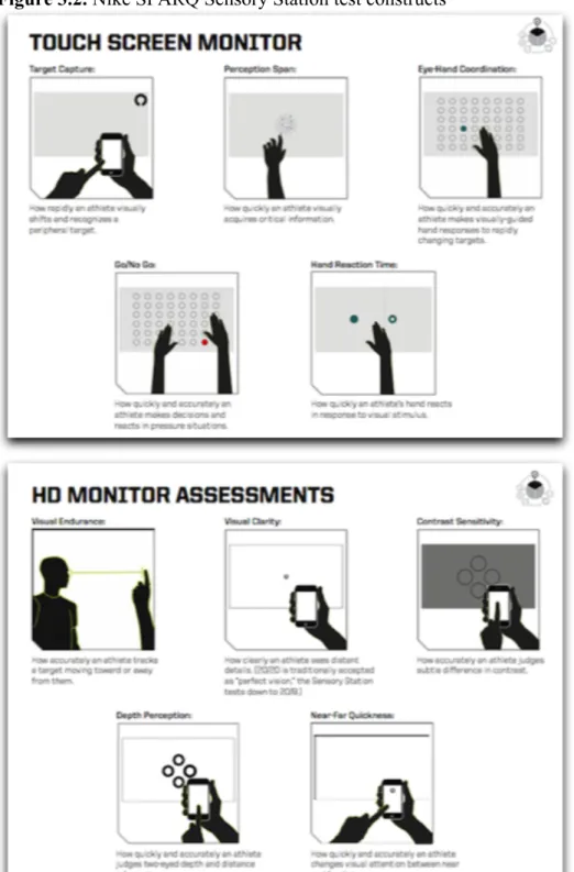
The Nike SPARQ Sensory Station was created as a functional measure of visual
clarity, near far quickness, target capture, reaction time, and eye hand coordination. The
SPARQ allows for an interactive testing environment that is able to identify deficits in
visual measures and reaction times.91 A single study has examined the reliability of the
SPARQ in a group of younger adults. There were no practice effects for the following
measures: visual clarity, contrast sensitivity, depth perception, target capture, perception
span, and reaction time. There were practice effects for near-far quickness, eye-hand
coordination, and go/no go. The motor response characteristics of these measures are a
possible explanation for the practice effect associated with near-far quickness, eye-hand
coordination, and go/no go.91 While Erickson et al. provided a preliminary estimation of
reliability for the SPARQ, their sample was not required to be physically active and
26
measure in college athletes, the reliability of the measure in healthy physically-active
college-aged individuals must be established.
King-Devick Test
The King-Devick (K-D) test is a sideline visual screening tool that assesses an
individual’s ability to read aloud single digit numbers on a screen or a card quickly and
efficiently. The test is based on measurement of the speed of rapid number naming
(reading single-digit numbers from 3 test cards in addition to any errors the individual
makes. The King-Devick test captures impairments of eye movements, attention,
language, and visual-motor functioning and it can be used to identify individuals with
dyslexia, learning disabilities, suboptimal brain or vision function, and it may be useful in
detecting visual impairments following concussion.96 In addition, it may be used as a
rapid sideline assessment for faulty or slow eye movements and other characteristics
following a potential brain injury.96 The King-Devick test has been used to accurately
diagnose concussions in some boxers and mixed-martial arts fighters.97 Similar results
were found in a collegiate athletic population.96 The lower scores recorded for individuals
who sustained a concussion suggest that the King-Devick test may be sensitive to visual
tracking deficits that occur post-concussion.
Why Test Vision?
It is important to examine vision following a head injury to identify deficits that
may go unnoticed in the absence of reported subjective symptoms. Blurred vision,
dizziness, sensitivity to light, and inefficient convergence are all symptoms commonly
experienced in concussed individuals and all could be indicative of suboptimal
27
measures of symptoms,91 postural sway,91 and neurocognitive functioning,91 there is
limited evidence establishing the reliability of visual performance measures in an athletic
collegiate population.
There is a gap in the literature regarding psychometric properties of visual
assessments that may be used in the evaluation and management of concussion. Reliable
change indices demonstrate the change in an individual’s score we should expect to see
between a first and second testing session. Reliable change indices for visual measures
will allow for a comparison in the change of concussed individual’s scores between a first
and second testing session and the change we would expect to see if that individual was
healthy. If the change in the patient’s scores is within the RCI for those measures, then
we would expect that the individual is healthy. If the change is the patient’s scores is
larger than the RCI for those measures, then we would expect that there is some type of
deficit.
Visual assessments should be involved in concussion evaluation. Athletes should
not return to full participation until visual performance measures have returned to
baseline. The addition of visual assessments to the composite evaluation may identify
deficits that are masked in the absence of symptoms and create a more conservative
28 CHAPTER III
METHODS Participants
We studied a convenience sample of 44 active, healthy college students (29 male
and 15 female; age = 19.90 ± 0.96 yrs; height = 172.03 ± 11.13 cm). Participants were
excluded from this study if they had known neurocognitive deficits or disorders, known
psychological conditions, color blindness, history of dizziness, imbalance or abnormal
vestibular function, or musculoskeletal abnormalities to the head, neck, shoulder, or back
that would disrupt normal range of motion. All participants read and signed consent
forms approved by our institution’s ethics review board. Participants also completed a
pre-participation form providing us with demographic, sleep habit, history of vision
problems, and concussion history.
Our third research question sought to explore the effect of concussion on vision.
Unfortunately, we were only able to capture 5 injuries, an insufficient number to yield
any meaningful data to address this research question. These 5 cases are instead used to
support some aspects of the discussion related to our other two research questions.
Instrumentation
Dynamic Visual Acuity Test
The Dynamic Visual Acuity Test (DVAT) was performed using the InVision
29
a 15-in flat panel high contrast liquid-crystal display monitor were used to display an
optotype (the letter “E”). The DVAT began with assessment of the participant’s static
visual acuity by having them identify the orientation of the optotype “E” on a computer
screen located 8 feet in front of them, as illustrated in Figure 3.1. Responses were
recorded using a handheld remote with buttons that indicated whether the “E” was
positioned to the right, left, upwards, or downwards. The size of the “E” was reduced if
the participant correctly identified 3 out of 5 “E”s of a given size. Static visual acuity was
defined based on the smallest “E” correctly identified, and measured in logMAR.
The participant’s dynamic visual acuity (DVA) was measured in three different
axes: the “yaw” (vertical axis rotation), “pitch” (medial-lateral rotation), and “roll”
(antero-posterior axis rotation). The participant wore a head harness (InterSense Inertia
Cube, Engineering Systems Technology, Kaiserslautern, Germany) with a sensor that
integrated the 3 axes the head was moving about to determine rotational velocity
(deg/sec) of the head. Participants were required to generate rotational head movements
at least 20 degrees from midline in each direction while still being able to correctly
analyze an optotype “E” of varying sizes. Each test allowed for a practice trial. The size
of the smallest optotype identified correctly while rotating the head faster than the
minimum velocity was recorded for results of the DVAT. The DVAT scores were
converted to visual loss by subtracting dynamic visual acuity from baseline static visual
acuity for each eye, averaging the two means, and reporting the outcome in logMAR.90
Gaze Stability Test
The InVision system (NeuroCom International,Clackamas, OR) used for the
30
acuity measured with the DVAT. The GST measures head movement velocities that the
participant achieves while maintaining their static visual acuity. Participants were
required to maintain gaze on the center of a computer screen, demonstrated in Figure 3.1,
and correctly identify the orientation of the optotype “E” while generating repetitive head
movements at varying velocities. Participants used the same headband with a 3-axis
integrating gyro to determine velocity for each trial. The participants were instructed to
perform a smooth sinusoidal head shake movement until the display screen was visible.
Two feedback bars gave the participant information on the velocity and amplitude of the
head and disappeared when the participant’s head velocity exceeded the required
minimum threshold for a trial. The optotype was displayed on the monitor until the
participant’s head velocity fell below the requirement for that trial or the participant
reached a maximum time. If three out of five “E”s were identified correctly, the
minimum head velocity that the participant must attain was increased. This was repeated
until the participant was unable to correctly identify three out of five “E”s, at which point
the velocity was reduced. If the participant was unable to achieve the minimum required
head velocity within 8 seconds from the start of head movement or unable to maintain
velocity for the required duration, or if the participant achieved and maintained the
required velocity but incorrectly identified the orientation, the trial was recorded as a
failure.90 The GST generated maximum head movement velocity (deg/sec) at which the
participant was able to maintain visual acuity in each of the three axes of movement
(yaw, pitch, and roll).
31
The Nike SPARQ Sensory Station is operated by a single computer controlling 2
high-resolution liquid crystal display monitors as shown in Figure 3.3. A handheld Apple
iPod touch (Apple Corporation, Cupertino, CA) was connected via wireless input to the
computer and was used to send the participant’s response to the computer software for
processing. Prerecorded instructions were automatically played at the start of each
assessment and repeated if the participant was unclear on the procedure. Participants
completed testing on the Nike SPARQ Sensory Station standing upright under ambient
lighting. Participants were aligned at 16 feet away from the monitor for Visual Clarity,
Contrast Sensitivity, Depth Perception, Near-Far Quickness, and Target Capture and
moved to within arm’s length distance from the monitor for Perception Span, Eye-Hand
Coordination, Go/No-Go, and Reaction Time assessments. More detailed descriptions of
each of the individual tests are included in Table 3.2.
King-Devick Test
This quick sideline assessment requires an individual to read aloud a series of
single digit numbers from left to right on three test cards. Standardized instructions are
used; the test requires less than 2 min to administer. The King-Devick test includes one
demonstration card and three test cards. Participants are asked to read the numbers on
each card from left to right as quickly as possible but without making any errors. The
sum of the three test card time scores constitutes the summary score for the entire test.
Numbers of errors made in reading the test cards are also recorded. The King-Devick test
can either be administered with paper cards or on portable computerized devices,
32 Procedure
Participants were tested under best-corrected vision condition. Participants
completed a questionnaire to ensure that all inclusion and exclusion criteria were met,
and to gather information about sleep patterns and cognitive load on the day of testing
(Appendix 1). All participants completed the Dynamic Visual Acuity Test (DVAT), Gaze
Stability Test, and the complete Nike SPARQ Sensory Station battery. Additionally, 40
of the 44 participants also completed the King-Devick Test. The order in which the test
batteries were administered was counterbalanced to control for order effect (Table 3.1).
The data collection session was concluded after the participant completed all three test
batteries. Participants reported to the research center for a total of two visits each with at
least 14 days, but no more than 19 days, between visits (mean time between testing
session: 14.6 ± 1.6 days). The second data collection session consisted of repeating the
four protocols in the same test order as the initial data collection session. Each testing
session lasted approximately one hour.
Data Reduction
We computed a number of outcome measures pertaining to our research study.
The two King-Devick Test measures included total completion time and number of
committed errors. The SPARQ Sensory Station measures included Visual Clarity
(measured in logMAR), Contrast Sensitivity (contrast ratio), Depth Perception (mean of
left and right threshold reached; measured in arc seconds), Near-Far Quickness
(frequency of trials completed within 30 seconds), Target Capture (threshold response
33
Coordination (time in milliseconds), Go/No-Go (number of correct responses minus
number of incorrect responses), and Reaction Time (consisting of Reaction Time,
Response Time, and Motor Movement Time; all in milliseconds). We calculated DVA
loss in logMAR by subtracting the dynamic visual acuity for each eye from the
participant’s static visual acuity, and then computed the average between the two sides.
We computed this in yaw, pitch, and roll directions. Similarly, the Gaze Stability Test
measured rotational velocity (in degrees/second) in the left and right directions; these
were averaged for each direction (yaw, pitch, and roll).
Data Analysis
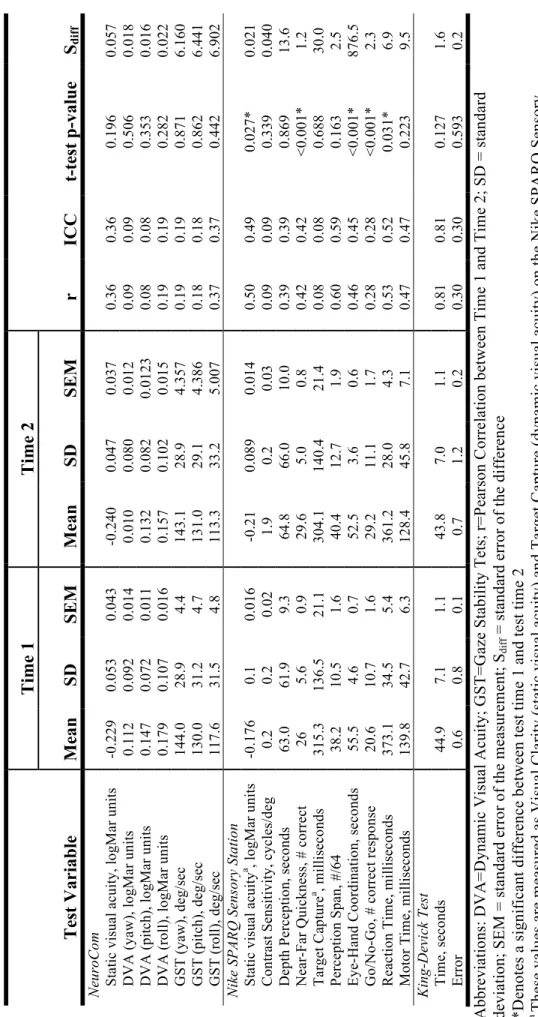
General descriptive statistics were computed for each clinical outcome measure
on the Gaze Stability Test, the Dynamic Visual Acuity Test, the King-Devick Test, and
the Nike SPARQ Sensory System in our sample of healthy participants. Additionally,
interclass correlation coefficients were computed using Pearson correlations to assess the
test-retest reliability of our measures. Since this technique is unable to identify systematic
differences, we also employed paired-samples t-tests comparing both test sessions for
each outcome measure. We employed this approach since the Pearson correlation
coefficient is a required step in determining the Reliable Change Index (RCI) for our
measures.
The RCI outcomes were computed using an identical and systematic approach
employed for each outcome measure. First, the correlation (r) between the two test
sessions was determined. Descriptive statistics included standard deviations (SD) for
34
compute the standard error of the measurements (SEM) for each test session using the
following formula:
!"#! !" ! ! !
Next, we computed the standard error of the difference (SEdiff):
!"!"## ! !"#!!!"#!
Lastly, the SEdiff was multiplied by the z scores associated with 80% (z = 1.282), 90% (z
= 1.684), and 95% (z = 1.96) confidence intervals to compute the RCI values for each of
the measures as follows:98,99
RCI = SEdiff x z score for associated confidence level
Data were analyzed using SPSS 19 (SPSS Inc.; Chicago, IL). An a priori ! level of
35
Table 3.1. Counterbalanced test order for all assessments. Testing
Order Option
First Assessment Second Assessment
Third Assessment
Fourth Assessment 1 Dynamic Visual
Acuity Test
Gaze Stability Test
Nike SPARQ Assessment
King-Devick Test* 2 Gaze Stability
Test
Dynamic Visual Acuity Test
Nike SPARQ Assessment
King-Devick Test*
3 Nike SPARQ
Assessment
King-Devick Test*
Gaze Stability Test
Dynamic Visual Acuity Test
4 Nike SPARQ
36 Table 3.2. Data analysis summary table.
Research Question
Description Data Source Comparison Method
1 Are athletes’ visual test performances reliable on Nike SPARQ, King-Devick Test, Neurocom gaze stability test and Neurocom dynamic visual acuity test?
IV: Time Test Session 1 Test Session 2 (14 days)
DV: SPARQ measures (10) DVT (3) GST (3) King-Devick (2)
Visual performance on SPARQ, GST, DVAT from testing session 1 to testing session 2
Paired-samples t-test Pearson correlations ICC3,1 values
2 What are the reliable
change indices between testing session 1 and testing session 2 on the measures?
IV: Time Test Session 1 Test Session 2 (14 days)
DV: SPARQ measures (10) DVT (3) GST (3) King-Devick (2)
Visual performance on SPARQ, GST, DVAT from testing session 1 to testing session 2
Sdiff RCI 80%, 90%, 95%
3 (Exploratory)
Is there a significant difference in scores on visual performance measures between individuals with a concussion and
matched controls across time?
IV: Group Healthy vs. Concussed Time
Asmptomatic/Full Participation
DV: SPARQ measures (10) DVT (3) GST (3) King-Devick (2)
Difference of scores between groups on SPARQ domains and between the two testing sessions.
2x2 mixed model repeated measures ANOVAs, with Tukey post hoc when the omnibus test for
38
39
40
41
CHAPTER IV
MANUSCRIPT
Introduction
Concussions are a public health concern in today’s society, with as many as 3.8
million reported each year in the United States from sports and recreational activity
alone.2 Many other suspected head injuries may go unreported.3 The frequency of head
injuries has seen a steady increase over the past 5-10 years,32,100 likely due to an
escalation in education efforts and improvements in concussion assessment tools and
treatments. Concussion, a form of mild traumatic brain injury, has been defined as a
complex pathophysiologic process affecting the brain and its functioning following an
injury to the head.4 Concussions result in debilitating symptoms including cognitive,
balance, and visual deficits that can last anywhere from 24 hours to several months after
the initial injury.4 While subjective assessments can expose acute symptoms such as
mental status deterioration, dizziness, headache, nausea, confusion, tinnitus, and blurry
vision,4 objective assessments are typically employed to expose cognitive, balance, and
vision disturbances.
The commonly used symptom, neurocognitive and balance assessments are well
established, reliable and sensitive for specific deficits,7-9 but do not address aspects such
as dynamic or static vision or gaze stability, which may be associated with concussion.
Recent literature suggests incorporating visual evaluations may lead to a more complete
42
plays for all athletes,12,13 and the sport-specific visual skills required to perform tasks
such as hitting a baseball, catching a football, or spotting a 4-inch balance beam. To
accomplish these tasks, athletes interpret visual information and pattern motor responses
dependent on the stimuli. There is proportional relationship between an athlete’s
perceptual ability and motor response.14 Successful athletes generally interpret visual
information better and, therefore, have sharper visual acuity, accuracy and
spatio-temporal awareness.14 If an athlete is not receiving visual feedback fast enough to assess
the situation and act on the information, performance will decline.14 Impaired visual
performance may inhibit the ability to anticipate potentially injurious mechanisms during
participation and, thus, increase injury risk.101,102 To account for the changing nature of
athletic performance, static visual acuity is an insufficient measure of visual and sensory
performance. We posit that dynamic visual acuity measures may be more appropriate to
expose deficits that could potentially affect an athlete’s functional visual ability during
sport participation. Given the potential interrelationship between performance and injury
prevention, we believe that visual performance measures may have a doubly important
role in the concussion management paradigm that has too long been limited to symptoms,
cognition, and balance testing.15 Such measures should include functional visual skills
such as depth perception, dynamic visual acuity, contrast sensitivity, and
vergence-accomodation12 If there are deficits in the visual system due to concussion, employing
objective measures of an athlete’s vision as part of the baseline-testing program will yield
helpful clinical information for the athlete’s post-injury care. Unfortunately, very little is
known about the potential clinical utility of vision and sensory performance measures in
43
Therefore, the purpose of this study was to determine the test-retest reliability and
reliable change indices for measures of visual and sensory performance in healthy college
participants. Reliable change methodology has been described in detail,103-106 and revised
over the years to encompass both the reliability and the practice effects of an
instrument.103 The RCI incorporates the reliability and variance of a measure to produce a
value that represents a clinically meaningful change, which can be defined as change that
occurred beyond the scope of measurement error or variability. Reliable change
methodologies have been used for various concussion assessment measures including
cognition and balance.19,99,107,108
Methods
Participants
We studied a convenience sample of 44 active, healthy college students (29 male
and 15 female; age = 19.90 ± 0.96 years). Participants were excluded from this study if
they had known neurocognitive deficits or disorders, known psychological conditions,
color blindness, history of dizziness, imbalance or abnormal vestibular function, or
musculoskeletal abnormalities to the head, neck, shoulder, or back that would disrupt
normal range of motion. All participants read and signed consent forms approved by our
institution’s ethics review board.
Instrumentation
Dynamic Visual Acuity Test
The Dynamic Visual Acuity Test (DVAT) was performed using the InVision
44
and a 15-in flat panel high contrast liquid-crystal display monitor were used to display an
optotype (the letter “E”). The DVAT began with assessment of the participant’s static
visual acuity by having them identify the orientation of the optotype “E” on a computer
screen located 8 feet in front of them seen in Figure 3.1. Responses were recorded on the
computer by the clinician after the subject indicated whether the “E” was positioned to
the right, left, upwards, or downwards. The size of the “E” was reduced if the participant
correctly identified 3 out of 5 “E”s of a given size. Static visual acuity was defined based
on the smallest “E” correctly identified, and measured in logMAR units.
The participant’s dynamic visual acuity (DVA) was measured in three different
axes: the “yaw” (vertical axis rotation), “pitch” (medial-lateral rotation), and “roll”
(antero-posterior axis rotation). The participant wore a head harness (InterSense Inertia
Cube, Engineering Systems Technology, Kaiserslautern, Germany) with a sensor that
integrated the 3 axes the head was moving about to determine rotational velocity
(deg/sec) of the head. Participants were required to generate rotational head movements
at least 20 degrees from midline in each direction while still being able to correctly
analyze an optotype “E” of varying sizes. Each test allowed for a practice trial. The size
of the smallest optotype identified correctly while rotating the head faster than the
minimum velocity was recorded for results of the DVAT. The DVAT scores were
converted to visual loss by subtracting dynamic visual acuity from baseline static visual
acuity for each eye, averaging the two means, and reporting the outcome in logMAR
45
Gaze Stability Test
The InVision system (NeuroCom International; Clackamas, OR) used for the
DVAT was also used for the Gaze Stability Test. The GST uses the same static visual
acuity measured with the DVAT. The GST measures head movement velocities that the
participant achieves while maintaining their static visual acuity. Participants were
required to maintain gaze on the center of a computer screen and correctly identify the
orientation of the optotype “E” while generating repetitive head movements at varying
velocities. Participants used the same headband, seen in Figure 3.1, with a 3-axis
integrating gyro to determine velocity for each trial. The participants were instructed to
perform a smooth sinusoidal head shake movement until the display screen was visible.
Two feedback bars gave the participant information on the velocity and amplitude of the
head and disappeared when the participant’s head velocity exceeded the required
minimum threshold for a trial. The optotype was displayed on the monitor until the
participant’s head velocity fell below the requirement for that trial or the participant
reached a maximum time. If three out of five “E”s were identified correctly, the
minimum head velocity that the participant must attain was increased. This was repeated
until the participant was unable to correctly identify three out of five “E”s, at which point
the velocity was reduced. If the participant was unable to achieve the minimum required
head velocity within 8 seconds from the start of head movement or unable to maintain
velocity for the required duration, or if the participant achieved and maintained the
required velocity but incorrectly identified the orientation, the trial was recorded as a