CONTRIBUTIONS OF HIPPOCAMPCAL AREA CA2 TO HIPPOCAMPAL OSCILLATORY NETWORKS
Logan Y. Brown
A dissertation submitted to the faculty at the University of North Carolina at Chapel Hill in partial fulfillment of the requirements for the degree of Doctor of Philosophy in the Neuroscience Curriculum.
Chapel Hill 2018
ABSTRACT
Logan Y. Brown: Contributions of Hippocampal Area CA2 to Hippocampal Oscillatory Networks (Under the direction of Serena M. Dudek)
Complex cognitive abilities, such as memory, require synchronized neural activity across large populations of cells. The hippocampus is a region of the brain required for the formation of long term episodic memories, which is our memory for autobiographical information. The hippocampus itself consists of four sub-regions, the dentate gyrus (DG), CA1, CA2, and CA3, which can be viewed as functionally specialized processing hubs that uniquely contribute to memory formation based on their distinct molecular, synaptic, and anatomical properties. Only together; however, does the collective activity of all four sub-regions provide the neurobiological underpinnings necessary for a functional memory system.
One mechanism for the coordination of neural networks is synchronization through oscillations. Neuronal oscillations reflect waves of synchronous action potentials and their presence in the
hippocampus is strongly associated with episodic learning and memory. Although much progress been made towards understanding how different frequencies of activity are generated and how they support hippocampal-based memory, relatively little is known about the role of CA2 in organizing oscillations. In this dissertation work, I use combinatorial electrophysiological and chemogenetic approaches to
genetically target and manipulate CA2 principal cells to investigate their role in coordinating hippocampal oscillatory networks in awake, behaving mice.
In Chapter 2, I use Designer Receptors Exclusively Activated by Designer Drugs (DREADDs) to manipulate the endogenous G protein-coupled receptor (GPCR) signaling pathways in CA2 while animals explore a novel spatial environment. These experiments revealed that activation or inhibition of CA2 pyramidal cells through the endogenous Gq- and Gi-coupled pathways, respectively, is sufficient to bi-directionally modulate synchronized hippocampal activity in the slow gamma and beta frequency ranges.
ACKNOWLEDGEMENTS
First, I would like to thank the University of North Carolina at Chapel Hill and the Curriculum in Neuroscience for providing me with an outstanding environment for my graduate studies. Many faculty were involved with, and contributed to, my progress as a graduate student and I am eternally grateful for the support provided.
I would especially like to thank my academic advisor, Dr. Serena Dudek, for all her guidance and support throughout my dissertation work. It has been an incredible journey and I am deeply grateful for her mentorship. I would also like to thank all the current, and former, members of the Dudek lab for their support, training, and wealth of expertise. I am especially grateful for the mentorship and oversight of Dr. Georgia Alexander, whom without, none of the work presented here would have been possible. Beyond the Dudek Lab, I would also like to thank Dr.’s Flavio Frohlich and Paul Manis who have both served as invaluable resources throughout my graduate studies. I am both honored and humbled to have had the privilege to work with so many brilliant, talented, and dedicated scientists.
The ways our earliest mentors and teachers shape our lives cannot be overstated and I have been truly privileged to have had several exceptional mentors throughout my academic career. Dr.’s William McDaniel and Larry Lynch were both pivotal in my trajectory towards a doctoral degree and, more importantly, for inspiring in me a life-long curiosity and love of philosophy and science.
TABLE OF CONTENTS
LIST OF FIGURES ... ix
LIST OF ABBREVIATIONS ... xi
CHAPTER 1. INTRODUCTION ... 1
1.1 The Hippocampus as an Episodic Memory Platform ... 1
1.2 Anatomical Characterization of the Rodent Hippocampus ... 2
1.3 CA2- A Distinct Hippocampal Region ... 5
1.4 The Local Field Potential and Synchronization Through Oscillations ... 7
1.5 Hippocampal Oscillations and Memory... 8
1.6 Chemogenetic Tools for Manipulation of Discrete Neural Circuits ... 13
1.7 Figures ... 16
CHAPTER 2. CHEMOGENETIC MODULATION OF Gq- AND Gi-COUPLED GPCR SIGNALING IN CA2 BI-DIRECTIONALLY MODULATES HIPPOCAMPAL SLOW GAMMA AND BETA OSCILLATIONS DURING RUNNING ... 17
2.1 Historical Context ... 17
2.2 Methods ... 18
2.3 Results ... 25
2.3.1 Amigo2-icreERT2+ mice enable genetic access to CA2 pyramidal cells ... 25
2.3.2 Acute chemogenetic activation of CA2 pyramidal cells induces hippocampal slow gamma oscillations and inhibits hippocampal beta oscillations ... 26
2.3.3 Acute chemogenetic inhibition of CA2 pyramidal cells inhibits hippocampal slow gamma oscillations and induces hippocampal beta oscillations ... 28
2.4 Discussion ... 29
2.5 Figures ... 32
3.1 Overview of Findings... 41
3.2 Methods ... 42
3.3 Results ... 47
3.3.1 Acute chemogenetic inhibition of CA2 pyramidal cells decreases hippocampal slow and fast gamma power in the pyramidal layer of CA1 during investigation of novel stimuli ... 47
3.3.2 Acute chemogenetic inhibition of CA2 pyramidal cells decreases hippocampal slow and fast gamma power in stratum oriens during investigation of novel stimuli ... 48
3.3.3 Acute chemogenetic inhibition of CA2 pyramidal cells decreases hippocampal fast gamma power in stratum radiatum during investigation of novel stimuli ... 50
3.3.4 Acute chemogenetic inhibition of CA2 pyramidal cells does not affect oscillatory power in stratum lacunosum-moleculare during investigation of novel stimuli ... 51
3.3.5 Hippocampal oscillations in the pyramidal cell layer of CA1 do not significantly differ during investigation of a novel animal relative to a novel object ... 52
3.3.6 Hippocampal oscillations in CA1 stratum oriens do not significantly differ during investigation of a novel animal relative to a novel object ... 53
3.3.7 Hippocampal oscillations in CA1 stratum radiatum do not significantly differ during investigation of a novel animal relative to a novel object ... 54
3.3.8 Hippocampal oscillations in CA1 stratum lacunosum-moleculare do not significantly differ during investigation of a novel animal relative to a novel object ... 55
3.3.9 Acute chemogenetic inhibition of CA2 pyramidal cells reduces average distance traveled ... 56
3.3.10 Acute chemogenetic inhibition of CA2 pyramidal cells does not affect time spent investigating novel stimuli ... 57
3.4 Discussion ... 57
3.5 Figures ... 61
CHAPTER 4. DISCUSSION ... 81
4.1 CA2 as a Generator of Hippocampal Gamma and Beta Oscillations for Spatial Cognition ... 81
LIST OF FIGURES
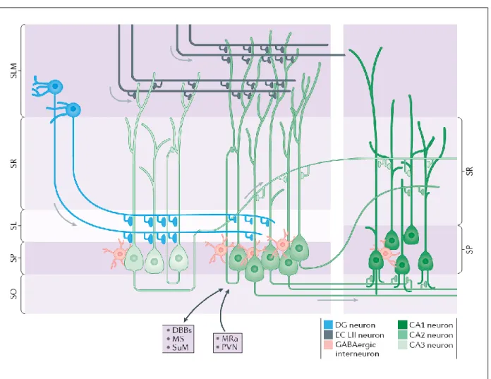
Figure 1.1: Anatomical Connectivity of Hippocampal Area CA2 ... 16 Figure 2.1: Expression of the Cre indicator, tdTomato, in Amigo2-icreERT2+;
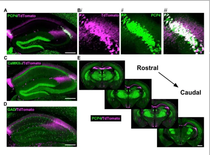
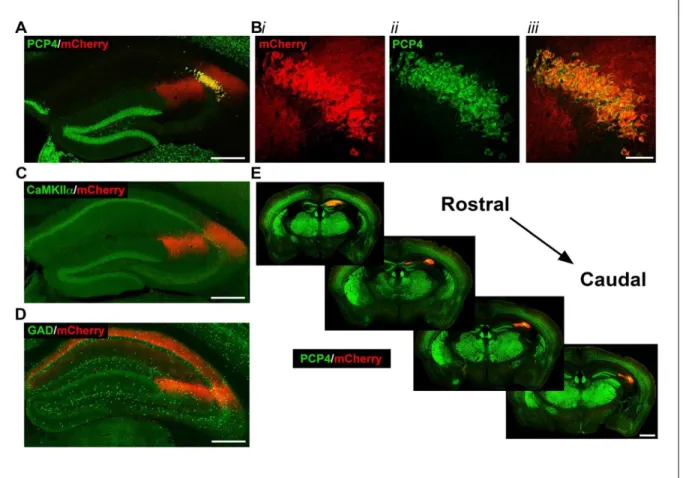
ROSA-tdTomato+ mice ... 32 Figure 2.2: Selective expression of hM3Dq-mCherry-tagged DREADD receptors in CA2
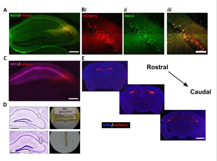
pyramidal cells of Amigo2-icreERT2+ mice. ... 33 Figure 2.3: Selective expression of hM4Di-mCherry-tagged DREADD receptors in CA2
pyramidal cells of Amigo2-icreERT2+ mice ... 34 Figure 2.4: CNO dose-dependently increases slow gamma power in the pyramidal cell
layer of the hippocampus in Amigo2-icreERT2+ mice infused with hM3Dq-mCherry. ... 36 Figure 2.5: Activation of CA2 pyramidal cells with CNO in hM3Dq-infused Amigo2
-icreERT2+ mice dose-dependently increases slow gamma power in the hippocampus ... 38 Figure 2.6: Inhibition of CA2 pyramidal cells with CNO in hM4Di-infused Amigo2
-icreERT2+ mice decreases slow gamma power in the hippocampus ... 40 Figure 3.1: Experimental timeline, recording paradigm, electrodes, and recording location ... 62 Figure 3.2: Inhibition of CA2 pyramidal cells with CNO in hM4Di-infused Amigo2
-icreERT2+ mice reduces slow and fast gamma power in the pyramidal cell layer of CA1
during investigation of novel stimuli ... 64 Figure 3.3: Inhibition of CA2 pyramidal cells with CNO in hM4Di-infused Amigo2
-icreERT2+ mice reduces slow and fast gamma power in stratum oriens of CA1 during
investigation of novel stimuli ... 66 Figure 3.4: Inhibition of CA2 pyramidal cells with CNO in hM4Di-infused Amigo2
-icreERT2+ mice reduces fast gamma power in stratum radiatum of CA1 during
investigation of novel stimuli ... 68 Figure 3.5: Inhibition of CA2 pyramidal cells with CNO in hM4Di-infused
Amigo2-icreERT2+ mice does not affect oscillations in stratum lacunosum-moleculare of CA1
during investigation of novel stimuli ... 70 Figure 3.6: Hippocampal oscillations in the pyramidal cell layer of CA1 do not significantly
differ during investigation of a novel animal relative to a novel object following treatment
with vehicle or CNO in hM4Di-infused Amigo2-icreERT2+ mice ... 72 Figure 3.7: Hippocampal oscillations in stratum oriens of CA1 do not significantly differ
during investigation of a novel animal relative to a novel object following treatment with
vehicle or CNO in hM4Di-infused Amigo2-icreERT2+ mice ... 74 Figure 3.8: Hippocampal oscillations in stratum radiatum of CA1 do not significantly differ
during investigation of a novel animal relative to a novel object following treatment with
Figure 3.9: Hippocampal oscillations in stratum lacunosum-moleculare of CA1 do not significantly differ during investigation of a novel animal relative to a novel object
following treatment with vehicle or CNO in hM4Di-infused Amigo2-icreERT2+ mice ... 78 Figure 3.10: Acute inhibition of CA2 pyramidal cells with CNO in hM4Di-infused Amigo2
LIST OF ABBREVIATIONS AAV Adeno-Associated Virus
ANOVA Analysis of Variance
AMIGO2 Adhesion Molecule with Ig-like Domain 2 AVPR1B Vasopressin 1B Receptor
BAC Bacterial Artificial Chromosome
BGH-F Bovine Growth Hormone-Forward primer
BP Base Pairs
CA1 Cornu Ammonis 1
CA2 Cornu Ammonis 2
CA3 Cornu Ammonis 3
CNO Clozapine-N-Oxide
DG Dentate Gyrus
DIO Doubled-floxed Inverted Open read
DREADD Designer Receptors Exclusively Activated by Designer Drugs EGFP Enhanced Green Fluorescent Protein
ER Estrogen Receptor
GABA Gamma Amino Butyric Acid GAD Glutamate Decarboxylase
GIRK G-protein Inward Rectifying potassium channel GPCR G-protein Coupled Receptor
HM3Dq Human Muscarinic 3 DREADD-Gq receptor HM4Di Human Muscarinic 4 DREADD-Gi receptor HSYN Human-Synapsin
ICREERT2 Inducible Cre-Estrogen Receptor- Tamoxifen-inducible 2 IP Intra-Peritoneal
LTP Long Term Potentiation PBS Phosphate Buffer Solution PCR Polymerase Chain Reaction
PLC Phospholipase C
PSD Power Spectral Density PSP Post-Synaptic Potential
CHAPTER 1. INTRODUCTION
1.1 The Hippocampus as an Episodic Memory Platform
In humans, the hippocampus serves as one part of an extended memory system residing in the medial temporal lobe of the brain. This distributed system encompasses several distinct regions including the hippocampus proper, the subiculum, and the entorhinal, perirhinal, and parahippocampal cortices (Anderson, 2007). The medial temporal lobe system is required for the encoding and consolidation of long term memories that take the form of facts and events, otherwise known as explicit or declarative memory (Squire, 1992). Explicit memory is one of two types of long-term memory in humans and refers to the conscious, intentional recollection of information, previous experiences, and concepts (Squire, 2004). Explicit memory can be further divided into two categories: episodic memory, which is our records for personal experiences, and semantic memory, which is our memory for factual information.
research with patient H.M., and other clinical cases like his, have cemented the idea that the
hippocampus is critical for episodic memory. At the same time, scientists have been utilizing molecular and electrophysiological tools in animal models to probe the biological mechanisms underlying episodic memory.
Much evidence for the spatial component of episodic memory comes from the discovery of place cells by O’Keefe and Dostrovsky in 1971 when they observed neurons in the rat hippocampus that selectively increased firing within a well-defined region of the animal’s environment (O'Keefe &
Dostrovsky, 1971). Almost fifty years later, the precise firing and spatial tuning properties of place cells have been extensively explored in a myriad of contexts and environments (Alexander et al., 2016; Lu, Igarashi, Witter, Moser, & Moser, 2015; Muller & Kubie, 1987; O'Keefe, Burgess, Donnett, Jeffery, & Maguire, 1998; D. M. Smith & Mizumori, 2006), leading to consensus that these cells, along with a few other classes of spatially selective cells, are responsible for generating the brain’s spatial representations of its environment. Between human clinical studies and work in animal models, these two disparate lines of inquiry into the nature of hippocampal function have led to considerable debate over whether the primary function of the rodent hippocampus is spatial cognition or a more general episodic memory platform, as is the case in humans (Eichenbaum, Dudchenko, Wood, Shapiro, & Tanila, 1999; O'Keefe, 1999; Redish, 2001). Much recent work has focused on trying to bridge the disconnect between these two views, and consensus for the rodent hippocampus as a more general memory system incorporating both spatial and other episodic elements has grown in recent years (Eichenbaum & Cohen, 2014). Here, we present evidence in support of the view of the rodent hippocampus as a more general episodic memory platform capable of storing information about a variety of environmental stimuli, rather than one exclusively reserved for spatial representations.
1.2 Anatomical Characterization of the Rodent Hippocampus
The organization of the hippocampus is highly conserved across mammalian species (Strange, Witter, Lein, & Moser, 2014) and much of our knowledge about the organization and connectivity of circuits within the hippocampus comes from the pioneering work of early twentieth century
fields one through three (CA1, CA2, and CA3) (Lorente de No, 1934) (Figure 1.1). Although there exist many hippocampal circuits, Ramon y Cajal established early on that the fields of the hippocampal formation are linked by a sequence of unique, and largely unidirectional, connections when he first described one important relay of synaptic transmission through the hippocampus, the tri-synaptic loop (Anderson, 2007). Later work by Andersen and colleagues provided an important extension of these early studies by proposing that the hippocampus is organized in parallel “lamellae,” or small strips (Andersen, Bliss, & Skrede, 1971). The lamellar hypothesis of hippocampal function posited that excitatory activity travels from the entorhinal cortex, and through the hippocampus, within a series of parallel hippocampal “slices” or “lamellae”. In this way, the temporal lobe interactions between the entorhinal cortex and the hippocampus were thought to be organized topographically, with different lamellae operating
independently. Although later work by Amaral and colleagues ultimately found the lamellae hypothesis to be an incomplete description of hippocampal connectivity based on the observation that excitatory collaterals originating from CA3 dispersed over a much wider region than originally proposed (Amaral & Witter, 1989; Ishizuka, Weber, & Amaral, 1990), the lamellar hypothesis of the hippocampus’s anatomical organization has had tremendous influence on the conceptualization of information processing in the hippocampus, and research today is still aimed at understanding how layer-specific interactions among subfields contribute to memory formation and retrieval.
diverse array of interneurons that primarily target local pyramidal cells, where they exert inhibition that contributes to the timing and organization of hippocampal activity (Klausberger & Somogyi, 2008).
Three major fiber systems are associated with input into and out of the hippocampus. The first is the angular bundle, which primarily carries fibers between the entorhinal cortex (EC) and the hippocampal fields (Anderson, 2007). The EC is a specialized region of cortex that serves as the primary interface between the hippocampus and the associational cortices of the neocortex (van Groen, Miettinen, & Kadish, 2003). The second is the fimbria-fornix pathway, through which the hippocampus is connected with the basal forebrain, hypothalamic, and brain stem regions. The third compromises the dorsal and ventral commissures, which connect the two hippocampi in each cerebral hemisphere. Here we will focus on a subset of pathways within the angular bundle, as this is main route taken by entorhinal cortex fibers projecting to the hippocampus, a key structure for the organization of hippocampal oscillations.
1.3 CA2- A Distinct Hippocampal Region
Episodic information is encoded and consolidated in the hippocampus through several parallel circuits and hippocampal area CA2 has become increasingly appreciated as a distinct region based on several unique synaptic (Chevaleyre & Siegelbaum, 2010; Zhao, Choi, Obrietan, & Dudek, 2007), molecular (Lein, Callaway, Albright, & Gage, 2005), and anatomical (Kohara et al., 2014) properties that suggest specific roles for CA2 hippocampal function. Previously believed to serve as a mere transition region between the CA3 and CA1 subfields, CA2 has now been shown to participate in several unique hippocampal circuits, receiving direct excitatory input from ECII and CA3 (Kohara et al., 2014), and from several subcortical nuclei, including the supramammillary and paraventricular nuclei (PVN) of the
hypothalamus, as well as the medial septum and diagonal band of broca (Cui, Gerfen, & Young, 2013). In turn, CA2 axons primarily target the basal dendrites of ipsilateral CA1 located in stratum oriens, where its terminals account for approximately 20% of synapses, but also send a minor projection to CA1 dendrites in stratum radiatum, as well as recursive input onto itself (Lorente de No, 1934; Tamamaki, Abe, & Nojyo, 1988), and back-projections to CA3 (Shinohara et al., 2012; Tamamaki et al., 1988).
The hippocampus supports a cognitive map of space and in the rodent, the hippocampus is strongly associated with the spatial aspects of episodic memory due in large part to the presence of place cells, which are hippocampal pyramidal cells that selectively increase their firing rate within a well-defined region of the animal’s environment. Only a subset of the place cells fire in any given environment;
however, and when the animal is moved to a different environment, place cells undergo remapping (Muller & Kubie, 1987), enabling the hippocampus to form distinguishable representations of large numbers of experiences in a large number of environments, even when events take place at the same spatial coordinates (Lu et al., 2015). Place cells also encode and store non-spatial forms of information (Aronov, Nevers, & Tank, 2017; Eichenbaum, 2018); however, and although all hippocampal subfields exhibit place cells, CA2 place cells appear to be the least spatially tuned pyramidal cells (Lu et al., 2015; Mankin, Diehl, Sparks, Leutgeb, & Leutgeb, 2015), suggesting a shift to other cognitive processes. Interestingly, CA2 place fields remap upon exposure to novel or familiar conspecific animals while CA1 place fields do not (Alexander et al., 2016), and several lines of evidence now indicate that CA2 is
may be mediated in part by vasopressinergic signaling, a peptide neurotransmitter derived from PVN neurons of the hypothalamus. The idea that vasopressin may be involved in the formation of memories containing socially relevant information is consistent with the fact that this neurotransmitter is involved in the regulation of social behaviors in many mammalian species (Insel, 2010).
In the rodent hippocampus, CA2 pyramidal cells exhibit a selective enrichment of mRNA for the vasopressin 1b receptor subtype (Avpr1b) (Young, Li, Wersinger, & Palkovits, 2006). Synaptic plasticity at the CA3 CA2 Schaffer collateral synapses is also uniquely regulated relative to the CA3 CA1 Schaffer collateral synapses, in that the CA2 synapses do not readily undergo high frequency stimulation-induced potentiation (Pagani et al., 2015). The ability of neurons to alter the strength of their synaptic connections has served as a candidate mechanism for a molecular correlate of memory storage (Bliss & Collingridge, 1993) since the discovery of long-term potentiation (LTP) in the early 1970’s (Bliss & Lomo, 1973), which is one long lasting form of synaptic plasticity. Synaptic potentiation can be revealed at the CA3CA2 synapses; however, with the use of certain neuromodulators, including an agonist for the Avpr1b subtype (Pagani et al., 2015). Of note, the Avpr1b is also coupled to the endogenous Gq- signaling pathway which we selectively target and manipulate in CA2 pyramidal cells in subsequently presented experiments, suggesting a link between vasopressin and our results demonstrating CA2’s role in coordinating synchronous hippocampal activity. In addition to the molecular and synaptic evidence indicating a role for CA2 in social memory, several studies have genetically targeted and manipulated Avpr1b expressing cells in CA2 and measured changes in behavior. Genetic deletion of the Avpr1b impairs a variety of social behaviors while leaving other hippocampal dependent behaviors intact (Caldwell, Wersinger, & Young, 2008; Stevenson & Caldwell, 2012), and optogenetic stimulation of the vasopressin-containing axon fibers in CA2 can extend the duration of social memory (A. S. Smith, Williams Avram, Cymerblit-Sabba, Song, & Young, 2016). Additionally, Hitti and Siegelbaum showed that CA2 activity is required for intact social recognition memory by using a tetanus toxin light chain construct to permanently silence CA2 output which abolished memory for conspecifics while leaving other
hippocampal dependent memories intact (Hitti & Siegelbaum, 2014).
demonstrated a role for CA2 in social memory via manipulation of the vasopressinergic system, in line with more traditional views of rodent hippocampal function, its participation in spatial cognition should also not be overlooked. Although much work has been done to identify the unique synaptic and molecular features that distinguish CA2, much less is known about the functional properties of CA2 neurons in vivo and how they might coordinate their activity with the rest of the hippocampus to integrate spatial and social cues into memory.
1.4 The Local Field Potential and Synchronization Through Oscillations
A microelectrode placed in neuronal tissue measures changes in voltage that originate from ionic currents flowing into and out of nearby cells. These transmembrane currents are driven by an imbalance of charge across neuronal membranes and, when measured in the extracellular space, are referred to as local field potentials (LFPs). Although the origin of the LFP can be complex, physically, it reflects the spatial summation of all electrical fields within recording range of the electrode. In neural tissue, this typically represents a mixture of fast activity generated by action potentials and slower activity generated by synaptic currents. After being low-pass filtered to remove the fast activity of action potentials, the LFP is thought to primarily reflect synaptic currents (Buzsaki, Anastassiou, & Koch, 2012), which are the same currents responsible for generating the post synaptic potentials. In signal processing, algorithms can be used to estimate the strength of different frequency components of a time-domain signal, referred to as the power spectrum. The LFP is such a time-domain signal that can be analyzed using spectral estimation methods because voltage fluctuations detected in the LFP frequently exhibit a periodic, or oscillatory, nature. These brain rhythms, or oscillations, are waves of neuronal activity that represent the synchronous activity of large groups of neurons, a process thought to be critical for cognitive operations which require coordinated activity of anatomically distributed cells, such as hippocampal based learning (Colgin & Moser, 2010; Fell & Axmacher, 2011).
oscillations (Engel, Fries, & Singer, 2001; Jutras & Buffalo, 2010; Womelsdorf et al., 2007). At the single cell level, if a neuron receives input from multiple regions, each of which oscillate at distinct frequencies and/or phases relative to one another, the receiving neuron may tune into one of those input streams if excitably is modulated at the proper phase relative to the preferred input (Fries, 2005, 2015). In this way, synchronization of oscillatory phases is thought to influence the relative timing of action potentials between regions and, consequently, shape the flow of information through neural circuits. Accordingly, the power spectrum of the LFP recorded from neural tissue serves as one measure of neuronal
synchrony in that network, and because currents that are synchronized in time sum to produce larger amplitude deflections, whereas asynchronous currents cancel out, increases in LFP power are thought to represent increases in synchronized input into the observed region.
1.5 Hippocampal Oscillations and Memory
In the hippocampus, the presence of oscillations is strongly correlated with episodic learning and memory (Colgin, 2016). Although a definitive explanation of how the hippocampus encodes and stores different types of memories is lacking, it is known that hippocampal based learning requires the
association of information through several corticohippocampal and intrahippocampal pathways, including both long-range and local microcircuits (Basu & Siegelbaum, 2015). In the rodent hippocampus, three frequencies of activity dominate the LFP power spectrum during awake behavior: theta (~4-12 Hz), gamma (~25-100 Hz), and ripples (~100-300 Hz) (Buzsaki et al., 2003; Buzsaki, Leung, & Vanderwolf, 1983).
The theta frequency (4-12 Hz) is a large amplitude, highly regular oscillation that is easily detectable in all areas of the hippocampus whenever an animal engages in translational motion
(Vanderwolf, 1969). In addition to a prominent increase in power during exploration, the theta oscillation is strongly linked to spatial processing due to its role in coordinating place cell firing, which is systematically related to the phase of the theta cycle (O'Keefe & Recce, 1993). As an animal moves through a place cell’s receptive field, the place cell alters its action potential timing to occur at successively earlier points in each cycle of theta, a phenomenon known as theta phase precession (Skaggs, McNaughton, Wilson, & Barnes, 1996). In addition to its role in organizing spatial representations, the hippocampal theta
information. Hippocampal theta oscillations transiently lock to the rhythmic neuronal activity that
accompanies vibrissa-based sensation in rodents during the sensory-gathering epoch of a discrimination task (Kleinfeld, Deschenes, & Ulanovsky, 2016). To ensure that observed shifts in theta power are not merely a reflection of increased whisking requires controlling for spikes in theta power that co-occur with individual whisking motions. In our experiments, because we did not have a camera capable of tracking this behavior, we excluded the theta band from our experimental analysis in chapter 3 when subjects were allowed to investigate novel stimuli. Similar to the relationship between place cell firing and the theta cycle, the gamma oscillation also tends to occur during specific phases of the theta cycle (Leung, 1998; Scheffer-Teixeira et al., 2012; Schomburg et al., 2014; Shirvalkar, Rapp, & Shapiro, 2010), although there is currently disagreement about what that phase is (Colgin, 2015).
In contrast to the large amplitude, highly-regular activity of the theta band, the gamma frequency is a small-amplitude, transient oscillation that occurs in bursts throughout behavior (Colgin & Moser, 2010). Although historically thought to be a single, continuous frequency band spanning the entire ~25-100 Hz range, there is now evidence indicating that the gamma oscillation may correspond to two functionally distinct bands, including a slow (~25-55 Hz) and fast (~55-100 Hz) band (Colgin, 2015; Lasztoczi & Klausberger, 2014). In physiological situations, for a local network to become entrained to the gamma cycle, a network of interconnected, peri-somatically targeting, fast-spiking interneurons is required (Bartos, Vida, & Jonas, 2007; Buzsaki & Wang, 2012). Basket cells are a well-defined type of soma-inhibiting, GABA-releasing interneuron, so named because of their formation of peri-somatic ‘baskets’ around target cell somata (Bartos et al., 2007). In the hippocampus, a large subset of basket cells exhibit a fast-spiking action potential and express the calcium-binding protein parvalbumin and, although the function of parvalbumin is not fully understood, its expression represents a reliable marker for interneuron identification (Bartos et al., 2007). When such a network of interneurons is innervated, these cells
synchronize their activity to generate the gamma rhythm. Although in vitro work and computational models have demonstrated that networks composed exclusively of interneurons (I-I networks) are
distal upstream region and does not have to be rhythmic itself (Bartos et al., 2002). When such a network of interneurons spike, synchronized IPSPs feedback onto pyramidal cell soma at the gamma frequency creating alternating periods of high & low inhibition which are thought to have several biophysical consequences for communication among participating neurons. Simultaneous paired intra- & extra-cellular recordings in vitro have determined that these IPSPs are the main electrophysiological event corresponding to the presence of hippocampal gamma oscillations in the LFP when recording in the pyramidal cell layer from actively spiking cells (Buzsaki & Wang, 2012). For this reason, gamma detected in the pyramidal cell layer serves as an electrophysiological signature for local network entrainment. In contrast, gamma oscillations recorded in synaptic layers are thought to represent excitatory input from distal upstream regions, which may or may not ultimately impact the timing of activity in the downstream network.
One prominent theory describing how gamma oscillations facilitate inter-regional communication is referred to as the communication through coherence (CTC) theory (Fries, 2005). When two separate networks that are each locally entrained to the gamma cycle interact, from the sending network’s
perspective, action potentials that are coordinated in time will result in larger EPSPs in the target dendritic domain of the receiving network, increasing the probability of depolarizing the receiving cells to threshold and generating an action potential. From the receiving network’s perspective, each cell’s membrane potential repeatedly moves closer to and away from action potential threshold, providing discrete windows in which appropriately timed inputs have an increased probability of inducing firing. If input is not
appropriately timed; however, arriving during heightened inhibition, the input may be ignored. Cells will be most susceptible to excitatory drive that is timed to arrive during the fading inhibition part of the cycle. Accordingly, if the sending network is to exert the maximum impact possible on the receiving network, it should deliver coordinated bursts of spikes at every falling inhibition phase of the gamma cycle. This can occur only if the gamma phases in each region are synchronized. In this way, the efficacy of
Rhythmic synchronization in the gamma frequency range is highly structured across brain areas, but the precise patterns of synchronization among layers and corresponding projections changes
dynamically with stimulation and behavioral context (Buzsaki et al., 2003; Buzsaki et al., 1983). Accordingly, the particular benefits of this oscillation depend on the function of the brain system that supports it. The hippocampus has long been recognized to be critical for episodic memory and, because shifts in gamma synchrony have been observed to correlate with performance in memory-dependent tasks, it is thought to aid memory formation and retrieval (Montgomery & Buzsaki, 2007; J. Yamamoto, Suh, Takeuchi, & Tonegawa, 2014). Current theories suggest gamma oscillations may support memory through several distinct mechanisms including input selection, grouping cells into functional ensembles, facilitating inter-regional communication, and dynamic routing of information (Colgin & Moser, 2010). Although a comprehensive model explaining how gamma supports memory does not yet exist, the observation of increased gamma synchronization during active behaviors can still be viewed as a proxy for a coordinated pattern of neural activity that supports memory acquisition and retrieval. While an exhaustive list linking changes in gamma synchronization with specific stages and types of memory remains lacking, several studies have begun to build such a framework.
Komorowski, Manns, Kopell, & Eichenbaum, 2009). Together, these studies illustrate that hippocampal networks, particularly at the CA3-CA1 interface, can be dynamically coupled by gamma oscillations according to the specific behavioral demands of a task. In addition to the progress that has been made toward understanding which frequencies of neuronal activity support distinct types of memory, the contributions of the different subfields to the generation and maintenance of these oscillations is also being explored. While all hippocampal sub-fields possess the cellular constituents required to exhibit gamma rhythmogenesis, gamma oscillations in vivo are not observed to occur spontaneously in all regions, but rather, are generated locally in some regions and propagate to others (Hajos & Paulsen, 2009).
One approach to probing the extent of involvement of a given region in the generation of gamma has been to disrupt the function of that region and observe how synchronization is altered. Such
Prior studies have examined the single-unit firing properties of individual CA2 pyramidal cells in spatial and social contexts (Alexander et al., 2016; Mankin et al., 2015), as well as its role in sharp-wave ripple generation (Kay et al., 2016; Oliva, Fernandez-Ruiz, Buzsaki, & Berenyi, 2016), but none have examined the role of CA2 in the organization ofgamma oscillations. Given that CA3 is recognized as a source of slow gamma oscillations in CA1, and CA1 also receives Schaffer collateral input from CA2 pyramidal cells (Kohara et al., 2014; Shinohara et al., 2012; Tamamaki et al., 1988), we asked whether hippocampal gamma oscillations also depend on CA2 output. Because studies of hippocampal gamma oscillations have historically focused on spatial memory, we first investigated the role of CA2 in
coordinating oscillations during periods of running, a behavior during which physiological gamma power is elevated (Ahmed & Mehta, 2012; Chen, Resnik, McFarland, Sakmann, & Mehta, 2011). Here, we show that acute, reversible manipulation of CA2 pyramidal cells using excitatory and inhibitory DREADDs (hM3Dq and hM4Di, respectively) (Alexander et al., 2009; Armbruster, Li, Pausch, Herlitze, & Roth, 2007) bidirectionally modulates hippocampal slow gamma oscillations in awake, behaving mice.
1.6 Chemogenetic Tools for Manipulation of Discrete Neural Circuits
The neural circuits underlying memory are complex and involve anatomically distinct sub-regions composed of many different cell types. We have begun to untangle the contributions of one of these sub-regions, CA2, by employing electrophysiological recording techniques in combination with chemogenetic tools to selectively target and manipulate CA2 pyramidal cells in vivo and observe the effects on
hippocampal synchrony as measured by oscillations detected in the LFP. Chemogenetics refers to the use of proteins that have been selectively modified to bind previously unassociated compounds (Roth, 2016). The most common chemogenetic tools used by neuroscientists are the Designer Receptors Exclusively Activated by Designer Drugs (DREADDs), which are modified G protein-coupled receptors (GPCRs) coupled to the canonical G alpha subunits (Gs, Gi, and Gq) (Armbruster et al., 2007). The first DREADD construct was a modified human muscarinic receptor that responded selectively to the inert ligand clozapine n-oxide (CNO) (Armbruster et al., 2007), and this is the system we have employed for our experiments.
potential. Notably, these changes in cellular activity are downstream of the receptor manipulation (Alexander et al., 2009). Thus, while G protein-coupled DREADDs enable targeted manipulations at the level of the receptor, they require the presence of endogenous signaling pathways to induce changes in neuronal activity, suggesting that this form of cellular manipulation may have more relevance to
physiological forms of activity relative to other tools, such as optogenetics. Initial assessments of the hM3Dq receptor revealed that bath application of CNO in acute slice preparation induced a depolarization of CA1 pyramidal neurons which, in turn, caused an increase in action potential firing, confirming that hM3Dq-mediated Gq stimulation increases action potential probability. This effect was blocked in the presence of a PLC inhibitor, indicating that the effects of hM3Dq relied on the canonical Gq-coupled signaling cascade (Alexander et al., 2009). The inhibitory effects of the Gi-coupled hM4Di, on the other hand, appear to rely on multiple classic signaling pathways. Hyperpolarization through hM4Di is
mediated, in large part, through activation of the G protein inward rectifying potassium (GIRK) channels by the Gβγ subunit (Armbruster et al., 2007).
DREADDs can be delivered to target cells using viral vectors that encode DREADD constructs (Roth, 2016). Although DREADDs can be delivered indiscriminately to all cells in a given region, they are also amendable to use with the Cre/lox system. Because expression of the Cre recombinase enzyme can be regulated by cell-specific promoters, this system allows access to genetically defined cell
populations which, when combined with DREADDs, provides a tool for remotely manipulating the activity of these populations. The Cre/lox system is a widely used, site-specific recombination system in which the enzyme Cre recombinase recognizes specific DNA base-pair sequences called lox sites (Carter, 2010). Lox sites are added to a transgene so that they flank a sequence of DNA on either side. Each lox site has an asymmetric sequence in the middle, providing the sequence with directionality, which is critical because Cre processes DNA based on the orientation of the lox sites. When DNA is located between two lox sites oriented in the same direction, Cre excises the DNA, whereas when it is located between two inverted sites, the sequence is also inverted. In this way, the Cre/lox system allows for complex rearrangement of DNA in transgenic organisms (Carter, 2010).
1.7 Figures
CHAPTER 2. CHEMOGENETIC MODULATION OF GQ- AND GI-COUPLED GPCR SIGNALING IN CA2 BI-DIRECTIONALLY MODULATES HIPPOCAMPAL SLOW GAMMA AND BETA OSCILLATIONS
DURING RUNNING 2.1 Historical Context
Many cognitive operations require dynamic coordination of activity across anatomically distributed cells. Several mechanisms exist for this purpose, but one of the most studied is synchronization through oscillations (Buzsaki & Draguhn, 2004; Engel et al., 2001; Schomburg et al., 2014; Senior, Huxter, Allen, O'Neill, & Csicsvari, 2008; Vanderwolf, 1969). Because the hippocampus is critically involved in the encoding and storage of episodic memories, hippocampal oscillations are thought to support memory acquisition and consolidation.
al., 2009). Interestingly, complete silencing of CA3 synaptic output with tetanus toxin light chain does not abolish slow gamma oscillations in the hippocampus, but causes a reduction in its magnitude by
approximately 30% (Middleton & McHugh, 2016), suggesting another source of slow gamma oscillations may exist.
Given that CA3 Schaffer collaterals are recognized as a necessary source of innervation for generation of hippocampal slow gamma oscillations in CA1, that CA2 Schaffer collaterals also project to CA1 (Kohara et al., 2014; Shinohara et al., 2012; Tamamaki et al., 1988), and that CA2 exhibits several network features thought to be required for spontaneous gamma rhythmogenesis, we asked whether hippocampal slow gamma oscillations also depend on CA2 pyramidal cell output. Because prior studies examining gamma oscillations in the hippocampus in vivo have largely focused on spatial memory, we first investigated the role of CA2 in coordinating oscillations during periods of running, a behavior during which physiological gamma power is normally elevated (Ahmed & Mehta, 2012; Chen et al., 2011).
Although much progress has been made toward understanding which frequencies of neuronal activity support different types of memory, understanding the contributions of each hippocampal sub-region to the generation, and maintenance, of these oscillations is less well understood. In this chapter, we present evidence that targeted, reversible manipulation of CA2 pyramidal cells using excitatory and inhibitory DREADDs (hM3Dq and hM4Di, respectively) is sufficient to bidirectionally modulate
hippocampal slow gamma and beta oscillations in awake, behaving mice. These findings provide support for the idea that CA2 is a unique hub capable of coordinating hippocampal neural synchronization through oscillations.
2.2 Methods
Mice: All animals (>8 weeks old) were maintained on a 12:12 light/dark cycle with ad libitum
Generation of Amigo2-icreERT2 mice: The BAC clone RP23-288P18 was used to generate the Amigo2-icreERT2 mouse line. To recombine the cDNA encoding an icreERT2 fusion protein (Hainer et al., 2015) into the BAC, we constructed a targeting vector from which we derived a targeting fragment for recombineering. The targeting fragment consisted of a 243 bp homology region (A-Box) immediately upstream of the ATG in the Amigo2 gene. The icreERT2 cassette was fused to the A-Box replacing the
Amigo2 ATG with the icre ATG preceded with a perfect KOZAK sequence. At the 3' end of the icreERT2
cassette a synthetic bovine growth hormone (BGH) polyadenylation signal was added after the STOP codon. For selection of recombined BACs, a flipase-site flanked neomycin resistance gene was incorporated into the targeting fragment following the icreERT2 cassette. Finally, the 3' end of the
targeting fragment contained a 263 bp homology region (B-Box) starting downstream of the Amigo2 ATG. Recombineering was performed according to a previously described protocol (E. C. Lee et al., 2001). In brief, the targeting fragment was electroporated into induced EL250 bacteria harboring the Amigo2 BAC. Recombined colonies were selected on Chloramphenicol/Kanamycin plates and screened by colony PCR. The neo gene was removed from the recombined BAC by arabinose driven flipase expression. Recombined BACs without the neo marker were linearized by restriction enzyme digestion, gel purified and electro-eluted from the gel slice. After filter dialysis with a Millipore VSWP02500 filter, the BAC fragment concentration was adjusted to 1 ng/µl and microinjected into pronuclei of B6SJLF1 mouse oocytes (Taconic, North America). Six independent founder mice resulted, which were bred to ROSA -tdTomato indicator mice. Resulting offspring that genotyped positive for both Cre and -tdTomato were treated with tamoxifen (Sigma, 100 mg/kg daily administration, IP, 7 days of treatment). At least one week following the final treatment with tamoxifen, mice were perfused with 4% paraformaldehyde and brains were sectioned and examined for tdTomato expression. Two lines showed adult expression of icreERT2 in CA2; one showed sparse expression in dentate gyrus and was not used in this study, another (line 1;
B6(SJL)-Tg(Amigo2-icre/ERT2)1Ehs) showed selective expression in CA2 within hippocampus as well as
mice was done using the following primers: BGH-F (forward primer) 5'-CTT CTG AGG CGG AAA GAA CC-3' and dAmigo4 (reverse primer) 5'-AACTGCCCGTGGAGATGCTGG-3'. PCR protocol is 30 cycles of 94°C 30 sec., 60°C 30 sec., 72°C 30sec. PCR product is 600bp.
Viruses: All AAV viruses were produced by the Gene Therapy Center Vector Core at the University of North Carolina at Chapel Hill and had titers of >1012 genome copies/mL. For chemogenetic
manipulation using hM3Dq-mCherry, mice were unilaterally infused with 0.5 µl of hsyn-hM3Dq-DIO-mCherry. For manipulation using hM4Di-mCherry, mice were bilaterally infused with 0.5 µl of AAV5-hsyn-DIO-hM4Di-mCherry into each hemisphere of the brain.
Stereotaxic virus infusions and tamoxifen treatment: For virus-infusion surgery, mice were anesthetized with ketamine (100 mg/kg, IP) and xylazine (7 mg/kg, IP), then placed in a stereotaxic apparatus while on a heated pad. Sedation was maintained at 1.5-2.0% isoflurane during surgery if animal regained reflex responses at any point throughout the procedure. Following 3 alternating swabs with betadine and 70% ethanol, an incision was made down the midline of the scalp, a burr hole was drilled above each target region and viruses were microinjected using a 1 µl Neuros Hamilton syringe at a rate of 100 nl/min. Following infusion, the needle was left in place for an additional 5-10 minutes to allow for diffusion of the virus before being slowly withdrawn. Injection coordinates for CA2 were (in mm: -2.30 AP, +/-2.50 ML, -1.90 DV from bregma). Amigo2-icreERT2 mice were infused unilaterally in the left hemisphere for the hM3Dq construct and bilaterally for hM4Di construct. The scalp was then sutured and animals were administered buprenorphine (0.1 mg/kg, SQ) for pain and returned to their cages. Two weeks following AAV infusion surgery, Amigo2-icreERT2 mice began daily tamoxifen treatments (100 mg/kg tamoxifen dissolved in warmed corn oil, IP) for a total of 7 days. At least one week following the last dose of tamoxifen, animals were euthanized and perfused with 4% paraformaldehyde for anatomical studies, or underwent electrode implantation for electrophysiology studies.
with betadine and 70% ethanol, an incision was made down the midline of the scalp and the skull was cleaned and dried. One ground screw (positioned approximately 4 mm posterior and 2 mm lateral to Bregma over the right hemisphere) and four anchor screws were secured to the skull and electrode arrays were slowly lowered into drilled holes over the target brain region. Electrode wires were connected to a printed circuit board (San Francisco Circuits, San Mateo, CA), which was connected to a miniature connector (Omnetics Connector Corporation, Minneapolis, MN). Electrodes wires were composed of stainless steel (44 μm) with polyimide coating (Sandvik Group, Stockholm, Sweden). Wires were bundled using high performance medical tubing (Microlumen).
Neurophysiological data acquisition and behavioral tracking: Neural activity was transmitted via a 32-channel wireless 10× gain head stage (Triangle BioSystems International, Durham, NC) and was acquired using the Cerebus acquisition system (Blackrock Microsystems, Salt Lake City, UT). Continuous LFP data were band-pass filtered at 0.3–500 Hz and stored at 1,000 Hz. Neurophysiological recordings were referenced to a silver wire connected to a ground screw secured in the posterior parietal bone (approximately 4 mm posterior to Bregma, 2 mm lateral). For behavioral tracking we used two
approaches. First, the wireless head stage was equipped with a light-emitting diode (for use with a color camera), and we also attached a piece of infrared reflective tape to the head stage (for use with an infrared camera). The X and Y coordinates in space of the light-emitting diode or infrared reflective tape were sampled at 30 Hz using the integrated Neuromotive Software (Blackrock Microsystems) and position data were stored with the neural data. These coordinates were used to calculate the subject’s
instantaneous velocity, which was then smoothed and periods of immobility extracted. To isolate periods of running, we analyzed behavioral videos offline using the behavioral tracking software suite Ethovision XT (Noldus, Wageningen, Netherlands). Samples in which the animal’s smoothed instantaneous velocity exceeded a threshold of ≥ 7 cm/sec were collected and designated as periods of running.
connected to a wire positioned in the cell body layer, as determined by the presence of ripples. Data collected during the 30 to 60 min time window following CNO administration were first divided into periods of running (>7 cm/sec) or resting (<0.5 cm/sec and limited to up to 20 sec once an animal has started moving <0.5 cm/sec). These LFP subsets were then z-scored to control for changes in overall signal amplitude (and, consequently, power) over the course of up to 2 weeks of recordings (in the case of hM3Dq animals in which multiple doses of CNO or vehicle were administered every 2 to 3 days). LFPs were then filtered using a zero-phase offset filter in the theta (5-10 Hz), beta (14-18 Hz), slow gamma (30-60 Hz) or fast gamma (65-100 Hz) range. The Chronux function mtspectrum, a multitaper spectral estimate, was used with 5 tapers, and resulting spectral values were smoothed. For all treatments, spectral measures were made during each of run and rest periods during the 30 to 60 minutes following treatment. Spectral density plots for each behavioral state, each treatment, and in each recording site were averaged across animals according to genotype and AAV infused. Plots shown in figures are measured in arbitrary units due to z-scoring of LFPs, as described above. Peak powers in each frequency range were collected to compare changes in peak theta, beta, low gamma or high gamma power
according to treatment. Power values measured for each treatment were compared using appropriate statistical tests, listed in text, after data were checked for normal distributions and equal variance. As a consequence of z-scoring LFPs for periods of run and rest separately, theta power appears to be similar between the two states. However, measurement of spectral power without z-scoring LFPs for run and rest shows that theta power is higher during periods of running than during rest (see figure 2.4). We did not observe a difference in time spent running or in running speed during recordings for any of the
treatments. For detection of ripples signals were denoised with an IIR notch filter at 60 and 180 Hz and filtered between 100 and 300 Hz with a 69-order FIR zero phase shift filter. Signals were then Hilbert transformed, and the absolute value envelopes were smoothed with a 50-msec window. Envelope amplitude deflections that exceeded 3 standard deviations from the mean amplitude (i.e., mean +3 standard deviations) for more than 30 msec were counted as ripple events.
additional 2 hours. For these experiments subjects could freely explore a custom-built, 5-sided (open top), black plexiglass open field arena (80 cm x 80 cm x 100 cm). Subjects always received vehicle for the first recording session and subsequently received increasing doses of CNO in three-day intervals. Room lights remained illuminated during data acquisition, but a curtain was placed around the open field chamber to minimize light exposure. For neurophysiology experiments using hM4Di-infused animals, subjects were administered either vehicle or CNO (5 mg/kg) and returned to their home cage for 30 minutes prior to the start of the experiment. For hM4Di experiments the room lights were turned off and red lights were illuminated following CNO or vehicle administration. Subjects were placed in the open field arena for recording just prior to the initiation of each experiment.
Electrode localization: Upon completion of electrophysiology studies, mice were perfused with 4% paraformaldehyde. Heads with electrodes remaining implanted were then submerged in 4%
paraformaldehyde for 24-48 h. Electrodes were carefully removed, brains were cryoprotected using 30% sucrose/PBS, and sectioned at 40 μm on a cryostat or vibratome. Slices were stained with cresyl violet for probe placement verification.
Sample size and group assignment: For all experiments, 35 Amigo2-icreERT2 mice (8 for histology, 29 for electrophysiology), 13 Amigo2-icreERT2; ROSA-tdTomato mice (all for histology), 3
Amigo2-icreERT2; GAD-eGFP; ROSA-tdTomato mice (all for histology) and 4 Amigo2-icreERT2; GAD
-eGFP mice (all for histology) were used. No statistical tests were used to determine sample sizes a priori, but sample sizes for histological, electrophysiological and behavioral studies were similar to those used in the field and based on the expected variances from previous electrophysiological experiments. For electrophysiology studies, pairs of Amigo2-icreERT2+ and Amigo2-icreERT2- animals were randomly selected from individual litters and, whenever possible, mice from the same litter were split across treatments to minimize differences in age. No specific method of randomization was used to assign groups. Animals were housed with same-sex littermates following weaning but before genotyping. Genotype information was unknown at the time of selection for AAV infusion. Pre-established criteria for excluding mice or cells from analysis included 1) missed injections or lack of viral expression, 2)
equipment failures during testing.
post-test was performed following significance with an ANOVA. All data were analyzed with GraphPad Prism 6 software.
2.3 Results
2.3.1 Amigo2-icreERT2+ mice enable genetic access to CA2 pyramidal cells
To gain selective genetic access to molecularly-defined CA2 pyramidal cells, we generated a tamoxifen-inducible mouse line, Amigo2-icreERT2. When combined with a Cre-dependent tdTomato reporter mouse line (Madisen et al., 2010), we observed robust expression of tdTomato in CA2 of brain sections from Amigo2-icreERT2+; ROSA-tdTomato+/- mice treated with tamoxifen (Figure 2.1 A). Expression of tdTomato colocalized with the CA2 pyramidal cell marker, PCP4 (Kohara et al., 2014) (Figure 2.1 B), a marker of hippocampal pyramidal neurons, and CaMKIIα (Figure 2.1 C), a marker for glutamatergic cells, but did not colocalize with glutamic acid decarboxylase (GAD) (Figure 2.1 D), a marker for inhibitory neurons. Expression of tdTomato was present throughout the entire rostro-caudal extent of the hippocampus (Figure 2.1 E). Expression of tdTomato was also observed in
extra-hippocampal brain structures and associated with vasculature. In control experiments,
Amigo2-icreERT2+; ROSA-tdTomato+/- animals treated with corn oil (tamoxifen vehicle) showed no tdTomato
pyramidal neurons in vivo with excitatory or inhibitory DREADDs and measure the resulting effects on hippocampal activity and behavior. One major advantage offered by the DREADD technology is that modification of neuronal activity is transient and reversible, reducing the potential for compensatory circuit reorganization.
2.3.2 Acute chemogenetic activation of CA2 pyramidal cells induces hippocampal slow gamma oscillations and inhibits hippocampal beta oscillations
To determine how engagement of the Gq-coupled signaling pathway in CA2 pyramidal cells affects hippocampal oscillatory activity, we infused Amigo2-icreERT2+ and control Amigo2-icreERT2- mice unilaterally with a Cre-inducible hM3Dq construct (Figure 2.2 A-D), treated with tamoxifen, and implanted wire-bundle electrodes targeted to the pyramidal cell layer of CA2/proximal CA1 (Figure 2.3 D). For these experiments subjects could freely explore an open field arena of their own volition. Subjects were treated with various doses of CNO or vehicle and hippocampal LFPs were assessed for effects of CNO treatment using Fourier based spectral analysis, focusing ontheta (5-10 Hz), beta (14-18 Hz), slow gamma (30-60 Hz), and fast gamma (65-100 Hz) frequency bands. As a first pass to assess the effects of CNO treatment in hM3Dq-infused animals, LFP spectrograms were generated for each dose level of CNO to examine how power varied with time in all frequencies ranging from 1-80 Hz. We found that CNO dose-dependently increased hippocampal oscillatory power in the slow gamma frequency range in
Amigo2-icreERT2+ (Figure 2.4 B), but not in Amigo2-icreERT2- mice (Figure 2.4 A). To determine
whether there was a state-dependent relationship between the animal’s behavior and the frequency of hippocampal neuronal activity, we isolated behavioral epochs in which subjects were running or at rest, generated power spectral densities for the 1-100 Hz frequency range, and looked for state-specific shifts in oscillatory power. Power measurements were quantified 30 to 60 minutes following CNO
one-p=0.0545; 4 mg/kg: p=0.0133, Holm-Sidak post hoc test for multiple comparisons) (Figure 2.5 D, right); however, no significant differences were detected for periods of running (F(2.274, 15.91)=2.91, p=0.0784, RM one-way ANOVA with Geisser-Greenhouse correction) (Figure 2.5 D, left). CNO also caused a significant increase in slow gamma power during running for all doses tested (F(1.904, 13.33)=9.457,
p=0.0030, RM one-way ANOVA with Geisser-Greenhouse correction; 0.5 mg/kg: p=0.0286; 1 mg/kg:
p=0.0286; 2 mg/kg: p=0.0286; 4 mg/kg: p=0.0191, Holm-Sidak post hoc test for multiple comparisons) (Figure 2.5 E, left) and during periods of rest for all doses tested except at the 0.5 mg/kg level (F(2.306, 16.15)=32.2, p<0.0001, RM one-way ANOVA with Geisser-Greenhouse correction; 0.5 mg/kg: p=0.1008; 1 mg/kg: p=0.0161; 2 mg/kg: p=0.0002; 4 mg/kg: p=0.0004, Holm-Sidak post hoc test for multiple
comparisons) (Figure 2.5 E, right). Fast gamma power in Amigo2-icreERT2+ mice was not significantly affected by CNO treatment compared with vehicle during either running (F(1.384, 9.69)=2.288, p=0.1602, RM one-way ANOVA with Geisser-Greenhouse correction) (Figure 2.5 F, left) or periods of rest (F(1.286, 9.003)=4.775, p<0.0501, RM one-way ANOVA with Geisser-Greenhouse correction) (Figure 2.5 F, right). In contrast to the effects observed in Amigo2-icreERT2+ mice, no significant effects of CNO were observed in control Amigo2-icreERT2- mice during periods of running for the theta
(F(1.782,5.347)=1.161, p=0.3734, RM one-way ANOVA with Greenhouse-Geisser), beta (F(1.61, 4.829)=0.8606, p=0.4547, RM one-way ANOVA with Greenhouse-Geisser), slow gamma (F(1.669, 5.006)=1.36, p=0.3281; RM one-way ANOVA with Greenhouse-Geisser correction), or fast gamma (F(1.895, 5.684)=0.5079, p=0.6175, RM one-way ANOVA with Geisser-Greenhouse correction)
frequency bands (data not shown). CNO also revealed no significant effects in Amigo2-icreERT2- during periods of rest for the theta (F(1.335, 4.006)=1.072, p=0.3875, RM one-way ANOVA with Geisser correction), beta (F(1.867, 5.600)=0.521, p=0.6086, RM one-way ANOVA with Greenhouse-Geisser correction), slow gamma (F(2.256, 6.769)=0.2001, p=0.8460; RM one-way ANOVA with
2.3.3 Acute chemogenetic inhibition of CA2 pyramidal cells inhibits hippocampal slow gamma oscillations and induces hippocampal beta oscillations
Based on our finding that engagement of the Gq-coupled signaling pathway in CA2 pyramidal cells of hM3Dq-expressing mice increased synchronized neuronal activity in the slow gamma frequency range, we hypothesized that acute inhibition of CA2 pyramidal neurons through engagement of the Gi-coupled singling pathway would have the opposite effect: a reduction in slow gamma power during running, a behavior in which endogenous hippocampal gamma power is elevated. To test our hypothesis, we recorded LFPs from the pyramidal cell layer of Amigo2-icreERT2+ and control Amigo2-icreERT2- mice bilaterally infused with hM4Di, treated with tamoxifen, and implanted with wire bundle electrodes. Using the same type of analysis as for hM3Dq experiments, we compared LFPs filtered in the theta (5-10 Hz), beta (14-18 Hz), slow gamma (30-60 Hz), and fast gamma (65-100 Hz) frequency ranges during periods of running and resting 30-60 minutes following CNO (5 mg/kg) or vehicle administration. Power spectral densities are shown for Amigo2-icreERT2- (Figure 2.6 A) and Amigo2-icreERT2+ (Figure 2.6 B) mice. In contrast to the observed increases in theta power in Amigo2-icreERT2+ mice as a result of activating CA2 pyramidal cells, acute inhibition of CA2 with CNO did not affect theta power during running (t(7)=0.7786, p=0.4617, two-tailed paired t-test) (Figure 2.6 C, left) or periods of rest (t(7)=2.214,
p=0.0625, two-tailed paired t-test) (Figure 2.6 C, right). Consistent with the observed increases in beta power following activation of CA2 pyramidal cells, inhibition of CA2 with CNO revealed a significant decrease in beta power during running (t(7)=2.401, p=0.0474, two-tailed paired t-test (Figure 2.6 D, left); however, no change was observed during periods of rest (t(7)=0.7453, p=0.0625, two-tailed paired t-test) (Figure 2.6 D, right). Consistent with our hypothesis, inhibition of CA2 with CNO revealed a significant reduction in slow gamma power during running (t(7)=4.408, p=0.0031, two-tailed paired t-test) (Figure 2.6 E, left); however, no change was observed during periods of rest (t(7)=0.4522, p=0.6648, two-tailed paired t-test) (Figure 2.6 E, right). Treatment with CNO did not significantly alter fast gamma power relative to vehicle during running (t(7)=2.029, p=0.0821, two-tailed paired t-test) (Figure 2.6 F, left) or periods of rest (t(7)=0.172, p=0.8683, two-tailed paired t-test)(Figure 2.6 F, right).In contrast to the effects observed in Amigo2-icreERT2+ mice, no significant effects of CNO were observed in control
tailed paired t-test), or fast gamma(t(4)=1.101, p=0.3329, two-tailed paired t-test) frequency bands (data not shown). Similarly, no significant effects of CNO were observed in control Amigo2-icreERT2- mice during periods of rest for thetheta(t(4)=0.3356, p=0.7540, two-tailed paired t-test), beta (t(4)=0.1184, p=0.9115, two-tailed paired t-test), slow gamma (t(4)=0.4089, p=0.7036, two-tailed paired t-test), or fast gamma (t(4)=0.4324, p=0.6878, two-tailed paired t-test) frequency bands (data not shown).
2.4 Discussion
In this study, we used excitatory and inhibitory DREADDs to reversibly modify activity of CA2 pyramidal cells and examined the effects on hippocampal neuronal oscillations in the pyramidal cell layer of CA2/proximal CA1. We found that increasing activity of CA2 pyramidal cells by engaging the Gq-coupled signaling pathway robustly increased hippocampal slow gamma power and reduced beta power. Conversely, inhibiting CA2 pyramidal cell output by engaging the Gi-coupled signaling pathway
decreased hippocampal slow gamma power during running by approximately 20% and increased beta power by a similar magnitude. These findings demonstrate that manipulation of the endogenous Gq- and Gi-signaling pathways in CA2 can bidirectionally modulate hippocampal oscillations in the beta and slow gamma frequency ranges, and suggest a role for CA2 in oscillations naturally induced by running.
When recording in the pyramidal cell layer of the hippocampus from actively spiking neurons, increases in gamma power detected in the LFP indicate increases in local network entrainment. Interestingly, the magnitude of the hM3Dq-mediated increase in slow gamma power appears to be attenuated during periods of running. This is apparent in the spectrograms presented in figure 2.4 B, in which periods of running can be identified by a robust increase in theta power (~8 Hz), as well as in the PSDs presented in figure 2.5 E, where the magnitude of the hM3Dq-induced slow gamma power at the 4.0 mg/kg CNO dose is approximately four-fold larger relative to vehicle during periods of rest, but only about 50% larger during periods of running. One possible explanation for this observation is that some portion of the same neurons are recruited for synchronization in the gamma frequency range regardless of the inputs or behavioral demands driving synchronization. The naturally occurring increases in gamma power observed during running may involve the same neurons engaged by the hM3Dq manipulation, resulting in a disruption of the synchronized timing imposed by hM3Dq activation. Because
that activation of cells using this approach could interfere with endogenous synchronization mechanisms. Accordingly, although the robust, long-lasting induction of gamma power through hM3Dq activation reported here does not resemble the short bursts of increased gamma power observed during natural behaviors (Colgin & Moser, 2010), the propensity of the network to readily synchronize in the slow gamma frequency range suggests a role for neural activity in this frequency range in CA2-mediated functions, including social and spatial memory.
Acute inhibition of CA2 through the endogenous Gi-signaling pathway reduced slow gamma power during running by approximately 20%. Slow gamma oscillations observed in CA1 have previously been shown to depend on Schaffer collateral input from CA3 pyramidal cells (Bragin et al., 1995). However, permanent silencing of CA3 neurotransmitter release with a genetically targeted tetanus toxin light chain construct produced only a 30% reduction in slow gamma power recorded in CA1 (Middleton & McHugh, 2016), thereby challenging the notion that CA3 is the sole origin of hippocampal slow gamma oscillations. Our results demonstrate that CA2 innervation also contributes to slow gamma generation and, although the magnitude of reduction was smaller compared to silencing CA3 with tetanus toxin light chain, hM4Di was previously reported to substantially, but not entirely, inhibit synaptic neurotransmitter release (Stachniak, Ghosh, & Sternson, 2014). Thus, complete silencing of CA2 may reduce slow gamma power by greater than the 20% observed here. The most likely scenario is that complete silencing of both CA2 and CA3 is required to abolish endogenous hippocampal slow gamma oscillations during running. Based on these findings, CA2 and CA3 together likely provide the excitatory drive required to generate hippocampal slow gamma oscillations; however, due to the brain’s plastic nature and ability to
dynamically reassign function following insult, it would not be unexpected that upon inhibiting CA2, CA3 or another source would be capable of compensating for the initial loss following some passage of time. Further, because CA2 and CA3 preferentially innervate the deep and superficial CA1 pyramidal neurons, respectively (Kohara et al., 2014; S. H. Lee et al., 2014), gamma oscillations arising from the two areas may be actively engaging distinct hippocampal circuits and, consequently, serve distinct cognitive functions based on the output of these two populations.
oscillations in the beta frequency band have been studied far less in the hippocampus than the theta and gamma oscillations, they are thought to contribute to hippocampal novelty detection, as beta power is increased upon exposure to a novel environment and decreases with subsequent exposures (Berke, Hetrick, Breck, & Greene, 2008; Grossberg, 2009). A role for CA2 in novelty detection is perhaps unsurprising, given the recent findings that CA2 place fields remap in response to exposure to novel environmental stimuli, social and otherwise (Alexander et al., 2016), as well as other studies
demonstrating CA2’s responsiveness to novelty (Lu et al., 2015; Wintzer, Boehringer, Polygalov, & McHugh, 2014), even when there is no social component involved in the task.
2.5 Figures
Figure 2.1: Expression of the Cre indicator, tdTomato, in Amigo2-icreERT2+;
ROSA-tdTomato+ mice. Cre-dependent tdTomato expression colocalized with PCP4 and CaMKIIα but not GAD. (A) Coronal sections showing expression of Cre protein from Amigo2-icreERT2+ mice
Figure 2.2: Selective expression of hM3Dq-mCherry-tagged DREADD receptors in CA2 pyramidal cells of Amigo2-icreERT2+ mice. Coronal sections from Amigo2-icreERT2+ mice infused unilaterally with AAV-hSyn-DIO-hM3Dq-mCherry and treated with tamoxifen. Cre-dependent expression of mCherry colocalized with PCP4 and CaMKIIα, but not GAD. (A) Expression of
Figure 2.3: Selective expression of hM4Di-mCherry-tagged DREADD receptors in CA2 pyramidal cells of Amigo2-icreERT2+ mice. Coronal sections from Amigo2-icreERT2+ mice infused bilaterally with AAV-hSyn-DIO-hM4Di-mCherry and treated with tamoxifen. (A) Co-expression of hM4Di and RGS14, a marker for CA2 pyramidal cells. (B) Expression pattern of (i) hM4Di, (ii) RGS14 and (iii) the merged image. No evidence of mCherry expression was found outside of the RGS14 expressing region. (C) Expression of hM4Di and WFS1, a marker for CA1 principal neurons. Cre-dependent expression of mCherry co-localized with expression of RGS14, but not WFS1. (D) Example electrode board (top right) and wire bundles (bottom right) used to acquire in vivo electrophysiology data and representative recording location in CA2/proximal CA1 (Nissl stain; tracks surrounded by black
Cre+
C
E
F
D
Rest
Run
A
B
Cre
-Slow Gamma
Fast Gamma Theta