POLY(2-OXAZOLINE) MICELLAR FORMULATION FOR CANCER THERAPY
Xiaomeng Wan
A dissertation submitted to the faculty at the University of North Carolina at Chapel Hill in partial fulfillment of the requirements for the degree of Doctor of Philosophy in the Division of Pharmacoengineering and Molecular Pharmaceutics in the UNC Eshelman School of Pharmacy.
Chapel Hill 2018
II
ABSTRACT
Xiaomeng Wan: Poly(2-oxazoline) micellar formulation for cancer therapy (Under the direction of Alexander Kabanov)
Most nanoparticles have a fairly low threshold for incorporation of such drugs. Previously, we have reported a nanosized polymeric micelles (PM) formulation based on highly defined amphiphilic triblock copolymers of poly(2-oxazoline)s (POx), poly(2-methyl-2-oxazoline-block-2-butyl-2-oxazoline-block-2-methyl-2-oxazoline) (P(MeOx-b-BuOx-b-MeOx)), that have greatly enhanced the solubility of single and multiple drug combinations. [1, 2] In particular, the POx-based PM of paclitaxel (PTX) with unprecedentedly high drug loading of nearly 45 % wt. and controllable ~30 to 40 nm size displayed reduced toxicity and superior efficacy in early and late stage breast cancer models compared to clinically approved Taxol and Abraxane. [3]
IV
We also propose that co-loading of etoposide (ETO) and platinate at synergistic drugs ratios in the POx-based drug delivery platform with “worm-like” shape micelles can safely and efficiently treat both SCLC and NSCLC lung cancer.
In summary, tri-block POx copolymer is a viable and promising platform for various chemotherapeutic agents, singly and multiply, delivery in cancer therapy.
VI
ACKNOWLEDGMENTS
First of all, I would like to express my sincere gratitude to my advisor Prof. Alexander Kabanov for his who offered me this great educational opportunity in his lab and constant support throughout my PhD journey, for his priceless guidance, patience, motivation, and extraordinary wisdom. He is an exceptional and remarkable mentor I am so blessed to have, and I will be forever thankful to his inspiration, mentorship in not only research but other aspect in life.
I would also like to thank my committee members, Prof. Leaf Huang, Prof. William Zamboni, Dr. Carey Anders and Prof. Elena Batrakova for their precious encouragement, guidance, support and help throughout the years. In addition, I would like to thank Dr. Marina Solkosky for her continual understanding, support and tremendous help in both my research studies and overcoming life issues in the years. I would like to extend my special appreciation to Dr. Rainer Jordan, Dr. Robert Luxenhofer, Dr. Yuanzeng Min, Dr. Andrew Wang, Dr. Zibo Li, Dr. Hui Wang, Dr. Herdis Bludau, Mr. Andrew Keith and former lab members Dr. Zhijian (Jimmy) for their collaboration and as my external advisors.
along with all past and present members of Kabanov Lab. I also want to thank my friends from other labs for their kind help throughout my study: Dr. Lei Miao, Dr. Hao Cai, Dr. Junghyun (Jay) Kim, Ms. Naihan Chen, Dr. Qiaoxi Li, Dr. Yu Mi and Dr. Feifei Yang.
My thesis work involves interdisciplinary efforts which is not possible to be conducted by myself. I want to express my sincere thanks to all the core facility managers who have kindly allowed me to use their instruments and trained me to use them with confidence. Dr. Bob Bagnell and Ms. Victoria (Vichy) Madden for their help with my confocal microscopy experiments; Dr. Wallace Ambrose and Dr. Amar S. Kumbhar for helping me with TEM and AFM to characterize my nanoparticles; Dr. Michael Jay, and Dr. Zibo Li for kindly letting me use their gamma counters for my pharmacokinetic analysis; Dr. Hong Yuan, Mr. Jonathan Frank, and Mr. Joseph Merrill for their help with my IVIS imaging and PET imaging in my projects.
VIII
TABLE OF CONTENTS
LIST OF TABLES ...xii
LIST OF FIGURES ... xiv
LIST OF ABBREVIATIONS AND SYMBOLS ... xvii
CHAPTER 1 INTRODUCTION ... 1
1.1 Clinical Sate of Polymeric Micelle (PM) on Cancer Treatment ... 2
1.1.1 Taxane-based chemotherapy (paclitaxel and docetaxel)... 2
1.1.1 Platinum-based chemotherapy (cisplatin and oxaliplatin) ... 6
1.1.2 Doxorubicin-based chemotherapy ... 7
1.1.3 Other compounds based chemotherapy ... 7
1.2 Polymeric micelles in anticancer therapy ... 9
1.2.1 High drug loading capacity ... 9
1.2.2 Controlled release ... 10
1.2.3 Biodistribution and pharmacokinetics profile ... 10
1.3 Polymeric micelles for multi-drug delivery in cancer ... 12
1.3.1 Combination index (CI) ... 12
1.3.2 Concurrent multiple drug delivery ... 13
1.3.3 Sequential multiple drug delivery... 13
1.4 The influence of nanoparticle shape on cancer drug delivery ... 15
1.4.1 Circulation ... 15
1.4.3 Cellular uptake ... 16
1.4.4 Anticancer efficacy and cytotoxicity ... 17
1.5 Conclusion ... 18
CHAPTER 2 : A HIGH CAPACITY POLYMERIC MICELLE OF PACLITAXEL: IMPLICATION OF HIGH DOSE DRUG THERAPY TO SAFETY AND IN VIVO ANTI-CANCER ACTIVITY 1... 21
2.1 Summary ... 21
2.2 Introduction ... 22
2.3 Materials and Methods ... 25
2.3.1 Materials ... 25
2.3.2 Preparation of POx/PTX polymeric micelles ... 25
2.3.3 In vitro complement activation, hemolysis, blood coagulation and cytotoxicity ... 27
2.3.4 Serum albumin quenching studies ... 27
2.3.5 Serum binding studies ... 28
2.3.6 In vivo studies ... 29
2.3.7 PK and biodistribution studies ... 31
2.3.8 PK and data analysis ... 32
2.4 Results ... 33
2.4.1 Characterization of PTX-loaded POx micelles ... 33
2.4.2 Serum binding studies ... 33
2.4.3 MTD and toxicology profiles of POx/PTX in nude or healthy mice ... 34
2.4.4 Drug PK and biodistribution of POx/PTX compared to Taxol ... 37
2.4.5 Antitumor efficacy and tumor accumulation of POx/PTX ... 40
2.5 Discussion ... 42
CHAPTER 3 : CO-DELIVERY OF PACLITAXEL AND CISPLATIN IN HIGH CAPACITY POLY(2-OXAZOLINE) POLYMERIC MICELLES IMPROVES TREATMENT OF OVARIAN AND BREAST CANCER 1 ... 71
X
3.2 Introduction ... 72
3.3 Materials and Methods ... 74
3.3.1 Materials ... 74
3.3.2 Synthesis of 64Cu-DOTA-POx ... 74
3.3.3 Synthesis and characterization of hydrophobic platinum (IV) prodrugs (CPs) ... 75
3.3.4 Preparation and characterization of POx PM ... 75
3.3.5 In vitro drug release ... 76
3.3.6 In vitro cytotoxicity assays... 77
3.3.7 Pharmacokinetics (PK) and tumor accumulation ... 77
3.3.8 Efficacy and tumor accumulation studies ... 78
3.3.9 Statistical Analysis ... 80
3.4 Results ... 81
3.4.1 Synthesis of CPs ... 81
3.4.2 Preparation and characterization of PTX and CP loaded POx PM ... 81
3.4.3 Drug co-loading in the PM slows down drug release compared to single-drug PM ... 82
3.4.4 Drug combination micelles exhibit synergy in ovarian cancer cells ... 83
3.4.5 PK and tumor accumulation in A2780/CisR ovarian tumor bearing mice... 84
3.4.6 In vivo efficacy ... 86
3.5 Discussion ... 90
CHAPTER 4 : DRUG COMBINATION SYNERGY IN WORM-LIKE POLYMERIC MICELLES IMPROVES TREATMENT OUTCOME FOR SMALL CELL AND NON-SMALL CELL LUNG CANCER 1 ... 115
4.1 Summary ... 115
4.2 Introduction ... 116
4.3 Materials and Methods ... 118
4.3.1 Materials ... 118
4.3.3 Preparation and characterization of drug loaded POx PM ... 119
4.3.4 In vitro drug release ... 120
4.3.5 Serum binding studies ... 120
4.3.6 In vitro cytotoxicity and combination index (CI) analysis ... 121
4.3.7 In vitro cell uptake ... 122
4.3.8 Maximum Tolerable Dose (MTD) determination ... 122
4.3.9 Toxicology studies ... 123
4.3.10 PK and tumor accumulation ... 123
4.3.11 In vivo tumor growth inhibition ... 124
4.3.12 Tumor sections histology ... 125
4.3.13 Statistical Analysis ... 126
4.4 Results ... 127
4.4.1 Synthesis of CPs ... 127
4.4.2 Preparation and characterization of drug-loaded micelles ... 127
4.4.3 Drug release and serum binding are decreased in co-loaded C6CP/ETO PM ... 130
4.4.4 Combination C6CP/ETO PM display high activity drug synergy in cancer cells... 132
4.4.5 Combination C6CP/ETO PM increase drug accumulation in cancer cells ... 133
4.4.6 Toxicology studies MTD and toxicology profiles of co-loaded C6CP/ETO PM ... 133
4.4.7 Combination C6CP/ETO PM have improved PK in tumor-bearing mice ... 134
4.4.8 Combination C6CP/ETO PM have superior anti-tumor activity in vivo ... 135
4.4.9 Combination C6CP/ETO PM increase DNA damage and apoptosis in tumors ... 137
4.5 Discussion ... 138
CHAPTER 5 SUMMARY AND FUTURE DIRECTIONS ... 177
XII
LIST OF TABLES
TABLE 1.1POLYMERIC MICELLES IN CLINICAL STUDIES. ...19
TABLE 2.1 DRUG CONCENTRATION, LOADING EFFICIENCY (LE), LOADING CAPACITY (LC), DRUG LOADED MICELLE SIZE AND PDI OF POX/PTX
FORMULATIONS. ...47
TABLE 2.2 SIZE AND PDI OF TWO POX/PTX FORMULATIONS BEFORE AND AFTER
LYOPHILIZATION. ...48
TABLE 2.3 SPE COLUMN SEPARATION OF POX/PTX 50/40. ...49
TABLE 2.4 SPE COLUMN SEPARATION OF PTX DISSOLVED IN ETOH/WATER...50
TABLE 2.5 SPE COLUMN SEPARATION OF SERUM SAMPLES INCUBATED WITH
POX/PTX 50/40, POX/PTX 50/20, OR TAXOL AND FREE PTX FOR 1H AND 4H...51
TABLE 2.6 SUMMARY OF CYTOTOXICITY TO LIVER HEP G2 AND KIDNEY LLC-PK1
CELLS. ...52
TABLE 2.7 MONITORING OF ANIMAL WEIGHT AND BEHAVIOR TO DETERMINE THE
MAXIMUM TOLERATED DOSE. ...53
TABLE 2.8 CLINICAL CHEMISTRY PARAMETERS OF BALB/C MICE RECEIVING
SALINE, POX AND POX/PTX AND TAXOL. ...54
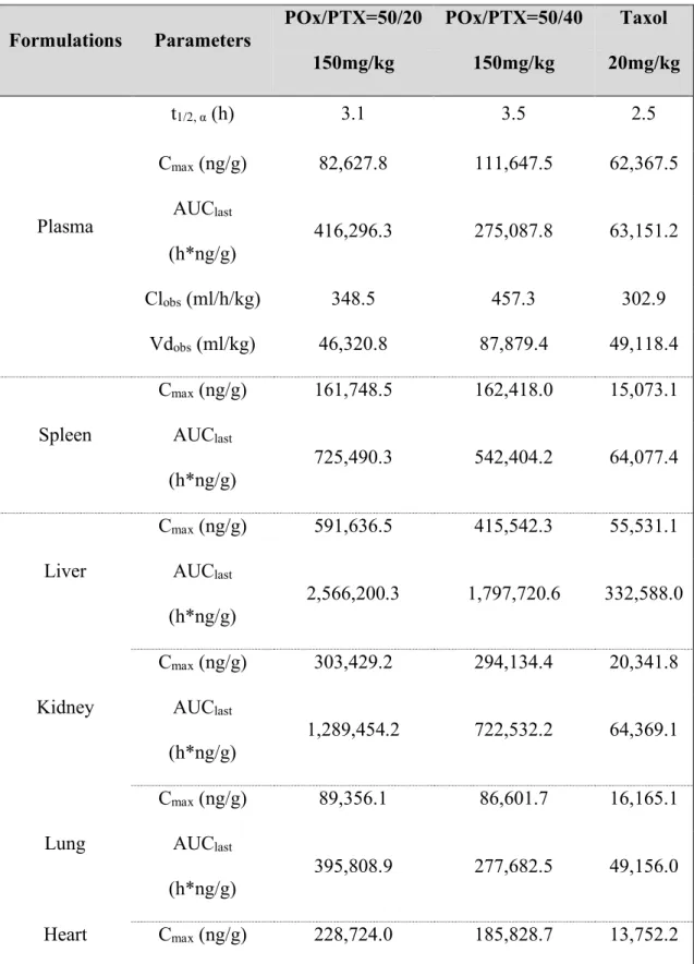
TABLE 2.9 PK PARAMETERS OF PTX FOR PLASMA AND ORGANS IN TUMOR-FREE
NUDE MICE. ...55
TABLE 2.10 PK PARAMETERS OF PTX FOR PLASMA AND ORGANS IN TUMOR
BEARING MICE. ...56
TABLE 3.1 CHARACTERISTICS OF C6CP/ETO PMA. ...95
TABLE 3.2 PK PARAMETERS OF C6CP AND PTX IN PLASMA AND TUMOR AFTER
ADMINISTERING PM IN A2780/CISR TUMOR BEARING MICE. ...97
TABLE 3.3 PK PARAMETERS OF C8CP AND PTX IN PLASMA AND TUMOR AFTER
ADMINISTERING PM IN A2780/CISR TUMOR BEARING MICE. ...98
TABLE 3.4 PK PARAMETERS OF POX POLYMER IN PLASMA AND TUMOR IN
A2780/CISR TUMOR BEARING MICE. ...99
TABLE 3.5 COMPARISON OF PK PARAMETERS OF DRUGS IN PM IN PLASMA AND
TABLE 3.6 STATISTICAL COMPARISONS OF TUMOR GROWTH BETWEEN GROUPS. (BY ONE-WAY ANOVA WITH HOLM-SIDAK POST-HOC TEST FOR MULTIPLE COMPARISONS AT A SIGNIFICANCE LEVELS OF P < 0.05 (*, P < 0.05, **, P < 0.01,
***, P < 0.001) ... 101
TABLE 3.7 STATISTICAL COMPARISONS OF ANIMAL SURVIVAL BY LOG-RANK (MANTEL-COX) TEST. ... 103
TABLE 4.1 CHARACTERISTICS OF ETO LOADED PM.A ... 142
TABLE 4.2 CHARACTERISTICS OF C4CP LOADED PM. ... 143
TABLE 4.3 CHARACTERISTICS OF C6CP LOADED PM. ... 143
TABLE 4.4 CHARACTERISTICS OF C8CP LOADED PM. ... 144
TABLE 4.5 CHARACTERISTICS OF C10CP LOADED PM. ... 144
TABLE 4.6 CHARACTERISTICS OF C6CP/ETO PM.A ... 145
TABLE 4.7 SERUM BINDING OF DRUGS IN PM. ... 146
TABLE 4.8 CYTOTOXICITY OF THE SINGLE DRUG AND CO-LOADED DRUG PM IN SCLC H69AR AND NSCLC 344SQ/LUC. CELL LINES. ... 147
TABLE 4.9 IC50 VALUES OF SINGLE CP PM AND FREE DRUG IN SCLC H69AR AND NSCLC 344SQ/LUC. CELL LINES. ... 148
TABLE 4.10 CLINICAL CHEMISTRY PARAMETERS OF H69AR TUMOR BEARING FEMALE NU/NU MICE. ... 149
TABLE 4.11 PK PARAMETERS OF ETO AND C6CP IN PLASMA AND TUMOR IN H69AR TUMOR BEARING MICE. (N=3)... 150
TABLE 4.12 STATISTICAL COMPARISONS OF TUMOR GROWTH BETWEEN GROUPS. ... 152
TABLE 4.13 STATISTICAL COMPARISONS OF ANIMAL SURVIVAL BY LOG-RANK (MANTEL-COX) TEST. ... 154
XIV
LIST OF FIGURES
FIGURE 1.1 SCHEMATIC EXAMPLES OF VARIOUS SHAPES OF NANOPARTICLES. ...20
FIGURE 2.1 STANDARD OPERATING PROCEDURE (SOP) FOR SMALL SCALE (1-5 MG) MICELLE PRODUCTION...58
FIGURE 2.2 CONSTRUCTION AND PHYSICOCHEMICAL PROPERTIES OF PTX NANOFORMULATIONS. ...59
FIGURE 2.3 CHARACTERIZATION OF POX/PTX FORMULATION. ...60
FIGURE 2.4 IN VITRO TOXICITY EVALUATION. ...61
FIGURE 2.5 MTD IN TUMOR BEARING NUDE MICE AND TOXICOLOGY PROFILES. ...62
FIGURE 2.6 TISSUE BIODISTRIBUTION AND PET/CT IMAGES OF TUMOR BEARING (A2780 XENOGRAFT) NUDE MICE OBTAINED AFTER I.V. INJECTION OF FORMULATIONS CONTAINING 0.2 MCI 64CU-POX. ...63
FIGURE 2.7 QUENCHING OF BSA FLUORESCENCE BY MICELLAR PTX...64
FIGURE 2.8 PHARMACOKINETICS, BIODISTRIBUTION AND TUMOR INHIBITION IN A2780 HUMAN OVARIAN TUMOR BEARING MICE. ...65
FIGURE 2.9 PK AND BIODISTRIBUTION IN TUMOR FREE NUDE MICE. ...66
FIGURE 2.10 PHARMACOKINETICS IN A2780 HUMAN OVARIAN TUMOR BEARING MICE. ...67
FIGURE 2.11 BIODISTRIBUTION OF POX AND PTX IN A2780 XENOGRAFT MICE. ...68
FIGURE 2.12 ANTITUMOR EFFICACY OF PTX FORMULATIONS IN A2780 TUMORS OF SMALL SIZE. ...69
FIGURE 2.13 ANTITUMOR EFFICACY OF VARIOUS PTX FORMULATIONS IN DIFFERENT TUMORS. ...70
FIGURE 3.1 STRUCTURE OF CP AND THEIR REDUCTION TO FREE CISPLATIN... 104
FIGURE 3.2 1H-NMR OF CISPLATIN PRODRUGS (CPS). ... 105
FIGURE 3.4 PHYSICOCHEMICAL CHARACTERIZATION OF PTX/CP PM. ... 108
FIGURE 3.5 DRUG RELEASE PROFILES OF PTX AND CP FROM SINGLE AND COMBINATION DRUG PM CONTAINING PTX AND (A, B) C6CP, (C) C8CP AND (D) C10CP. ... 109
FIGURE 3.6 IN VITRO CYTOTOXICITY OF PTX/CP PM IN A2780/CISR HUMAN OVARIAN CANCER CELL LINES. ... 110
FIGURE 3.7 IN VITRO CYTOTOXICITY OF SINGLE AND COMBINATION DRUG PM IN (A, B) A2780/CISR AND (C) A2780 CELL LINES. ... 111
FIGURE 3.8 IN VITRO CYTOTOXICITY OF PTX/CP PM IN A2780 HUMAN OVARIAN CANCER CELL LINE... 112
FIGURE 3.9 PHARMACOKINETICS AND BIODISTRIBUTION IN A2780/CISR HUMAN OVARIAN TUMOR BEARING MICE. ... 113
FIGURE 3.10 ANTI-TUMOR EFFECTS OF THE SINGLE AND COMBINATION DRUG POLYMERIC MICELLES. ... 114
FIGURE 4.1 SCHEME OF CO-DELIVERY OF ETOPOSIDE AND CISPLATIN PRODRUG VIA POLY (2-OXAZOLINE) CO-POLYMERIC MICELLE. ... 156
FIGURE 4.2 PREPARATION OF BINARY PM CONTAINING ETO AND CPS... 157
FIGURE 4.3 CHARACTERIZATIONS OF C6CP/ETO PM FORMULATIONS. ... 158
FIGURE 4.4 SOLUBILIZATION OF ETO LOADED POX PM. ... 159
FIGURE 4.5 CHARACTERIZATION OF SINGLE CPS LOADED MICELLE. ... 160
FIGURE 4.6 TYPICAL AFM IMAGES AND PARTICLE AREA HISTOGRAMS FOR EMPTY POX PM AND DRUG LOADED MICELLES, C6CP/ETO PM (4/8/10). ... 161
FIGURE 4.7 AFM SIZE MEASUREMENT FOR EMPTY POX PM. ... 162
FIGURE 4.8 AFM SIZE MEASUREMENT FOR DRUG CO-LOADED C6CP/ETO PM (4/8/10). ... 163
FIGURE 4.9 AFM POPULATION ANALYSIS FOR DRUG CO-LOADED C6CP/ETO PM (4/8/10). ... 164
XVI
FIGURE 4.11 POPULATION SEPARATION BY CIRCULARITY OF PARTICLES IN AFM
IMAGES FOR DRUG CO-LOADED C6CP/ETO PM (4/8/10). ... 166
FIGURE 4.12 DRUG RELEASE PROFILES FOR SINGLE AND CO-LOADED DRUG PM... 167
FIGURE 4.13 CYTOTOXICITY, SYNERGY AND DRUG UPTAKE FOR SINGLE AND CO-LOADED DRUG PM IN (A, B) SCLC H69AR AND (C-F) NSCLC 344SQ/LUC. CELL
LINES. ... 168
FIGURE 4.14 IN VITRO CYTOTOXICITY OF SINGLE CPS LOADED MICELLES. ... 169
FIGURE 4.15 HISTOLOGICAL EXAMINATION KIDNEY AND TUMOR TISSUES BY
HEMOTOXYLIN & EOSIN (H&E) STAINING. ... 170
FIGURE 4.16 PK AND TUMOR ACCUMULATION OF C6CP AND ETO IN H69AR SCLC
BEARING MICE. ... 171
FIGURE 4.17 BIODISTRIBUTION AFTER SINGLE I.V. INJECTION OF VARIOUS POX
MICELLE FORMULATIONS AT 1HR P.I. (N=3) ... 172
FIGURE 4.18 ANTI-TUMOR EFFECTS OF THE SINGLE AND CO-LOADED DRUG PM IN
NCSLC AND SCLC ANIMAL MODELS. ... 173
FIGURE 4.19 EFFICACY OF VARIOUS DRUG FORMULATIONS IN 344SQ-LUC LUNG
CANCER MODEL. ... 174
FIGURE 4.20 KAPLAN-MEIER SURVIVAL PLOT SHOWING ANTI-TUMOR EFFECTS OF THE SINGLE AND CO-LOADED DRUG PM IN 344SQ/LUC. NCSLC ANIMAL
MODEL. ... 175
LIST OF ABBREVIATIONS AND SYMBOLS
ABC accelerated blood clearance
ACN acetonitrile
AFM atomic force microscope
ALP alkaline phosphatase
ALT alanine aminotransferase
AML acute myeloid leukemia
ASCO American Society of Clinical Oncology
Asp aspartic acid
AUC area under the curve
BC breast cancer
BD biodistribution
BMS Bristol-Myer Squibb
BSA bovine serum albumin
BUN blood urea nitrogen
CI combination index
CLSM confocal laser scanning microscopy
Cmax maximum concentration
CMC critical micelle concentration
CNT carbon nanotube
CP cisplatin prodrug
DAPI 4',6-diamidino-2-phenylindole
DC drug concentration
Deff effective diameter
DLS dynamic light scattering
DMEM Dulbecco’s Modified Eagle Medium
DMF dimethylformamide
DMSO dimethyl sulfoxide
DMPC 1,2-dimyristoyl-sn-glycero-3-phosphocholine
DNA deoxyribonucleic acid
DOPA 1,2-dioleoyl-sn-glycero-3-phosphoethanolamine
DOPC 1,2-dioleoyl-sn-glycero-3-phosphocholine
DOTA tetraazacyclododecane-1,4,7,10-tetraacetic acid
DOX doxorubicin
DSPE
1,2-distearoyl-sn-glycero-3-phosphoethanolamine
DTX docetaxel
ECM extracellular matrix
EDC 1-ethyl-3-[3-(dimethylamino)propyl] carbodiimide
EDTA ethylenediaminetetraacetic acid
EP/PE etoposide and platinum drug combination
EPR enhanced permeability and retention
ER estrogen receptor
EtOH ethanol
FBS fetal bovine serum
FDA Food and Drug Administration
GEM genetically engineered mouse
GPC gel permeation chromatography
h hours
HEPES 4-(2-hydroxyethyl)-1-piperazineethanesulfonic acid
H&E hematoxylin & eosin
HPLC high-performance liquid chromatography
ICP-MS inductively coupled plasma mass spectrometry
IC50 half maximal inhibitory concentration
ID injected dosage
i.p. intraperitoneal
i.v. intravenous
IVIS international veterinary information service
LC loading capacity
LE loading efficiency
MBC metastatic breast cancer
MDR multiple drug resistance
MeOH methanol
mg milligram
mL milliliter
Mn number-average molecular weight
mPEG-PLA methoxy poly(ethylene glycol)-b-poly(lactide)
MP1U mouse phase 1 unit
MS mass spectrum
MTD maximum tolerated dose
MTT 3-[4,5-dimethylthiazol-2-yl]-2,5-diphenyl tetrazolium bromide
MW molecular weight
Mw weight-average molecular weight
NCI National Cancer Institute
NIH National Institute of Health
ng nanogram
NMR nuclear magnetic resonance
NSCLC non-small cell lung cancer
OC ovarian cancer
OST orthotopic syngeneic transplant
PBS phosphate buffered saline
pBuOx poly(2-butyl-2-oxazoline)
PCL poly-carprolactone
PDI polydispersity index
PDLLA Poly(D,L-lactic acid)
PEG-PLA methoxy poly(ethylene glycol)-block-poly(D,L-lactic acid)
PEI polyethylenimine
PFS the randomization to relapse or death
P-gp P-glycoprotein
PK pharmacokinetics
PLA poly(D,L-lactic acid)
PLGA poly(lactic-co-glycolic acid)
PLL poly(L-Lysine)
PM polymeric micelle
pMeOx poly(2-methyl-2-oxazoline)
POx poly(2-oxazoline)s
PRINT particle replication in non-wetting templates
Pt platinum
PTX paclitaxel
q4d x 4 every 4 days injection for 4 injections in total
Ref. reference
RES reticuloendothelial system
RT room temperature
s.c. subcutaneous
SCID severe combined immune-deficiency
SCLC small cell lung cancer
SEM standard error of the mean
SNHS N-hydroxysulfonosuccinimide
SPARC secreted protein acidic and rich in cysteine
SPE solid phase extraction
t1/2 half-life
TEM transmission electron microscopy
TFA trifluoroacetic acid
THF tetrahydrofuran
TIC tumor initiating cells
TNBC triple negative breast cancer
TV tumor volume
µg microgram
µL microliter
USP United States Pharmacopeia
Vd volume of distribution
Vdss steady state volume of distribution
VEGF vascular endothelial growth factor
vs. versus
WOR wortmannin
CHAPTER 1 INTRODUCTION
Most drug candidates in pre-clinical development display poor solubility, bioavailability, suboptimal pharmacokinetics and biodistribution profiles and systemic toxicity and can be administrated only through non-patient-centric administration routs. Finding the solution that gives a particular drug the desired bioavailability, safety and efficacy profile is still a major challenge.
Nano formulations address these challenges by developing efficient encapsulation and solubilization techniques for the drug candidates and improve bioavailability, PK and
biodistribution and reduce systemic toxicity through: 1) safe delivery of the loaded carrier to the target organ and/or cell in the body, and 2) on-site triggered release of the payload.
new millennium the focus has evolved from solubilization of the hydrophobic drugs to developing drug delivery tools which provide sustained release of the incorporated cargo, improve metabolic stability, circulation time in the blood stream and enhance accumulation of the cargo in the target site through passive targeting (enhanced permeability effect). Later, the focus has shifted to design of new polymers and materials specifically tailored for the drug delivery purposes, such as high capacity carriers, carries for several drug candidates and carriers for charged drug candidates. To address PK and biodistribution issues the attention shifted towards targeted carriers, stimuli responsive materials, which release the cargo in response to environmental clues in the targeted environment and alternative routes of administration.
However, following repeating administration, the issue of acquired resistance arises and presents a major drawback to successful clinical treatment. Overcoming drug resistance through the selection of the carrier has being additional approach to overcoming drug development
challenges. In this chapter, we discuss resent developments of different aspects of overcoming drug development challenges with polymeric micelles.
1.1 Clinical Sate of Polymeric Micelle (PM) on Cancer Treatment
Various chemical compounds loaded polymeric micelles are currently in clinical use and Phase I, II, III and IV clinical studies. [6, 7] Among all the anti-cancer agents, paclitaxel, docetaxel and cisplatin are the most loaded drugs. In this section, we discussed the current polymeric micelle formulations in market and being investigated in clinical studies for cancer therapies. (Table 1.1)
1.1.1 Taxane-based chemotherapy (paclitaxel and docetaxel)
Genexolâ PM
recently. [8] The paclitaxel loading capacity in Genexolâ PM is more than 10%, 10-fold
increasing, compared to Taxolâ with only 1% paclitaxel loading capacity. The pharmacokinetic
studies demonstrated a similar AUC with free PTX, however, a 3-fold higher Cmax in tumor,
liver, spleen, lung and kidney. [9] Clinical studies determined Genexolâ PM with a MTD 390 mg/m2 every 3 weeks or 120 mg/m2 every week, and in the Phase II studies, it was proved to be safer and more effective with high response rates in patients in advanced pancreatic cancer and breast cancer. [10] Moreover, Genexolâ PM showed significant activity in combined use with
cisplatin in non-small cell lung cancer patients. However, this formulation lacks good stability when present in the circulation and fast extracted from the polymeric micelle core, which compromised the antitumor efficacy. [11-13]
Nanoxelâ M
Another PM formulation produced by Samyang Biopharm and approved clinical use in South Korea is Nanoxelâ M. [14-17] Nanoxelâ M was originally developed to find an alternative
for Taxotere, a conventional docetaxel formulation and is composed of mPEG-b-PDLLA block copolymers using the solid dispersion method. The PMs have an average diameter of 25 nm with around 10% loading capacity. It has completed a phase I trial for advanced solid tumors and a Phase II trial in patients with metastatic head and neck cancer and a Phase III trial in patients with bladder cancer are now recruiting in South Korea.
Paclicalâ
Paclical/Apealea is a vitamin A derivative XR17, an excipient technology of a Swedish company Oasmiaã, based micellar paclitaxel formulation for the treatment of lung, breast and
combination with carboplatin, Paclicalâ combined with carboplatin, where paclitaxel in
combination with platinum containing compound has emerged as a first line treatment in patients with ovarian cancer, showed an extended PFS (the period from randomization to relapse or death). [18]
NK-105
NK-105 is a PTX-loaded micellar formulation based on PEG-b-poly(aspartate) block copolymer. The average diameter of NK-105 PM is 85 nm with 23% (w/w) of loading capacity. [20] Phase I study showed NK-105 with a 15-fold higher plasma AUC, compared to PTX. It was demonstrated with modest tolerate and activity against advanced gastric cancer in a Phase II clinical trial. [21] However, in a recent Phase III clinical trial compared with PTX that the statistical noninferiority of PFS (progression-free survival) could not be achieved, suggesting a promising PM formulation failed to provide greater efficacy in clinical trials. [13]
BIND-014
BIND-014 is a DTX formulation, currently under development by BIND therapeutics. It is a targeted PEG- poly(D,L-lactide-co-glycolide) (PLGA) micelles using an RNA aptamer accurin as targeting moiety that binds to the extracellular domain of the prostate-specific membrane antigen (PSMA) on the surface of prostate cancer cells. [22-24] BIND-014 has been shown to have enhanced cellular uptake compared to their non-targeted counterparts in vitro and in vivo. The micelles have a particle size about 90 nm and drug loading close to 11%. The
BIND-014). It was evaluated in NSCLC and mCRPC. Currently BIND therapeutics released
preliminary data from a Phase II trial at the 26th the European Organization for Research and Treatment of Cancer, the National Cancer Institute and the American Association for Cancer Research (EORTC-NCI-AACR) meeting.
The clinical results indicated that BIND-014 was well tolerated and had promising anti- tumor activity in 40 patients with advanced or metastatic NSCLC at a dose of 60 mg/m 2 q3w. This dose, lower than conventional Taxotere®, also showed activity in patients with KRAS mutant tumors, which normally poorly respond to Taxotere® treatment. Five patients (13%, N=40) achieved a PR with a median PFS of 2.7 months and 2 of 9 confirmed KRAS mutant patients experienced an objective response (22%) with median PFS of 2.7 months. In addition, BIND-014 treated patients experienced substantially reduced toxicities such as neutropenia, anemia, neuropathy, and alopecia commonly associated with Taxotere®.
CriPecâ doxetaxel
CriPecâ doxetaxel is developed to overcome the side effects of Taxotere.[26] DTX is
covalently conjugated to cross-linked polymeric micelles (DTX-CCL-PMs) with mPEG-b-poly(HPMAm-Lacn) block copolymer, with an average diameter of 66 nm and a 12% (w/w) DTX loading. As a result of a cross-linked conjugation of drug to the polymer, CriPecâ
1.1.1 Platinum-based chemotherapy (cisplatin and oxaliplatin)
NC-6004 (Nanoplatinä)
NC-6004 was first reported in 2003 that originally developed by Prof. Kazunori Kataoka’s group at University of Tokyo. It is a cisplatin incorporated via polymer-metal
complex-formation, PEG-b-poly(Glu) copolymeric micellar formulation, with an average size of 28 nm in diameter with high drug loading (39%). [28] The in vivo study of NC-6004 showed prolonged circulation and a 20-fold higher tumor accumulation. The common neurotoxicity and nephrotoxicity caused by cisplatin were found significant lower in the NC-6004 treated patients. [28-31] The most significant antitumor activity for NC-6004 was proved in colon cancer that it led to tumor regression in mice. [28] In clinical study, NC-6004 provides sustained release of
platinum with a 230-fold increase in plasma T1/2 and 8.5-fold increase in AUC. The phase II study suggested the MTD to be identified as 120 mg/m2. A Phase I/II clinical trial of NC-6004 combined with gemcitabine (GEM) was conducted in patients with pancreatic cancer, with a relatively well tolerated and exhibited modest antitumor efficacy. [32]
NC-4016
Similar as NC-6004 (Nanoplatinä), NC-4016 uses the same polymer as in Nanoplatinä,
however, it encapsulates a platinum derivation dichloro(1,2-diaminocyclohexane)platinum(II) (DACH-Pt), which overcomes the acute toxicity and acquired resistance caused by cisplatin. [33] NC-4016 PM has an average size of 40 nm with an extreme high platinum loading of 75% (w/w). It has great stability that no drug releases in water and 60% released after 96 h of
incubation with phosphate buffered saline at 37°C, and as a result in vivo, NC-406 elevates the
better antitumor efficacy and 25-fold higher accumulation in tumor, compared to oxaliplatin free drug.
1.1.2 Doxorubicin-based chemotherapy
NK-911
NK-911 is a DOX-loaded polumeric micelle formulation composed of PEG-b-poly(Asp) block copolymers with DOX conjugated to the core block (poly(Asp)) with an average diameter of 40 nm. A Phase I study showed a moderate response in patients with metastatic pancreatic cancer. [35] However, the plasma clearance of NK-911 was demonstrated 400-fold higher compared with Doxil®, suggesting that the NK-911 has lower stability in the bloodstream.
SP1049C
SP1049C is a DOX-loaded micelle formulation based on the class of Pluronic® polymers, which is the first Pluronic-based polymeric micelle formulation of DOX entering clinical trials. [36, 37] SP1049C PM has been demonstrated with higher antitumor effects compared with free drug. [36, 37] SP1049C has successfully completed Phase I and II clinical trials, obtained a SPA on a single approvable Phase III trial in refractory upper GI adenocarcinoma and has obtained an orphan drug designation in adenocarcinoma of the esophagus from US FDA. [38] In late 2016 patent rights to SP1049C, which expire in 2028, were acquired by Canadian SoftKemo Pharma Corp. to complete the final development of the novel anticancer therapeutic now code named SKC1049.
1.1.3 Other compounds based chemotherapy
NK-012
(w/w) of drug loading. [39] In several Phase I/II clinical trials, NK-012 showed promising
antitumor activity in patients with advanced solid tumors.[40, 41] The safety and efficacy of NK012 for advanced and metastatic triple negative breast cancer treatment in a Phase II study
demonstrated efficacious and presented a satisfactory safety profile, as well as, for colorecal cancer treatment in combination with 5-fluorouracil in a Phase I study. [41]
NC-6300
The formulation NC6300 consists of epirubicin covalently conjugated to the carboxylic acid groups of PEG-b-poly(aspartate) block copolymers via an acid-labile hydrazone bond. NC-6300 PM has an average size of 40-80 nm in diameter with 14.7% (w/w). [42-48] In pre-clinical studies, NC-6300 showed a prolonged blood circulation with preferential accumulation in human liver and breast cancer xenograft model. [45] It also showed extended antitumor effects with reduced epirubicin cardiotoxicity in mice bearing Hep3B liver orthotopic tumors. [47] A Phase I trial is underway in Japan in patients with advanced solid tumors since 2013 to evaluate
1.2 Polymeric micelles in anticancer therapy
Block copolymer micelles (PM) encapsulating anticancer compounds have been
developed for around 40 years and have been widely used as a drug delivery platform for cancer therapy. [13, 14, 50, 51] At low concentration, the amphiphilic polymers exist as monomers, but self-assembly micelles are formed when the concentration increases. [52] The concentration at which the micelles are formed is critical micelle concentration (CMC). Compared to low MW
excipients, amphiphilic copolymers usually have a much lower CMC. [53]
Polymeric micelles have several advantages over other nanosized drug delivery systems, such as 1) small size enabling effective EPR accumulation (diameter = 10-200nm), 2) high structural stability, 3) high drug loading, 4) low toxicity, 5) incorporation of various chemical species, 6) agents can be formulated in a non-covalent way, 7) biodegradability, and 8) simple manufacturing. [54] In this section, we summarize the outstanding features of polymeric micelle nanoformulation mainly in the following aspects.
1.2.1 High drug loading capacity
The polymeric micelles are originally designed for enhancing the solubility of
hydrophobic agents. Consequently, incorporation of these agents in micelles avoid the toxic side effect causing by the conventional solubilizing adjuvants, such as Cremophor EL. [55-57]
LC = Mdrug/ (Mdrug + Mexcipient) ´ 100%, (a)
LE = Mdrug/ (Mdrug added) ´ 100%, (b)
PMs are optimized formulations having high drug to polymer ratios enabling
solubilization of drugs up to 10000-fold higher their aqueous solubility. [8, 20, 58] As discussed in the previous section, the PMs under clinical trial exhibits high drug loading capacity 10% to 75% (w/w). (Table 1.1) Notably, the poly (2-oxazoline) (POx) tri-block co-polymer platform used in cancer therapy developed by our lab exhibits extreme high loading capacities of various
hydrophobic drugs, such as PTX, ETO and 17-AAG. [2, 3] However, a major challenge with PM is the design of the formulation act as true drug carriers but not only as solubilizers, or in another way, the in vivo stability as nanoparticle in blood circulation. [59]
1.2.2 Controlled release
Drug release from micelles mostly depends on the rate of micelle stability, drug
diffusion, and the rate of biodegradation of the copolymers. [60] The concern of the micelles with poor stability reflect on the fast release post intravenous administration. However, for those micelles as considered carriers (good stability), a controlled-pattern of release have been observed in many studies. [61] Moreover, the drug concentration in micelles, the length of the hydrophobic polymer, the molecular weight and the physicochemical characteristics of the drug and the localization of the drug within the micelles. [12]
1.2.3 Biodistribution and pharmacokinetics profile
1.3 Polymeric micelles for multi-drug delivery in cancer
In this section, we summarize recent research on polymeric micelles for multi-drug delivery in cancer therapy. Tumors are micro-environmentally diverse due to the variety of expression rate of biomarkers, proliferation profile and vasculature distribution. Other factors like age, sex, genetics vary the tumor heterogeneity as well. [64-68] Clinically, the strategy of combining anticancer drugs with various antitumor mechanism would possibly overcome tumor heterogeneity, thus improve the efficacy of oncology. [69] Delivery of multiple drugs in one vehicle raises the hopes for antitumor efficacy and also with reduced toxicity. However, multi-drug delivery has several considerations, such as, synergistic effect, concurrent or sequential delivery, pharmacokinetics and biodistribution, toxicity and safety, and anticancer efficacy. [70] In sequential drug delivery, polymeric micelles participate in pretreatment strategies that “prime” solid tumors and enhance the penetration of secondarily administered anticancer agent or
nanocarrier. The improved delivery of multiple poorly water-soluble anticancer agents by polymeric micelles via concurrent or sequential regimens offers novel and interesting strategies for drug combinations in cancer treatment. [70]
1.3.1 Combination index (CI)
To evaluate and quantify the multiple drug effect, the Chou-Talalay method was
values between Fa = 0.2 and Fa = 0.8 are considered valid. CI values less than 1 or more than 1 demonstrate synergism or antagonism of drug combinations, respectively. [74]
1.3.2 Concurrent multiple drug delivery
Poorly water-soluble anticancer agents can be loaded chemically or physically into polymeric micelles for concurrent multi-drug delivery. [75-78] The chemically conjugation include 1) mixed prodrug polymeric micelles, 2) PEG-b-poly(aspartic acid), 3) covalent linkage and hydrophobic interaction. [70] Physically trapped could be realized by 1) hydrophobic interaction, 2) multiple drug solubilization, 3) PEG-b-PLA.
Multi-drug polymeric micelles may release physically loaded drugs by disassembling in blood or by diffusion mostly with similar rate (concurrent delivery). [77] Briefly, Bae et al. has reported a PEG-b-poly(Asp-Hyd) block copolymer micellar formulation incorporate DOX with wortmannin (WOR) or GDM-OH. The multi-drug micelles exhibit lower IC50 on breast cancer cell and higher antitumor efficacy on breast cancer model on mice. [79] Shin et al. reported a 3-in-1 of PTX, rapamycin and 3-in-17-AAG co-loaded PEG-b-PLA micelles. [77] The micelles show significant better in vivo efficacy in non-small cell lung cancer and breast cancer xenograft models. [76] Many more studies have shown multi-drug co delivery in PEG-PLGA, poly (2-oxazoline) (POx), PEG-b-poly(carbonate-co-lactic acid) (PEG-b-(CB-co-LA)), PEG-b-PCl, PEG-DSPE/TPGS and PEG-b-PGlu-b-PPhe) block copolymers. These results exhibit concurrent multi-drug delivery enhance antitumor efficacy with reduced toxicity. [2, 80-86]
1.3.3 Sequential multiple drug delivery
1.4 The influence of nanoparticle shape on cancer drug delivery
According to previous studies, the antitumor efficacy of nanoparticle is not only influenced by the particle size, the shape of nanoparticle is also a crucial element influencing both cell uptake and pharmacokinetics. However, most formulations for anticancer therapy are in the spherical form. Various non-spherical nanoparticle platforms have been explored in several studies exhibiting beneficial properties compared to spherical shapes. In this section of
introduction, we summarize the influence of nanoparticle shape on biocirculation,
pharmacokinetics, cellular uptake and anticancer efficacy by comparing spherical and non-spherical nanoparticles. A list of nanoparticle shapes in recent studies in anticancer therapy is summarized in Fig. 1.1. [89]
1.4.1 Circulation
The anticancer therapeutic efficacy of nanoparticles is highly influenced by the
affected under rapid blood flow and spleen filtration (nflow > 5 µms-1). [98] Other studies of
nanorods shaped formulation also demonstrated prolonged circulation time. [99]
(Equation 1) 1.4.2 Biodistribution and target delivery
Biodistribution plays a crucial role in determining anticancer drug delivery and
therapeutic efficacy. To maximize therapeutic efficacy, anticancer drugs should be delivered to the targeting biological site with minimal of-site distribution. A study done by Christian et al. has demonstrated high accumulation of filamentous micelles in mouse xenograft tumors. [92] The higher distribution in tumor site of filomicelles has also been supported by other studies. [100, 101] These founds of high concentration in tumors of worm-like micelles suggest a promise in reducing dose with high anticancer efficacy. [102, 103] Tumor targeting accumulation has been observed in other shapes of nanoparticles as well. CNT (carbon nanotubes), nanorods and elliptical disks nanoformulaitons have been observed with higher accumulation in mice bearing orthotopic tumors. [104-109]
1.4.3 Cellular uptake
The cellular uptake is also influenced by the shape of nanoparticles. Studies have
been studied with enhanced ability to cross the endothelial cell membrane and deliver siRNA into the cytoplasm, resulting in higher cellular uptake. [115] Overall, previous studies support an enhanced cellular uptake of non-spherical nanoparticles, however, the reason has not been clearly explored. [89]
1.4.4 Anticancer efficacy and cytotoxicity
There’s no clear evidence on the relationship between the nanoparticle shape and cytotoxicity, with conflicting reports from literatures. [116-119] However, clear evidences from literature demonstrate non-spherical nanoparticles have higher MTD (maximum tolerated dosage), antitumor efficacy in mice tumor models.[92, 120, 121] For example, DOX loaded worm-like micelles via pH-sensitive bonds have been found to have higher cytotoxicity on MCF-7 cells and in vivo antitumor efficacy on tumor model. [122, 123] Moreover, gold nanorods and
1.5 Conclusion
With decades of research directed towards understanding the polymeric micelle, we can now take advantage of this phenomenon to develop anti-cancer drug encapsulated formulation for their delivery. We summarized the current clinical applications of polymeric micelles on cancer therapy and more PM formulations appear to be encouragingly applicable in the near future. By combining various rational polymers and drugs with different shapes in order to optimize thermodynamic and kinetic stability, enhancing drug loading capacity and enabling possible targeting. One possible next step could be combination therapy with various mechanism of anti-cancer treatments.
Table 1.1 Polymeric micelles in clinical studies.
Polymeric micelle Block copolymer Drug Size (nm) LC (w/w) Indication Clinical phase Company Ref.
Genexol®-PM PEG-b-p(D,L-lactide) Paclitaxel 20-50 11.5% Breast, lung and ovarian cancer Approved Samyang Biopharm, South Korea [8, 10, 126-128]
Nanoxel®-M mPEG-b-p(D,L-lactide) Docetaxel 10-50 11.2%
Breast, non-small cell lung, prostate, ovarian
cancer
Approved Samyang Biopharm, South Korea [14-17]
Paclical® derivatives XR-17 Vitamin A Paclitaxel 20-60 N/A Ovarian cancer (completed) III
Oasmia Pharmaceutical AB,
Sweden [18, 19] NK-105 phenyl-1-butanol) PEG-b-p(Asp-4- Paclitaxel 85 23% stomach and Advanced
breast cancer III
NanoCarrier/Nippon Kayaku, Japan
[20, 21, 129-134] NC-6004
(Nanoplatinä) PEG-b-p(Glu) Cisplatin 16-30 39%
Pancreatic, lung, bladder, bile duct and head and neck cancer
III NanoCarrier, Japan/Orient EuroPharma (co-development) [135, 136]
NK-911 PEG-b-p(Asp-DOX) Doxorubicin 40 50% colorectal cancer Pancreatic and II Nippon Kayaku, Japan [35, 58] NK-012 PEG-b-p(Glu-SN-38) SN-38 20 20% colorectal cancer Breast, lung and II Nippon Kayaku, Japan [126, 137]
SP1049C Pluronics® Doxorubicin 30 8.20%
Advanced adenocarcinoma of esophagus, gastroesophageal junction and stomach cancer
III Supratek Pharma Inc. [138, 139]
BIND-014
PEG-p(D,L-lactide-b-glycolide) (PLGA),
with Accurin that targets PSMA
Docetaxel 95 11% Solid tumors II Bind Therapeutics [140, 141]
NC-6300 PEG-b-p(Asp-hydrazone) Epirubicin 40-80 14.7% Solid tumors I
NanoCarrier, Japan/Kowa
(co-development) [42-48] NC-4016 PEG-b-p(Glu) DACH-Pt 40 32% Solid tumors and lymphoma I NanoCarrier, Japan [33, 34, 142]
CriPec® mPEG5000-b- Docetaxel 66 12% Solid tumors I Cristal Therapeutics, [26, 57,
CHAPTER 2: A HIGH CAPACITY POLYMERIC MICELLE OF PACLITAXEL: IMPLICATION OF HIGH DOSE DRUG THERAPY TO SAFETY AND IN VIVO
ANTI-CANCER ACTIVITY 1
2.1 Summary
The poor solubility of paclitaxel (PTX), the commercially most successful anticancer
drug, has long been hampering the development of suitable formulations. Here, we present
translational evaluation of a nanoformulation of PTX, which is characterized by a facile
preparation, extraordinary high drug loading of 50% wt. and PTX solubility of up to 45 g/L,
excellent shelf stability and controllable, sub-100 nm size. We observe favorable in vitro and in
vivo safety profiles and a higher maximum tolerated dose compared to clinically approved
formulations. Pharmacokinetic analysis reveals that the higher dose administered leads to a
higher exposure of the tumor to PTX. As a result, we observed improved therapeutic outcome in
orthotopic tumor models including particularly faithful and aggressive “T11” mouse claudin-low
breast cancer orthotopic, syngeneic transplants. The promising preclinical data on the presented
PTX nanoformulation showcase the need to investigate new excipients and is a robust basis to
translate into clinical trials.
1
2.2 Introduction
Paclitaxel (PTX) [144] is a powerful antineoplastic agent against metastatic breast cancer,
non-small cell lung cancer, advanced ovarian cancer, head and neck cancer and other
malignancies.[145] By interfering with tubulin polymerization, thus perturbing microtubule
dynamics, PTX leads to chromosome missegregation on multipolar spindles.[146] Apart from
excellent potency, PTX is characterized by an extremely low solubility in aqueous media (<1
mg/L),[147] thereby demanding delivery vehicles for parenteral administration. Three
formulations are currently clinically approved, two of which by the FDA. Both are blockbusters
and make PTX the best-selling chemotherapeutic in history.[146]
The first clinical formulation of PTX was Taxol. It is characterized by very low drug
loading (1% wt.), thus, the amount of the excipient, Cremophor EL/ethanol, necessary to deliver
effective doses of PTX is substantial. Excipient plasma concentration can reach 0.4% (v/v) and
persist above 0.1% (v/v) for over 24 hours.[148] Cremophor EL causes severe allergic,
hypersensitivity, anaphylactic reactions and nephro- and neurotoxicity in animals and humans,
which significantly limits dosing and requires clinical intervention.[55, 149]
The clinical demand for alternative formulations led to the development of Abraxane, a
nanoparticle formulation (hydrodynamic diameter ≈ 130 nm) comprising human serum albumin
and ca. 10% wt. PTX. Evidenced advantages of Abraxane vs. Taxol such as increased antitumor
activity and tumor accumulation in several mice xenograft models, significantly higher
maximum tolerated doses (MTD) in human, as well as approximately 74% increase of response
rates in metastatic breast cancer patients ultimately led to the clinical approval of this new
formulation.[150] However, its clinical trials also revealed an increased peripheral neuropathy as
compared to weekly Abraxane in combination with Bevacizumab as first-line therapy for locally
recurrent or metastatic breast cancer indicates that Abraxane offers no benefits to
progression-free survival compared to Taxol, while inducing greater hematologic toxicity and sensory
neuropathy.[152] Thus, there clearly remains a clinical demand for a formulation of PTX with
improved safety profile and therapeutic outcome.
Besides other approaches, polymeric micelles were investigated to formulate PTX[9, 20,
21]For example, Genexol-PM,[9] a formulation comprising a block copolymer of poly (ethylene
glycol) (PEG) and poly(DL-lactide) (PLA) is clinically approved in South Korea. NK105, a
formulation of PTX using a block copolymer of PEG and modified poly(aspartate) recently
successfully completed a phase II clinical study.[21] However, even these developmental
formulations only in part overcome the common limitations of PTX formulations. The MTD of
Genexol-PM identified as 50 and 60 mg/kg in non-tumor bearing female SPF C3H/HeNcrj mice
and nude mice respectively is only 2-3x higher compared to Taxol.[9] NK105 could be safely
administered at 100 mg/kg in balb/c female nude mice but only at a rather low concentration
(0.12 mg/mL), requiring prolonged intervals of administration.[20] A superior dosage form, which
exhibits high drug loading, desirable pharmacokinetics (PK) and tumor accumulation, and low
toxicity while increasing therapeutic efficacy, remains elusive.
Here we present self-assembled nano-sized polymeric micelle formulation based on
amphiphilic poly(2-oxazoline) (POx) block copolymers with unique polysoap structure[1, 2]
fulfilling unmet needs in formulation of PTX. This nanoformulation of PTX exhibit a facile
preparation and unprecedentedly high drug loading of 50 % wt., with excellent shelf stability as
well as controllable sub-100 nm size. In addition, absolute drug concentrations of 45 g/L could
much higher maximum tolerated dose compared to clinically approved formulations were
observed. Pharmacokinetic analysis revealed higher exposure of the tumor to PTX as compared
to Taxol. Subsequently, we observed improved therapeutic outcome in orthotopic tumor models
including particularly faithful and aggressive “T11” mouse claudin-low breast cancer orthotopic,
2.3 Materials and Methods
2.3.1 Materials
Two batches of amphiphilic triblock copolymers P(MeOx33-b-BuOx26-b-MeOx45), Mn
= 10.0 kg/mol, Đ (Mw/Mn) = 1.14 and P(MeOx47-b-BuOx21-b-MeOx36), Mn = 9.9 kg/mol, Đ
(Mw/ Mn) = 1.19 were synthesized as described in the previous study[2]. PTX was purchased from
LC Laboratories (Woburn, MA). All other materials were from Fisher Scientific Inc. (Fairlawn,
NJ) and all reagents were HPLC grade. The A2780 cells were originally obtained from
Sigma-Aldrich. Cells were cultured in DMEM medium (Gibco 11965-092) supplemented with 10%
FBS and 1% pen-strep.
2.3.2 Preparation of POx/PTX polymeric micelles
POx/PTX micelles were prepared by a thin film method (Fig. 2.1). Briefly,
pre-determined amounts of POx and PTX (stock solution 10-20 g/L in ethanol) were dissolved in
ethanol (5-10 g/L) and mixed, followed by complete removal of volatiles. We tested and
optimized small (1-5mg scale, air flow at 40 °C) and large scale (200 mg scale, rotary
evaporator) production methods to control the thin film formation process (Fig. 2.1). Appropriate
amounts of deionized (DI) water or normal saline were used to rehydrate the dried thin-film
under heating at 50-60 C for up to 20 min in order to obtain drug loaded polymeric micelles. The
resulting micelle formulation was stored as aqueous solution in refrigerator for up to 2 weeks or
as lyophilized powder.
The drug concentrations in POx micelles were measured by reverse-phase HPLC method
with a Nucleosil C18 - 5µm column (250 mm ´ 4.6 mm) in an Agilent 1200 HPLC equipment.
Each sample was diluted 20 times in mobile phase (ACN/water; 55/45, v/ v) and 20 mL diluted
was 30 °C. A standard curve range from 5 mg/ mL to 200 mg/mL was used to calibrate the
quantity of PTX.
The drug loading capacity (LC), loading efficiency (LE) and drug loading (DL) were
calculated using following equations (a)-(b): where Mdrug and Mexcipient are the weight amounts of
the loaded (solubilized) drug and polymer excipient in the dispersion, while Mdrug added is the
weight amount of the drug initially added to the dispersion.
LC = Mdrug/ (Mdrug + Mexcipient) ´ 100%,
(a)
LE = Mdrug/ (Mdrug added) ´ 100%,
(b)
A Nano-ZS (Malvern Instruments Inc., UK) DLS equipment was used to measure size
distribution of POx micelles. Briefly, each sample was diluted 50 times with DI H2O or 10 mM
NaCl to yield 1 g/L final polymer concentration before the measurement. The hydrodynamic
diameters of POx/PTX micelles was determined by intensity-mean z-averaged particle size
(effective diameter) and the polydispersity index (PDI) from cumulate analysis. Results were
obtained from the average of three independent micelle samples.
The morphology of POx/PTX micelles was studied using a LEO EM910 TEM operating
at 80 kV (Carl Zeiss SMT Inc., Peabody, MA). Digital images were obtained using a Gatan
Orius SC1000 CCD Digital Camera in combination with Digital Micrograph 3.11.0 software
(Gatan Inc., Pleasanton, CA). One drop of each diluted POx/ PTX micelle solution (dilute 500 or
1000 times using DI H2O) was deposited on a copper grid/carbon film for 5 min and excess
solution was wicked off using fine filter paper. Then one drop of staining solution (1% uranyl
studies for POx/PTX micelles were performed using membrane dialysis method against
phosphate buffered saline (PBS, pH 7.4) at 37 C. Briefly, POx/PTX micelles were diluted in PBS
to achieve approximately 100 mg/mL of PTX final concentration. Subsequently, 100 mL of the
diluted micelle solutions were added into floatable Slide-A-Lyzer MINI dialysis devices (100
mL capacity, 3.5 kDa MWCO; Thermo Scientific) and suspended in 20 mL PBS in compliance
with the sink conditions. Three devices were used for every time point. At each time point the
samples were withdrawn from dialysis device and quantified by HPLC to obtain remaining drug
amount of sample. Drug release profiles were constructed by plotting the amount of PTX drug
released from POx/PTX micelles over time.
2.3.3 In vitro complement activation, hemolysis, blood coagulation and cytotoxicity
These studies were carried out following the protocols established and published by the
Nanotechnology Characterization laboratory (http://ncl.cancer.gov/working_assay-cascade.asp).
2.3.4 Serum albumin quenching studies
Fluorescence emission spectra were obtained using a PelkinElmer LS 55 Fluorescence
spectrometer equipped with a thermostatted cell holder, a Xenon Flash lamp, a Monk-Gillieson
type monochromator, and a variable slit system. Emission spectra were recorded in phosphate
buffered saline (PBS: 140 mM NaCl, 1.9 mM NaH2PO4, 8.1 mM Na2HPO4, pH 7.4) from 300 to
440 nm (data shown up to 410 nm) after excitation at 295 nm. Both excitation and emission slits
were set at 10 nm. BSA stock solution of 2.5 µM was freshly prepared by dissolving bovine
serum albumin in 150 mM PBS. To ensure proper mixing all samples were gently mixed by
using a laboratory vortex. The samples were then incubated for 30 min at 25 °C before
measured at different sample concentrations. Presented data represents average of triplicate
samples.
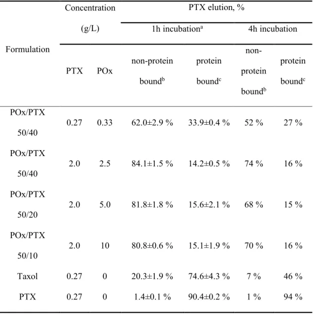
2.3.5 Serum binding studies
Reverse-phase Thermo Scientific™ SOLA™ HRP solid phase extraction (SPE)
cartridges were used for separation and determination of micellar (encapsulated) and
non-micellar (free) paclitaxel (PTX) in serum based upon the selective retention of non-micellar
(encapsulated) PTX and non-micellar (protein bound) PTX on the cartridge. The former
exhibited no retention, while the latter was retained on the stationary phase and eluted only with
acidified methanol.
Sample preparation was performed as follows. The formulations and PTX solutions (100
µL POx/PTX 50/40, 50/20, 50/10, Taxol and free PTX (dissolved in small amount of EtOH)
comprising 3H labeled PTX (2.5 µCi/mg PTX) were added to 2 mL of fetal bovine serum (FBS),
incubated at 37 °C, and 200 µL samples were collected from the mixture solution at 1hr and 4hr.
Each 200 µL serum sample was added to 200 µL of POx solution (2 mg/ml) in phosphate
buffered saline, pH 7.4.
To separate micellar and non-micellar (free) paclitaxel the following procedure was used:
(A1) Column conditioning: Add 0.5 mL methanol (to waste)
(A2) Equilibrate: Add 0.5 mL water (to waste)
For the next steps, collect the effluent of A3 and A4 as this contains the micellar
paclitaxel fraction. This fraction requires further clean up, described below.
(A3) Application: load pre-treated sample (collect)
(A4) Wash 1: 2 × 0.25 mL POx in phosphate buffered saline (2 mg/ml), pH 7.4 added
sequentially (collect)
(A6) Elution: 0.5 mL methanol + 0.1% formic acid (collect)
2.3.6 In vivo studies
All animal procedures were performed in compliance with federal animal welfare
regulations, and protocols were approved by the Institutional Animal Care and Use committee.
All animals used in PK, biodistribution, MTD, toxicology and efficacy studies were allowed to
acclimate for at least 72 h in the animal facilities before experiments. Animals were exposed to a
12 h light/dark cycle and received food and water ad libitum throughout the studies. Dosages of
POx/PTX micelle formulations or commercial drugs Taxol and Abraxane are expressed as the
quantity of PTX administered.
2.3.6.1 MTD studies
MTDs for Taxol, Abraxane and POx/PTX micelles were determined in a dose escalation
method in female nude mice (tumor-free 6-8 weeks of age). Animals (n=3 per group) received
i.v. injections of POx/PTX micelles (20, 40, 60, 90, 120, 150, 175 and 200 mg/kg), Taxol (20,
25, and 30 mg/kg), Abraxane (30, 60, 90 and 120 mg/kg), and saline as a control (q4d x 4). Mice
survival and changes in body weight were observed daily over two weeks in all groups. The
highest dose that did not cause toxicity (as defined by a median body weight loss of 15 % of the
control or abnormal behavior including hunched posture and rough coat) was defined as MTD.
Changes in histopathology such as inflammation, or presence of necrotic cells were used to
assess cytotoxicity occurring after treatment.
Healthy Balb/c mice received POx alone, POx/PTX and Taxol at MTD dose. The
following day mice were sacrificed and blood were withdrawn and a comprehensive blood
chemistry panel were performed. Major organs including heart, liver, kidney, spleen, lung and
addition, tumor bearing animals were sacrificed two weeks post fourth injection and organs were
harvested according to the same procedures as healthy mice.
2.3.6.2 Efficacy and POx/PTX tumor accumulation studies
A2780 ovarian cancer xenograft model
Female athymic nude mice (6-8 weeks) were subcutaneously inoculated in the right flank
with 8 x 10⁶ human A2780 ovarian cancer cells (Sigma Aldrich) resuspended in 50% growth
medium and 50% Matrigel. Two sets of experiments were performed: early stage tumor
treatment starting after tumor sizes reached ca. 100-200 mm3; or late stage tumor treatment when
tumor sizes reached ca. 400 mm3. Animals were randomized into groups of seven mice such that
the mean tumor volumes were similar between groups and then administered with following
formulations: 1) Normal saline; 2) Taxol (20 mg/kg PTX at determined MTD dose); 3) Abraxane
(45 and 90 mg/kg PTX at determined ½MTD and MTD doses); 4) PTX loaded micelles (75 and
150 mg/kg at determined ½MTD and MTD doses). The formulations were administered via tail
vein following q4d x 4 regimen (on the days 0, 4, 8, 12). Tumor growth was monitored twice
weekly for 15 weeks or earlier end-points defined by tumor volume (> 2000 mm3), animal
weight loss (> 15%), or animals becoming moribund. Tumor length (L), width (W) were
measured and tumor volume (TV) was calculated as TV =1/2 ´ L ´ W2. Survival and body
weight were monitored daily. Tumors were removed at the end of the observation and subjected
to histopathological examination.
Orthotopic model of LCC6-MDR human TNBC
The LCC6-MDR cells (obtained from Dr. R. Clarke, Georgetown University Medical
School, Washington DC) expressing high levels of P-glycoprotein (P-gp) were originated from
LCC6-WT cells were derived from estrogen receptor (ER)-negative, aggressive and metastatic
MDA-MB-435 cells. The orthotopic model was obtained by directly transplanting LCC6-MDR cells
into mice mammary fat pad (5 million cells/mouse).
T11 orthotopic, syngeneic transplant (OST) cancer model
T11 model mice are assessed using described practices of the Mouse Phase 1 Unit
(MP1U) of UNC (e.g., tumor regression, large cohort size, etc.). When tumors were noted to be
approximately 10-50 mm3 in size, animals were treated as described and tumor response was
assessed by weekly caliper measurements. Data in Fig. 2.6 are normalized to tumor size at the
time of therapy initiation, with volumes calculated using previously mentioned formula.
Tumor-bearing mice were euthanized at the indicated times for morbidity, tumor ulceration, or tumor
volume more than 3000 mm3.
2.3.7 PK and biodistribution studies
2.3.7.1 Tumor-free mice
Female Balb/c mice (6-8 weeks of age) were administered a single dose of Taxol (20
mg/kg) or POx/PTX micelles (150 mg/kg) containing 3H-labelled PTX (5 µCi/mouse) via tail
vein. At various sampling times (0.083, 0.5, 1, 2, 6, 24, 72, and 168 h post) a group of animals
(n=3) were euthanized and blood collected from cardiac puncture were analyzed for PTX plasma
concentration by measuring radioactivity. The tissues (brain, lung, kidney, spleen, liver, and
heart) were also removed, washed in ice-cold saline, weighted and homogenized in a glass tissue
homogenizer (TearorTM, BioSpec Products, Inc.), followed by radioactivity level determination
2.3.7.2 Tumor-bearing mice.
Female nude mice (6-8 weeks of age) were implanted with 8x10⁶ A2780 ovarian cancer
cells in 50% growth medium and 50% Matrigel (BD Biosciences) by subcutaneous injection.
When tumors were about 200 mm3 volume, mice were randomized (n=4 per group) such that the
mean and medium tumor weights were similar between groups. Mice were then administered a
single dose of above-mentioned formulations. At various time points, blood and tissue samples
were obtained accordingly.
2.3.8 PK and data analysis
PK parameters were assessed with Phoenix WinNonlin (version 6.0) using
non-compartmental analysis. Statistical comparison of efficacy and tumor accumulation data is
one-way ANOVA with Holm-Sidak post-hoc test for multiple comparisons at a significance level of
p<0.01 (Graphpad Prism, version 5.1.). If groups fail the normality or equal variance test,
2.4 Results
2.4.1 Characterization of PTX-loaded POx micelles
The POx/PTX formulation was prepared by a very simple, robust and highly reproducible
thin film hydration method[1, 2, 153] (Fig. 2.2A and B, SupplementaryFig.2.1). POx/PTX micelles
(< 100 nm effective diameter) with drug loading of approx. 50 wt.% spontaneously self-assemble
during rehydration of the dry film (Fig. 2.1B, Table 2.1).[153] In this work, we concentrated on
two formulations, comprising 50 g/L excipient and 40 or 20 g/L PTX, subsequently termed
POx/PTX 50/40 and POx/PTX 50/20, respectively. As opposed to Abraxane, the micelle
formulations are almost clear solutions with micelles of about 20-80 nm effective diameter and a
narrow size distribution (Fig. 2.2C,D). These solutions are directly lyophilizable and redispersed
in desired aqueous buffer, exhibit a neutral ζ-potential and excellent stability (Fig. 2.3; Table
2.2). In addition, these formulations can be directly injected due to their low viscosity.
2.4.2 Serum binding studies
Micelles are dynamic structures that can exchange both the surfactant and the drug with
the surroundings. Partitioning of the PTX from the micelles to the aqueous media should be very
low due to low drug solubility (approx. 1 mg/L). However, in the blood the drug can bind with
the serum proteins. Therefore, we studied interaction of POx/PTX with the bovine serum
albumin (BSA) by determining the quenching of the fluorescence of albumin tryptophan.[154]
While the polymer alone had little effect on the BSA tryptophan fluorescence, the POx/PTX
formulation produced a marked fluorescence quenching, which was increased as the drug
concentration increased (Fig. 2.7). Importantly, the fluorescence quenching was more
pronounced at a higher POx/PTX ratio.
unbound drug, but not the micelles. First, we demonstrated that in the absence of serum most of
the drug (~83-88 %) applied to the column in POx/PTX eluted in the micelle fraction (Table
2.3). The size of the particles in this fraction was 50 nm by DLS, corroborating the presence of
intact drug loaded micelles. Only ~9 to 15 % PTX were retained in the column and subsequently
extracted by the acid-methanol wash. On the contrary, when plain PTX was loaded onto the
columns, the paclitaxel was almost exclusively found in the fraction corresponding to the
acid-methanol wash (Table 2.4). Next, various POx/PTX formulations were mixed at different ratios
with 2 ml of the fetal bovine serum (FBS) to mimic the conditions that may realize upon
injection of the drug in the blood and incubated for either 1h or 4h before the column separation
(Table 2.5). In this case at the lower drug concentration ([PTX] = 0.27 mg/ml that correspond to
20 mg/kg PTX dose in the animal studies) only ~62% of PTX eluted with the micelles while the
rest was bound to the column and was only partially recovered. As the drug (and polymer)
concentration increased ([PTX] = 2 mg/ml, similar to 150 mg/kg PTX in the animal studies) the
portion of the drug in the micellar fraction (as well as the overall drug recovery) was also
increased markedly to > 80%. In contrast, only about 20% of PTX in Taxol formulation was
eluted in the micellar fraction. Free PTX, without excipient, was found to bind to serum proteins
almost quantitatively.
2.4.3 MTD and toxicology profiles of POx/PTX in nude or healthy mice
We investigated MTD by dose escalation in NCI-Nu/Nu mice in a regimen of every
fourth day for a total of 4 injections (q4d x 4), which was also employed in subsequent antitumor
efficacy studies. For the clinically approved formulations our studies confirmed MTDs reported