POST-TRANSCRIPTIONAL REGULATION OF HISTONE MRNAS AT THE LEVEL OF DECAY AND 3’ END PROCESSING
SHAWN MICHAEL LYONS
A Dissertation submitted to the faculty of The University of North Carolina at Chapel Hill in partial fulfillment of the requirements for the degree of Doctor of Philosophy in
the Department of Biology.
Chapel Hill 2013
Approved by:
William F. Marzluff
Robert J. Duronio
© 2013
ABSTRACT
Shawn Michael Lyons: Post-Transcriptional Regulation of Histone mRNAs at the Level of Decay and 3’ End Processing (Under the Direction William F. Marzluff)
Replication-dependent histone mRNAs are the only cellular eukaryotic mRNA which do not terminate in the canonical poly(A) tail. Instead, they end in a conserved 26 nucleotide stem-loop. This structure consists of a 6 base pair stem topped by a 4
nucleotide loop. This cis-element is bound by two trans factors: (1) stem-loop binding protein (SLBP) and (2) 3’ to 5’ histone exonuclease (3’hExo/Eri1). These two proteins play roles in regulating nearly all actions in the life cycle of histone mRNAs including direction of 3’ end cleavage of histone mRNA, nuclear export, translation and
degradation of the mRNA. Critical to this regulation is specific recognition of the histone stem-loop by these trans factors. In this thesis, I show the sequence and structural
ACKNOWLEDGMENTS
I owe immeasurable thanks to Bill for allowing me to work in his lab for these projects. His seemingly unending enthusiasm for all aspects of science is an inspiration. He has been a model of how to approach work in the lab. Anyone that has worked in any lab knows there are constant setbacks. Bill’s constant enthusiasm allowed me to continue through times when every experiment seemed to fail.
Most importantly, this work would not have been possible without the loving support of my wife Jessica. I owe her more than I could ever repay in helping me
TABLE OF CONTENTS
LIST OF TABLES………
LIST OF FIGURES. ……… vi
CHAPTER I: INTRODUCTION………1
Overview………..1
The Eukaryotic Cell Cycle………...2
The Two Classes of Histone Genes……….3
Cell-cycle Regulation of Replication Dependent Histone Genes in Mammals……….5
Cell-Cycle Regulation of SLBP……….14
SLBP’s Role in Translation of Histone mRNAs………...15
3’hExo is the 2ndtrans-factor that binds the histone stem-loop….16 3’ end Processing of polyadenylated mRNAs………...22
3’ end Processing of Histone mRNAs……….…..24
General Mechanism of mRNA Decay………...28
Deadenylation………28
Nucleases and Accessory Factors Involved in mRNA Degradation.. ………32
Decay Via mRNA Surveillance Pathways………36
No-Go Decay and Non-Stop Decay………..39
Histone mRNA Degradation………42
CHAPTER II: SEQUENCE REQUIREMENTS FOR BINDING OF SLBP AND 3’HEXO TO THE HISTONE STEM-LOOP Introduction……...……….52
Materials and Methods………..…….54
RNA-MITOMI………...54
HITS-CLIP of SLBP……….………….57
In vitro transcription of stem-loop substrates for crosslinking and electrophoretic mobility shift assays (EMSAs) ………...57
Expression and Purification of Recombinant SLBP and 3’hExo from Sf-9 Cells………...59
Electrophoretic Mobility Shift Assays………...…60
Crosslinking Assays………..61
Results…..………..……61
Sequence Requirements for SLBP binding to the histone stem-loop………61
The four-nucleotide loop of the histone stem-loop has sequence requirements for both SLBP and 3’hExo…..….71
SLBP and 3’hExo binding is not inhibited by oligouridylation………..…83
Discussion…..………89
SLBP’s binding is more pliable than previously suspected………89
Resolving ambiguities in HITS-CLIP derived CIMs as a result of homopolymer stretches and overlapping binding
sites………92
The histone stem-loop as a platform for assembling muti-subunit complexes………..94
CHAPTER III: ASSEMBLY OF THE HISTONE MRNA DEGRADATION COMPLEX AND THE ROLE OF LSM4 IN HISTONE MRNA DEGRADATION Introduction……...……….…………96
Materials and Methods………...…………..102
Protein Purification………..101
Transcription and purification of stem-loop probes…….102
Electrophoretic mobility shift assays………...103
siRNA knockdown……….…..103
GST Pulldown………..104
Cloning of Flag Vector………104
Production of Lentivirus for RNAi………..…105
Immunoprecipitation………105
Histone mRNA Degradation assay………..106
Results…..………106
OGFOD1 is involved in Poly(A) but not histone mRNA metabolism………...…106
eRF3 and SLBP directly interact……….113
SLIP1 remains in the initial RNA degradation complex..……….………….117
Amino acids in the unstructured C-terminal tail of Lsm4
are required for binding to SLBP ………..……….126
3’hExo interacts with Lsm4 and Lsm6………129
Mutants in the C-terminal tail of Lsm4 assemble into the Lsm1-7 Complex……….………133
Discussion…..………..………140
Lsm4 plays a specific role in histone mRNA degradation………...143
Transitioning histone mRNAs from translation mRNPs to degradation mRNPs……….………144
CHAPTER IV: POLYADEYLATION OF A SUBSET OF HISTONE MRNAS IN NON-DIVIDING CELLS Introduction……...………...……148
Materials and Methods………...151
RNA extraction from mouse tissue………..151
Preparation of protein lysate from mouse tissues………152
Extraction of Genomic DNA from NIH3T3 Cells……..152
S1 nuclease protection assay………153
Selection of Poly(A)+ mRNA………..154
Northern Blotting……….………158
Biotinylated RNA pulldown………158
Results…..………159
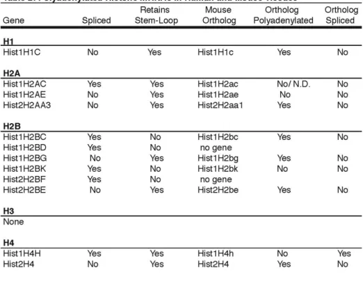
A subset of histone mRNAs are expressed in human tissue……….……….……….…….159
the murine liver and is exclusively polyadenylated…….166 G0 cells do not express polyadenylated histone
mRNA……….……….…169 Expression of histone specific factors in mouse tissues..172 Polyadenylation status of other core histone mRNAs….174 Discussion…..………..……177
Loss of U7 snRNA and SLBP in mouse liver leads to polyadenylation of histone mRNAs………183
LIST OF TABLES
Table 1 - RNA motif library for RNA-MITOMI………66
Table 2 - Generation of S1 nuclease protection probes………..156
Table 3 - Primers used in RT-PCR experiments……….157
LIST OF FIGURES
Figure 1 - Cell cycle regulation of histone mRNAs………...…7 Figure 2 - Histone mRNAs have a unique 3’ end……….…11 Figure 3 - Model of Histone mRNA metabolism………...…….….13 Figure 4 - Models of Active translation complexes for histone mRNAs and poly(A)
mRNAs……….………18
Figure 5 - SLBP and 3’hExo are histone specific trans factors that bind the histone stem- loop………..……….…20 Figure 6 - Eukaryotic degradation pathways……….………...30 Figure 7 - The initial steps in histone mRNA degradation is uridylation of the stem-
loop……….……….….….50 Figure 8 - Schematic of the RNA-MITOMI protocol………….………..57 Figure 9 - Numbering convention of the histone stem-loop………….………64 Figure 10 - Structure is largely sufficient to allow for SLBP binding to the stem-
loop………..…….………69 Figure 11 - Conventional low-throughput EMSA confirms RNA-MITOMI data……...73 Figure 12 - Sequence comparison of human stem-loops……….…….76 Figure 13 - LU1 and LU3 make direct contact with SLBP and are required for
binding………..78 Figure 14 - LU2 makes direct contact with 3’hExo and is required for binding…………83 Figure 15 - Uridylation does not affect the binding of SLBP or 3’hExo to the stem-
Figure 17 - OGFOD1 is not involved in histone mRNA metabolism, but does have a role
in poly(A) mRNA metabolism………...………….112
Figure 18 - eRF3 makes direct contact with SLBP ……….…116
Figure 19 - SLIP1 remains in an RNAse-insenstive complex at the initiation of histone mRNA degradation……….………120
Figure 20 - Lsm4 interacts with the RNA-binding domain of SLBP………...…...125
Figure 21 - The C-terminal tail of Lsm4 is required for binding to SLBP …………...129
Figure 22 - 3’hExo interacts with Lsm4………..123
Figure 23 - Exogenous Flag-tagged Lsm4 mutants are imcorporated into the Lsm1-7 complex ……….……….…138
Figure 24 - The C-terminal tail of Lsm4 is required for efficient histone mRNA degradation ……….………142
Figure 25 - A subset of histone mRNAs are expressed in mouse and human tissues….162 Figure 26 - MmuHist1H2BC is spliced and polyadenylated in mouse livers …….……166
Figure 27 - Expression of histone specific processing factors in mouse tissue…….…..171
Figure 28 - Expression of H2a and H3 genes in the mouse livers………...……175
Figure 29 - Phylogenetic analysis of promoter sequences of mouse histone genes……184
Figure 30 - The SLBP interactome………..188
Figure 31 - Current model for histone mRNA degradation……….191
Figure 32 - Lsm4 may help remove SLBP from the stem-loop………...196
CHAPTER I: INTRODUCTION
Overview
The genome of eukaryotes is composed of linear DNA molecules that are complexed with basic proteins called histones. Together these two make up the
chromosomes, which are composed of equal amounts of DNA and histone protein. The primary function of histones is to compact DNA in the nucleus, but also a role in
regulation of gene expression. Collectively, the histone:DNA complex is known as chromatin, of which the main subunit is a nucleosome. Each nucleosome is comprised of 146 base pairs of DNA wrapped around a core octamer of histone protein. A histone octamer contains two molecules each of the histone proteins H2A, H2B, H3 and H4. A fifth histone protein, H1, is not part of the core octamer, but plays an enigmatic role in helping to further compact chromatin. The globular central domains of histone proteins interact with each other to form the core of the nucleosome and are involved in
compaction of the DNA into chromatin (Luger et al. 1997); however, histone proteins contain an unstructured N-terminal tail that may be modified in order to regulate gene expression (Strahl and Allis 2000). Largely, this is accomplished by acetylation and methylation of lysine and arginine residues; however, mono-ubiquitination,
As cells are preparing to divide into two daughter cells, they replicate their genome. This newly synthesized genome must be rapidly packaged into chromatin in order to maintain genomic integrity. To accomplish this, all eukaryotic cells rapidly upregulate the production of histone mRNAs in order to produce histone protein to package newly replicated DNA into chromatin. Also, after the completion of DNA synthesis, the mRNAs encoding histone mRNAs are rapidly degraded. My work aims to better understand the regulation of these mRNAs. I will show what the mRNA sequence requirements are for two separate trans-factors (SLBP and 3’hExo) to bind the mRNA (Chapter 2). I will show how these two proteins participate in the degradation of the message through interactions with general mRNA degradation factors (Chapter 3). Finally, I show how cells are able to continue to maintain appropriate levels of histone proteins after they have exited the cell cycle (Chapter 5).
The Eukaryotic Cell Cycle
The growth and division of eukaryotic cells is governed by the eukaryotic cell cycle. The cell cycle can be divided into 4 distinct phases: (1) G1-phase, (2) S-phase, (3) G2-phase and (4) M-phase. Collectively G1- and G2-phases are known as “gap phases” and occur between the times when the genome is duplicated, S-phase, and when the cell divides, M-phase and cytokinesis. Progression through the cell cycle is a highly
least 8 CDKs and dozens of cyclins (although all do not change levels through the cell cell cycle). CDK-inhibitors (CKIs), which fall into two families, CIP/Kip (p21, p27, p57) and Ink4 (p15, p16, p18, p19), can also bind CDKs and restrict their activity.
As the synthesis of large amounts of histone proteins is only required as the cell is duplicating its genome, the expression of these mRNAs is restricted to S-phase. At least two cyclin/cdk complexes are directly involved in maintaining the proper regulation of these genes, Cyclin E/Cdk2 and Cyclin A/Cdk1. CycE/Cdk2 is required for entry into S-phase. The kinase activity of Cdk2 is required for upregulation of histone mRNA synthesis at the G1/S boundary, which is accomplished by phosphorylation of the transcription factor NPAT. CyclinA/Cdk1 phosphorylates SLBP, a critical histone mRNA specific trans-factor, at the completion of S-phase to promote proteasome-mediated destruction of the protein.
The Two classes of Histone Genes
There are two classes of histone genes: (1) replication-dependent and (2) replication-independent. As their names would suggest, the replication-independent histone genes are expressed constitutively throughout the cell cycle while the replication-dependent histone genes are expressed during S-phase in order to help package the newly synthesized genome. Nearly 95% of all of the histone in dividing cells is comprised of replication-dependent histone protein (West and Bonner 1980).
Hartwell 1986). The 146 base pairs of DNA wraps itself 1.7 times around the octamer. Between one nucleosome and the next there a linker region of DNA of variable length and may involve the binding of the 5th canonical histone, H1.
In all organisms, the different histone genes are clustered. In mammals, the replication-dependent histones are expressed from multiple different non-allelic genes clustered across the mammalian genomes (Marzluff et al. 2002). They are located in one major histone cluster (Hist1) and three minor histone clusters (Hist2, Hist3 and Hist4). The HIST1 cluster is located on chromosome 6 (6p21-22) in humans and 13 (13qA3.1) in mice and contains 55 and 51 genes, respectively. Most of the H2A, H2B, H3 and H4 genes and all of the H1 genes can be found in this cluster. The Hist2 cluster is located on chromosome 1 (1q22) in humans and chromosome 3 (3qF2.1) in mice and contains 11 and 10 genes, respectively. The Hist3 cluster contains 3 genes in both humans and mice and is located on chromosomes 1 (1q24) and 11 (11qB1.3), respectively. Finally, the Hist4 cluster is comprised of a single H4 gene and is located on chromosome 12 (12p13.1) in humans and chromosome 6 (6qG1) in mice.
The expression of replication-independent histone genes is not tied to the cell cycle. Instead, they are expressed at low levels throughout the cell cycle. These genes are located throughout the genome and are expressed as normal polyadenylated mRNAs. These genes play two general roles in the cell. The simplest task they may play is as replacement histones. The primary example of this type of histone is H3.3. This gene is expressed from a poly(A) encoding and intron containing gene. The second role for replication-independent histone genes is to serve as variants that often play specialized roles in regulation of chromatin structure.
Cell-cycle regulation of replication-dependent histone genes in mammals
The expression of replication-dependent histone mRNAs is tightly coupled to the cell cycle, as new histone protein is needed to package the newly synthesized genome (Figure 1). Failure to couple these processes leads to S-phase arrest and subsequent genomic instability or cell death. As cells approach S-phase, a Cyclin E dependent process allows for the upregulation of histone mRNA transcription. Transcription of histone genes is constitutive throughout the cell cycle (DeLisle et al. 1983), but transcription is enhanced during S-phase by the phosphorylation of nuclear protein ataxia-telangiectasia locus (NPAT), which presumably acts as a transcription factor for the histone genes (Ma et al. 2000; Ye et al. 2003). NPATs ability to activate transcription of histone genes is dependent upon phosphorylation by Cyclin E/CDK2 and interaction with the DNA binding factor Hinf-P (Miele et al. 2005).
Figure 1. Regulation of histone mRNAs and SLBP throughout the cell cycle. Cell
mRNA stability (Harris et al. 1991a). Specific regulation of histone mRNAs is attributable to a specialized 3’ end and a histone specific trans-factor, SLBP. The histone stem-loop and associated trans-factors
All eukaryotic mRNAs terminate in a polyadenosine (Poly(A)) tail except for the replication-dependent histone mRNAs (Adesnik and Darnell 1972; Adesnik et al. 1972). In order to add this tail, mRNAs are co-transcriptionally endonucleolytically cleaved and poly(A) polymerase (PAP) adds the poly(A) tail to the expose 3’ end.. After the addition of about 250 adenosines, polymerization will terminate as PAP can no longer
communicate with a complex of proteins known as the cleavage and polyadenylation specificity factor (CPSF complex)(Wahle 1995). This tail participates in all stages in the life cycle of mRNAs. It is required for mRNA export from the nucleus, enhances translation of poly(A) tailed mRNAs and protects the mRNA from degradation.
In order to perform these tasks, the poly(A) tail is bound by the RRM containing protein, polyadenylate-binding proteins (PABP). There are two distinct poly(A) binding proteins, one that is cytoplasmic (PABPC1) and the other that is nuclear (PABPN1). During polyadenylation, the poly(A) tail is bound by PABPN1, which facilitates nuclear export. Upon localization to the cytoplasm, PABPN1 exchanges with PABPC1 which allows for robust translation of its bound mRNA. PABPC1 also interacts with
eukaryotic release factor 3 (eRF3/GSPT1), an interaction that has been implicated in regulating both translation and mRNA degradation (Uchida et al. 2002; Hosoda et al. 2003).
Birchmeier et al. 1982), which serves many of the same functions that the poly(A) tail does in other mRNAs (Figure 2). Like the poly(A) tail of other eukaryotic mRNAs, the histone stem-loop plays a critical role in regulating the life cycle of histone mRNAs. The stem-loop is necessary for directing 3’ cleavage of histone messages, nuclear export of mRNAs, translation of histone mRNA and degradation of the mRNA. The histone stem-loop is the cis-element responsible for the post-transcriptional component of cell cycle regulation of the mRNAs (Harris et al. 1991a).
In mammals, the actions of the stem-loop are mediated by two trans-factors: (1) stem-loop binding protein (SLBP) and (2) 3’ to 5’ histone exonuclease (3’hExo/Eri1). In humans, SLBP is a 31 kDa (270 amino acid) protein and 3’hExo is a 40 kDa (349 amino acid) protein (Figure 3).
SLBP was first identified in a yeast triple hybrid screen conducted simultaneously by Dr. Zeng-Feng Wang in the Marzluff lab and Dr. Frank Martin in the Schümperli lab (Wang et al. 1996; Martin et al. 1997) using the histone stem-loop as bait. This protein contains a unique RNA binding domain located in the 73 amino acid region between amino acids 125 and 197 (Wang et al. 1996; Tan et al. 2013). SLBP is the trans-factor primarily responsible for the mRNA regulation conferred by the histone stem-loop. SLBP is required for proper histone mRNA 3’ processing (Dominski et al. 2001) and mRNA export (Sullivan et al. 2009a), translation of the mRNA in the cytoplasm
Figure 2. Histone mRNAs have a unique 3’ end. (A) Cleavage by CPSF-73 between
Figure 3. Histone mRNA metabolism.
Histone mRNAs are upregulated at the G1/S boundary as a result of phosphorylation of NPAT by CycE/Cdk2 and by increased stability and 3’ end processing due to synthesis and binding of SLBP. Endonucleolytic cleavage by CPSF-73 as directed by the U7 snRNP produces the mature mRNA, which is exported from the nucleus to the cytoplasm in a TAP dependent manner. Formation of a closed-loop translation complex is
facilitated in the cytoplasm by bridging the 5’ and 3’ mRNPs by SLIP1. Histone mRNA degradation is dependent upon ATR signaling from the nucleus. During this process, translation termination becomes inefficient. As a result, the NMD factor Upf1 is
single SLBP protein remains associated with a single histone mRNA throughout the life of that mRNA as there are not distinct nuclear and cytoplasmic isoforms of SLBP. Cell Cycle Regulation of SLBP
Like histone mRNAs, the expression of SLBP is tied to the eukaryotic cell cycle. However, unlike histone message, whose regulation is dictated by mRNA 3’ end
processing and mRNA degradation, SLBP protein expression is controlled by
translational regulation and proteolysis. The levels of SLBP protein mirror that of histone mRNA. Protein levels rapidly increases just before the cell enters S-phase and rapidly dissipate at the S/G2 border (Whitfield et al. 2000b). At the end of S-phase, Cyclin A/Cdk1 binds SLBP at amino acids 95 through 98 (KRKL) and phosphorylates threonine-61 (Zheng et al. 2003a; Koseoglu et al. 2008a). Following this
phosphorylation, casein kinase 2 (CK2) phosphorylates threonine-60. Phosphorylation of these two adjacent threonine residues is necessary for the proteasome-dependent cell cycle proteolysis of SLBP protein at the end of S-phase.
Unlike SLBP protein, SLBP mRNA levels change little throughout the cell cycle (Figure 1) (Whitfield et al. 2000b). Dr. Lian-xing Zheng tested whether SLBP was rapidly degraded when the cell was not in S-phase or whether translation of SLBP mRNA was inhibited. Cells were synchronized treated with MG132, an inhibitor of the
the level of proteolysis, but by translational regulation of the mRNA through an as of yet undetermined mechanism.
SLBPs Role in Translation of Histone mRNAs
The requirement of SLBP for efficient translation remains clear (Sànchez and Marzluff 2002). SLBP can stimulate the translation of stem-loop containing mRNAs both in vitro and in vivo. Dr. Ricardo Sanchez showed that the DWXSAVEE motif, located between amino acids 73 – 80 in humans, is required for the translation
stimulation. SLBP plays a similar role to PABPC1 in facilitating efficient translation. For polyadenylated mRNAs, efficient translation requires association of the 5’ and 3’ ends of the mRNA that form a closed-loop (Munroe and Jacobson 1990; Tarun and Sachs 1996; Martineau et al. 2008). It is thought that this configuration allows for more
efficient recycling of translation factors, particularly ribosomes. The association of three proteins spanning the 5’ and 3’ ends facilitates the formation of the closed-loop structure (Wells et al. 1998). At the 5’ end, eukaryotic translation initiation factor 4E (eIF4E) binds the 7-methylguanosine cap of the mRNA. eIF4E then interacts eukaryotic
translation initiation factor 4G (eIF4G) forming the eIF4F complex along with the RNA helicase eIF4A. To connect the ends of the mRNA, eIF4G directly interacts with
PABPC1. Using atomic force microscopy, the Sachs group was able to show that eIF4E, eIF4G and PABPC1 were sufficient to form a closed-loop of polyadenylated mRNA. (Wells et al. 1998).
(Gallie et al. 1996). For histone mRNAs, SLBP plays a critical role in the formation of the closed-loop complex along with eIF4F at the 5’ end of the message (Figure 4). SLBP remains bound to the stem-loop during translation (Whitfield et al. 2004). However, SLBP does not interact directly with eIF4G as PABPC1 does. To form the closed-loop, SLBP interacts with one or two accessory factors. By a yeast two-hybrid screen, Dr. Nihal Cakmakci discovered a novel protein, which was termed SLBP interacting protein 1 (SLIP1) (Cakmakci et al. 2008) that binds the sequence in SLBP required for efficient histone mRNA translation. This protein also interacts with eIF4G allowing for bridging of the 3’ and 5’ ends of the mRNA. Recently, a second protein, CBP80/20-dependent translation initiation factor (CTIF) has been implicated in assisting circularization the histone mRNAs through interaction with SLBP (Choe et al. 2013). In this case, the 5’ cap is bound by cap binding protein 20 (CBP20) and cap binding protein 80 (CBP80). Additionally, CTIF binds SLIP1 (J. Trotman & S. Meaux, Unpublished data). The exact contributions to SLIP1-dependent and CTIF-dependent histone mRNA translation are unclear and are currently being investigated in the Marzluff lab.
3’hExo is the 2nd trans-factor that binds the histone stem-loop
Figure 4. Models of translation complexes. (A) Histone mRNAs utilizing SLIP1 to
Figure 5. SLBP and 3’hExo bind the histone stem-loop
exonucleases is characterized by four invariant amino acids (three aspartates and one glutamic acid), which also give the group its name. 3’hExo is unique among other DEDDh exonucleases in that it also contains a 35 amino acid SAP domain. The SAP domain was first identified as a DNA binding domain (Aravind and Koonin 2000); however, in 3’hExo, this domain is required for RNA binding along with the interdomain spacer region located C-terminal to the SAP domain (Yang et al. 2006).
Initially, the only activity described for 3’hExo was its ability to remove the last three nucleotides from the mature histone mRNAs. In humans, after 3’ cleavage to form the mature message, 5 nucleotides (ACCCA) remain 3’ of the base of the stem. Using in vitro nuclease assays in the presence of SLBP, Dr. Zbigniew Dominski determined that 3’hExo would remove the final CCA leaving only two nucleotides (AC) after the base of the stem (Dominski et al. 2003). If these experiments were done in the absence of SLBP, 3’hExo was able to degrade the entire 3’ half of the stem to the loop. Later, using
circular RT-PCR, Dr. Thomas Mullen found that in the cytoplasm, histone mRNAs terminate in AC rather than the 5 nucleotide ACCCA formed after 3’ cleavage suggesting that 3’hExo is responsible for removing these nucleotides in vivo (Mullen and Marzluff 2008). Unfortunately, no role can be ascribed to the removal of the final 3 nucleotides.
More recently, the Heissmeyer lab was able to knockout the 3’hExo gene in mice, from which they derived fibroblasts for various experiments that they have used to elucidate many functions of the protein. They have demonstrated that it has a role in ribosomal RNA processing as the 5.8S rRNA contains an extended 3’ end in the knockout mouse (Ansel et al. 2008). They have also shown that it has a role in
they confirmed that 3’hExo is responsible for removing the final 3’ nucleotides from the 3’ end of histone mRNAs as histone mRNAs in the knockout mouse end in ACCCA. More importantly, they demonstrated that 3’hExo functions in the initial steps of histone mRNA degradation (Hoefig et al. 2013). They found that after inhibition of DNA synthesis by hydroxyurea (HU), histone mRNA degradation was slowed in 3’hExo knockout mouse cells. Normal degradation kinetics could be restored by the transfection of wild type 3’hExo, but not by transfection of 3’hExo containing point mutants to the catalytic core.
3’ end processing of polyadenylated mRNAs
(DSE), required for efficient polyadenylation is located downstream of the PAS and 3’ cleavage site. As suggested by its name, it is particularly G/U rich with a consensus sequence of YGUGUUYY (Gil and Proudfoot 1984; McLauchlan et al. 1985). Cleavage typically occurs between the AAUAAA and GU-rich element, about 20 - 30 nucleotides upstream of the GU-rich element following a CA dinucleotide (Chen et al. 1995b).
In order to make this cleavage, a large multisubunit protein complex must co-transcriptionally assemble on the nascent mRNAs. In mammals, there are 4 large multisubunit complexes that work to effect cleavage and polyadenylation: Cleavage and polyadenylation specificity factor (CPSF), Cleavage stimulatory factor (CstF),
Mammalian cleavage factor I (CF Im) and Mammalian cleavage factor II (CF IIm) (Mandel et al. 2007).
The CstF complex contains three individual proteins, 50, 64 and CstF-77, that dimerize to form a hexameric complex. CstF-64 is an RRM containing protein that has been shown to interact with the downstream G/U rich element by UV
crosslinking (Wilusz and Shenk 1988). The other components of CstF, CstF50 and CstF77, have been shown to interact with the C-Terminal domain (CTD) of the largest subunit of RNA polymerase II, likely coordinating transcription and 3’ end processing (McCracken et al. 1997).
Symplekin, a HEAT-repeat contain protein (Kennedy et al. 2009), is also a critical component in cleavage and polyadenylation despite not being contain specifically in either CPSF or CstF (Zhao et al. 1999). Symplekin is thought to function as a scaffold that coordinates the interaction between CstF and CPSF (Takagaki and Manley 2000). 3’ end processing of histone mRNAs
As histone mRNAs are not polyadenylated and contain no introns, the only enzymatic reaction needed to create the mature mRNA is 3’ cleavage. Histone mRNAs also utilize two cis-elements to direct 3’ end cleavage. Rather that the PAS and G/U rich region, the histone downstream element (HDE) and the previously discussed histone stem-loop direct histone mRNA cleavage. The mammalian HDE is a loosely conserved 11 nucleotide sequence with the consensus of AAAAGAGCTGT (Marzluff et al. 2002) (Figure 2).
complex with an unknown set of Sm-proteins, contains the trimethylated cap
characteristic of snRNAs and is required for the endonucleolytic cleavage of mammalian histone genes as well (Gick et al. 1986; Strub and Birnstiel 1986). Characterization of the U7 snRNA revealed that it contains a stem-loop at its 3’ and a single stranded region at its 5’ end. This single stranded region is complementary to the HDE and binding between the two in necessary of directing endonucleolytic cleavage (Schaufele et al. 1986; Bond et al. 1991). The location of the binding of U7 to the HDE acts as a
molecular ruler by directing the location of endonucleolytic cleavage (Scharl and Steitz 1994; Scharl and Steitz 1996; Yang et al. 2009c).
While both are unique to histone mRNA metabolism, the majority of study has focused on Lsm11, as it is the largest subunit and the primary subunit that interacts with specific processing factors in the U7 snRNP. Lsm11 is a unique Sm-protein in that it contains two separate Sm folds that are separated by a linker region. Also, it contains a long N-terminal extension, but no C-terminal extension. Lsm11 has a predicted
molecular weight of 36 kDa in contrast to other Sm-proteins that typically range from 10 – 15 kDa. In mammals, the U7 snRNP is stabilized on the HDE by an interaction
between Lsm11 and SLBP that is mediated by another histone specific protein, ZFP100 (Dominski et al. 2002; Azzouz et al. 2005; Wagner and Marzluff 2006). ZFP100
interacts with helix B in the SLBP RNA binding domain. This region had been shown to be required for efficient 3’ end processing of histone messages. ZFP100 also interacts with N-terminal domain of Lsm11.
fail to process histone mRNA in vitro and depletion of the Drosophila homolog of FLASH results in misprocessed histone mRNA in vivo.
After assembling the U7 snRNP complex with SLBP and ZPF100, the histone mRNA is cleaved after a CA dinucleotide in the same manner as with poly(A) mRNAs. Surprisingly, the same enzyme, CPSF-73, performs both endonucleolytic cleavages (Dominski et al. 2005). By using RNAs containing phosphothioates to slow the cleavage reaction, Dr. Zbigniew Dominski was able to crosslink CPSF-73 to the histone pre-mRNA and showed that it was necessary for both cleavage (Dominski et al. 2005; Yang et al. 2009b). Purification of the heat labile factor provided even more connections between histone mRNA processing and poly(A) mRNA processing. The heat labile factor was characterized by as being absolutely required for histone processing but distinct from the U7 snRNP and being susceptible to heat inactivation (Gick et al. 1987; LÅscher and SchÅmperli 1987; Luscher and Schumperli 1987). By biochemical fractionation, Dr. Nikolay Kolev partially purified the activity of the heat labile factor (Kolev and Steitz 2005). They confirmed the results from Dr. Dominski by showing that CPSF-73 was contained within the HLF, but also found that the polyadenylation factors CPSF-100, CPSF-160, CPSF-30, Fip1, CstF64, CstF-77, and Symplekin, which proved to be the factor susceptible to heat treatment. They later confirmed that a complex of CPSF-73, CPSF-100 and Symplekin was able to cleave histone mRNAs and this was also shown to be true in Drosophila (Kolev et al. 2008; Sullivan et al. 2009b). Further
General Mechanisms of Eukaryotic mRNA Degradation
Historically, mRNA transcription was assumed to be the primary contributor in establishing the abundance of mRNAs in a cell. However, the steady state levels of mRNA are a product of both the rate of transcription and rate of mRNA degradation. As the relative rates of transcription between various genes can be different, the rates of degradation between different mRNAs also vary (Perry and Kelley 1973; Singer and Penman 1973). The half-life of an mRNA is determined by cis-elements in the mRNA and RNA binding proteins and miRNAs that interact with the mRNA.
The pioneering studies of Dr. Roy Parker and coworkers have elucidated the pathways and factors involved in mRNA degradation in the budding yeast Saccromycces cerevisiae. Mammalian cells contain orthologs of the yeast factors, which play important roles in mRNA degradation (Figure 6).
Deadenylation
Figure 6. Outline of Cellular degradation pathways
(A) Deadenylation dependent decay relies on removal of the poly(A) tail by one of the
three deadenylases. Decay can then continue in a 5’ to 3’ direction after binding of Lsm1-7 stimulates decapping by a NUDIX fold containing protein, of which Dcp2 is the most well-studied member. Removal of the cap deprotects the 5’ end, leaving a 5’ phosphate that Xrn1 can use as a substrate for 5’ to 3’ exonucleolytic degradation.
Alternatively, following deadenylation, the exosome may degrade the mRNA in a 3’ to 5’ direction. Mounting evidence in mammalian systems indicates that these two pathways are not mutually exclusive and bidirectional decay may be commonplace.
(B) Endonucleolytic cleavage dependent mRNA decay relies on one of many cellular
poly(A) tail which is then completed by Ccr4-Not, the major deadenylase in eukaryotic cells. (Yamashita et al. 2005). Pan2 contains a DEDD nuclease domain that has poly(A) nuclease activity, while Pan3 contains a PABC1-interacting domain (PAM2 domain). Pan3 will interact with the poly(A) tail through its interaction with PABPC1 while the Pan2 subunit will begin to hydrolyze the poly(A) tail. Pan2-Pan3 has a lower affinity for the poly(A) tail after removal of about 100 adenosines because of the reduction in
PABPC1 molecules coating the tail. At this point, the Ccr4-Not complex will finish the deadenylation reaction.
The major deadenylase in all organisms is Ccr4-Not. It is a multisubunit complex of which five members are considered canonical: (1) Ccr4, (2) Caf1/Pop2, (3) Not1, (4) Not2, (5) Not3/5 (Albert et al. 2000; Temme et al. 2010). Both Ccr4 and Caf1 possess deadenylase activity with Ccr4 containing an EEP domain that coordinates two essential Mg2+ ions and Caf1 containing a DEDD exonuclease domain similar to the active site of Pan2. The HEAT domain containing protein Not1 acts as a scaffold upon which the other subunits of Ccr4-Not assemble. It also coordinates interactions with trans-factors that target specific mRNAs. For example, during AU-rich element (ARE) mediated degradation (AMD) (discussed in following sections), tristetraproline (TTP) binds to target mRNA and then directly interacts with Not1 to trigger deadenylation dependent degradation of ARE-containing mRNAs (Sandler et al. 2011).
The third vertebrate deadenylase is Poly(A)-specific ribonuclease (PARN), which is also a DEDD family exonuclease (Wu et al. 2005). PARN is unique in that it not only has affinity for the poly(A) tails of mRNAs, but also has an affinity for the
thought to help target PARN to non-translating mRNAs as the cap of translating mRNA should be bound by translation initiation factors such as eIF4E.
Nucleases and accessory factors involved in mRNA degradation
Following deadenylation, mRNAs can be degraded in a 5’ → 3’ direction, a 3’→5’ direction or bidirectionally. In special cases, degradation can be initiated as a result of cleavage by an endonuclease followed by degradation of the resulting two RNA fragments.
The multisubunit exonuclease complex called the exosome degrades mRNAs in the 3’ → 5’ direction and also has roles in RNA maturation (Mitchell et al. 1997). The core exosome, called Exo9, consists of 9 invariant subunits, six of which – Rrp41, Rrp42, OIP2 (Rrp43), PM/Scl-75 (Rrp45), Rrp46 and Mtr3 – form a hexameric torus. The remaining three core subunits – Rrp4, Rrp40 and Csl4 – form a trimeric cap atop of the hexameric base. The subunits contained within the trimeric cap contain S1 RNA binding domains and KH RNA binding domains, while the subunits of the hexameric ring contain RNAse PH domains. However, none of the subunits have ribonuclease activity due to mutations in the active sites of the RNAse PH domains. Instead, the ribonuclease activity of the exosome is conferred by the addition of auxiliary subunits, Dis3 (Rrp44) and Pm/Scl-100 (Rrp6), which, added to the Exo-9 complex, forms the Exo-10 exosome (Liu et al. 2006; Dziembowski et al. 2007). Dis3 is located on the opposite side of the
activity conferred by the PIN domain located at the N-terminus (Lebreton et al. 2008; Schaeffer et al. 2009; Schneider et al. 2009). The Exo-10 complex may also interact with a second exonucleolytic subunit, Pm/Scl-100 (Rrp6), in order to form the Exo-11
mammals, but inactivation of the protein still only stabilized a subset of mRNAs, albeit, a different subset as was stabilized with Dcp2 inactivation (Song et al. 2010a). More recently, six additional nudix containing proteins – Nudt 2, Nudt3, Nudt15, Nudt 17, Nudt19 – have been identified and confirmed to contain decapping activity, bringing the total number of decapping enzymes in mammals to eight (Song et al. 2013).
Decapping activity is enhanced by a variety of accessory proteins such as the Edc proteins. However, the critical complex in initiating degradation is the Lsm1-7 complex and this complex will be the most germane to the work presented in this thesis. This complex is a heptameric ring that is restricted to the cytoplasm (Bouveret et al. 2000; Tharun et al. 2000). Each protein in the ring contains a canonical Sm-fold for which they are named. The Sm-fold is characterized by 5 β-sheets and a single terminal α-helix. This structure is sufficient to bind RNA. Since most Lsm proteins are relatively small, often, the majority of the protein is contained within the Sm-fold. There are a few
notable exceptions that contain longer N-terminal or C-terminal extensions, which will be particularly relevant to my work. Six proteins in the Lsm1-7 complex are shared with the nuclear Lsm2-8 complex, which is a component of the U6 snRNP and plays major roles in RNA splicing as well as roles in nuclear RNA turnover (Salgado-Garrido et al. 1999).
The Sm-folds of the individual Lsm proteins provide the RNA-binding site. Lsm1-7 has strong affinity for both oligoadenylated and oligouridylated RNA.
unknown mechanism. The ability to distinguish between oligo(A) and poly(A) tails prevents Lsm1-7 from aberrantly targeting poly(A) tail containing messages for degradation. In yeast, deletion of Lsm1 impairs the rate of RNA degradation without affecting deadenylation, confirming that Lsm1-7 targets deadenylated mRNAs (Boeck et al. 1998; Schwartz and Parker 2000). Furthermore, mRNAs that contain an unadenylated 3’ end generated by ribozyme cleavage are not targets for 5’→3’ degradation mediated by Lsm1-7 and are instead degraded in a 3’ → 5’ direction by the exosome (Meaux and van 2006; Chowdhury and Tharun 2008).
The Lsm1-7 complex is typically considered to be in a stable complex with the protein Pat1b in yeast; however, a thorough biochemical investigation of this interaction has not been completed at this point, and no studies have been done on proteins bound to Lsm1-7 in mammalian cells. Pat1 is an activator or decapping as its deletion in yeast results in a partial defect in decapping. However, Pat1 may play a more important role in facilitating translational repression prior to the initiation of degradation. Pat1
coimmunoprecipitates with translation factor eIF4E and PABP in a RNA-dependent manner and a portion of the protein can be found on polysomes, which suggests that it interacts with the mRNA while it is still being actively translated and plays a role in mediating the transition from actively translated mRNA to one which is targeted for degradation (Tharun et al. 2000; Wyers et al. 2000).
translationally competent to being targeted for degradation. Pat1 acts to coordinate this complex with Lsm1-7 at the 3’ end of the mRNA (Braun et al. 2010; Ozgur et al. 2010). The Rck/p54, Hedls, hEdc3 complex can also include Dcp1and Dcp2 (Fenger-Gron et al. 2005). Thus, Pat1 may function to connect the decapping activity with deadenylation by coordinating Lsm1-7 binding to the 3’ end of deadenylated mRNAs with the decapping activity of Dcp2 at the 5’ end of mRNAs.
Following decapping, mRNAs are rapidly degraded in the 5’ → 3’ direction by Xrn1 (Hsu and Stevens 1993; Muhlrad and Parker 1994). The active site of Xrn1 requires coordination of at least one Mn2+ ion for activity (Chang et al. 2011). In mammals, Xrn1 is the major 5’→3’ exonuclease and is structurally and functionally related to the nuclear Xrn2/Rat1, which degrades downstream cleavage products in a 5’→3’ direction following 3’ cleavage (Luo et al. 2006). Utilization of Xrn1 in addition to the exonucleolytic activity of the exosome allows for mRNAs to be degraded by either decapping and 5’ to 3’ degradation, deadenylation followed by 3’ to 5’ degradation or a combination of the two processes.
Decay via mRNA surveillance pathways Nonsense Mediated Decay
In order to protect itself from possibly cytotoxic proteins produced from
production of C-terminally truncated proteins that might act as dominant negatives. These PTC containing mRNAs may arise from retained introns, errors in splicing, chromosomal rearrangements or mutations in genes. Additionally, it has been reported that 3 - 20% of mRNAs contain features that make them natural targets for NMD (Karam et al. 2013). In particular, the levels of many RNA binding proteins are autoregulated by coupling NMD with alternative splicing. For example, the polypyrimydine tract binding protein alters spicing of its own mRNA creating an mRNA sensitive to NMD.
In mammals, the creation of an NMD sensitive mRNA results from the location of deposited exon junction complexes (EJCs) in relation to the stop codons along the body of the mRNA. Following splicing of an mRNA, a multisubunit complex is deposited 24 nucleotides upstream of where two exons where joined together marking a successful splicing event. This core of this complex contains the proteins Y14, Magoh, eIF4III and RNPS1. Normal mRNAs or those that are not natural targets for NMD contain their stop codons within their last exon. Therefore, the presence of an EJC more than 50
nucleotides downstream of a stop codon indicates a premature termination codon and makes the mRNA a target for NMD. This is because this EJC is not removed by a translocating ribosome during translation. Additionally, translation termination at PTC appears to mechanistically different than at normal termination codons indicating that PTC dependent termination is inefficient. Toe print analysis of human β-globin mRNA that had a normal termination codon or a premature termination codon demonstrated that the NMD targets contained a toe print at the termination codon while normal termination did not (Peixeiro et al. 2012). These ribosome toe prints indicated that there was
were not as efficient at translation termination. Additionally, the drug ataluren (PTC124) preferentially causes read-through of PTC but not of normal termination codons, again, indicating a mechanistic difference between translation termination at PTC versus normal termination codons (Welch et al. 2007).
The primary effectors of NMD are the Upf proteins, which were originally discovered in yeast as suppressors of nonsense mutations, and later shown to be conserved in humans. There are three main Upf proteins, Upf1, Upf2 and Upf3, with there also being a second Upf3 variant, Upf3x. All three Upf proteins can form a
complex on the EJC with Upf3 being bound to the EJC, Ufp1 being recruited to PTC and Upf2 bridging the two proteins by interacting with both of them. Upf1 is a large protein (130 kDa) consisting of two major domains: a zinc-finger domain (CH domain) and a helicase domain (Cheng et al. 2007; Chakrabarti et al. 2011). Both domains are required for activating NMD (Weng et al. 1996; Bhattacharya et al. 2000). The helicase domain is required to “push” protein components off of the mRNA to be degraded (Franks et al. 2010). In cells with an ATPase-dead Upf1 protein, NMD mRNA intermediates are unable to be degraded due to NMD proteins remaining bound to the 3’ fragment of the mRNA. Upf1 also participates in other mRNA degradation pathways, particularly, histone mRNA degradation (Kaygun and Marzluff 2005a) and staufen- (stau1) mediated degradation (SMD) (Kim et al. 2005). In SMD, stau1 recognizes inter- and
intramolecular dsRNA helices and recruits Upf1 to target certain mRNAs for degradation.
phosphorylation and dephosphorylation of Upf1. Smg1 is a kinase that phosphorylates many residues in the N-terminal and C-terminal domains of Upf1 (Denning et al. 2001; Pal et al. 2001; Yamashita et al. 2001). The kinase activity of Smg1 is regulated by its two binding partners Smg8 and Smg9 (Yamashita et al. 2009). Smg5 and Smg7 regulate Upf1 dephosphorylation by recruiting protein phosphatase 2a (PP2a) (Chiu et al. 2003; Fukuhara et al. 2005). The role of this dephosphorylation is poorly understood, but presumably functions to recycle NMD components. Phosphorylation of threonine-28 provides a platform for Smg6 binding, which is unique among the Smg proteins (Okada-Katsuhata et al. 2012). Rather than affecting the phosphorylation of Upf1, Smg6 contains an endonucleolytic PIN1 domain like the one found in Dis3 (Glavan et al. 2006).
Experiments by the Lykke-Andersen and Izaurralde labs have demonstrated that Smg6 cleaves mRNAs targeted for NMD in both humans and Drosophila (Huntzinger et al. 2008; Eberle et al. 2009). Following this endonucleolytic cleavage, the two resulting halves of mRNA are degraded by Xrn1 in a 5’→3’ direction and the 3’→5’ direction by the exosome.
No-Go Decay and Non-Stop Decay
mimicking eRF1 and eRF3. Following ribosome release, the mRNA is targeted for degradation. In NSD, which has only been identified in S. cerevisiae, the unoccupied A-site of the stalled ribosome is recognized by the Hbs1/Dom34 complex. In this case, Hbs1 mimics eRF3 to promote ribosome release and Dom34 endonucleolytically cleaves the mRNA, allowing for degradation of the two halves of the mRNA by Xrn1 and the exosome.
Decay via cis-element mediated degradation
The half-life of every mRNA is determined by sequences in the mRNA, usually by its 3’ untranslated region. In addition, it is often necessary to regulate the half-lives of particular messages by either stabilizing them or initiating degradation. This task is accomplished by alteration of the trans-factors that interact with the cis-elements
encoded in the mRNA sequences. One of the best-studied examples of regulated mRNA stability by cis-elements is A+U rich element (ARE) mediated degradation (AMD). AREs are characterized by 50 – 150 nucleotide stretches of sequence that are particularly rich in adenosine and uridine residues and contain multiple AUUUA motifs. They are typically in the 3’ UTRs of target genes. The first characterized ARE was in the
granulocyte-macrophage colony stimulating factor (GM-CSF) mRNA (Shaw and Kamen 1986). They inserted a 58 bp sequence containing the suspected mRNA sequence
element into the 3’ UTR of a recombinant rabbit β-globin gene. After transcriptional shut off by treatment with actinomycin D the ARE containing β-globin gene had a half-life of less than 30 minutes, while β-globin constructs containing mutations to the
1998). Shaw and Kamen also pointed out that many genes that code for proto-oncogenes, lyphokines and cytokines contain similar AU-rich sequences in their 3’UTRs.
Subsequent studies have determined that many of these initial candidates are bone fide
AMD targets including TNF-α (Lai et al. 1999), c-fos (Chen et al. 1995a), p21 and interleukin-2 (Bhattacharya et al. 1999). Some estimates suggest that between 5 – 8% of transcripts contain AREs (Halees et al. 2008).
The ARE cis-elements function through a variety of trans-factors, often referred to as ARE-binding proteins (AUBPs). Some of these AUBPs function to promote degradation of ARE containing messages such as AUF1/hnRNP D or TTP. However, binding of some factors, such as HuR has been shown to stabilize ARE-containing mRNAs and binding of T-cell intracellular antigen 1 (TIA-1) or TIA-1-related protein (TIAR) functions to modulate translation. These situations are further complicated by the fact that a single ARE may bind multiple AUBPs, thus fine-tuning the expression of the target gene.
A less studied though equally intriguing cis-element that modulates mRNA stability are GU-rich elements (GREs). As their name suggests, these elements are similar to AREs except they contain guanosine and uridine residues as opposed to adenosine and uridine residues. They have a 11 nucleotide cis-element:
UGUUUGUUUGU (Vlasova et al. 2008). This element binds members of the CUGBP (CELF) family, which has at least 6 members (CELF1 – 6) (Vlasova and Bohjanen 2008).
the stem-loop is both necessary and sufficient for histone mRNA degradation (Pandey and Marzluff 1987). In fact, as AREs can be moved to non-ARE containing genes and alter their stability, the histone stem-loop can be inserted at the 3’end of reporter genes to regulate their half-lives (Pandey and Marzluff 1987; Su et al. 2013).
Histone mRNA degradation
The degradation of histone mRNAs utilizes many of the same factors used in general mRNA degradation; however, as their degradation is cell cycle dependent and required that there not be a poly(A) tail, there are some surprising aspects unique to histone mRNA degradation.
Histone mRNAs are rapidly degraded at the end of S-phase (Harris et al. 1991b; Morris et al. 1991) or when DNA synthesis is inhibited (Sittman et al. 1983; Baumbach et al. 1984; Graves and Marzluff 1984). This disappearance of these mRNAs is largely due to a change in the half-life of the messages (DeLisle et al. 1983; Sittman et al. 1983; Graves and Marzluff 1984). Following the inhibition of DNA synthesis by hydroxyurea, the half-life of histone mRNAs changes from 45 – 60 minutes to about 10 minutes. The initiation of degradation of these mRNAs occurs 5 – 10 minutes after the addition of HU (Graves and Marzluff 1984; Su et al. 2013).
Kaygun and Marzluff 2005b) or during G1-phase (Stimac et al. 1984; Harris et al. 1991a) if protein synthesis has been inhibited. The first direct evidence for translation being necessary for histone mRNA degradation came from the work of Dr. Reed Graves and colleagues (Graves et al. 1987). Their work outlined three critical findings that would help underpin our knowledge of histone mRNA degradation. Using chimeric histone genes transfected into mouse tk- L cells, they confirmed previous work showing that the histone stem-loop is the critical determinant for directing histone mRNA degradation (Alterman et al. 1985; Luscher et al. 1985; Pandey and Marzluff 1987). When histone mRNAs ended in poly(A) tails instead of stem-loops, their degradation was not regulated. Furthermore, this stem-loop needs to be at the 3’ end of the mRNA. If an mRNA
contained a stem-loop but terminated in a poly(A) tail, it was not subject to degradation. By removing coding regions of the histone mRNAs in 100 nucleotide blocks, they also determined that there are not sequences within the open reading frame of the histone mRNA. This observation was particularly useful, as later studies would swap the histone ORF with ORFs of other genes to use as a reporter.
translation of the histone message itself, rather than protein synthesis in general, was required for histone mRNA degradation.
Dr. Handan Kaygun followed up these experiments 20 years later by utilizing the iron response element (IRE) to modulate histone mRNA translation (Kaygun and
Marzluff 2005b). The IRE is a cis-element consisting of a 30-nucleotide stem-loop structure found in the 5’ UTRs of ferritin mRNAs that binds aconitase 1 (Aco1/IREB1). Binding of Aco1 to the ferritin mRNA inhibits translation of the bound mRNA and the ability of Aco1 to bind is dictated by the intracellular levels of iron. By inserting an IRE in the 5’ UTR of a recombinant histone mRNA and varying the intracellular levels of iron by treatment with deferoxamine, an iron chelator, or hemin, an iron-containing porphyrin molecule, Dr. Kaygun was able to modulate the translation of particular histone mRNAs. As suggested by previous work, the stability of an individual histone mRNAs is
dependent upon whether or not it is being translated. Furthermore, transfection of a mutant of SLBP that is not capable of maintaining translation (SLBP-SAVEE) stabilized histone message if endogenous SLBP mRNA what knocked down by RNAi.
This led her to investigate whether NMD factors might be required for histone mRNA degradation and she showed that Upf1 was required for histone mRNA degradation (Kaygun and Marzluff 2005a). Knockdown of Upf1, but not Upf2, led to stabilization of histone mRNA after the inhibition of DNA synthesis and during a normal cell cycle. She also confirmed that the helicase and ATP binding domains of Upf1 were necessary for its functions in histone mRNA degradation. She also determined that Upf1 was a direct player in histone mRNA degradation through her immunoprecipitation experiments. She transfected haemagglutinin (HA)-tagged SLBP and performed immunopreciptiation experiments before and after HU treatment. Immunoprecipitates were tested by western blot for the presence of Upf1 and she found that after the initiation of histone mRNA degradation, Upf1 associates with SLBP indicating that it is directly associated with histone mRNAs during degradation. In contrast to NMD, other Upf proteins are not involved in histone mRNA degradation. However, Smg1, the kinase that phosphorylates Upf1, is necessary (Tom Mullen, unpublished data).
He found that the 3’ end often contained a short non-genomically encoded oligo(U) tail (Figure 7). He hypothesized that this oligo(U) tail provided a binding site for Lsm1-7.
The addition of non-templated oligo(U) tails to a specific subset of mRNAs was a particularly novel finding. However, at the time of Dr. Mullen’s work, there were some other reports of uridylation being involved in RNA degradation. In Arabadopsis
thaliana, the addition of non-templated oligo(U) tails had been reported on the 5’ mRNA fragment after miRNA mediated cleavage (Shen and Goodman 2004). Oligoadenylation had also been seen as a post-transcriptional regulator of gene expression. In the nucleus, addition of short oligo(A) tails by the TRAMP complex is a trigger for degradation of aberrant RNAs (Allmang et al. 1999; van Hoof et al. 2000; Kuai et al. 2004; LaCava et al. 2005). The oligouridylation of histone mRNAs to trigger degradation also bears some similarity to the general mechanism of degradation in bacteria. Bacterial mRNAs
typically end in a stem-loop structure. Their degradation is triggered by the addition of an adenylate tail that provides a platform for binding of Hfq, a hexameric ring which is homologous to the eukaryotic Sm-proteins (Hajnsdorf and Regnier 2000; Le Derout et al. 2003). Following the publication of Dr. Mullen’s work, cellular uridylation has become a much more highly investigated topic particularly in the field of miRNA biogenesis. In particular, the Let7 miRNA has been shown to be uridylated as both a mature miRNA and as a pre-miRNA (Heo et al. 2008; Heo et al. 2009; Newman et al. 2011).
Wickens 2007). These were subsequently named Tut1 – Tut7. Using siRNAs directed to each putative Tutase, Dr. Mullen determined that knockdown of both Tut1 and Tut3 (PAPD5) resulted in stabilization of the histone message after inhibition of DNA synthesis. Following this study, Tut4 (ZCCHC11) was suggested as a possible effector of histone mRNA uridylation (Schmidt et al. 2011). This study also reported that
knockdown of either Tut1 or Tut3 had no effect of histone mRNA stability. Additionally, immunofluorescence of endogenous Tut3 showed that this protein was primarily nuclear and contained adenylation activity only (Rammelt et al. 2011). Follow up work in our lab has indicated that the mRNA stabilization seen by Tut1 knockdown was likely due to a cytotoxic effect or cell cycle defect and not due to a direct effect on histone mRNAs (Patrick Lackey, unpublished data). Following this contradictory data, a third study demonstrated that knockdown of Tut4 has stabilizing effects on histone mRNAs (Su et al. 2013). However, this study demonstrated that Tut4 was required for histone mRNA degradation in the absence of hydroxyurea treatment. Much more work is required to determine the exact molecular events that occur leading to uridylation of the histone mRNA, but, it seems likely that multiple TUTases may play combinatorial or redundant roles.
The other interesting finding from Dr. Mullen’s investigation of histone mRNA degradation was that an individual molecule was subject to both 3’→5’ and 5’→3’ degradation. Knockdown of either Xrn1 or components of the exosome (Pm/Scl-100 or Rrp41) stabilized histone mRNAs as did knockdown of Lsm1-7. More intriguingly, cRT-PCR of the Hist2H2aa3 gene demonstrated that a single molecule was degraded
presented data that ARE containing messages are degraded bidirectionally (Murray and Schoenberg 2007). Combined, these data suggest that in mammals, bidirectional decay may be the typical mechanism of degradation as opposed to yeast, which typically degrade mRNAs 5’ → 3’.
In this thesis, I will present data that will demonstrate the sequence and structural requirements for SLBP and 3’hExo binding as determined by utilizing two high
Figure 7. The initial step in histone mRNAs is oligouridlylation of the 3’ end. (A)
Schematic of circular RT-PCR (cRT-PCR) protocol. (B) Electropherogram of oligouridylated 3’ end ligated to 5’ end. (C) Knockdown of Lsm1 stabilizes the degradation of histone mRNAs
CHAPTER II: SEQUENCE ELEMENTS REQUIRED FOR BINDING OF SLBP AND 3’hEXO TO THE HISTONE STEM-LOOP
Introduction
The histone stem-loop is the critical cis-element that governs all aspects of the histone mRNAs life cycle. The two proteins that bind this structure, SLBP and 3’hExo, directly act on the histone mRNAs or provide both a platform for other proteins to act on these mRNAs. As these interactions are highly specific, the sequence and structural elements of the stem-loop that govern binding must be specifically organized. In fact, the remarkable evolutionary conservation of the stem-loops in the 65 individual replication-dependent histone genes in not only the human genome, but in all metazoans except for a single change in C.elegans, is a testament to this fact. Both SLBP and 3’hExo were isolated by their abilities to interact directly and specifically with the histone stem-loop (Wang et al. 1996; Martin et al. 1997; Dominski et al. 2003). However, prior to the discovery of the identity of these trans factors, the RNA sequences required for mature histone mRNA formation had been well studied. The sequence requirements for SLBP binding had been studied for nearly a decade before SLBP was identified.
as well as flipping the two base pairs at the base of the stem also prevented accumulation of the histone mRNAs. Finally, he showed that a C-G base pair at the top of stem as opposed to the conserved A-U base pair reduced the expression of histone mRNAs. He determined that the failure to express these recombinant mRNAs was due to an inability to properly process the mRNA at the 3’ end.
Dr. Anthony Williams followed up this work as he demonstrated by UV crosslinking that a protein with an apparent size of 45 kDa, which would later be identified as SLBP, would bind the stem-loop in both nuclear and cytoplasmic extracts (Pandey et al. 1991; Williams et al. 1994; Williams and Marzluff 1995). Building on Dr. Pandey’s work, he showed that the stem-loop mutants that did not express in CHO cells were defective in binding to SLBP by EMSA. He also determined that the upstream flanking region of the stem-loop was necessary for binding to SLBP. Mutation of the conserved adenosine nucleotides 5’ to the stem reduced SLBP binding to the stem-loop. An indepth study of SLBP’s binding affinity for the histone stem-loop was conducted 2001 by Dr. Dan Battle while in the laboratory of Dr. Jennifer Doudna (Battle and Doudna 2001). He determined that SLBP has subnanomolar affinities for the wild-type histone stem-loop and first demonstrated that the 2nd base-pair in the stem (G2-C15) drastically reduced affinity for SLBP for the stem-loop.
together reduced 3’hExo’s affinity for the stem-loop. He also found that the 3’ flanking region was more important for 3’hExo binding that the 5’ flanking region. Note that the importance of the flanking regions are opposite for SLBP and 3’hExo. High affinity binding required that the final nucleotide be an adenosine and that there be 5 nucleotides following the base of the stem.
While these initial investigations of the stem-loop sequence requirements that allow for binding of SLBP and 3’hExo provided several important insights, an investigation of the full sequence space has not been completed. No more than 16
Materials and Methods
RNA-MITOMI
The protocol for Mechanically Induced Trapping of Molecular Interactions (MITOMI) was modified to allow for measurements of protein binding to RNA rather than DNA (Maerkl and Quake 2007; Gerber et al. 2009). This modified protocol was termed RNA-MITOMI (Martin et al. 2012). An in-depth description of RNA-MITOMI along with validation of the technique can be found in Martin et al. 2012. The RNA-MITOMI experiments were carried out in Dr. Howard Chang’s laboratory at Stanford University by Lance Martin using recombinant proteins that I provided. Lance and I designed the RNA motif library we used and I subsequently validated the results by mobility shift experiments.
Briefly, on a 640-well wafer, stem-loop mRNAs were transcribed using T7 RNA polymerase (Figure 8). The stem-loops were extended on the 3’ end to contain a A24-tail. This oligo(A) tail was used to capture the RNA in the well via an oligo(dT) DNA oligo labeled with FAM-fluorophore. The oligo(dT) capture DNA was biotinylated on it’s 5’ end to allow for attachment to the well RNA capture. RNA capture was confirmed by lack of quenching after flowing an oligo(dA) primer covalently linked to an Iowa Black dark quencher. If RNA was effectively captured through its oligo(A) stretch, the Iowa Black-d(A) primer would not bind the oligo(dT) and there would not be quenching.
To measure binding of SLBP to the stem-loops, GST-SLBP that had been pre-incubated to TxRed-α-GST was flowed over captured RNA. After washing, fluorescent intensity of TxRed was measured to determine the amount of protein bound. The
Figure 8. RNA-MITOMI workflow. (A) An RNA motif library is designed to test
protein binding against (B) A cDNA microarray of templates that each serve as a template for RNA expression are spotted. (C) A microfluidic device is placed on top of the microarray such that each cDNA spot is compartmentalized in a unique champber. (D) A FAM-labeled poly(T) DNA capture probe is immobilized to the microarray surface
protein/RNA). Relative affinity was then calculated by setting the protein:RNA ratio of SLBP:SLWT interaction to 1.
HITS-CLIP of SLBP
HITS-CLIP was completed as described in (Licatalosi et al. 2008) by Lionel Brooks III in Dr. Mike Whitfield’s lab at Dartmouth University. HeLa-S3 cells were grown in
monolayer to 80 percent confluency on 100 mm polystyrene culture plates. Prior to UV crosslinking, cells were washed with 10 ml ice-cold PBS. The washed plates were then placed on ice and irradiated with 7400 mJ of 254 nm UV light. Cells were immediately lysed in NP-40 lysis buffer (0.5 % NP-40, 150 mM NaCl, 10 mM Tris [pH 7.5]) and concurrently exposed to micrococcal nuclease and DNase. The micrococcal nuclease digestion was stopped by chelation of Ca2+ ions by the addition of EGTA. Polyclonal α -SLBP antibody was used for immunoprecipitation of -SLBP complexes. As a negative control, α-GFP was used. Three α-SLBP IPs were performed and three negative control IPs were performed. Adapter ligation and RNA fragment purification were performed as according to manufacturers instructions except illumina sRNA adapters were used. The CLIP libraries were sequenced at the UNC genomics core facility on a GAII to yield 36 base pair single-end reads.
In vitro transcription of stem-loop substrates for crosslinking assays and
electrophoretic mobility shift assays (EMSAs)
with the reverse complement of the T7 promoter (Milligan et al. 1987; Pandey and Marzluff 1987). These oligonucleotides were annealed to an oligonucleotide which coded for the T7 promoter by mixing 10 pmol of each in a 10 µL solution containing
10mM Tris-HCl [pH 7.9], 10 mM MgCl2, 50 mM NaCl, 1 mM DTT. The reaction was heated to 100oC for 10 minutes and then placed on ice for 10 minutes. The resulting DNA product is shown below and transcription initiates at the underlined G and extends to the end of the template:
5’ TAATACGACTCACTATAGGG 3’
3’ ATTATGCTGAGTGATATCCC GGGTTTTCCGAGAAAAGTCTCGGTGGGT 3’
To this annealed DNA template, 5 µL of 10X Transcription buffer (40 mM Tris [pH 7.9],
6 mM MgCl2, 10 mM DTT, 2 mM spermidine), 5 µL of 10 mM rATP, rGTP, rUTP (3.3
mM each), 10 µL of 3.3 µM [α-32P]-3000 Ci/mmol-CTP, 1 µL of Ribolock (40U/µL)
(Fermentas), 2 µL of 50U/µL T7 RNA polymerase (New England Biolabs) and 17 µL of
dH2O were added on ice. The transcription reaction mixture was incubated at 37oC. After 2 hours, DNA template was removed by adding 1 µL of 1U/µL RQ1 DNAse
(Promega) for 15 minutes. Unincorporated nucleotides were removed by running the total reaction through Illustra G-25 sephadex microspin columns (GE Healthcare lifesciences). Eluate was ethanol precipitated with 1 µL of 15 mg/mL glycoblue
(Ambion). RNA pellet was resuspended in 20 µL of RNA loading dye (98% Formamide,
transcription products. Products were excised from gel and frozen for 1 hour at -80oC. Transcription products were eluted from gel overnight in RNA elution buffer (20 mM Tris, 250 mM Sodium Acetate, 1 mM EDTA, 0.25% SDS) at room temperature while rotating. The following day, the liquid phase was removed to a new eppendorf tube and extracted with phenol/chloroform followed by ethanol precipitation. The pellet was resuspended in 50 µL of dH2O. Concentration was calculated following determination of
cpm on scintillation counter.
For stem-loops used in crosslinking studies, protocol was the same except, in the transcription reaction, 10 µL of 3.3 µM [α-32P]-3000 Ci/mmol-UTP was used in place of
CTP, 10mM rATG, rGTP, rUTP (3.3 µM each) mix was replaced with 10 µM rATG,
rGTP, rCTP (3.3 µM each) and 100 µM rCTP was replaced with 100 µM rUTP. This
resulted in stem-loops labeled with UTP rather than CTP.
Expression and purification of recombinant SLBP and 3’hExo from Sf-9 cells
Full-length SLBP, RNA binding domain (amino acids 125 – 223) of SLBP and full length 3’hExo was cloned into pFastBac HTa. To produce bacmid, these pFastBac was transformed into DH10BAC E. coli and grown for 48 hours at 37oC on LB-agar plates supplemented with 10 µg/ml tetracycline, 7 µg/ml gentamicin, and 50 µg/ml
kanamycin 40 µg/ml of IPTG and 100 µg/mL Bluo-gal (Invitrogen). Incorporation of