How Diverse Cells Position Themselves in an Embryo: Variations on a Common Cytoskeletal Theme
Jessica Ragas Harrell
A dissertation submitted to the faculty of the University of North Carolina at Chapel Hill in partial fulfillment of the requirements for the degree of Doctor of Philosophy in
the Department of Cell and Developmental Biology
Chapel Hill 2010
Approved by:
Advisor, Dr. Bob Goldstein
Co-Advisor, Dr. Mohanish Deshmukh
Reader, Dr. Frank Conlon
Reader, Dr. Ken Jacobson
ii
© 2010
iii ABSTRACT
Jessica Ragas Harrell: How Diverse Cells Position Themselves in an Embryo: Variations on a Common Cytoskeletal Theme
(Under the direction of Dr. Bob Goldstein and Dr. Mohanish Deshmukh)
Understanding morphogenesis, the spatial and temporal distribution of cells
during development of an organism, is a key goal in studies in developmental
biology. Throughout diverse developmental systems, only a few cytoskeletal
mechanisms are used to achieve the cell shape changes and movements that are
required for development to proceed properly. Among these mechanisms is apical
constriction, the active narrowing of the apical side of a cell to drive its movement.
This mechanism is critical during development of C. elegans, sea urchins,
Drosophila, and Xenopus, including neural tube closure in vertebrates. The organisms use a variety of patterning mechanisms to spatially regulate this common
cytoskeletal movement. We have found that apical constriction is used reiteratively
in cells of distinct lineages to internalize during gastrulation in C. elegans. This presents an opportunity to examine the diversity of patterning mechanisms that
regulate cell movements within a single organism. Our results show that these cells
in distinct lineages use different fate regulators and surprisingly different cell polarity
regulators to control the timing of internalization during gastrulation. We conclude
that while diverse organisms utilize diverse patterning mechanisms to regulate
iv
cytoskeletal mechanisms for cell internalization, diverse patterning mechanisms can
v
vi
ACKNOWLEDGEMENTS
Writing the acknowledgement section of my thesis seems surreal to me.
Didn’t I just start graduate school yesterday? Well ok, I admit, it doesn’t really feel
like it was yesterday. It has actually been a long road. I feel like there are countless
people that I need to thank for getting me through this. From the difficult times
during and after Hurricane Katrina to the difficult decision to change labs, as well as
the challenges of being a working mom added to the normal stresses of being a
graduate student, I have had the most wonderful support system. I’m going to try
not to get too mushy and sentimental, but sometimes I just can’t help myself.
UNC has been a wonderful place to do my graduate work. I’m going to begin
by thanking Dr. Vytas Bankitis, the chair of the cell biology department, for believing
in me. When I came to the crossroads of graduate school at the end of my third
year, he made an investment in me because he was convinced that I would
succeed, even though I no longer believed in myself. Dr. Sharon Milgram also
offered an enormous amount of support during this time, and I appreciated all of her
advice as well. Their encouragement led me to seek out a new lab, and I am
especially grateful to Dr. Bob Goldstein for accepting me into his lab.
I don’t think Bob knows how thankful I am that he gave me a chance to join
his lab and continue graduate school. He had no idea who I was when I showed up
vii
lab adopted me and made me part of their family. I also don’t think that I will ever
be able to thank him enough for his support of me as a graduate student-parent. He
was supportive from the moment that I told him that I was adding to my family, and
being the father of two adorable and energetic boys, he has offered many valuable
pieces of parenting advice to me as I have navigated from infancy to the terrible
twos. Bob has also been extremely supportive of my career. I am tremendously
thankful for the teaching opportunity that I had this semester, and it was only
possible because of his assistance. Not many students are fortunate enough to
have such an accommodating advisor in graduate school and I don’t take for granted
how lucky I have been.
I would also like to thank the members of the Goldstein lab. I have truly
enjoyed working with all of them. Their encouragement, support, and advice for one
another is unrivaled, and I am so fortunate to have spent several great years with
them. Dr. Jenny Tenlen has been the most wonderful resource for anything and
everything – from worms to careers, and I appreciated her willingness to help me,
even at 1am. Dr. Jessica Sullivan-Brown has been very encouraging during the last
several months as I have juggled many different tasks. She’s been a wonderful
friend, and I appreciated her attendance in the cheering section of my first lecture in
my genetics class. Dr. Gidi Shemer and Dr. Dan Marston are no longer in the lab,
but both of them deserve thanks as well. Gidi has been a wonderful resource this
semester as I taught my first genetics class, and his encouragement has been
invaluable. As for Dan, I probably owe him a six-pack of beer or something for
viii
My fellow graduate students in the trenches with me were what kept me going
through this process. Erin McCarthy Campbell has graduated and moved on, but
she was a wonderful source of inspiration as I watched her finish her graduate
career with a smile. Jacob Sawyer always knew when I was having a bad day, but
as the best storyteller around, he could easily cheer me up. Adam Werts kept me
laughing, even when he wasn’t trying, and I appreciated the comic relief when I
couldn’t seem to get a simple PCR to work. Minna Roh-Johnson was a brief
labmate when she rotated in the Schaller lab, and I was so happy to be working with
her again in the Goldstein lab. I really admire her work ethic and drive – I think she
has a long and successful career ahead of her. She has been an amazing friend
through all of this, and I truly don’t think I would have made it without her. She was
always willing to listen and often offered alternative perspectives when I thought a
failed experiment was the end of the world.
I would like to thank the members of my committee that encouraged me
through this process: Dr. Mohanish Deshmukh, Dr. Ken Jacobson, Dr. Dave Reiner,
and Dr. Frank Conlon. I have high regard for all of them as scientists and each of
them brought a unique perspective to my project. They were challenging, yet
encouraging, at every committee meeting, and I have appreciated their contributions
and guidance.
I feel as though I would be remiss if I did not acknowledge Dr. Mike Schaller.
As the graduate studies director of the cell biology department at the time that I
applied to graduate school, he played a large part in recruiting me to UNC. While I
ix
would agree that things have worked out better for both of us. I have done very well
in the Goldstein lab and I have heard that he is enjoying his position in West Virginia.
There are a couple of people from my past that I would like to thank. Dr.
Samir El-Dahr hired me as his technician at Tulane and was extremely generous in
the opportunities and experiences he offered me. He was very encouraging during
the application process to graduate school and has continued to be interested in my
success.
I would not be getting my Ph.D. if it wasn’t for a professor when I was an
undergraduate at LSU that took the time to tell me about research and gave me the
chance to work in his lab. I am extremely thankful to Dr. Patrick Limbach and all of
the opportunities he gave me when I worked for him. The experiments, the paper,
the meetings, and the guidance were all invaluable to my career. I appreciate that
he is still just a phone call away.
Lastly, I would like to thank my family. I need to thank my parents, Mom,
Bobs, and Dad, who have always believed in me and encouraged me to challenge
myself. They always told me that I could do anything, and I am thankful for the
confidence they instilled in me. They all made sacrifices to ensure that I received a
good education, which allowed me to take advantages of many opportunities that
have been instrumental in getting me where I am today. My sister Tobi has always
kept me on my toes and always gave me a good laugh when I needed one. I just
wish that she lived closer. I need to mention my grandparents, who played a large
role in my upbringing – they have been wonderful role models. My grandmother was
x
(and there were a lot of them). She has always been my biggest fan. I have
countless aunts, uncles, and cousins, (I’m from south Louisiana, after all) that
cheered me on during this process as well, and I feel very blessed to come from
such a large and loving family. I would also like to thank my in-laws for their
willingness to help whenever we needed them to, and for their interest in my
success.
Finally, I need to thank the two most important men in my life. My husband
Patrick has been my rock through all of this. He didn’t always understand exactly
what I was doing in the lab, but whenever I needed a little bit of encouragement or
support, he was there to keep me going. I can hear him singing the Rocky song to
me now. Without the sacrifices that he has made for me, none of this would be
possible. He has given me the best gift of all, my son Peyton, who brightens my
world every day and has taught me about what truly matters in life. I can’t wait to
see what the future holds for all of us.
xi PREFACE
How did I end up here? I often ask myself this very question. I didn’t grow up
thinking that I was going to get my Ph.D. I didn’t wake up one day and decide to get
one either. However, after a series of events, decisions, and influences from others,
here I am.
I have always liked science. One of my first science teachers, Miss Williams,
asked us to draw a picture of a scientist. I drew an old man in a white lab coat, his
face covered with a beard and his eyes hidden behind goggles. Most of my
classmates drew the same type of picture. Miss Williams wanted to know why we all
thought you had to be a man to be a scientist. She told us about her friend that was
a scientist – a young female. She wasn’t old, and she certainly didn’t have a beard.
I suppose that was the stereotype at the time, and I’m actually not sure that it has
changed much, despite the fact that half of my graduate school class getting their
Ph.Ds in science are women. Nonetheless, Miss Williams’s point was that any of us
could be a scientist, and her class was where my love of science began.
When I started my undergraduate studies at LSU, I wanted to major in
biomedical engineering. Then I thought about being a physical therapist. A nurse.
A medical technician. Obviously, I liked science but I needed direction. One of the
majors I was considering required me to take an analytical chemistry class, and I
xii
and I felt that Dr. Limbach was approachable. I visited him in office hours one day
when I had some questions about a homework problem I was working on. He asked
me what I was majoring in. I didn’t really have a clear answer and he asked me if I
had thought about doing research and majoring in biochemistry. The word
biochemistry actually scared me. We talked for quite awhile that afternoon and he
told me all about a career in research, what people in the lab did, the types of
classes that I could take, and the idea of going to graduate school. He even offered
to let me do research in his lab. That day was probably a defining moment for me in
my career. I certainly hadn’t gone to his office to figure out what I was going to do
with my life, but I felt like things were starting to take shape when I left.
The next semester, the spring of 1999, I declared biochemistry as my major
and I started undergraduate research in Dr. Limbach’s lab. While a member of his
lab, I worked with a fantastic graduate student, Tracey Simmons, on an independent
project comparing different techniques to purify DNA for analysis. I was able to
attend several meetings to present my research and wrote and published a paper on
my work. It was a wonderful opportunity and I’m extremely thankful that Dr.
Limbach generously offered to let me start my research career in his lab.
While still an undergraduate, I was encouraged to participate in a summer
research program at another university to get a feel for a different environment and a
different field of research. In the summer of 2000, I became a member of the
summer undergraduate research program at Rockefeller University in New York.
That was a fantastic summer. I had already taken biochemistry and studied
xiii
any of these types of experiments myself. I remember putting together that first
digest and running my first gel and how exciting it was. I could actually see the
bands on the gel and they were the right size! (At the time, no one told me that it
doesn’t always work correctly the first time!) That summer, I fell in love with my
project and developmental biology. I worked in a neuroscience lab, specifically one
that was working on the function of the hair cells in hearing. I was examining the
expression patterns of two very similar genes in developing zebrafish embryos. I
took some beautiful pictures of the hair cells and neuromast cells in these embryos.
I enjoyed the biological application of the research that I was doing that summer,
and this experience really influenced my next step.
I graduated from LSU in May 2001, but I didn’t think I was quite ready for
graduate school. I really liked biology and model organisms but most of my
research background had been in chemistry, except for the short summer at
Rockefeller. I wanted to get more biological research experience before starting
graduate school.
I worked at Tulane Medical School as a technician for two years. The first
year I worked in a yeast lab examining telomere chromatin structure. The second
year I worked for Dr. Samir El-Dahr in a developmental biology lab analyzing the role
of p53 and cell adhesion molecules in kidney development. Dr. El-Dahr was very
encouraging and willing to give me many opportunities to learn new techniques. I
considered staying in his lab to do my graduate work, but ultimately decided that I
really liked the environment and research atmosphere at UNC. For a New Orleans
xiv
a summer project was easy – I knew I’d be back but this move away to another state
would be long-term, perhaps even permanent. Luckily, I had a loving partner, my
husband Patrick, that fully supported this move.
In the fall of 2003, I started my graduate program in the Cell and
Developmental Biology Department at the University of North Carolina in Chapel Hill.
I rotated through three labs my first year and ultimately decided to join the laboratory
of Dr. Mike Schaller. I wanted to understand the role of Focal Adhesion Kinase
(FAK) in breast cancer, since many breast cancer cells have more FAK than normal
cells. However, for a number of reasons, some of which were out of my control,
things did not go well in the Schaller lab. I pondered my life quite a bit in my third
year of graduate school. Did I want to stay in graduate school and change labs?
Did I want to find something completely different to do? If so, what? I don’t make
decisions very quickly and this was a major decision that I certainly didn’t take
lightly.
I was in the process of preparing for my proposal exam at the beginning of my
third year when Hurricane Katrina struck the gulf coast in August 2005. This was
one of the worst periods of my life. My mother and stepfather evacuated to North
Carolina to stay with us and we spent that week trying to assess the damage, figure
out where everyone was, what they were doing, and what was next. It was several
agonizing days before we knew that everyone was safe. Meanwhile, I was
supposed to be writing up my thesis proposal, and that didn’t seem to be going very
far. It was hard to get away from the sensational news coverage and the ringing
xv
allowed me to delay my proposal exam for a couple of weeks. While a very difficult
time, I decided that this was not the best time for me to make a decision about my
future. I didn’t want what was going on at home to cloud my judgment about my
career.
After several months of considering my options, I finally determined in July
2006 that I needed to make a decision about my future. The results of the FAK and
cancer project that I had been working on had just been published by another lab,
and I was going to have to start over no matter what. With the influence and
guidance of several wonderful professors, including those from my past, I decided
that it was in my best interest to change labs. This was not a decision I made lightly.
I didn’t like giving up on something and I certainly didn’t want to hurt anyone in the
process. But I knew the reality was that I would not finish graduate school in the lab
that I started in. There were just too many confounding factors. I had come to UNC
to get my Ph.D. and I still wanted to get my degree.
As I pondered where and how to start over, I thought a lot about what I
needed in my next lab. I knew that I needed to be in a lab with a lot of good energy,
a good learning environment, and I needed to be doing something that I was excited
about. I asked for and received much advice as I took this next step. I was fairly
certain that I wanted to teach when I finished graduate school, and one suggestion I
received was to choose a lab with a good teaching model organism. There were
several to choose from, but I was particularly intrigued by C. elegans. I had heard a
xvi
campus and I started reading a little more about each of them. One of Dr. Bob
Goldstein’s papers caught my attention, and I was impressed by the simplistic yet
elegant approach he used to answer a question about gut cell specification in C. elegans embryos. Since then, I have used this paper a number of times in journal clubs that I have led in a variety of classes – it still remains a favorite of mine.
I contacted Bob, explained who I was, gave him a little background on my
situation, and asked him if I could meet with him. I waited several agonizing days to
hear back from him and I was ecstatic when he agreed to talk with me. It really
wasn’t until much later that I learned he had no intention of taking me into his lab.
Actually, it was best that I didn’t know this at the time. We met in his office and he
told me about the different projects in the lab. I was especially intrigued by the
gastrulation projects and only became more excited about the possibility of working
there. Bob was kind of vague at the end of that meeting about whether he had any
space available, but he suggested that I might want to meet with the different
members of the lab and hear more about their individual projects.
I returned to Bob’s lab the next day and spent time with each graduate
student and post-doc, still not really knowing where this was headed. It was quite an
exhausting day and I felt like my head was going to explode from information
overload by the time it was over. Bob mentioned that he wanted me to come back
the following day and tell him more about what I had been working on in the Schaller
lab. Unfortunately, I wasn’t going to be able to, so I offered to tell him about it right
then and there. I pulled out the dry erase markers, started drawing on the board,
xvii
so I found this part to be pretty easy. Again, I left his office without really knowing if
there was space available for me. We agreed to meet the next week. In the
meantime, Bob did a background check, called my references, (I’m quite certain he
googled me), and I read a few papers on C. elegans gastrulation. I showed up in Bob’s lab the next week, and I was really hoping he was going to let me stay
permanently. Looking back, I was really optimistic that Bob was going to let me set
up shop in his lab because I didn’t have any other plans for that day. Fortunately for
me, he decided to give me a chance in his lab, and this was yet another defining
moment on my way to my Ph.D.
The first chapter of this thesis is a portion of a published review on apical
constriction that many lab members contributed to. This chapter represents my
contribution to the review. The second chapter represents my project in the
Goldstein lab. Having already spent a number of years in graduate school, I chose a
straight-forward project to begin when I joined the Goldstein lab. Much work had
been done in our lab and by Dr. Jeremy Nance that characterized the internalization
of the endodermal precursor cells, the beginning of gastrulation in C. elegans.
However, there are many more cells that need to internalize – cells that contribute to
the germline, muscle cells, pharynx, etc. Failure of any of these cells to internalize
would be a tragic ending for these embryos too. Not much attention had been given
to these other cells, so my goal was to understand how these cells were internalizing
and enlighten the rest of the world about gastrulation in C. elegans.
Many people had the misconception that the endodermal precursor cells
xviii
were often forgotten about and I decided that it was time that people gave them the
recognition that they deserved. I remember telling Dr. John Wallingford, a respected
scientist studying Xenopus morphogenesis, about my project when he visited UNC, and even he hadn’t realized that there were other gastrulating cells in C. elegans. He told me that he thought my project was “bitchin’” and I walked around on cloud
nine for weeks afterwards.
In theory, my project was straightforward, but that didn’t make it easy. One of
the first experiments that I attempted was immunostaining staged embryos for
phosphorylated myosin regulatory light chain. I think I tried that experiment two or
three times a week for the first 18 months I was in the lab. There were so many
places where things could go wrong in that protocol and I managed to fail at every
single one of them at some point. Dan Marston, a post-doc in the Goldstein lab, had
developed the protocol in our lab, and finally we sat side-by-side one day doing the
protocol and trading experimental slides at specific places in the protocol to narrow
down the trouble spots. This helped tremendously and I stopped cursing him for
developing the protocol in the first place.
Dan’s help exemplified why I loved the Goldstein lab so much. I worked with
some of the smartest people I have ever met, but for me the best part was how
generous and supportive all of them were. Being a part of this lab was like being
part of a family – we helped each other troubleshoot, we prepared each other for
important talks, we gave feedback on papers and proposals, and we celebrated
xix
Bob has many Bobisms, and one of my favorites is, “It’s your career!” He
feels like we are in graduate school to be trained for what we want to do next, and
that next step is different for each person. I think Bob may be one of the few PIs that
thinks this way. Many feel as though we are here to do research and only research,
and I can understand and respect where they are coming from too. But I was lucky
to have Bob as an advisor because he has been very understanding and supportive
of my desire to teach. He has allowed to me to determine what would be best for my
career and has allowed me to pursue my teaching interests, even when it distracted
from my research. I have managed to acquire quite a bit of teaching experience and
I have loved every minute of it. I am excited about what the future holds, and I can’t
wait to share everything I’ve learned with the next generation of students.
There were many defining moments for me on my way to getting my Ph.D.
Some of these moments had a positive impact, others sent me reeling in the other
direction. I have learned a great deal about myself in this process. There were
many, many times where I doubted myself and thought that I wouldn’t make it. I’ve
cried a lot, but now as I stand at the finish line, I think I’ve taught myself that with
xx
TABLE OF CONTENTS
Page
LIST OF FIGURES ...xxi
LIST OF ABBREVIATIONS AND SYMBOLS ...xxii
CHAPTER 1. INTRODUCTION ... 1
1.1 SEA URCHIN GASTRULATION: MULTIPLE MECHANISMS MAY DRIVE TISSUE BENDING……….3
1.2 BOTTLE CELLS IN XENOPUS GASTRULATION: ROLES FOR MICROFILAMENTS AND MICROTUBULES... 6
1.3 C. ELEGANS GASTRULATION: CELL MANIPULATIONS AND GENETICS MEET TO IDENTIFY KEY REGULATORS... 8
REFERENCES ... 17
CHAPTER 2. INTERNALIZATION OF MULTIPLE CELLS DURING C. ELEGANS GASTRULATION DEPENDS ON COMMON CYTOSKELETAL MECHANISMS BUT DIVERSE CELL POLARITY AND CELL FATE REGULATORS ... 20
2.1 INTRODUCTION………...22
2.2 MATERIALS AND METHODS... 26
2.2.1 Strains and Worm Maintenance... 26
2.2.2 DIC and Confocal Time-Lapse Microscopy ... 26
2.2.3 Cell Fate Transformation ... 27
2.2.4 Polarity Regulators... 27
2.2.5 Laser Delay ... 28
2.2.6 Immunostaining... 28
2.3 RESULTS ... 30
2.3.1 Identifying each of the cells that gastrulate ... 30
2.3.2 Different Lineages Use Distinct Fate Regulators to Control Internalization ... 32
2.3.3 Apical PAR Proteins that Function in Endodermal Precursor Polarization and Internalization are Not Required for Timely Internalization of Mesodermal Lineages ... 34
2.3.4 Internalizing Cells Accumulate Myosin at Their Apical Surface, And Apical Myosin Becomes Activated ... 37
2.3.5 Is Internalization as Sister Cell Pairs Most Efficient? ... 38
2.4 DISCUSSION ... 40
xxi
LIST OF FIGURES
Page Figure 1.1. Rhumbler’s 1902 drawings of cell shape changes driving morphogenesis (Rhumbler,
1902). ... 13
Figure 1.2. Scanning electron micrographs of apically constricting cells in sea urchins and X. laevis (Kimberly and Hardin, 1998; Keller, 1981).... 14
Figure 1.3. Schematic diagrams of bottle cell formation (Hardin and Keller, 1988)... 15
Figure 1.4. C. elegans gastrulation. ... 16
Figure 2.1 Known requirements for endodermal cell internalization ... 44
Figure 2.2 Gastrulation in C. elegans... 45
Figure 2.3 MS descendants use distinct fate regulators to control internalization ... 47
Figure 2.4. Lineage and internalization timing in individual tbx-35;ceh-51 mutant embryos... 49
Figure 2.5 Germ line descendants use distinct fate regulators to control internalization ... 50
Figure 2.6. Lineage and internalization timing in individual mes-1 mutant embryos... 52
Figure 2.7 PAR proteins are not required for timely internalization of MS and D lineage cells... 53
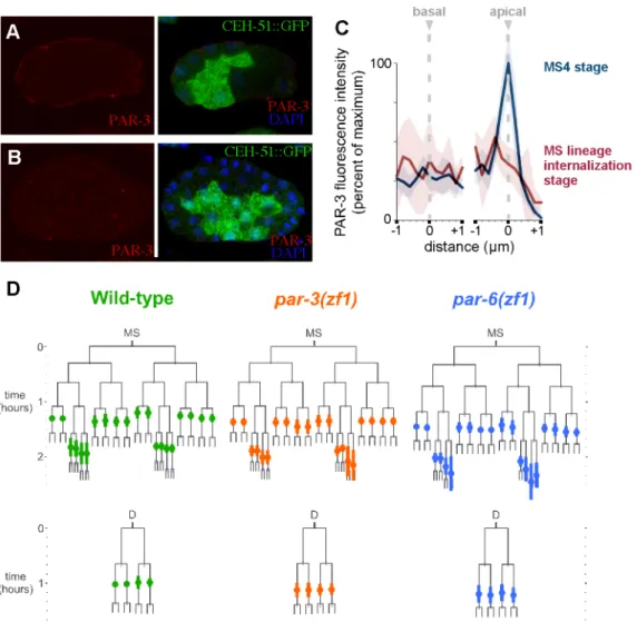
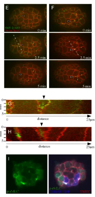
Figure 2.8 Internalizing mesodermal and germ line precursor cells accumulate myosin at their apical surfaces... 55
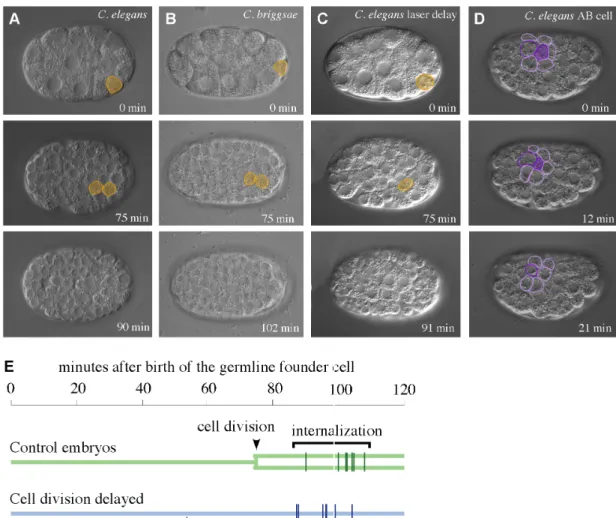
Figure 2.9 Cells generally move in as sister pairs but this is not required for efficient internalization ... 57
xxii
LIST OF ABBREVIATIONS AND SYMBOLS
AB AB lineage
C C lineage
C. briggsae Caenorhabditis elegans C. elegans Caenorhabditis elegans
DIC Differential Interference Contrast
D D lineage
E endoderm
MS mesoderm
GEF Guanine nucleotide Exchange Factor
GFP Green Fluorescent Protein
GTPase Guanine Triphosphate hydrolase
min minute
n number
N2 wild-type C. elegans strain
NMY-2 Non-muscle Myosin-2
p p-value
P4 germ line precursor
PAR Partitioning Defective
PH pleckstrin homology
PLC Phospholipase C
p-RMLC phosphorylated regulatory myosin light chain
xxiii
µm micrometer
WT wild-type
ZF1 Zinc Finger 1
Z2 primordial germ cell
Z3 primordial germ cell
< less than
> greater than
δ delta
CHAPTER 1 INTRODUCTION
This chapter represents my contribution to a review on apical constriction that has been accepted for publication in Developmental Biology (Sawyer et. al. 2010).
Organisms use a variety of mechanisms to achieve the morphogenetic
movements that shape the final animal. One conserved mechanism that is used
repeatedly in a variety of organisms to drive cell rearrangements and movements is
apical constriction, the narrowing of the apical surface of a cell. In 1902, Rhumbler
first proposed the importance of the constriction of the apical sides of cells to drive
the bending of cell sheets in a variety of developmental systems (Rhumbler, 1902).
Later, an epithelial sheet of cells was modeled using brass bars and rubber bands.
The addition of rubber bands increased tension on one side and resulted in a
concave shape, much like the bending of a sheet of cells (Lewis, 1947). This model
showed that increased tension on one side could result in bending, supporting the
hypothesis first proposed by Rhumbler.
Many years after Rhumbler and Lewis’s models, there are many known
examples of the use of apical constriction during morphogenesis, including during
2
(Sawyer et al., 2010). In addition, many historical and fundamental experiments on
the mechanisms driving cell shape changes have been completed in sea urchins,
3
1.1 SEA URCHIN GASTRULATION: MULTIPLE MECHANISMS MAY DRIVE TISSUE BENDING
Perhaps surprisingly, given the large repertoire of classes of morphogenetic
movements available to embryos, many organisms have evolved a role for apical
constriction in gastrulation (Stern, 2004). In gastrulating sea urchin embryos, cells
on the vegetal surface of the embryo become columnar, forming the vegetal plate.
The surface of this plate bends inward, a process termed primary invagination
(Figures. 1.1 and 1.2). Primary invagination is accompanied by a number of other
movements; here we discuss only the primary invagination, which has been
proposed to be driven by apical constriction (for review see Davidson et al., 1995;
Kominami and Takata, 2004).
The cells that undergo primary invagination form the archenteron, or future
gut. Computer modeling suggests that apical constriction of cells in the vegetal plate
could feasibly drive primary invagination, so long as the extracellular matrix can be
deformed easily—about as easily as the cells can be deformed (Davidson et al.,
1995). In principle then, changes of individual cell shapes can drive tissue bending,
although other mechanisms for bending a cell sheet are possible (Davidson et al.,
1995). Forces generated within the vegetal plate are sufficient to drive tissue
bending, as invagination can occur normally in a dissected vegetal plate (Moore and
Burt, 1939; Ettensohn, 1984). The cells proposed to undergo apical constriction
have bands of actin microfilaments associated with apical adherens junctions and
also spanning across the inside of each cell's apical surface, as might be expected
4
in cells that do not undergo such shape changes. Hence the presence of such an
apical microfilament network does not necessarily indicate that it will bend a cell
sheet (Ettensohn, 1984).
In certain species of sea urchin, a ring of cells along the edges of the vegetal
plate has been recognized to undergo more pronounced apical constriction, as
judged by scanning electron micrographs (Nakajima and Burke, 1996; Kimberly and
Hardin, 1998, Fig. 2). Cells in this ring have been referred to as bottle cells, a term
coined by Ruffini (1907) for amphibian embryonic cells that are shaped like bottles,
with dramatically constricted apical sides and enlarged basolateral areas. Bottle
cells in sea urchin embryos have a greater enrichment of apical arrays of F-actin
than do other cells in the vegetal plate (Nakajima and Burke, 1996). Laser ablation
of bottle cells interferes with normal invagination, whereas laser ablation of
neighboring cells does not (Kimberly and Hardin, 1998), consistent with the notion
that apical constriction may drive primary invagination. RhoA is required for the
initiation of primary invagination (Beane et al., 2006), as it is for apical constriction
and resulting tissue bending in other systems discussed below. How are specific
cells driven to apically constrict during primary invagination? This is not yet clear,
although calcium signaling (Nakajima and Burke, 1996), Wnt/Frizzled signaling
(Croce et al., 2006), a transcriptional gene regulatory network (Davidson et al., 2002;
Wu et al., 2008), and FGF signaling (Rottinger et al., 2008) have all been implicated
in regulating primary invagination. The links between these regulators and RhoA
5
One key result is at odds with the model that actomyosin- dependent apical
constriction is the key driver of primary invagination: cytochalasin treatment, which
should depolymerize F-actin networks, fails to fully disrupt primary invagination in
sea urchins (Lane et al., 1993). This result suggests the possibility that other
mechanisms may provide force, either alone or redundantly with actin-based
mechanisms. Interestingly, among the mechanisms proposed to drive apical
constriction and tissue bending in sea urchins during primary invagination is one in
which vegetal plate cells secrete extracellular matrix components into a multi-layered
structure, in a calcium regulated manner (Lane et al., 1993). In this model,
later-deposited matrix, secreted into a layer between the cells and the earlier layers of
matrix, swells as it hydrates, driving bending of the matrix and hence the attached
epithelial sheet. This is similar to the way in which the thermal expansion of a layer
of metal in a thermostat's bimetallic strip can bend the entire strip. In Lane et al.'s
model, the proposed source of force is extracellular, driving cell shape changes by
bending of the matrix, rather than mediated by intrinsic cell shape changes, an
interesting departure from traditional models. As an experimental model, sea urchin
primary invagination leaves a variety of possible mechanisms for tissue shape
change and some valuable tools for dissecting the contributions to forces made by
6
1.2 BOTTLE CELLS IN XENOPUS GASTRULATION: ROLES FOR MICROFILAMENTS AND MICROTUBULES
The amphibian archenteron also includes bottle cells at the site where
invagination begins (Holtfreter, 1943). Early embryologists believed that amphibian
bottle cells functioned in gastrulation because of the cells' unique shapes
(Figure 1.2). Rhumbler (1902) suggested the possibility that these cells were
actively migrating toward the interior of the embryo. Experiments by Holtfreter
(1944) were consistent with this hypothesis, as isolated bottle cells could stretch in a
polarized manner on a glass substrate, similar to migrating cells. While no live
imaging evidence exists for the active migration of these bottle cells in vivo, vital dye tracings demonstrate that these cells do migrate to the interior of the embryo in
Ambystoma mexicanum (Lundmark, 1986). In addition, cell tracing experiments in which labeled bottle cells from Xenopus laevis were grafted into unlabeled host embryos demonstrate that bottle cells spread out and form the anterior of the
archenteron (Hardin and Keller, 1988).
X. laevis bottle cells (Figure 1.3) are a potentially valuable model for studying mechanisms of cell shape change in morphogenesis, as the cells are large and
readily treated with inhibitors. These cells can be manipulated in culture much as
sea urchin cells can be, and the potential exists to identify key molecular players
using genetic screens in the model frog Xenopus tropicalis. Blastopore initiation begins and proceeds on schedule in explants that include the bottle cells (Hardin
7
cells still occurs, but archenteron length is compromised (Keller, 1981). Therefore,
bottle cells appear to initiate blastopore formation and to contribute to the full
extension of the archenteron in X. laevis.
A number of distinct mechanisms control cell shape in X. laevis bottle cells.
In vivo, the apical surfaces of these cells shrink while the apicobasal sides lengthen. Isolated, cultured bottle cells contract uniformly around the entire cell surface,
suggesting that contraction is an intrinsic behavior but that the apicobasal elongation
seen in vivo depends on contact with surrounding cells (Hardin and Keller, 1988). This likely reflects a cellular mechanism that distinguishes the basolateral and apical
sides of bottle cells, or surfaces contacting other cells and free surfaces, perhaps
similar to a mechanism that has been outlined in C. elegans, discussed below. F-actin and activated myosin accumulate at the apical surfaces of bottle cells just
before the apical surfaces narrow, consistent with a role for F-actin and myosin in
apical constriction (Lee and Harland, 2007). Furthermore, pharmacological
inhibitors of F-actin or myosin demonstrate that they are both required for bottle cell
formation. Interestingly, treatment with a microtuble depolymerizing drug,
nocodazole, prevents full apical constriction of bottle cells and invagination without
affecting apicobasal cell lengthening, and without apparent effects on F-actin or
activated myosin distribution (Lee and Harland, 2007). This result suggests that
microtubules may have an as yet undefined role in apical constriction in Xenopus
8
1.3 C. ELEGANS GASTRULATION: CELL MANIPULATIONS AND GENETICS MEET TO IDENTIFY KEY REGULATORS
Unlike gastrulation in sea urchins or Xenopus, where entire cell sheets are internalized, gastrulation in C. elegans involves the internalization of many cells or groups of cells at distinct times. C. elegans gastrulation begins at the 26-cell stage when two endodermal precursor cells move from the perimeter to the inside of the
embryo (Figure 1.4). This event is followed later by internalization of mesoderm and
germline precursors (Sulston et al., 1983; Nance and Priess, 2002). Internalization
of the endodermal precursors has been most thoroughly studied and is the focus of
our discussion here.
Cell movements associated with C. elegans gastrulation can occur in vitro, allowing mechanisms to be explored by cell manipulation experiments as in sea
urchins and Xenopus (Lee and Goldstein, 2003). One revealing finding from such studies is that very few cells are required for the movements of C. elegans
gastrulation to occur: even a line of embryonic cells in culture arranged in single file
will fold at the time of gastrulation (Lee and Goldstein, 2003). This makes clear that
mechanisms requiring large numbers of cells to work in concert, such as
multicellular purse string mechanisms, are not essential for cell movements in C. elegans gastrulation. Some of the strengths of this system lie in the ability to combine such manipulations with live cell microscopy and genetics, and to study
mechanisms of morphogenesis at the level of individual cells, in a developmental
9
Apical constriction plays a key role in C. elegans gastrulation. Just before endodermal precursor cells internalize, the cell surface that faces the perimeter of
the embryo on each of these cells (the apical surface) flattens, and myosin II
becomes enriched at this surface(Nance and Priess, 2002). Although the apical
surfaces become smaller until they disappear at the time of cell internalization, these
cells do not become noticeably bottle-shaped. Contraction of apical cell surfaces
was revealed by tracking the movements of fluorescent, microscopic beads placed
on the surfaces of the endodermal precursor cells (Lee and Goldstein, 2003). The
observed surface movements exclude the possibility that shrinking of the apical
surface reflects only a flow of apical surface to lateral positions—a possibility that is
difficult to exclude in many systems. Myosin has been implicated in driving apical
constriction because pharmacological inhibitors of myosin activity prevent the
endodermal precursors from internalizing (Lee and Goldstein, 2003). In addition,
apical myosin becomes activated near the time that gastrulation begins: apically
localized myosin regulatory light chain is phosphorylated at a residue that in other
systems unkinks myosin heavy chains, allowing myosin complexes to bundle into
bipolar filaments, which can bind to and walk on actin filaments (Lee et al., 2006;
Somlyo and Somlyo, 2003). These results suggest that local activation of myosin
shrinks the apical actin mesh. Actin architecture is likely to be important as well.
Indeed, the Arp2/3 actin-nucleating complex has been reported to localize to the cell
cortex in gastrulating embryos, and depletion of this complex results in failure of
endodermal precursor cells to internalize on schedule (Severson et al., 2002;
10
Do neighboring cells contribute to internalization of the endoderm precursors?
When neighboring cells were removed and reassociated with endodermal precursor
cells in various orientations, the neighboring cells still moved in a direction consistent
with the hypothesis that apical constriction in endodermal precursors drives the
movement of the neighboring cells, suggesting that neighboring cell polarity is not
important for the bulk of their movement (Lee and Goldstein, 2003). However, short,
actin-rich extensions form on three of the six neighboring cells of the ring that closes
beneath the endoderm precursors, and Arp2/3-depleted embryos that fail to
gastrulate also fail to produce these extensions, raising the possibility that the
extensions might contribute to completion of endodermal internalization in vivo
(Nance and Priess, 2002; Roh-Johnson and Goldstein, 2009).
C. elegans genetics has identified multiple regulatory inputs that are important for gastrulation, including inputs that specify which cells should enrich myosin to one
side, inputs that specify to which side of a cell this enrichment should occur, as well
as a signaling input that directs activation of myosin. Cell fate specification genes
including genes encoding endodermal GATA factors are necessary for early cell
internalization, and embryos with ectopically specified endoderm have ectopic early
cell internalization, suggesting that endoderm fate is both necessary and sufficient
for early cell internalization (Lee et al., 2006). One aspect of endodermal cell fate is
a gap phase uniquely introduced to the cell cycle of endodermal progenitors one cell
cycle after the endoderm precursor cell is born, which is near the time of cell
internalization (Sulston et al., 1983; Edgar and McGhee, 1988). This pause is
11
actomyosin cytoskeleton that normally accompanies cell division (Lee et al.,2006;
Oegema and Hyman, 2006).
For the endodermal precursor cells to accumulate myosin near their apical
surfaces, an apical surface must be established. PAR proteins function in
anteroposterior polarization of the embryo first, and are known to become
apicobasally polarized later, starting at the four cell stage (see Goldstein and
Macara, 2007 for review. To test whether PAR proteins function in apicobasal
polarization, Nance and colleagues devised a clever method for degrading the
polarity proteins PAR-3 or PAR-6 specifically in somatic cells, adding a motif from
another protein that becomes degraded in somatic cells. They demonstrated in this
way that PAR-3 and PAR-6 are required for apical flattening, apical myosin
enrichment, and timely cell internalization (Nance et al., 2003). Elegant cell
manipulation experiments revealed that these PAR proteins' localization depends on
where cells contact each other: only contact-free membranes accumulate apical
PAR proteins, establishing an apical domain at the contact-free surface (Nance et
al., 2003). Myosin later accumulates at apical domains, and this is dependent on
apical PAR proteins (Nance et al., 2003). Once myosin becomes enriched apically,
it becomes activated downstream of a Wnt–Frizzled–Dishevelled signaling pathway
that causes regulatory light chain phosphorylation, through an unidentified kinase
(Lee et al., 2006).
These results paint the outlines of a potentially generalizable mechanism for
cell internalization by apical constriction: Among cells that polarize PAR proteins
12
where the apical PAR proteins become localized—at contact-free surfaces.
Activation of myosin can then result in shrinking the myosin-enriched, contact-free
surfaces of any such cells, pulling neighboring cells across the free surfaces and, as
a result, displacing the apically constricting cells toward the interior. The ability to
shrink any exterior surface of specific cells could, in theory, make it possible for a
cell to internalize regardless of which specific surfaces initially contact other cells.
How then do certain PAR proteins become enriched only apically in response
to cell contacts? Anderson et al. (2008) screened for genes required for cell
contact-dependent PAR protein localization and identified a key intermediate, a RhoGAP
domain-containing protein, PAC-1. PAC-1 localizes to the cell cortex at cell–cell
contact zones, where it has been proposed to inactivate CDC-42 at these zones,
potentially restricting the active form of CDC-42 to contact-free cell surfaces. Active
CDC-42 interacts with a semi-CRIB domain in PAR-6,and through this interaction is
thought to establish apical localization of PAR-6 and PAR-6 complex members in
these cells. PAC-1 localization to contact zones is therefore the earliest known step
in recognizing contact zones as unique, spatial information that is critical to PAR
protein and myosin localization. How PAC-1 becomes localized to contact zones is
an interesting topic for future study.
While much work has been done to characterize the patterning mechanisms
that lead to apical constriction in the endodermal precursor cells, very little
information existed previously about the internalization of the other cells that must
13
Figure 1.1. Rhumbler’s 1902 drawings of cell shape changes driving morphogenesis (Rhumbler, 1902).
Top: A sea urchin embryo undergoing primary invagination. The vegetal-most part of
the embryo bends inward (arrowhead). Bottom: “Theoretical gastrulation scheme, to
show that invagination (b) of a cell plate (a) necessarily must take place if each cell
changes from form a1 (due to higher pressure on the pigmented side) to the form
b1. The invagination effect is significant even though the change in cell form from a1
to b1 is very small” (translation of figure legend in Rhumbler, 1902). We have
inverted some parts of this figure to match the orientation of tissue bending between
14
Figure 1.2. Scanning electron micrographs of apically constricting cells in sea urchins and X. laevis (Kimberly and Hardin, 1998; Keller, 1981).
A) Sea urchin vegetal plate (Kimberly and Hardin, 1998), B) X. laevis midsagittal section at early gastrula showing bottle cells (BC) and involuted mesodermal cell
stream (MCS) (Keller, 1981). Arrowheads mark bends in epithelia at proposed sites
15
Figure 1.3. Schematic diagrams of bottle cell formation (Hardin and Keller, 1988).
A) Prior to gastrulation, the prospective anterior mesoderm (darker shading) and
posterior mesoderm (lighter shading) comprise the deep marginal zone. B) The
bottle cells have undergone apical constriction. Arrows indicate movements
hypothesized to result. C) This causes reorientation of the vegetal edge of the
marginal zone (anterior mesoderm) such that it is now leading the movement into
16 Figure 1.4. C. elegans gastrulation.
A) Illustrations of embryos just before (top) and during (bottom) endodermal
internalization. Green, endodermal progenitors. Two neighboring cells are marked in
purple. Renderings by J. Iwasa based on confocal sections of phalloidin-stained
embryos (Lee et al., 2006). B) Diagram showing where apical constriction occurs
(arrowheads). C) Myosin is activated in the apical cortex of the internalizing cells.
17 REFERENCES
Anderson, D. C., Gill, J. S., Cinalli, R. M. and Nance, J. (2008). Polarization of the C. elegans embryo by RhoGAP‐mediated exclusion of PAR‐6 from cell contacts. Science 320, 1771‐1774.
Beane, W. S., Gross, J. M. and McClay, D. R. (2006). RhoA regulates initiation of invagination, but not convergent extension, during sea urchin gastrulation. Dev Biol 292, 213‐225.
Croce, J., Duloquin, L., Lhomond, G., McClay, D. R. and Gache, C. (2006). Frizzled5/8 is required in secondary mesenchyme cells to initiate archenteron invagination during sea urchin development. Development 133, 547‐557.
Davidson, E. H., Rast, J. P., Oliveri, P., Ransick, A., Calestani, C., Yuh, C. H., Minokawa, T., Amore, G., Hinman, V., ArenasMena, C. et al. (2002). A provisional regulatory gene network for specification of endomesoderm in the sea urchin embryo. Dev Biol 246, 162‐190. Davidson, L. A., Koehl, M. A., Keller, R. and Oster, G. F. (1995). How do sea urchins invaginate? Using biomechanics to distinguish between mechanisms of primary invagination. Development 121, 2005‐2018.
Ettensohn, C.A. (1984). Primary invagination of the vegetal plate during sea urchin gastrulation. Amer Zool 24, 571‐588.
Edgar, L. G. and McGhee, J. D. (1988). DNA synthesis and the control of embryonic gene expression in C. elegans. Cell 53, 589‐599.
Goldstein, B. and Macara, I. G. (2007). The PAR proteins: fundamental players in animal cell polarization. Dev Cell 13, 609‐622.
Hardin, J. and Keller, R. (1988). The behaviour and function of bottle cells during gastrulation of Xenopus laevis. Development 103, 211‐230.
Holtfreter, J. (1943). A study of the mechanics of gastrulation. Part I. J Exp Zool. 94, 261‐318. Holtfreter, J. (1944). A study of the mechanics of gastrulation. Part II. J Exp Zool. 95, 171‐212. Keller, R. E. (1981). An experimental analysis of the role of bottle cells and the deep marginal zone in gastrulation of Xenopus laevis. J Exp Zool 216, 81‐101.
18 Kominami, T. and Takata, H. (2004). Gastrulation in the sea urchin embryo: a model system for analyzing the morphogenesis of a monolayered epithelium. Dev Growth Differ 46, 309‐326. Lande M.C., Koehl, M.A., Wilt, F., Keller, R. (1993). A role for regulated secretion of apical extracellular matrix during epithelial invagination in the sea urchin. Development 117, 1049‐1060. Lee, J. Y. and Goldstein, B. (2003). Mechanisms of cell positioning during C. elegans gastrulation. Development 130, 307‐320.
Lee, J. Y. and Harland, R. M. (2007). Actomyosin contractility and microtubules drive apical constriction in Xenopus bottle cells. Dev Biol 311, 40‐52.
Lee, J. Y., Marston, D. J., Walston, T., Hardin, J., Halberstadt, A. and Goldstein, B. (2006). Wnt/Frizzled signaling controls C. elegans gastrulation by activating
actomyosin contractility. Curr Biol 16, 1986‐1997.
Lewis, W.H. (1947). Mechanics of invagination. Anat Rec 97, 139‐156.
Lundmark, C. (1986). Role of bilateral zones of ingressing superficial cells during gastrulation of Ambystoma mexicanum. J Embryol Exp Morphol 97, 47‐62.
Moore, A.R., Burt, A.S. (1939). On the locus and nature of the forces causing gastrulation in the embryos of Dendraster excentricus. J Exp Zool 82, 159‐171.
Nakajima, Y. and Burke, R. D. (1996). The initial phase of gastrulation in sea urchins is accompanied by the formation of bottle cells. Dev Biol 179, 436‐446.
Nance, J. and Priess, J. R. (2002). Cell polarity and gastrulation in C. elegans. Development 129, 387‐397. Nance, J., Munro, E. M. and Priess, J. R. (2003). C. elegans PAR‐3 and PAR‐6 are required for apicobasal asymmetries associated with cell adhesion and gastrulation. Development 130, 5339‐5350.
Oegema, K. and Hyman, A. A. (2006). Cell division. WormBook, 1‐40.
Rhumbler, L. (1902). Zur mechanik des gastrulationsvorganges, insbesondere der invagination. Arch Entw mech 14, 401.
RohJohnson, M. and Goldstein, B. (2009). In vivo roles for Arp2/3 in cortical actin organization during C. elegans gastrulation. J Cell Sci 122, 3983‐3993.
19
morphogenesis [corrected] and regulate gastrulation during sea urchin development.
Development 135, 353‐365.
Sawyer, J. M., Harrell, J. R., Shemer, G., SullivanBrown, J., RohJohnson, M. and Goldstein, B. (2010). Apical constriction: A cell shape change that can drive
morphogenesis. Dev Biol. In Press.
Severson, A. F., Baillie, D. L. and Bowerman, B. (2002). A Formin Homology protein and a profilin are required for cytokinesis and Arp2/3‐independent assembly of cortical microfilaments in C. elegans. Curr Biol 12, 2066‐2075.
Stern, C. (2004). Gastrulation: from cells to embryos. Cold Spring Harbor Laboratory, New York.
Somlyo, A. P. and Somlyo, A. V. (2003). Ca2+ sensitivity of smooth muscle and nonmuscle myosin II: modulated by G proteins, kinases, and myosin phosphatase.
Physiol Rev 83, 1325‐1358.
Sulston, J. E., Schierenberg, E., White, J. G. and Thomson, J. N. (1983). The embryonic cell lineage of the nematode Caenorhabditis elegans. Dev Biol 100, 64‐119.
CHAPTER 2
INTERNALIZATION OF MULTIPLE CELLS DURING C. ELEGANS
GASTRULATION DEPENDS ON COMMON CYTOSKELETAL MECHANISMS BUT DIVERSE CELL POLARITY AND CELL FATE REGULATORS
Understanding the links between developmental patterning and
force-producing cytoskeletal mechanisms is a central goal in studies of morphogenesis.
Gastrulation is the first morphogenetic event in the development of diverse
organisms. Gastrulation involves the internalization of surface cells, often driven by
the contraction of actomyosin networks that are deployed with spatial precision, in
specific cells and in specific positions within each cell. These cytoskeletal
mechanisms in diverse organisms rely on a multitude of cell polarity and cell fate
regulators. C. elegans gastrulation presents an opportunity to examine the diversity of mechanisms used within a single organism by dozens of cells that become
internalized at distinct times. Nearly half of the cells of the C. elegans embryo are born on the embryo’s surface and move to the interior. Here, we present an
identification of all of the cells that become internalized in C. elegans gastrulation. To gain mechanistic insights into how diverse cells internalize, we have manipulated
cell polarity, cell fate and cytoskeletal regulators and determined the effects on cell
21
actomyosin-based mechanisms to gastrulate, but different cell fate regulators, and,
surprisingly, different cell polarity regulators. Apical PAR proteins that function in
apical myosin localization and internaliztion of endodermal cell precursors do not
function in cell internalization in other lineages. We conclude that much as diverse
organisms employ diverse patterning mechanisms to regulate common cytoskeletal
mechanisms for cell internalization, diverse patterning mechanisms can be
22 2.1 INTRODUCTION
Morphogenesis is the process by which cells in an embryo become an
organism with ordered shape and structure. One key goal in studying
morphogenesis is to understand the links between patterning mechanisms controlled
by cell fate and cell polarization and the cytoskeletal mechanisms that drive the
embryonic shape changes (Wieschaus, 1995). Such links are being identified in
diverse developmental systems. One theme emerging is that diverse animals
employ a handful of common cytoskeletal mechanisms, and these are regulated by
diverse and often organism-specific patterning mechanisms. For example,
contraction of an apical actomyosin network internalizes cells in diverse systems. In
Drosophila gastrulation, an actomyosin network is spatially regulated by an apically-localized guanine nucleotide exchange factor for Rho (RhoGEF). Recruitment of this
RhoGEF to the apical sides of cells depends on the secreted protein Fog and the
transmembrane protein T48 (Barrett et al., 1997; Nikolaidou and Barrett, 2004;
Rogers et al., 2004; Kolsch et al., 2007). Fog and T48 homologs are not known in
vertebrates, where the F-actin binding protein Shroom3 acts instead as an apical
determinant. Shroom3 recruits a myosin activator, Rho kinase, to the apical sides of
cells, and also affects directly or indirectly the apical localization of myosin and
F-actin (Hildebrand and Soriano, 1999; Haigo et al., 2003; Hildebrand, 2005;
Nishimura and Takeichi, 2008). C. elegans uses yet another mechanism for apico-basal polarization of similar actomyosin based forces. Here, a putative GTPase
activating protein for Cdc42 localizes basolaterally in response to cell contacts,
where it prevents the localization of apical PAR proteins that are important for apical
myosin localization (Nance et al., 2003; Anderson et al., 2008). These cases
illustrate that different apico-basal patterning mechanisms can control apical
23
fate regulators control the apical constriction machinery in specific cells in diverse
animal systems (see Sawyer et al., 2010 for review).
The extent to which such diversity exists within each organism is less clear.
There are cases where different cells internalize by morphologically distinct
processes in a single animal system, for example between gastrulation and tracheal
tube formation in Drosophila. In these cases, different patterning mechanisms are indeed known to act upstream of common cytoskeletal mechanisms (Leptin and
Grunewald, 1990; Brodu and Casanova, 2006). But the extent to which such
diversity of spatial patterning mechanisms might regulate repeated, superficially
similar morphogenetic processes within a single organism is not well known. When
many cells move in the same direction and by superficially similar patterns, do they
use the same cytoskeletal mechanisms to do so, and are these mechanisms
associated with common upstream spatial patterning mechanisms? Answering this
will be a key component to understanding the links between patterning mechanisms
and the cytoskeletal mechanisms that drive morphogenesis. Answering this will also
provide insight into which specific mechanisms discovered in model systems will be
more or less likely to be informative to understanding morphogenesis-related birth
defects in humans. We consider this to be important because
morphogenesis-related birth defects like neural tube closure defects are common human birth
defects (Copp and Greene, 2010)
Gastrulation is the earliest morphogenetic process in many animal embryos.
During gastrulation, cells that will establish the internal germ layers – the endoderm,
the mesoderm and the germ line – move from the outside of the embryo to the
interior, leaving just the ectoderm at the surface. C. elegans gastrulation is a valuable model for studying the cellular and molecular mechanisms involved in cell
internalization in part because gastrulation begins early in development, soon after
24
an understanding of the regulation of gastrulation at the level of individual cells is
possible. Many cells internalize at distinct times during C. elegans gastrulation, and nearly all of these cells do so from the ventral surface of the embryo (Sulston et al.,
1983; Nance and Priess, 2002). As we will show here, nearly all of these cells
internalize as sister cell pairs. Therefore, C. elegans gastrulation also presents an unusual opportunity to study the links between patterning and cytoskeletal
mechanisms for a multitude of cells that are internalized at distinct times but in
superficially similar ways.
C. elegans gastrulation begins with the internalization of two endoderm precursor cells, a process that is driven at least in part by actomyosin-dependent
apical constriction (Nance and Priess, 2002; Lee and Goldstein, 2003; Nance et al.,
2003; Lee et al., 2006; Anderson et al., 2008; Roh-Johnson and Goldstein, 2009).
Cell fate, cell polarity and cytoskeletal mechanisms comprise three known
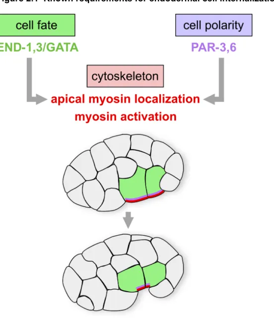
requirements for timely internalization of the endoderm (Figure 2.1). First,
endodermal cell fate must be specified properly for the endodermal precursors to
internalize on schedule. Mutant embryos defective in endodermal cell fate
specification fail to internalize the cells that would normally become endodermal
(Zhu et al., 1997; Maduro et al., 2005; Lee, 2006). In addition, in mutants that
produce ectopic endoderm, the ectopic endodermal precursors internalize soon after
the normal endodermal precursors do (Lee, 2006). Second, apicobasal polarity is
regulated by apically-localized PAR proteins. These PAR proteins are required for
apical flattening and enrichment of a nonmuscle myosin II protein (NMY-2) on the
apical surface of each endodermal precursor cell (Nance, 2003). Third, these
endodermal precursor cells must undergo an actomyosin-driven apical constriction,
which is dependent on a Wnt-Frizzled-Disheveled signaling pathway (Lee, 2006).
Phosphorylation of the regulatory light chains of myosin at a conserved site is