95
HISTO-ANATOMICAL AND CHROMATOGRAPHIC RESEARCHES ON
CAMPANULA PERSICIFOLIA
L. (
CAMPANULACEAE
) SPECIES
GEORGE DAN MOGOŞANU1
, CORNELIA BEJENARU2, LUDOVIC EVERARD BEJENARU1, ANDREI BIŢĂ1, ANTONIA BLENDEA2, IULIA DARIA SCOREI3*
1
Department of Pharmacognosy & Phytotherapy, Faculty of Pharmacy, University of Medicine and Pharmacy of Craiova; E-mail: [email protected]
2
Department of Vegetal & Animal Biology, Faculty of Pharmacy, University of Medicine and Pharmacy of Craiova; E-mail: [email protected]
3
BioBoron Research Institute, S.C. Natural Research S.R.L., Craiova; E-mail: [email protected]
Keywords: Campanula persicifolia L., Campanulaceae, histo-anatomy, polyphenols, thin-layer chromatography.
ABSTRACT
The paper presents the histo-anatomical researches on root, rhizome,
aboveground stem and leaf of
Campanula persicifolia L.
(Campanulaceae) species, together with
the thin-layer chromatography analysis of the polyphenols content of Campanulae persicifoliae herba. Chlorogenic acid (108.6 μg/mL) was identified in the 20% methanolic extract of the aerial parts.
INTRODUCTION
Campanula persicifolia L.,
Peach-bells, Peach-leaf bell-flower
(Campanulaceae), is a common plant in
Romania’s flora, blooming in July– August, through mesophilic meadows and forests, from the plain to the subalpine area. It is cultivated as ornamental through parks and gardens [5, 10].
From the phytochemical point of view, Campanula species contain a wide range of active principles, as follows: flavonosides (isoquercitrin, diosmin, rutin), anthocyanosides (pelargonidin, delphinidin and cyanidin derivatives), coumarins (fraxoside), phenylpropane derivatives (barbatosides A–D), essential oil (linalool, α-terpineol, lavandulyl acetate, allo-ocimene, β-pinene, α -cadinene, β-farnesene, β-caryophyllene), polyacetylenes (lobetyol, lobetyolin), acylated triterpenoids, phenolic acids, sterols (β-sitosterol), fructosans, sugar alcohols (myo-inositol), cyanogenic heterosides, piperidine alkaloids (lobelin, campedin), fatty oil, resins, enzymes [4,
6, 9, 11, 16].
Various extracts (aqueous, methanolic), essential oils and diterpenoid components obtained from
Campanula species have analgesic,
anti-inflammatory, antioxidant and anti-microbial properties [6, 12, 15]. For the calming, sedative and haemostatic effects, in the Romanian ethnopharmacology are used several extracts from C. abietina, C. patula and
C. trachelium. Leaves, roots and freshly
harvested flowers of C. persicifolia are also used for food purposes in the form of salads [13].
There is no information about the histo-anatomy of C. persicifolia, in the specialized papers we consulted. The histo-anatomical analyses of the roots, rhizomes, aboveground stems and leaves
of C. persicifolia, as well as the
preliminary thin-layer chromatography investigation of polyphenols content from the aerial parts (Campanulae persicifoliae
96
MATERIALS AND METHODS
Histo-anatomical analysis
For Campanula persicifolia species,
the vegetal material was collected in June 2015, in the blooming period, from the “Alexandru Buia” Botanical Garden, University of Craiova, Dolj County (southwestern Romania).
A 70% ethanol solution was used for the fixation and preservation of the biological material (roots, rhizomes, aboveground stems, leaves). The cross-sections were obtained using a botanical razor.
After washing with distilled water, the sections were clarified in Javel water (a 10% sodium hypochlorite solution). The sequential washing of the sections was made also with distilled water to remove the clarification agent [2].
Congo red–chrysoidine mixture (Genevese reagent) was applied for the sections’ staining, obtaining several colors, according to the chemical composition of cell membranes: pink-red for cellulose and mucilages, pale red for cytoplasm, yellow for suberin, and brown for lignin [2].
Krüss binocular photon microscope (objectives ×4, ×10, ×20, ×40) was used for the analysis of stained and mounted sections. Nikon Eclipse 55i binocular microscope and Nikon DS–Fi1 high definition video camera were applied to take photos. Image-Pro Plus ver. 6.0 software package (Media Cybernetics) was employed for images acquisition and processing.
The histo-anatomical analysis was achieved starting from a reference work [14].
Thin-layer chromatography
(TLC) investigation
Using CAMAG (Muttenz, Switzerland) system, the preliminary TLC investigation of polyphenols from the
aerial parts of C. persicifolia species
(Campanulae persicifoliae herba) was
accomplished in the following experimental conditions [1, 3, 7, 8]:
▪ stationary phase: TLC silica gel 60 F254 (Merck, Darmstadt, Germany)
10×10 cm precoated glass plates, prewashed with chloroform–methanol (1:1, v/v) and activated by oven-drying (1100C, 30 minutes);
▪ mobile phase: ethyl acetate– formic acid–methanol–water (15:1:0.1:1, in volumes);
▪ 10 mL of mobile phase were added in the developing twin-chamber (CAMAG) and then oversaturated for 20 minutes;
▪ sample: 20% methanolic extract
of Campanulae persicifoliae herba;
▪ standards (Merck): 0.05% methanolic solutions of caffeic acid, chlorogenic acid, quercetin and rutin;
▪ migration distance: 62 mm (sample application line 8 mm, solvent front 70 mm);
▪ sample (8 μL, 10 μL) and standards (2 μL) application: CAMAG Linomat 5 semi-automatic system – spray gas nitrogen, syringe volume 100 μL, dosage speed 150 nL/s, predosage volume 0.2 μL, bands length of 8 mm;
▪ plate drying: 5 minutes, at 250
C (cold air dryer);
▪ photographing the chromatographic plate: UV light (λ 254 nm);
▪ detection: CAMAG TLC Scanner 3 photodensitometer, for densitogram and
in situ UV light (λ 280 nm) spectra,
without derivatization, deuterium– tungsten lamp, scanning speed 40 mm/s, data resolution 200 μm/step, measurement mode absorption;
97
RESULTS AND DISCUSSIONS
Histo-anatomical analysis
Root
In cross-section, in the lower third area, the root has round shape and secondary structure due to the two secondary meristems: subero-phellodermic cambium (phellogen) and libero-ligneous cambium. The following histological sequence was evidenced in cross-section, from the outside towards the inside of the root: Peridermis consists of suber, phellogen and phelloderm. The suber is made up of 4–5 layers of large, flattened, suberin-impregnated cells. From place to place, it is exfoliated. The subero-phellodermic cambium consists of one layer of antero-posterior flattened cells, with thin walls, of which the radial walls are slightly curled. The phelloderm is made up of 4–5 layers of cells with
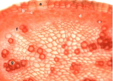
cellulosic thin walls. The conducting tissues are arranged on two concentric rings. Phloem tissue forms a thin, external ring, consisting of sieve tubes, phloem parenchyma and annex cells. A single layer of libero-ligneous cambium is found between the xylem and phloem tissues. The central area of the root is occupied by the xylem tissue, consisting of few metaxylem vessels of different calibers, with disordered layout in the libriform tissue mass, pushing to the center the small diameter protoxylem vessels accompanied by xylem parenchyma. The medullary rays are multicellular, uniseriate and cellulosic, into the phloem tissue, and multicellular, uniseriate and slightly lignified, at the level of xylem tissue. The medullary parenchyma is absent (Figure 1).
Figure 1. Cross-section through C. persicifolia root: (a) suber; (b) phellogen; (c) phelloderm; (d) cortical parenchyma; (e) phloem tissue; (f) libero-ligneous cambium; (g) xylem tissue
(Congo red–chrysoidine staining, ×200).
Rhizome
At lower third level, in cross-section, the rhizome has circular shape and secondary structure due to the presence of the subero-phellodermic cambium (phellogen) and libero-ligneous
98
consists of 7–8 layers of large, flattened cells, impregnated with suberin. The phellogen has one layer of antero-posterior flattened cells, with thin walls and the radial walls slightly curled. The phelloderm has 2–3 cell layers with cellulosic thin walls. The conducting tissues are disposed on two concentric rings. A thin, external ring, made up of sieve tubes, phloem parenchyma and annex cells represents the phloem tissue. At the phloem tissue level, the medullary rays are multi-cellular, uniseriate and cellulosic. Between the xylem and phloem tissues, one circular layer of libero-ligneous cambium is found. The xylem tissue forms a thick, inner ring, composed of few metaxylem vessels with different calibers, unevenly spread in the
well-represented libriform tissue. The metaxylem has reticulate thickenings highlighted in the longitudinal-radial sections. The medullary rays are multicellular, uniseriate and lignified, at the level of xylem tissue. To the center of the rhizome, the metaxylem is placed in radial strings and is accompanied by xylem parenchyma. Located in the vicinity of the medullary parenchyma, the protoxylem is poorly represented by few xylem vessels having small diameter and by xylem parenchyma. At the level of metaxylem and of protoxylem, the medullary rays are multicellular, uniseriate and cellulosic. The medullary parenchyma is poorly developed (Figures 2 and 3).
Figure 2. Cross-section through C. persicifolia rhizome: overview (Congo red–chrysoidine staining, ×40).
99
Aboveground stem
In the upper third, in cross-section, the aboveground stem has round-ribbed shape and secondary structure due to the libero-ligneous cambium. From the outside towards the inside of the aboveground stem, the following histological sequence is highlighted in cross-section: The epidermis has quasi-isodiametric cells; a thick cuticle with toothed relief covers the thickened external wall. The epidermal cells are slightly tangential elongated, with thin radial walls and thickened tangential external and internal walls. Stomata are found in patches. The bark is organized in 5–6 layers of angular collenchyma at the ribs level and 2–3 layers of chlorenchyma between the ribs. The bark inner area is parenchymatous and comprises a single layer of endodermis made up of large cells provided with Casparian thickenings. The conducting tissues are organized into numerous collateral-open libero-ligneous
fascicles of various sizes. Sieve tubes, phloem parenchyma and annex cells define the phloem tissue. At this level, the medullary rays are multicellular, multiseriate and cellulosic. One layer of libero-ligneous cambium with circular-sinuous shape was found. The secondary xylem tissue consists of well-represented libriform tissue, placed near the intrafascicular cambium, and of metaxylem vessels with different calibers, arranged in radial strings. The xylem vessels exhibit reticulate and helical thickenings, in longitudinal-radial sections. Few primary xylem vessels and xylem parenchyma are specific for the poorly represented primary xylem tissue. At the xylem level, the medullary rays are multicellular, multiseriate and lignified. The medullary parenchyma is well developed, of meatus type. In the central area, the aboveground stem has a medullary lacuna (Figures 4–6).
100
Figure 5. Cross-section through C. persicifolia aboveground stem: (a) libero-ligneous cambium; (b) libriform tissue; (c) metaxylem; (d) protoxylem; (e) medullary ray; (f) medullary parenchyma
(Congo red–chrysoidine staining, ×200).
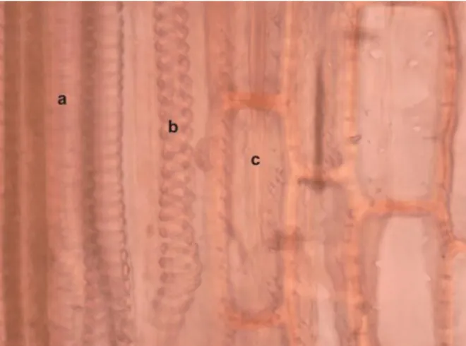
Figure 6. Longitudinal-radial section through C. persicifolia aboveground stem: (a) reticulate xylem vessel; (b) helical xylem vessel; (c) xylem parenchyma
(Congo red–chrysoidine staining, ×400).
Leaf’s limb
In cross-section, from the outside towards the inside of leaf’s limb, the following histological sequence is evidenced: The upper epidermis is made up of large, flattened cells, with thickened tangential external and internal walls and thin radial walls. The external walls are bulged and covered by a thick cuticle with toothed relief. The mesophyll consists of one layer of palisade parenchyma, with large and elongated cells, rich in chloroplasts, but also of 3–4 layers of lacunose parenchyma, having small cells with disordered layout and aeriferous spaces. Numerous small libero-ligneous conducting fascicles, surrounded by assimilatory sheaths, are found into the mesophyll. The plant presents C4
101
Figure 7. Cross-section through C. persicifolia leaf’s limb: (a) upper epidermis; (b) palisade parenchyma; (c) lacunose parenchyma; (d) libero-ligneous fascicle; (e) assimilatory sheath;
(f) lower epidermis; (g) stomate; (h) angular collenchyma (Congo red–chrysoidine staining, ×100).
Figure 8. Cross-section through C. persicifolia leaf’s limb: (a) upper epidermis; (b) cuticle; (c) palisade parenchyma; (d) lacunose parenchyma; (e) libero-ligneous fascicle; (f) assimilatory
sheath; (g) lower epidermis; (h) stomate (Congo red–chrysoidine staining, ×200).
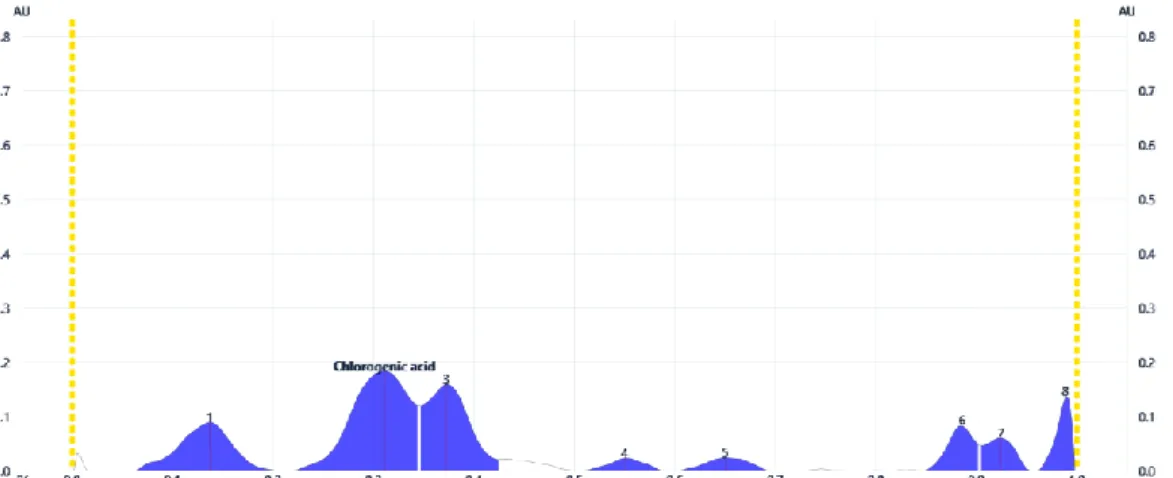
TLC investigation
The experimental data about the preliminary TLC investigation of polyphenols from Campanulae
persicifoliae herba are shown in Figures
9–11. In the 20% methanolic extract,
starting from the eight fingerprint chromatographic bands, chlorogenic acid (Rf 0.31) was quantified in an amount of
102
Figure 9. TLC chromatogram of polyphenols from Campanulae persicifoliae herba 20% methanolic extract (UV 254 nm, without derivatization). From left to right: first four
bands – standards (2 μL); last two bands – sample (8 μL and 10 μL).
Figure 10. Densitogram of polyphenols (UV 280 nm, without derivatization) separated from Campanulae persicifoliae herba 20% methanolic extract. Chlorogenic acid was
identified at Rf 0.31.
103
separated from the analyzed sample.
CONCLUSIONS
For Campanula persicifolia
species, the histo-anatomical analysis of root, rhizome, aboveground stem and leaf, as well as the preliminary TLC investigation of polyphenols from the aerial parts were accomplished. In the lower third, the root and also the rhizome have round shape and secondary
structure. In the upper third, the aboveground stem has round-ribbed shape and secondary structure. The leaf’s limb has a bifacial, dorsiventral, hypostomatic structure. Chlorogenic acid was quantified in the 20% methanolic extract.
BIBLIOGRAPHY
1. Altemimi, A., Watson, D.G., Kinsel,
M., Lightfoot, D.A., 2015 – Simultaneous
extraction, optimization, and analysis of flavonoids and polyphenols from peach and pumpkin extracts using a
TLC-densitometric method, Chem. Cent. J.
9:39.
2. Andrei, M., Paraschivoiu, R.M., 2003
– Microtehnică botanică, Ed. Niculescu,
Bucureşti, 2003, 222 pag.
3. Bojić, M., Simon Haas, V., Sarić, D.,
Maleš, Z., 2013 – Determination of
flavonoids, phenolic acids, and xanthines
in mate tea (Ilex paraguariensis St.-Hil.),
J. Anal. Methods Chem. 2013:658596.
4. Brandt, K., Dötterl, S., Francke, W.,
Ayasse, M., Milet-Pinheiro, P., 2017 –
Flower visitors of Campanula: are
oligoleges more sensitive to host-specific
floral scents than polyleges? J. Chem.
Ecol. 43(1):4–12.
5. Ciocârlan V., 2000 – Flora ilustrată a
României. Pteridophyta et
Spermatophyta, ediţia a 2-a revizuită şi
adăugită, Ed. Ceres, Bucureşti, 1138 pag.
6. Dumlu, M.U., Gurkan, E., Tuzlaci, E.,
2008 – Chemical composition and
antioxidant activity of Campanula
alliariifolia, Nat. Prod. Res. 22(6):477–
482.
7. Gîrd, C.E., Nencu, I., Costea, T.,
Duţu, L.E., Popescu, M.L., Ciupitu, N.,
2014 – Quantitative analysis of phenolic compounds from Salvia officinalis L.
leaves, Farmacia 62(4):649–657.
8. Jug, U., Glavnik, V., Kranjc, E., Vovk,
I., 2018 – High-performance thin-layer chroma-tography and high-performance
thin-layer chromatography–mass
spectrometry methods for the analysis of
phenolic acids, J. Planar Chromatogr.
31(1):13–22.
9. Kim, H.J., Son, D.C., Kim, H.J., Choi,
K., Oh, S.H., Kang, S.H., 2017 – The
chemo-taxonomic classification of Korean Campanulaceae based on triterpene,
sterol, and poly-acetylene contents,
Biochem. Syst. Ecol. 74:11–18.
10. Koutsovoulou, K., Daws, M.I.,
Thanos, C.A., 2014 – Campanulaceae: a
family with small seeds that require light
for germination, Ann. Bot. 113(1):135–
143.
11. Ouzounis, T., Fretté, X.,
Rosenqvist, E., Ottosen, C.O., 2014 –
Spectral effects of supplementary lighting on the secondary metabolites in roses,
chrysanthemums, and campanulas, J.
Plant Physiol. 171(16):1491–1499.
12. Park, S.H., Sim, Y.B., Lim, S.S.,
Kim, J.K., Lee, J.K., Suh, H.W., 2010 –
Anti-nociception effect and mechanisms of Campanula punctata extract in the
mouse, Korean J. Physiol. Pharmacol.
14(5):285–289.
13. Pârvu, C., 2002 – Enciclopedia
plantelor. Plante din flora României. Vol.
104
14. Toma, C., Rugină, R., 1998 –
Anatomia plantelor medicinale. Atlas, Ed.
Academiei Române, Bucureşti, 320 pag.
15. Usta, C., Yildirim, A.B., Turker,
A.U., 2014 – Antibacterial and antitumour
activities of some plants grown in Turkey,
Biotechnol. Biotechnol. Equip. 28(2):306– 315.
16. Vergauwen, R., Van den Ende, W.,
Van Laere, A., 2000 – The role of fructan
in flowering of Campanula rapunculoides,