R E V I E W A R T I C L E
Adverse eff ects of radiotherapy on oral tissues: A review
K. Shwetha Nambiar, Vanishri C. Haragannavar, Dominic Augustine, S. V. Sowmya, Roopa S. Rao
Department of Oral Pathology and Microbiology, M S Ramaiah University of Applied Sciences, Bengaluru, Karnataka, India
Abstract
Oral cancer is a major health problem in developing countries like India that is attributed mainly due to tobacco chewing habit. Oral squamous cell carcinoma (OSCC) accounts to about 90% of all oral cancers and holds 3rd place in South Central Asia. OSCC is managed by surgery, radiotherapy, or chemotherapy, or a combination of any of these modalities. Radiotherapy is a part of cancer treatment where high doses of radiation are delivered to large areas of oral cavity that includes the lesional areas as well as surrounding structures. This may result in several undesired reactions that manifest during or after the completion of therapy. The acute and chronic eff ects of radiation on oral tissues are discussed in detail here. The irreversible damage caused to the oral tissues is related to the dosage of radiation, the fi eld of irradiation, the degree of hypovascularity/hypocellularity of tissues, the age of the patient, and the wound healing capacity. Majority of the patients undergoing treatment have a compromised quality of life as a side eff ect of radiation therapy. Hence, in-depth knowledge of radiation exposure, adequate dosage, modality, general state, and prognosis of each case is essential to evaluate personalized treatment plan. The severity of the complications can be minimized by implementing oral care protocols before, during, and after radiation therapy. A multidisciplinary treatment plan with dental surgeons, radiotherapists, speech therapists, nutritionists, and psychologists is required for patient management.
Keywords: Mucositis, oral cancer, osteoradionecrosis, taste dysfunction, trismus, xerostomia
Correspondence
Dr. K. Shwetha Nambiar, Department of Oral Pathology and Microbiology, M S Ramaiah University of Applied Sciences, Bengaluru – 560 054, Karnataka, India. Phone: +91-7259469783. E-mail: [email protected]
Received 29 July 2016; Accepted 02 September 2016
doi: 10.15713/ins.ijcdmr.105
How to cite the article:
K. Shwetha Nambiar, Vanishri C. Haragannavar, Dominic Augustine, S. V. Sowmya, Roopa S. Rao, “Adverse eff ects of radiotherapy on oral tissues: A review,” Int J Contemp Dent Med Rev, vol.2016, Article ID: 020816, 2016. doi: 10.15713/ins.ijcdmr.105
Introduction
Radiotherapy is a curative medical intervention in cancer therapeutics. High doses of radiation used to destroy cancer cells can cause side eff ects because radiation can damage healthy cells and tissues near the zone of radiation. Total body irradiation and irradiation to the head and neck cause several adverse eff ects that manifest during or after the course of treatment. Today, major advances in radiotherapy have made it more precise with fewer side eff ects. The multidisciplinary management of oral cancer is essential to minimize eff ects of ionizing radiation on oral tissues. This paper reviews the adverse eff ects of radiation therapy on oral tissues.
Rationale of Radiotherapy of Oral Cavity
Radiation therapy is part of the oral cancer treatment. Most of the aggressive oral cancers require radiotherapy either as a primary mode of treatment, pre-surgery/post-surgery, part of radiochemotherapy, or as palliative therapy.[1] The radiation dose is assessed by tumor variables such as size, location, invasion, histological diagnosis, radiosensitivity, and invasion into adjacent structures.[2] Radiation therapy is indicated when
the lesion is radiosensitive, advanced or deeply invasive, and cannot be approached surgically.[3]
Classifi cation of Eff ects of Radiation Therapy on Oral Cavity
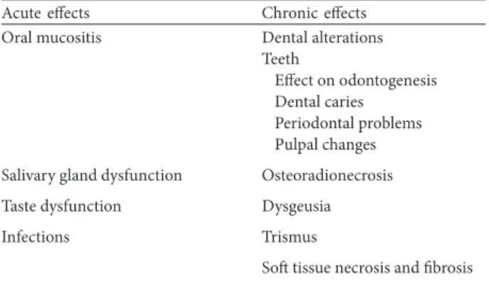
The eff ects of radiation on oral tissues can be classifi ed as acute and chronic as given in Table 1.
Acute eff ects
Mucositis
Radiotherapy induced mucositis is an infl ammatory reaction of the mucous membrane of the oral/oropharyngeal area during radiation therapy.[4] The basal mucosal layer is composed of rapidly dividing radiosensitive cells that begin to show areas of redness and infl ammation (mucositis). Radiation mucositis is considered to be an inevitable but transient side eff ect of therapeutic head and neck irradiation.[5]
Clinical presentation
sensitivity to hot and spicy food.[6] As the therapy continues the irradiated mucous membrane shows redness, infl ammation, it begins to break down, with the formation of white to yellow psuedomembrane (the desquamated epithelial layer). Thus erythematous areas may develop into elevated white desquamative patches and subsequently into painful ulcers which becomes secondarily infected. This impairs the nutrition and fl uid intake, resulting in malnutrition and dehydration.[6]
Scales developed by the National Cancer Institute and the World Health Organization,[7] based on common toxicity criteria [Table 2].
Pathophysiology
The acute mucosal response to radiotherapy is due to basal cell death of the mucosal epithelium, compromising its capacity to regenerate itself thus leading to thinning of epithelium and ulcerations.[8] It also damages the endothelium of the blood vessels.[9]
Histopathology
It shows atrophy of epithelium, absence of vascular damage, and juxtaepithelial dense infl ammatory infi ltrate. Degenerative changes such as homogenization of the collagen and mucoid degeneration are seen. The submucosa will gradually increase in collagen content and become less vascular and more fi brotic.[10]
Salivary gland pathoses
Parenchymal component (salivary acini) is radiosensitive. Serous cells are more radio-sensitive than mucous cells thus parotid glands are more sensitive than submandibular or sublingual glands. Radiation thus tends to aff ect the parotid gland earlier to the other major salivary glands and therefore residual saliva is more viscous.
Initial radiation induced changes include degeneration or destruction of acinar tissue with subsequent infl ammation and marked loss of salivary secretion (hyposalivation) in fi rst few weeks. Months after irradiation infl ammatory response becomes more chronic and glands demonstrate progressive fi brosis, adiposis, loss of fi ne vasculature, and concomitant parenchymal degeneration, thus accounting for xerostomia. There is usually diffi cult and painful swallowing due to loss of lubricating properties of residual saliva. Low concentration of Ca+2 in individuals with xerostomia leads to greater solubility of tooth structure and reduced remineralization.[3,4]
Hyposalivation
The fi rst few weeks after the initiation of radiotherapy, a marked and progressive loss of salivary secretion is seen. The mouth becomes dry and tender. Swallowing is diffi cult and painful as saliva also loses its normal lubricating properties. Four phases of loss of salivary gland function induced by radiation in rat parotid gland were observed by Coppes et al. They are as follows:[3,4] • The fi rst phase (0-10 days): Characterized by decrease in
salivary fl ow rate without any changes in amylase secretion or cell number of acini
• The second phase (10-60 days): Decrease in amylase secretion and acinar cell loss
• The third phase (60-120 days): Salivary fl ow rate, amylase secretion with no change in acinar cell numbers
• The fourth phase (120-240 days): Deterioration of salivary gland function with poor tissue morphology but increase in number of acinar cells.
The fi nal degree of radiation-induced hyposalivation depends on patient characteristics, such as patient age, gender, and pre-irradiation salivary gland function.[11,12]
Rating scales can be used to describe the degree of xerostomia. The Radiation Therapy Oncology Group uses two scales: One for acute reactions; the other for late or delayed reactions[13] as shown in Table 3.
Alterations in composition of saliva
Change in salivary composition makes it very viscous. The salivary pH, buff ering capacity reduces, electrolyte levels are altered and changes are seen in immune/non-immune antibacterial systems. The average pH decreases from 7 to 5 which can initiate the decalcifi cation of normal enamel. Since the overall immunity is compromised, alterations are seen in the oral microbial fl ora of the patients undergoing radiotherapy.[14]
Histopathology
Initial changes include the degeneration or destruction of acinar tissue with subsequent infl ammation. Chronic exposure leads Table 1: Classifi cation of eff ects of radiation therapy on oral cavity
Acute eff ects Chronic eff ects Oral mucositis Dental alterations
Teeth
Eff ect on odontogenesis Dental caries
Periodontal problems Pulpal changes
Salivary gland dysfunction Osteoradionecrosis
Taste dysfunction Dysgeusia
Infections Trismus
Soft tissue necrosis and fi brosis
Table 2: Systems for rating mucositis severity
Score National Cancer Institute rating WHO rating
0 None No symptoms
1 Painless ulcers, erythema, or mild soreness
Sore mouth, no ulcers
2 Painful erythema, edema, or ulcers; however patient can eat solid food
Sore mouth with ulcers but able to eat normally
3 Painful erythema, edema, or ulcers; patient cannot eat solid food
Liquid diet only
4 Requires parenteral or enteral support (such as gastric feeding tube) to provide nutrition
to fi brosis, adiposis, loss of fi ne vasculature, and concomitant parenchymal degeneration.[15]
Dry mouth/xerostomia, burning sensation, increased thirst, taste alterations, diffi culties in oral functioning and wearing dentures, soft tissue alterations, disturbances in oral microfl ora, oral discomfort at night, radiation induced caries, mucus accumulation, and gingival/periodontal disease are the consequences of radiation-induced hyposalivation in the oral cavity.[16]
Taste dysfunction
Taste could be defi ned as a chemical sensation related to specialized receptors, selectively stimulated by molecules and ions of solutions in contact with them.[4] Taste buds are radiosensitive and gets almost completely destroyed during therapy.[17]
Ionizing radiations cause extensive degeneration of normal architecture of salivary glands and taste buds leading to taste alterations during 2nd and 3rd week of radiotherapy. With irradiation of the posterior 2/3rd of tongue, bitter and acid fl avors are more severely aff ected, while irradiation of the anterior 1/3rd of tongue aff ects salt and sweet fl avors. Recovery of taste buds to near normal level takes some 60-120 days after irradiation.[18]
Scoring systems
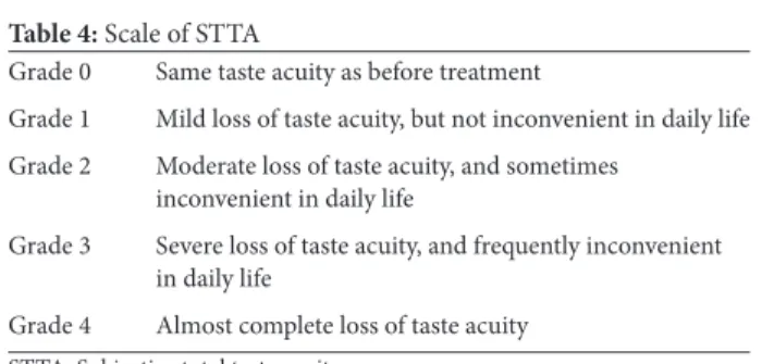
Multiple systems have been developed for grading the adverse eff ects of cancer treatment and several classifi cations have been used for describing the radiation-induced alterations. A subjective total taste acuity (STTA) scale, modifi ed from the late eff ects of normal tissues/subjective, objective, management, analytic scoring system, is used to evaluate, in a specifi c way, the STTA[19] as given in Table 4.
People with cancer who lose 10% or more of their normal body weight do not live as long as those with similar cancers at similar stages who remain well nourished. The multiple oral complications caused by stomatitis, xerostomia, and taste changes, maintaining adequate appetite, and nutrition is a challenge.[18]
Infections
The reduction in salivation from radiotherapy coincides with a shift in the oral microfl ora, with a prominence of cariogenic microorganisms. This shift shows an increase in Streptococcus mutans, Lactobacillus, and Candida albicans, with a decrease in Streptococcus sanguinis, Neisseria, and Fusobacterium.[14] This increase in oral Gram-negative enterobacteria and Pseudomonas is an aggravating factor for developing oral mucositis. Candidiasis is the most common infection aff ecting the oral cavity during radiotherapy.[20]
Bacterial infections may also occur early in the course of head/neck radiation. Herpes virus infections may also occur in patients who are seropositive prior to head and neck radiation due to virus re-activation.[21]
Late eff ects
Dental alterations
During the course of radiotherapy, increased dental sensitivity is experienced by many patients to temperature and taste variations due to loss of protective layer of saliva.[4]
Eff ects on teeth
Retardation of growth of teeth is seen when the oral cavity of the child is irradiated during the developing years. Before calcifi cation, radiation leads to the destruction of the tooth bud while irradiation after calcifi cation causes inhibition of cellular diff erentiation, causing malformations and arrest in general growth. However, the structure of enamel, dentin, or cementum does not alter. Solubility of the teeth does not increase due to radiotherapy. Pulpal tissue demonstrates long-term fi bro-atrophy after irradiation. The eruptive mechanism of teeth is relatively resistant to radiation eff ects and the irradiated teeth with alterations in root structure still erupt into the oral cavity.[22]
Eff ects on odontogenesis
The extent of retardation of tooth growth depends on the radiation dosage and the stage of tooth development. Table 3: Radiation Th erapy Oncology Group radiation morbidity scoring criteria for xerostomia
Acute reactions
0: No change over baseline
1: Mild mouth dryness/slightly thickened saliva/may have slightly altered taste, such as metallic taste/changes are not refl ected by alteration in baseline feeding
2: Moderate to complete dryness/thick, sticky saliva/markedly altered taste
3: Not used
4: Acute salivary gland necrosis
Late reactions
0: No change over baseline
1: Slight dryness of mouth/good response on stimulation
2: Moderate dryness of mouth/poor response on stimulation
3: Complete dryness of mouth/no response on stimulation
4: Fibrosis
Table 4: Scale of STTA
Grade 0 Same taste acuity as before treatment
Grade 1 Mild loss of taste acuity, but not inconvenient in daily life
Grade 2 Moderate loss of taste acuity, and sometimes inconvenient in daily life
Grade 3 Severe loss of taste acuity, and frequently inconvenient in daily life
Grade 4 Almost complete loss of taste acuity
Odontogenic cells are more radiosensitive in the pre-formative and diff erentiation phases than cells in the secretory/mature stage. Kaste et al. stated that the maturing ameloblasts may be permanently damaged with as little as 10 Gy, and ameloblastic activity ceases after exposure to 30 Gy.[23]
Radiation caries
Radiation caries is rampant, may occur in individuals who receive a course of radiotherapy that includes exposure to salivary glands. It causes rapid destruction of tooth structures.[24]
Clinically, three types of radiation caries exist which may occur in combinations in some patients. They are as follows: 1. Widespread superfi cial lesions attacking buccal, occlusal,
incisal, and palatal surfaces – Most common
2. Lesions involving primarily cementum and dentin in cervical region. These lesions may progress around the teeth circumferentially and result in loss of the crown
3. Dark pigmentation of entire crown and wearing away of the incisal edges.[25]
Pathogenesis
Radiation caries could be a direct or indirect eff ect of radiotherapy. Several studies claim that radiation caries occurs due to the presence of main salivary glands within the radiation fi eld causing hyposalivation. This change in the salivary fl ow rate brings changes in the normal microbial fl ora and immunologic factors, thus increasing the caries incidence in irradiated patients.[24,25]
Periodontal problems
Post irradiation, decrease in the vascularity and acellularity of the periodontal membrane is seen. Disorientation and rupture of Sharpey’s fi bers, thickening, and widening of the periodontal space have been reported. The cementum gets acellular, and its repair and regeneration capacity is severely reduced.[22]
These changes that occur in the periodontal ligament and cementum may predispose individuals to radiation-induced hyposalivation, increased plaque accumulation, shift in oral microfl ora, which may lead to infection. The potential of the periodontium to regenerate following surgery will be reduced.[22,26]
Pulpal defects
Pulpal tissue will demonstrate long-term fi broatrophy after irradiation. Patients may exhibit hypersensitivity, pulpal pain, and necrosis.[25]
Osteoradionecrosis
Marx and Johnson in 1987 and Constantino et al., 1995,defi ned osteoradionecrosis as “bone death secondary to radiotherapy.” The incidence of osteoradionecrosis is more common in mandible.[27]
Osteoradionecrosis of the jaws occurs due to radiation, followed by trauma, and then infection.[4] Portal of entry for oral
bacteria into the underlying bone occurs due to trauma. Due to compromised vascularity and minimal regenerative abilities, the infection rapidly progresses and spreads throughout the bone. The primary damage to mature bone results from radiation-induced damage to the vasculature of the periosteum and cortical bone, which is normally already sparse.[27,28]
Marx (1983) described the following steps in the development of osteoradionecrosis. They are as follows:[29]
• Hypoxic-hypovascular-hypocellular tissue: Bone loses its ability to replace normal collagen
• Tissue breakdown: Synthesis and cellular replication is exceeded by collagen lysis and cell death
• Chronic non-healing wounds: Occurs due to increase in energy, oxygen, and metabolic demands that exceeds the supply.
Non-healing wound in irradiated bone occurs in the oral cavity due to decreased vascularity of the mandible and increased infection. It is more common in mandible than maxilla, probably because of the richer vascular supply to the maxilla and also due to the fact that mandible is more frequently irradiated. As the radiation dose absorbed by the bone is increased, the risk of osteoradionecrosis is also increased.[28,29]
Histopathology
The initial changes in the bone components - Osteocytes, osteoblasts, and osteoclasts result from injury or damage to the remodeling system. There is decrease in the formation and increase in the lytic activity because osteoclasts tend to be more radioresistant than osteoblasts. Radiation injury aff ects the bone vascularity and tissues in the vicinity. This leads to hyperemia, followed by endarteritis and thrombosis. Ultimately, this leads to obliteration of small vessels and fatty degeneration. Atrophy of endosteum occurs with loss of active osteoblasts.[27,28]
Trismus
During radiotherapy, if masticatory muscles and/or the temperomandibular joint (TMJ) comes in the path of radiation, trismus can occur. Muscle fi brosis/scarring, fi brosis of the TMJ ligaments, pterygo-mandibular raphes can scar in response to radiation injury. Oral hygiene, speech, nutritional intake of the patient is compromised due to limited jaw opening interferes. Trismus develops in most patients within 3-6 months after radiotherapy and frequently becomes a long lasting problem.[30]
Soft tissue necrosis and fi brosis
Conclusion
Radiation therapy plays a signifi cant role in cancer therapy. As a result, various changes are induced in oral tissues. The resulting sequelae cause substantial problems and may aff ect the patient’s quality of life. Larger prospective trials that include the prevention and treatment of radiation-induced damage to oral tissues are needed to improve management and enhance better prognos is.
References
1. Beumer J 3rd, Curtis T, Harrison RE. Radiation therapy of the oral cavity: Sequelae and management, part 2. Head Neck Surg 1979;1:392-408.
2. Ow TJ, Myers JN. Current management of advanced resectable oral cavity squamous cell carcinoma. Clin Exp Otorhinolaryngol 2011;4:1-10.
3. Imanimoghaddam M, Rahrooh M, Tafakhori Z, Zahedanaraki S, Homaeieshandiz F. Changes of parotid and submandibular glands caused by radiotherapy - An ultrasound evaluation. Dentomaxillofac Radiol 2012;41:379-84.
4. Vissink A, Jansma J, Spijkervet FK, Burlage FR, Coppes RP. Oral sequelae of head and neck radiotherapy. Crit Rev Oral Biol Med 2003;14:199-212.
5. Sonis ST, Eilers JP, Epstein JB, LeVeque FG, Liggett WH Jr, Mulagha MT, et al. Validation of a new scoring system for the assessment of clinical trial research of oral mucositis induced by radiation or chemotherapy. Mucositis Study Group. Cancer 1999;85:2103-13.
6. Köstler WJ, Hejna M, Wenzel C, Zielinski CC. Oral mucositis complicating chemotherapy and/or radiotherapy: Options for prevention and treatment. CA Cancer J Clin 2001;51:290-315. 7. Pavlatos J, Gilliam KK. Oral care protocols for patients
undergoing cancer therapy. Gen Dent 2008;56:464-78.
8. Redding SW. Cancer therapy-related oral mucositis. J Dent Educ 2005;69:919-29.
9. Barasch A, Coke JM. Cancer therapeutics: An update on its eff ects on oral health. Periodontol 2000. 2007;44:44-54. 10. Handschel J, Prott FJ, Sunderkötter C, Metze D, Meyer U, Joos
U. Irradiation induces increase of adhesion molecules and accumulation of beta2 - integrin - expressing cells in humans. Int J Radiat Oncol Biol Phys 1999;45:475-81.
11. Pinna R, Campus G, Cumbo E, Mura I, Milia E. Xerostomia induced by radiotherapy: An overview of the physiopathology, clinical evidence, and management of the oral damage. Th er Clin Risk Manag 2015;11:171-88.
12. Coppes RP, Vissink A, Konings AW. Comparison of radiosensitivity of rat parotid and submandibular glands aft er diff erent radiation schedules. Radiother Oncol 2002;63:321-8. 13. Dreizen S, Brown LR, Handler S, Levy BM. Radiation - induced
xerostomia in cancer patients. Eff ect on salivary and serum electrolytes. Cancer 1976;38:273-8.
14. Brown LR, Dreizen S, Handler S, Johnston DA. Eff ect of radiation - induced xerostomia on human oral microfl ora. J Dent Res 1975;54:740-50.
15. Hamlet S, Faull J, Klein B, Aref A, Fontanesi J, Stachler R, et al. Mastication and swallowing in patients with postirradiation xerostomia. Int J Radiat Oncol Biol Phys 1997;37:789-96. 16. Bäckström I, Funegård U, Andersson I, Franzén L, Johansson I.
Dietary intake in head and neck irradiated patients with permanent dry mouth symptoms. Eur J Cancer B Oral Oncol 1995;31B:253-7.
17. Conger AD. Loss and recovery of taste acuity in patients irradiated to the oral cavity. Radiat Res 1973;53:338-47. 18. Baharvand M, ShoalehSaadi N, Barakian R, Moghaddam EJ.
Taste alteration and impact on quality of life aft er head and neck radiotherapy. J Oral Pathol Med 2013;42:106-12.
19. Ruo Redda MG, Allis S. Radiotherapy-induced taste impairment. Cancer Treat Rev 2006;32:541-7.
20. Ramirez-Amador V, Silverman S Jr, Mayer P, Tyler M, Quivey J. Candidal colonization and oral candidiasis in patients undergoing oral and pharyngeal radiation therapy. Oral Surg Oral Med Oral Pathol Oral Radiol Endod 1997;84:149-53. 21. Scully C, Epstein JB. Oral health care for the cancer patient. Eur
J Cancer B Oral Oncol 1996;32B:281-92.
22. Kassim N, Sirajuddin S, Biswas S, Rafi uddin S, Apine A. Iatrogenic Damage to the Periodontium Caused by Radiation and Radiotherapy. Open Dent J 2015;9:182-6.
23. Kaste SC, Hopkins KP, Jenkins JJ 3rd. Abnormal odontogenesis in children treated with radiation and chemotherapy: Imaging fi ndings. AJR Am J Roentgenol 1994;162:1407-11.
24. Karmiol M, Walsh RF. Dental caries aft er radiotherapy of the oral regions. J Am Dent Assoc 1975;91:838-45.
25. Gupta N, Pal M, Rawat S, Grewal MS, Garg H, Chauhan D, et al. Radiation-induced dental caries, prevention and treatment - A systematic review. Natl J Maxillofac Surg 2015;6:160-6. 26. Andrews N, Griffi ths C. Dental complications of head and neck
radiotherapy: Part 2. Aust Dent J 2001;46:174-82.
27. Th orn JJ, Hansen HS, Specht L, Bastholt L. Osteoradionecrosis of the jaws: Clinical characteristics and relation to the fi eld of irradiation. J Oral Maxillofac Surg 2000;58:1088-93.
28. Lambade PN, Lambade D, Goel M. Osteoradionecrosis of the mandible: A review. Oral Maxillofac Surg 2013;17:243-9. 29. Lyons A, Ghazali N. Osteoradionecrosis of the jaws: Current
understanding of its pathophysiology and treatment. Br J Oral Maxillofac Surg 2008;46:653-60.
30. Bensadoun RJ, Riesenbeck D, Lockhart PB, Elting LS, Spijkervet FK, Brennan MT, et al. A systematic review of trismus induced by cancer therapies in head and neck cancer patients. Support Care Cancer 2010;18:1033-8.