R E V I E W A R T I C L E
The composition, function and role of saliva in maintaining
oral health: A review
Brij Kumar1, Nilotpol Kashyap1, Alok Avinash1, Ramakrishna Chevvuri2, Mylavarapu Krishna Sagar3, Kumar Shrikant3
¹Department of Pedodontics and Preventive Dentistry, Rungta College of Dental Sciences and Research, Bhilai, Chhattisgarh, India, ²Department of Public Health Dentistry, Rungta College of Dental Sciences and Research, Bhilai, Chhattisgarh, India, ³Department of Pedodontics and Preventive Dentistry, People’s Dental Academy, Bhopal, Madhya Pradesh, India
Abstract
Background: Saliva is certainly one of the most important components in the oral environment and an integral component to oral health. Aim: The components of saliva, its functions in maintaining oral health and the main factors that cause alterations in salivary secretion will be reviewed, the importance of saliva in caries development and bacterial plaque formation will be discussed, and its role as an aid to diagnosing certain pathologies will also be discussed here. Conclusion: Saliva aids in maintaining mucosal integrity and in digestion through salivary enzymes. Saliva is essential information
of the pellicle, which protects the tooth after eruption. Saliva has several oral benefits including buffering, remineralization, and lubrication. Clinical Significance: Saliva is
an important biological fluid that aids in mechanically removing food debris and bacteria from the oral cavity and teeth, reduced salivary flow causes ill effects to the oral tissues.
Keywords: Caries, dental plaque, saliva, salivary flow rate, salivary proteins, xerostomia
Introduction
A critical component of the oral environment is saliva, a dilute aqueous solution containing both inorganic and organic constituents. Saliva plays an essential role during mastication, in swallowing and in speech. The substances dissolved in saliva during mastication are transported to stimulate taste receptors for taste perception.
The salivary amylase is a digestive enzyme responsible for the initial stage in starch and glycogen breakdown, and salivary lipase secreted by lingual salivary glands (Von Ebner’s glands)
may play a significant role in fat digestion. In many animals
evaporation of saliva spread on fur or while panting is important in temperature regulation during heat stress.[1]
The functions of saliva are to protect the oral tissues by keeping them moist and by providing a lubricating mucoid
secretion, by maintaining a fluid environment with high calcium and phosphate concentrations and the power of buffering acids and to initiate the digestion of starch. Impaired salivary secretion
(hyposalivation) increases the risk of oral diseases such as dental caries and oral candidal infection.[2]
Recently, additional functions of salivary glands have been uncovered. Salivary glands have been shown to contain, and possibly secrete, a large number of physiologically active substances, such as nerve growth factor, vasoactive peptides, and regulatory peptides. Thus salivary glands may have a role in functions not normally associated with that traditional alimentary function.[3,4]
Composition of Saliva [Table 1][5] Functions of saliva in humans Digestive functions
Although amylase is a major component of the parotid secretion
and is present at an appreciable level in the submandibular fluid
as well, its salivary role in the digestion of carbohydrates is really
minimal. The only effective conversion of starch to maltose that occurs in the oral cavity is in food-retentive sites, and this benefits
primarily the plaque bacteria. Most of the food is swallowed rather quickly, and in the stomach salivary amylase would be
minimally effective, given the low pH and high proteolytic
activity of the gastric juice.[6]
Correspondence
Dr. Brij Kumar, Department of Pedodontics and Preventive Dentistry, Rungta College of Dental Sciences and Research, Bhilai, Chhattisgarh, India. Phone: +91-8827800994. E-mail: [email protected]
Received: 30 October 17 Accepted: 15 December 17
doi: 10.15713/ins.ijcdmr.121
How to cite this article:
The high water content of the parotid secretions moistening the food and the mucins generated by the submandibular, sublingual, and minor salivary glands coating
the food combine effectively to facilitate ingestion. Other
lubrication molecules, such as the parotid proline-rich glycoprotein albumin complex, may also participate in the rate of food passage by becoming incorporated into pellicle, thus providing a lubricating interface between teeth and facilitating mastication.
Saliva also plays a gastronomic role by solubilizing many of the food components and acting as a medium for interaction
with the receptors on the taste cell. It has been proposed that a specific zinc-binding salivary protein, gustin, and mediates
taste sensation. Saliva also enhances taste perception by its relatively low level of salts and its very low concentration of sugar; thus, in a sense, it does not compete with exogenous taste modalities.[7]
Protective Functions
Lubrication and demulcent properties
From an evolutionary point of view, the oldest function of salivary glands is to supply lubrication molecules, to coat not
only the food but the oral soft tissue as well. The lubrication film
allows for ready phonation as well as food passage and provides for smooth tissue surfaces that exhibit minimal friction and are comfortable as well as functional. The lubricating properties of saliva have always been ascribed to the mucin glycoproteins
because they can provide fluid layers with high film strength,
and there are several experimental models to show that salivary
mucin and similar molecules do indeed have effective lubricating properties. Very recently, Hatton et al. (1985) showed that the proline-rich glycoprotein of parotid saliva, when complexed
with albumin, was also an extremely effective lubricant. The
distribution of this complex in the oral cavity remains to be established, but it should be functional on teeth as part of
pellicle, and on mucous membranes as well. It could also be part
of the food coating but is probably overshadowed by the much more adhesive mucin.
Maintenance of Mucous Membrane Integrity
The salivary mucins possess rheological properties which include low solubility, high viscosity, elasticity, and adhesiveness, which enable them to concentrate on the oral mucosal surface, where they
provide an effective barrier against desiccation and environmental
insults. The molecular structure of salivary mucins enables them
to bind water effectively, and hence their presence on the mucous membrane surfaces serves as natural “waterproofing” and helps to
maintain these tissues in a hydrated state.
Mucins have been shown to be important in the control of permeability of mucosal surface, and the presence of a salivary
film is important in limiting penetration of a variety of potential
irritants and toxins in foods and beverages as well as of potentially hazardous agents from tobacco smoke and other sources.[8]
A variety of proteolytic enzymes is generated in the bacterial plaque around the teeth and in the crevicular area, especially in people with periodontitis. Proteases are also generated by polymorphonuclear leukocytes, the numbers being related to the
level of inflammatory disease. The bacterial and PMN proteases
(e.g., elastase, collagenase, and cathepsin) have the potential of
affecting the integrity of the mucous membranes and causing
ulceration. Mucins are protective in this regard since their glycosylated regions are very resistant to proteolysis.
There is also a second line of defense against protease activity, cysteine-containing phosphoproteins, in particularly high concentration in submandibular saliva, which are identical to Cystatin S. Cystatins S is an inhibitor of cysteine proteinases, especially Cathepsin C. This antiprotease activity is augmented
by antileukoprotease, an effective inhibitor of granulocyte elastase
and Cathepsin G, present in both parotid and submandibular glands, but to a much greater extent in the former.[9]
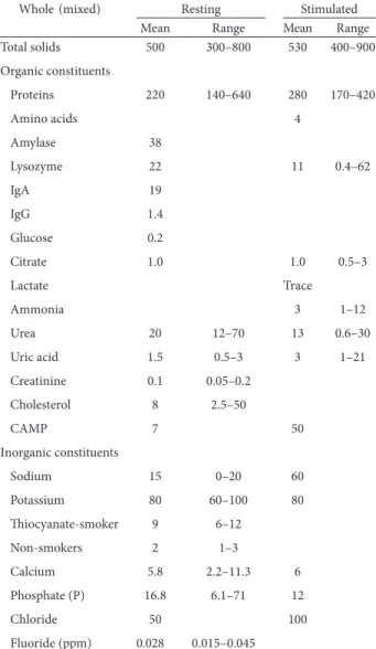
Table 1: Composition of saliva: (mg/100 ml)
Whole (mixed) Resting Stimulated
Mean Range Mean Range
Total solids 500 300–800 530 400–900
Organic constituents
Proteins 220 140–640 280 170–420
Amino acids 4
Amylase 38
Lysozyme 22 11 0.4–62
IgA 19
IgG 1.4
Glucose 0.2
Citrate 1.0 1.0 0.5–3
Lactate Trace
Ammonia 3 1–12
Urea 20 12–70 13 0.6–30
Uric acid 1.5 0.5–3 3 1–21
Creatinine 0.1 0.05–0.2
Cholesterol 8 2.5–50
CAMP 7 50
Inorganic constituents
Sodium 15 0–20 60
Potassium 80 60–100 80
Thiocyanate‑smoker 9 6–12
Non‑smokers 2 1–3
Calcium 5.8 2.2–11.3 6
Phosphate (P) 16.8 6.1–71 12
Chloride 50 100
Soft Tissue Repair
Licking one’s wounds may be more than metaphor. The presence of nerve growth factor and epidermal growth factor in the submandibular saliva may accelerate wound-healing. Epidermal growth factor is present in human saliva but at much
lower levels. The effect of saliva in oral wound-healing in humans
remains to be established.
An alternate role for saliva in wound-healing is suggested by a paper of Volker (1942), in which he showed that saliva speeds
blood coagulation, both by affecting the anticoagulant valuable
property in an area where rough food or traumatic injury can
induce bleeding and bleed readily due to inflammatory disease.[10]
Maintenance of Ecological Balance
Colonization of tissue surfaces, adherence is a critical event for the survival of many bacteria, and interference with this process, bacterial clearance, by mechanical, immunological, and non-immunological means is one of the major functions of the salivary defense mechanism. The ability of saliva to maintain an appropriate ecological balance in the oral cavity is an important evolutionary force in the long period of human existence before plaque control.
Debridement and Lavage
The physical flow of saliva augmented by the muscular activity of the lips and tongue effectively removes a large number of potentially
harmful bacteria from teeth and mucosal surfaces. This clearance mechanism is similar to tearing and blinking in the eye, blowing the nose, and coughing and expectorating to clear the lungs.[11]
Aggregation
In addition to physical effects, saliva can interfere with bacterial
adherence by more direct means that depend on molecular interactions. The ability to inhibit bacterial attachment is a
major characteristic of the secretory IgA system and is the rationale for the interest in an oral vaccine against caries. In
addition to these made to order antibodies, there is a variety of
ready-to- wear macromolecules, some very specific in action,
which mask bacterial adhesins or compete with them for attachment sites on tissue. They may also function by clumping or aggregating bacteria to the point where they can no longer
effectively adhere to hard or soft tissue and are expectorated
or swallowed. Most attention in experimental studies of aggregation has been given to the high molecular weight mucins. The presence of multiple complex oligosaccharide side chains and a characteristic micro heterogeneity provide a wide range of possibilities for interactions with many bacteria. The
amount of covalently bound lipid can also affect the properties of mucins and can influence their propensity for bacterial
interaction.[12,13]
Direct Antibacterial Properties
A group of salivary proteins lysozyme, lactoferrin, and lactoperoxidase working in conjunction with other components
of saliva can have an immediate effect on oral bacteria, interfering
with their ability to multiply or killing them directly. Lysozyme can cause lysis of bacterial cells, especially Streptococcus mutans by interacting with anions of low charge density chaotropic ions (thiocyanate, perchlorate, iodide, bromide, nitrate, chloride, and
fluoride), and with bicarbonate. It has recently been shown that
another cationic peptide in saliva the histidine-rich peptide of
parotid saliva has growth-inhibitory and bactericidal effects on oral bacteria. The histidine-rich peptides appear to be effective
antifungal agent as well, able to inhibit growth and kill Candida albicans at very low concentration.[14]
Lactoferrin, the exocrine gland equivalent of transferrin, is
effective against bacteria that require iron for their metabolic processes. It can compete with the bacterial iron-chelating
molecules, and deprive the bacteria of this essential element.
Lactoferrin is also capable of a bactericidal effect that is distinct
from simple iron deprivation.
Salivary peroxidase is part of an antibacterial system which involves the oxidation of salivary thiocyanate by hydrogen peroxide (generated by oral bacteria) to hypothiocyanite and
hypothiocyanous acid. These products, in turn, affect bacterial
metabolism (especially acid production) by oxidizing the sulfhydryl groups of the enzymes involved in glycolysis and sugar
transport. The antimicrobial effect of salivary peroxidase against
S. mutans is significantly enhanced by interaction with secretory IgA. The protective potential of all the antibacterial proteins
can be extended by interaction with mucin which can serve to concentrate this defense force at the interface of the mucosa and the inhospitable external environment.
When teeth are present, especially if some gingivitis exists,
the oral fluids will be augmented by a contribution from the gingival crevice area, the gingival crevicular fluid. This fluid can
contribute to the oral defense system by providing: (a) Serum
antibodies against oral bacteria, especially IgG antibodies, (b) phagocytic cells (PMN’s), and (c) antibacterial products
liberated from the phagocytic cells (e.g. lysozyme, lactoferrin, and myeloperoxidase).[15]
Maintenance of pH
Saliva is effective in helping to maintain a relatively neutral pH
in the oral cavity, in the bacterial plaque, and on swallowing, in
the esophagus as well. In the oral cavity and the esophagus, the major regulation of pH, especially during eating or drinking, is
the salivary bicarbonate, the level of which varies directly with
flow rate.
In the bacterial plaque, where acid production is the natural
sequela to bacterial metabolism of carbohydrates, saliva helps
diffused into the plaque. Urea from saliva is converted by bacterial
urease to ammonia, which can neutralize the acid. Amino acids and peptides can be decarboxylated to form monoamines and polyamines, a process which consumes hydrogen ions. Arginine and arginine peptides can form ammonia as well as the
polyamine, putrescine, and thus can be particularly effective in elevating plaque pH.
Maintenance of Tooth Integrity
In addition to helping to counter plaque acidity, saliva helps
protect the teeth in a number of other ways. This protective function begins immediately after tooth eruption into the oral cavity. Although the crown of the tooth is fully formed morphologically when it erupts, it is crystallographically
incomplete. Interaction with saliva provides a post-eruptive maturation through diffusion of ions such as calcium, phosphorus, magnesium, and fluoride, as well as other trace components, into
the surface enamel. This maturation increases surface hardness, decreases permeability, and has been experimentally shown to increase resistance to caries.[16]
Once tooth begins to function in the mouth, its developmental
cuticle or pellicle is rapidly worn away and is replaced by a
constantly replenished salivary film, the acquired pellicle. This
selectively adsorbed coating of proteins and lipids provides a
protective barrier and a lubricating film against excessive wear, a diffusion barrier against acid penetration, and a limitation
against mineral egress.[17]
One of the major contributions of salivary research during
the past decade has been the characterization of the system that regulate the ionic environment in the plaque and oral cavity. A group of phosphoproteins acidic proline-rich proteins, statherin, and cysteine-containing phosphoproteins provide
this protection by effectively binding calcium and helping to
maintain saliva in a state of supersaturation with respect to calcium phosphate salts. They bind to the surfaces of early crystal nuclei and retard crystal growth.[18]
Excretory Functions
Many drugs, as well as alcohol, are excreted into the saliva, which
could theoretically serve as a route of elimination. However,
since most of the saliva generated is swallowed rather than
expectorated, it is a very inefficient disposal system, since the
substances would be absorbed and recycled. Clearly this is a minor function.
Water Balance
Salivary glands are part of a control system for maintaining an
appropriate level of hydration. Thirst and need for fluid intake
are usually signaled by dry mouth. This sensation results from a diminution in resting secretion and activation of receptors in the oral cavity. The signals to salivary glands result from
osmotic changes detected in hypothalamus or volemic changes operating through the renin-angiotensin system of the kidney. Thirst satiation and cessation of drinking are initiated by sensory messages passing into the brain from taste receptors in the mouth.
Hormonal Function
Several studies have shown that the polypeptide hormone known as an epidermal growth factor, is identical with human
urogastrone. Nerve growth factor and transforming growth factor may be closely related as well. Human urogastrone, found
in very high concentrations in urine, is readily measurable in the submandibular saliva. The major properties of urogastrone include gastric cytoprotection and inhibition of gastric acid secretion.[19]
Saliva and Periodontal Health
Role of saliva in oral diseases is most apparent when salivary flow
is markedly reduced. With respect to periodontal health, saliva plays a role in two ways.
Pellicle and plaque formation
Saliva influences supragingival plaque deposition and activity in a variety of ways. It is predominantly involved in the first step of
plaque formation, i.e., deposition of a pellicle (or cuticle) which is a four stage process.
a. Bathing of the tooth surfaces by salivary fluids which contain
abundant proteins.
b. Selective adsorption of certain negatively and positively charged glycoproteins which act as an agglutinating base. c. Loss of solubility of the adsorbed proteins by surface
denaturation and acid precipitation.
d. Alteration of the glycoproteins by enzymes from bacteria and the oral secretions.
Now, this pellicle which is formed is actively involved
in the second stage of plaque formation which is “bacterial
colonization.”
1. The initial step in dental plaque formation involves the adherence of bacteria to the salivary-coated tooth surface. 2. The bacteria that adhere to the pellicle initially are called the
“primary” colonizers which are usually Streptococcus sanguis species followed by S. mutans.
3. Once an initial layer of bacteria attaches to the pellicle surface,
plaque can progress at a rapid pace, as many “secondary
colonizers” adhere to the primary bacteria and carve an “ecological niche” for themselves. When we ask patients to
maintain a good oral hygiene, we are actually targeting to counter this step.
4. In the third or maturation stage, saliva continues to
5. Salivary proteins and carbohydrates serve as a substrate for metabolic activity of the bacteria. Salivary calcium, phosphates, magnesium, sodium, and potassium become part
of the gel-like consistency of plaque and begin to influence
its mineralization and demineralization. Mineral precipitate results from a local rise in degree of saturation of calcium
and phosphates in saliva due to (a) increase of pH of saliva
(b) colloidal and particles in saliva which bind to calcium and phosphates making super-saturated solution.[14,20-22]
Plaque mineralization and calculus formation
Salivary proteins such as esterase, pyrophosphates possibly acid phosphatase and lysozyme play a role. Calcium crystals are served by saliva. Persons who are heavy calculus formers are known to have higher levels of salivary proteins (glycoproteins). Therefore, oral hygiene instructions should be tailor-made to suit each child’s need.
Recent concepts
The fact that α-amylase is found in acquired enamel pellicle suggests that it has a role in bacterial adhesion. If it is so then α-amylase is double danger, because it binds to bacteria in
the plaque, hold them together, and at the same time provide additional glucose to those plaque microorganisms, and that too include proximity to tooth surface. The resulting lactic acid is added to the pool of acid that is already present.[23]
Saliva and Dental Caries
Evidence shows that saliva from caries-free individuals has higher values of the following: (Compared to caries prone).
1. The increased rate of flow increased pH and increased buffering.
2. Higher calcium and phosphorous concentrations. 3. Higher ammonia concentration.
4. High concentrations of ATP and fructose diphosphate. 5. Increased aldolase activity and O2 uptake of bacteria.
6. Increased opsonin activity.
7. Increased general antibacterial activity.
8. Increased antibacterial activity specific to lactobacilli and
streptococci.
9. Higher number of intact leukocytes.
10. Difference in proportion of epithelial cells to leukocytes.[23]
The saliva influences on the caries process which is fundamental, and the saliva also affects all the three components
of Keye’s classic Venn diagram of etiology of caries (tooth, plaque,
and substrate). The saliva also affects flow rates and clearance, pH and buffer capacity, calcium phosphate homeostasis and effects on bacterial metabolism. The obvious manifestation
of the saliva/caries interactions includes adsorption to oral tissues and elimination from the oral cavity. There are many studies which had been done to relate certain aspects of salivary output and composition to caries susceptibility. Most of those studies were focused at either the physico-chemical properties
of saliva (flow rate, buffer capacity) or definite components of saliva with antimicrobial activity, such as salivary IgA, lactoferrin,
lysozyme and the salivary peroxidase-hypothiocyanite system. The consensus relationship between caries experience and the activity of any of the salivary antimicrobial proteins cannot be
validated, except hypothiocyanite system and IgA.[24]
The proline-rich proteins of the human parotid saliva exhibits inherited polymorphisms [34] that have led to a number of analysis done to correlate the genetic phenotype with caries severity. Results have commonly been inconsistent in terms of determining a relationship between prevalence of dental caries (DMFS) and genetic phenotype of saliva.[13]
There are number of cross-sectional studies had been done to
correlate the compositions and flow of saliva with dental caries. However, a basic flaw of any such study is that the use of the DMF
index for evaluation of caries activity. The DMF index may be a lifetime collective index of dental disease and treatment and should have very little bearing activity at the precise purpose in time. Similarly, a one-time determination of whole stimulated
salivary flow rate does not pass muster as a comprehensive analysis of salivary function. Other more sensitive, indicator
of salivary activity have to be compelled to be used, probably combining in vivo demineralization/remineralization models with
different indicators of dental caries activity along with recurrent,
standardized and comprehensive secretion collections.[25]
The use of saliva as a diagnostic fluid
There has been a continuing hope over many years that saliva could provide an alternative to blood for tests which might help in the diagnosis of disease. This could occur only if a blood component was transferable across the salivary gland epithelium in proportion to its concentration in blood. This is true for
urea, glucose and the unbound steroid hormones. Only for the
some of these has saliva proved a useful medium for monitoring concentrations. Glucose is used by the gland cells and appears in saliva at around one-hundredth of its plasma concentration - too low for easy measurement.[26]
Change in salivary composition in salivary gland disease
Sodium and chloride concentrations in the affected gland secretions are raised in sialadenitis, suggesting that inflammation particularly affects the ductal system. This possibility is
reinforced by the observation that phosphate concentrations are usually lowered.
Sjogren’s syndrome is a connective tissue disorder which usually involves reduced secretion from the lacrimal and salivary glands. Reduced salivary secretion causes a dry mouth and is termed xerostomia. Again the principal changes in ionic composition of saliva are increased concentration of sodium and
chloride and decreased concentration of phosphate. In addition,
there are changes in the relative concentrations of some minor salivary proteins. These may have some diagnostic value. Salivary gland neoplasms are uncommon, and there are few data available
Radiation used in the treatment of neoplasm in the head and neck region often results in radiation-induced damage to salivary
gland. This is seen as a marked reduction in flow rate, leading
to xerostomia. The small amount of saliva secreted has high concentrations of sodium, chloride, calcium, and magnesium but low concentrations of hydrogen carbonate. The low volume of
saliva and its poor buffering power are associated with rampant
dental caries in these patients.
The best treatment for xerostomia patients seems
to be frequent use of mouthwashes with a high fluoride
concentration.[27]
Conclusion
Clearly, saliva has profound effects on the mouth, however, as
Sreebny has noted, few dental practitioners hassle to raise the required queries or build the required observations and/or measurements to work out whether or not there’s any level of
exocrine gland hypofunction in their patients. Unfortunately, a
minimum of a part of the matter is that, for many half, there’s
not an easy fix to a broken salivary flow. We will solely hope that
future trends in biological science analysis might permit the
xerostomic patient to regain perform. Until then, dentists ought
to be additional alert to their patient’s salivary function embrace additional disciplined preventive practices to discourage the
negative effects of reduced salivary flow.
References
1. Cawson RA, Porter OS. Essentials of Oral Pathology and
Oral Medicine. 7th ed. London: Churchill Livingstone Elsevier
Science; 2002.
2. Anderson P, Hector MP, Rampersad MA. Critical pH in resting and stimulated whole saliva in groups of children and adults. Int J Paediatr Dent 2001;11:266‑73.
3. Alamoudi N, Farsi N, Faris J, Masoud I, Merdad K, Meisha D. Salivary characteristics of children and its relation to oral micro‑organism and lip mucosa dryness. Int Clin Pediatr 2004;28:239‑48.
4. Baskar SN. Orban’s Oral Histology and Embryology. 11th ed. St.
Louis: Harcourt Asia PIE Ltd., Mosby; 1997.
5. Berkenbilt DA, Bahn AN. Development of antibodies to cariogenic streptococci in children. J Am Dent Assoc 1971;83:332‑7.
6. Humphrey SP, Williamson RT. A review of saliva: Normal composition, flow, and function. J Prosthet Dent 2001;85:162‑9. 7. Jenkins GN. Physiology and Biochemistry. Oxford: Blackwell
Scientific Publications; 1977.
8. Lovelle CL. Applied Oral Physiology. 2nd ed. London:
Butterworth’s & Co., Publishers Ltd.; 1988.
9. Mandel ID. The functions of saliva. J Dent Res 1987;66:623‑7.
10. Tencate AR. Oral History, Development, Structure and
Function. 6th ed. St. Louis: Mosby Press; 2004.
11. Sreebny LM. Saliva in health and disease: An appraisal and update. Int Dent J 2000;50:140‑61.
12. Rayment SA, Liu B, Soares RV, Offner GD, Oppenheim FG, Troxler RF. The effects of duration and intensity of stimulation on total protein and mucin concentrations in resting and stimulated whole saliva. J Dent Res 2001;80:1584‑7.
13. Tukia‑Kulmala H, Tenovuo J. Intra‑and inter‑individual
variation in salivary flow rate, buffer effect, Lactobacilli, and
Mutans streptococci among 11 to 12 year old school children. Acta Odontol Scand 1993;51:31‑7.
14. Morinushi T, Murayama M, Kinjyo S. Mutans streptococci,
Lactobacilli in saliva and acidity for microorganisms in dental plaque: Changes after restorative treatment. J Clin Pediatr Dent 2004;28:327‑32.
15. Tenovuo J. Antimicrobial function of human saliva‑
how important is it for oral health? Acta Odontol Scand 1998;56:250‑6.
16. Van Nieuw Amerongen A, Bolscher JG, Veerman EC. Salivary proteins: Protective and diagnostic value in cariology? Caries Res 2004;38:247‑53.
17. Azen EA. Genetics of salivary protein polymorphisms. Crit Rev Oral Biol Med 1993;4:479‑85.
18. Nekrashevych Y, Hannig M, Stösser L. Assessment of enamel erosion and protective effect of salivary pellicle by surface roughness analysis and scanning electron microscopy. Oral Health Prev Dent 2004;2:5‑11.
19. Rantonen PJ, Penttilä I, Meurman JH, Savolainen K, Närvänen S, Helenius T. Growth hormone and cortisol in serum and saliva. Acta Odontol Scand 2000;58:299‑303.
20. Bimstein E, Small PA Jr, Magnusson I. Leukocyte esterase and protein levels in saliva, as indicators of gingival and periodontal diseases in children. Pediatr Dent 2004;26:310‑5.
21. Saxén L, Tenovuo J, Vilja P. Salivary defense mechanisms in juvenile periodontitis. Acta Odontol Scand 1990;48:399‑407. 22. Shafer WG, Hine MK, Levy BM. A Text Book of Oral Pathology.
4th ed. Philadelphia, PA: W.B. Saunder’s Company; 1983.
23. Sullivan A, Borgström MK, Granath L, Nilsson G. Number of Mutans streptococci or Lactobacilli in a total dental plaque sample does not explain the variation in caries better than the numbers in stimulated whole saliva. Community Dent Oral Epidemiol 1996;24:159‑63.
24. Gelbier MJ, Winter GB. Absence of salivary glands in children with rampant dental caries: Report of seven cases. Int J Paediatr Dent 1995;5:253‑7.
25. Moore PA, Guggenheimer J, Etzel KR, Weyant RJ, Orchard T. Type 1 diabetes mellitus, xerostomia, and salivary flow rates. Oral Surg Oral Med Oral Pathol Oral Radiol Endod 2001;92:281‑91. 26. Nordgarden H, Jensen JL, Storhaug K. Oligodontia is associated
with extra‑oral ectodermal symptoms and low whole salivary flow rates. Oral Dis 2001;7:226‑32.
27. Guggenheimer J, Moore PA. Xerostomia: Etiology, recognition and treatment. J Am Dent Assoc 2003;134:61‑9.