Development of Envelope Protein Antigens To Serologically
Differentiate Zika Virus Infection from Dengue Virus Infection
Lakshmanane Premkumar,
aMatthew Collins,
aStephen Graham,
aGuei-Jiun Alice Liou,
aCesar A. Lopez,
aRamesh Jadi,
aAngel Balmaseda,
bJames A. Brackbill,
cReynaldo Dietze,
dErwin Camacho,
eAruna D. De Silva,
f,g*
Camila Giuberti,
dHelena Lucia dos Reis,
dTulika Singh,
hHolly Heimsath,
hDaniela Weiskopf,
gAlessandro Sette,
gJorge E. Osorio,
iSallie R. Permar,
hMichel J. Miley,
cHelen M. Lazear,
aEva Harris,
jAravinda M. de Silva
aaDepartment of Microbiology and Immunology, University of North Carolina School of Medicine, Chapel Hill, North Carolina, USA
bLaboratorio Nacional de Virología, Centro Nacional de Diagnóstico y Referencia, Ministry of Health, Managua, Nicaragua
cDepartment of Pharmacology, University of North Carolina School of Medicine, Chapel Hill, North Carolina, USA
dNúcleo de Doenças Infecciosas, Centro de Ciências da Saúde, UFES, Vitoria, Brazil
eUniversidad de Sucre, Sincelejo, Sucre, Colombia
fGenetech Research Institute, Colombo, Sri Lanka
gDivision of Vaccine Discovery, La Jolla Institute for Allergy and Immunology, La Jolla, California, USA
hDepartment of Pediatrics and Human Vaccine Institute, Duke University Medical Center, Durham, North Carolina, USA
iDepartment of Pathobiological Sciences, University of Wisconsin, Madison, Wisconsin, USA
jDivision of Infectious Diseases and Vaccinology, School of Public Health, University of California, Berkeley, Berkeley, California, USA
ABSTRACT
Zika virus (ZIKV) is an emerging flavivirus that can cause birth defects
and neurologic complications. Molecular tests are effective for diagnosing acute ZIKV
infection, although the majority of infections produce no symptoms at all or present
after the narrow window in which molecular diagnostics are dependable. Serology is
a reliable method for detecting infections after the viremic period; however, most
serological assays have limited specificity due to cross-reactive antibodies elicited by
flavivirus infections. Since ZIKV and dengue virus (DENV) widely cocirculate,
distin-guishing ZIKV infection from DENV infection is particularly important for diagnosing
individual cases or for surveillance to coordinate public health responses. Flaviviruses
also elicit type-specific antibodies directed to non-cross-reactive epitopes of the
in-fecting virus; such epitopes are attractive targets for the design of antigens for
de-velopment of serological tests with greater specificity. Guided by comparative
epitope modeling of the ZIKV envelope protein, we designed two recombinant
anti-gens displaying unique antigenic regions on domain I (Z-EDI) and domain III (Z-EDIII)
of the ZIKV envelope protein. Both the Z-EDI and Z-EDIII antigens consistently
de-tected ZIKV-specific IgG in ZIKV-immune sera but not cross-reactive IgG in
DENV-immune sera in late convalescence (
⬎
12 weeks postinfection). In contrast, during
early convalescence (2 to 12 weeks postinfection), secondary DENV-immune sera
and some primary DENV-immune sera cross-reacted with the Z-EDI and Z-EDIII
anti-gens. Analysis of sequential samples from DENV-immune individuals demonstrated
that Z-EDIII cross-reactivity peaked in early convalescence and declined steeply over
time. The Z-EDIII antigen has much potential as a diagnostic antigen for
population-level surveillance and for detecting past infections in patients.
KEYWORDS
comparative epitope mapping, computational prediction, ELISA, Zika
virus, antibody-binding region, cross-reactivity, dengue virus, flavivirus, serological
diagnosis, surveillance
Received15 September 2017 Returnedfor modification12 October 2017 Accepted11 December 2017
Acceptedmanuscriptpostedonline20 December 2017
CitationPremkumar L, Collins M, Graham S, Liou G-JA, Lopez CA, Jadi R, Balmaseda A, Brackbill JA, Dietze R, Camacho E, De Silva AD, Giuberti C, dos Reis HL, Singh T, Heimsath H, Weiskopf D, Sette A, Osorio JE, Permar SR, Miley MJ, Lazear HM, Harris E, de Silva AM. 2018. Development of envelope protein antigens to serologically differentiate Zika virus infection from dengue virus infection. J Clin Microbiol
56:e01504-17. https://doi.org/10.1128/JCM
.01504-17.
EditorAlexander J. McAdam, Boston Children's Hospital
Address correspondence to Lakshmanane Premkumar, [email protected], or Aravinda M. de Silva, [email protected].
*Presentaddress:ArunaD.DeSilva,
Z
ika virus (ZIKV) is an enveloped, positive-sense, single-stranded RNA virus in the
Flavivirus
genus, which includes other medically important viruses, such as dengue
virus
(DENV),
West
Nile
virus,
and yellow
fever
virus
(1).
ZIKV
infection
has
become a
major
global health
concern
because
it
can
disseminate rapidly
in
naive
populations
and
lead to
neurologic sequelae, such as
a
Guillain-Barré-like
syndrome,
in
otherwise
healthy individuals. ZIKV also has the unusual ability among human
flaviviruses to be
transmitted
through
sexual
contact
and
from
mother
to
fetus
during
pregnancy
(2).
Congenital
ZIKV
infection
can
cause
developmental
abnormalities,
including
ocular
damage, microcephaly, and fetal death (2–5). People at risk of DENV infection are also
at
risk of ZIKV infection,
as
both viruses
are transmitted
by
Aedes
mosquitoes (3).
Accurate
diagnosis
is
critical to many
aspects of
the public
health response
to the
Zika disease epidemic (6) but is complicated by multiple
factors.
Clinically,
it is
impos-sible
to
discern among myriad
causes
of
acute
fever
and/or
rash. Molecular
tests
are
useful
for
detecting symptomatic
flavivirus infections
during
the brief period
immedi-ately following infection (7).
However, most
individuals with ZIKV infection never seek
medical
attention
because
they
are
asymptomatic
or
experience
only
a
mild,
self-limited illness (8, 9). Beyond this acute period, serological tests are necessary to detect
ZIKV
infections
and
to
support
public health
efforts,
such
as
prenatal
evaluation
and
management, risk reduction counseling,
and surveillance and outbreak investigations.
Unfortunately, most serological tests lack specificity due to cross-reactive antibodies
elicited
by flavivirus
infections. Neutralization assays,
which are
more
specific but less
widely
available
due
to
their
resource-intensive
nature,
may
or
may
not
clarify
IgM
results
that
suggest
ZIKV
or
DENV
infection,
leaving
many
weeks
of
waiting
for
a
diagnosis
or
giving
the
ambiguous
designation
“recent
flavivirus
infection”
(10,
11).
Patient serum collected 5 or more days after the onset of symptoms contains a complex
mixture
of
antibody
populations
against
the
viral
envelope
(E)
protein,
directed
to
epitopes that are unique to the infecting virus as well as to epitopes that are conserved
among
flaviviruses
(12, 13). Consequently,
assays
that employ the whole virus
or E as
antigen
do
not
reliably
distinguish
infections
caused
by
ZIKV
from
those
caused
by
DENV (14). Recombinant ZIKV antigens containing epitopes recognized by type-specific
but
not
cross-reactive
antibody
are
needed
for
the
development
of
serological
diag-nostic
assays
with greater specificity for ZIKV infection.
The
surface
of
the
ZIKV
virion
is
decorated
by
180
copies
of
E
with
icosahedral
symmetry
(12,
15–19).
Each
E
protein
monomer
is
composed
of
an
amino-terminal
ectodomain
(E80;
amino
acids
[aa]
1
to
403),
two
amphipathic
␣
-helices,
and
two
carboxy-terminal
membrane-spanning
␣
-helices
(17–19). The
surface-exposed E80
re-gion comprises three distinct domains (EDI, EDII, and EDIII), with EDI in the center. EDI
(aa 1
to
49,
136
to
195,
and 286 to
302) and EDII
are
noncontiguous in sequence and
are connected by a flexible hinge region (EDI/II hinge), whereas EDIII (aa 303 to 403) is
a continuous domain extending from
EDI (Fig.
1).
Here
we
present
the
design,
production,
and
evaluation
of
ZIKV
EDI
and
EDIII
antigens (referred to here as Z-EDI and Z-EDIII, respectively) for serological diagnosis of
ZIKV by use of well-characterized early- and late-convalescent-phase immune sera from
individuals
infected
by ZIKV, DENV, or
both.
MATERIALS
AND
METHODS
Humansubjectsandclinicalspecimens.(i)SamplesfromNorthCarolina(15samples).Serawere collectedfromNorthCarolinaresidentsorvisitorswithpossibleorconfirmedDENVorZIKVinfection basedonself-reportedsymptomsandtraveltoorpriorresidenceinareaswhereflavivirusesareendemic. Allspecimensweredeidentified. AllUniversityofNorthCarolina(UNC)donationswerecollectedin compliancewiththeInstitutionalReviewBoard(IRB)ofUNC-ChapelHill(protocol08-0895).
FIG 1Identification of putative virus-specific antigenic regions on ZIKV E protein. We performed mapping of type-specific (A) and cross-reactive (B) epitopes on E protein by using experimentally determined antibody complex structures available in the Protein Data Bank. Contact residues observed at the interface between E protein and antibody in the complexes are shown as spheres (purple or magenta). (C) Mapping of the degrees of conservation of amino acid positions among eight clinically relevant flaviviruses. The color scale (cyan, variable region; and maroon, conserved region), as described in ConSurf (33, 51), is shown at the top. Three highly variable regions that overlap type-specific antibody-binding regions in panel A were identified as putative ZIKV-specific antibody-binding regions (orange circles), and the corresponding amino acid residues within this region are shown as spheres.
forDENVorZIKVinfectionorpresentingwithundifferentiatedfebrileillness.AllsuspectedZikadisease caseswereconfirmedbyRT-PCRanalysisofserumand/orurine,usingtriplexassaysthatsimultaneously screenforZIKV,DENV,andCHIKVinfections(ZCDassay[21]orCDCTrioplexassay)or,insomecases,the CDCZIKVmonoplexassay(22)inparallelwithaDENV-CHIKVmultiplexassay(23).Asecondsetof19 specimenswasobtainedfromaprospective,hospital-basedstudyofDENV(1998topresent;Nicaraguan HospitalInfantilManualde JesúsRivera).Childrenof 6monthsto14years ofage withsuspected flavivirusinfection(⬍7daysofillness)wereenrolled(24)anddiagnosedbyRT-PCR,andbloodwas obtainedattheacute(days1to6)andconvalescent(days14to28)phasesaswellas3,6,12,and18 monthsfollowinginfection.OnlysamplesobtainedpriortotheintroductionofZIKVintoNicaraguawere used.AllstudieswereapprovedbytheIRBsoftheNicaraguanMinistryofHealthandtheUniversityof California,Berkeley.Parentsorlegalguardiansofallsubjectsprovidedwritteninformedconsent,and subjectswhowereⱖ6yearsoldprovidedassent.
(iii)Samples fromColombia(6samples).SerawerecollectedinSincelejo,Colombia,between December2015andMarch2016,aspartofafieldinvestigationoftheZIKVoutbreakandanarbovirus surveillanceprogramconductedbytheUniversityofSucre.Allparticipantsprovidedinformedconsent priortobloodcollection,asdescribedintheUniversityofSucreBioethicsCommittee-approvedprotocol. Sampleswerecollectedduringtheconvalescentphase(3monthsaftersymptomonset)from partici-pantswhoreportedZIKV-relatedsymptoms.
(iv)SamplesfromBrazil(9samples).Acohortofpregnantwomenwithconfirmedorsuspected ZIKVinfectionduringpregnancyinVitoria,EspíritoSantoState,Brazil,wereenrolledin2016inaclinical study tofollow ZIKV andother relatedviruses by RT-PCR,serology,and clinicaloutcomes forthe mother-infantpair,underaprotocolapprovedbythenationalandlocalIRBs.
(v)SamplesfromSriLanka(13samples).Serawerecollectedintheconvalescentphasefrom patientswithconfirmedDENVinfection.AcuteinfectionwasconfirmedbydetectionofDENVRNAand/or thepresenceofDENV-specificIgMandIgGintheserum.Sampleswerecollected2to12weeksafter infection,aspreviouslydescribed(25).TheIRBsofboththeLaJollaInstituteforAllergyandImmunology andtheMedicalFaculty,UniversityofColombo(servingasanNIH-approvedIRBforGenetech),approved allprotocolsdescribedforthisstudy.
Serawereheatinactivatedat56°Cfor30min.Theserostatusesofspecimenswerecategorizedas primaryorsecondaryinfectionbyuseofneutralizationassays(seeTablesS1andS4inthesupplemental material)aspreviouslydescribed(26).Fivefold-dilutedseraweremixedwith50to100focus-forming units of DENV1, DENV2, DENV3, DENV4, or ZIKV per well in Dulbecco’s modified Eagle medium supplementedwith2%fetalbovineserum(FBS).Virus-antibodymixtureswereincubatedfor 1 h at 37°C, transferredtoa confluentmonolayerof Verocells,andthenoverlaidwith mediumcontaining1% methylcellulose. Infected cell foci were detected 48 h after infection, following fixation with 4% paraformaldehydeandincubationwith500ng/mlofflavivirus-cross-reactivemousemonoclonal anti-bodyE60(27) for 2 h at roomtemperature.Afterincubationfor1hwitha1:5,000dilutionofhorseradish peroxidase(HRP)-conjugatedgoatanti-mouseIgG(Sigma),fociweredetectedbyadditionofTrueBlue substrate(KPL).FociwereanalyzedwithaCTLImmunospotinstrument.Fiftypercentinhibitory con-centration(IC50)valueswerecalculatedusingthesigmoidaldose-response(variableslope)equationin Prism7(GraphPadSoftware).ReportedvalueswererequiredtohaveanR2valueof⬎0.75,ahillslope of⬎0.5,andanIC50withintherangeoftheassay.
ofthedonor.SpecimenswithneutralizingantibodiestoanyoneserotypeofDENVortoZIKV,with minimalcross-neutralizingantibodies,weredefinedashavingprimaryflavivirusinfections(meaning thattheIC50forasingleDENVserotypeorZIKVwas⬎4-foldhigherthanthatforanyothervirus tested).Inmostcases,theperson’stravelhistorycorroboratedtheimmunestatus.Serathathad highlevelsofneutralizingantibodyto⬎2flavivirusesweredefinedashavingsecondaryflavivirus infections.Mostsecondaryinfectionsampleswerefrompersonswhohadresidedincountrieswhere DENVorZIKVisendemic.Thecharacteristicsofallthesamplesusedinthestudyarepresentedin TablesS1toS4andS6.
Proteinproduction.Acodon-optimizedgeneencodingZ-EDIorZ-EDIIIfromZIKVstrainH/PF/2013 (28)wasclonedintothepETPPLHis6MBPexpressionvector(2K-T)byuseofaligation-independent cloningmethod(29).The2K-TplasmidwasagiftfromScottGradia(Addgeneplasmid37183).Maltose bindingprotein(MBP)fusedtoZ-EDIorZ-EDIIIwasexpressedinEscherichiacoliBL21(DE3)pLysSand purifiedusingamyloseaffinityresin.TheZIKVE80antigen(aa1to404)wasexpressedintheExpi293 transient expressionsystemand purifiedby use of Ni-nitrilotriaceticacid (Ni-NTA) affinity resinas previouslydescribed(30,31).
IgGELISA.HumanserumIgGbindingwasmeasuredbyenzyme-linkedimmunosorbentassay(ELISA) aspreviouslydescribed(32).RecombinantZIKVE80antigen(500ng/well)wasusedtocoattheplate, blockedwith3%milk,andincubatedwithhumanserumattheindicateddilutionat37°Cfor1h.Z-EDIII andZ-EDIsandwichELISAswerethesameasdescribedabove,exceptthattheantigens(200ng/well) werecapturedbyuseofamurineanti-MBPmonoclonalantibody(NewEnglandBioLabs).BoundIgGwas detectedwithanalkalinephosphatase-conjugatedanti-humansecondaryantibodybyincubationwith ap-nitrophenylphosphatesubstrate(Sigma),andabsorbanceat405nmwasmeasuredonanEpoch plate reader(BioTek). The meanbindingsignal foreach serum was calculatedfromduplicatesby subtractingthemeanabsorbanceof thebackgroundsignalobtainedfrompositive serumwithno antigen (for ZIKVE80) orMBP (for Z-EDIIIandZ-EDI). Statisticalanalysiswasperformed usingthe Mann-WhitneyUtestinPrism7.0bfornonparametriccomparisonofrecombinantantigenreactivities betweenserafromZIKVandDENVpatients.
Molecularmodelingandstructuralanalysis.ForaminoacidconservationanalysisbyConSurf(33), eightflavivirusEprotein sequences(fromZIKV,fourserotypes ofDENV,St.Louisencephalitisvirus, Japaneseencephalitisvirus,andyellowfevervirus)wereused.TheConSurfalgorithmassignsarelative conservationscoretoeachresidueandnormalizesthescoresuchthattheaverageiszeroandnegative andpositivedeviationsdenotethedegreesof conservationandvariation,respectively.Therelative conservationscoreswerethenconvertedtovaluesrangingfrom1to9(1formostvariable[cyan],5for average[white],and9formostconserved[purple])togenerateaheatmapthatwasusedtocolorthe molecularsurfaceoftheZIKVEproteinstructure.
For type-specificepitope mapping, structures of monoclonal antibody complexeswith E orE fragments(ProteinDataBank[PDB]IDs4UIF[34],5A1Z[34],4UIH[34],3IYW[35],4C2I[36],3J05[37], 3J6U[36],3UAJ[38],3UC0[38],and1ZTX[39])werealignedtothereferenceEproteinstructurebyuse of PyMol (ThePyMOLMolecularGraphics System,version 1.8; Schrödinger,LLC).Forcross-reactive epitopemapping,antibodystructurecomplexeswithEorEfragments(PDBIDs4UT9[40],4UT6[40], 4UTA[40],3I50[41],2R29[42],3UZQ[43],4FFY[44],5AAM[45],4L5F,4BZ2,4AL8[46],3UYP[43],3UZE [43],and3UZV[43])werealignedtothereferenceEproteinstructurebyuseofPyMOL.Contactresidues intheEprotein-antibodyinterfacewerethenidentifiedbya5.0-Åcutoffdistancebetweenanyatoms inEandanyatomintheantibody.AllmolecularfiguresweredrawnwithPyMOL.
RESULTS
Computational
prediction
of
ZIKV-specific
antibody-binding
regions.
ZIKV
E
protein shares 55 to 58% sequence
identity with DENV E proteins and contains highly
conserved epitopes that
are
responsible
for
extensive cross-reactivity
with polyclonal
serum
antibodies
(47).
However,
people infected
with ZIKV
develop
some
antibodies
that
neutralize
ZIKV
but
not
DENV,
demonstrating
the
presence
of
epitopes
that
are
unique to ZIKV (26, 48, 49). To identify E protein antigenic regions that may be targets
for ZIKV-specific antibodies, we generated and compared surface maps of known DENV
antibody
epitopes
and
a
map
of
surface
amino
acid
conservation
between
different
flaviviruses,
including
ZIKV
and
the
4
DENV
serotypes
(Fig.
1).
Surface
amino
acid
sequence
conservation
analysis
has
been
used
to
identify
conserved
and
variable
regions
between
proteins
(50).
Our
rationale
is
that
such
conservation
analysis
com-bined with the knowledge of conformational epitopes of E protein can guide prediction
of ZIKV-specific antigenic
regions.

-strands A, B, E, and G (Fig. 1B). Next, we used the ConSurf algorithm (33, 51) to obtain
a conservation score for each amino acid position across 8 different E proteins from
clinically relevant flaviviruses (Fig. 1C). Projecting the ConSurf conservation scores onto
the molecular surface of the ZIKV E structure showed that most of the solvent-exposed
outer surface is variable between flaviviruses, whereas the surface adjacent to the stem
region, the transmembrane helices, and the regions contributing to intermolecular
assembly are largely conserved. The correlations between cross-reactive epitopes and
the conserved regions and between virus-specific epitopes and variable regions were
evident across the maps. Accordingly, we identified three regions that we predicted
would be recognized by ZIKV type-specific antibodies: a region around the
solvent-exposed “glycosylation loop” on EDI and the edge of EDI, a region on the outer surface
of the flexible hinge region formed between EDI and EDII, and a region on the “lateral
ridge” of EDIII (Fig. 1C).
Expression of ZIKV recombinant antigens.
Following our prediction that epitopes
recognized by ZIKV type-specific antibodies are located mainly on EDI and EDIII, we
designed two constructs of Z-EDI and Z-EDIII fused to maltose binding protein (MBP)
for periplasmic expression in
E. coli
. Soluble recombinant Z-EDI and Z-EDIII were readily
purified by amylose affinity chromatography, with yields of
⬃
3 mg of purified protein
from 1 liter of bacterial culture (Fig. 2A and B). Size exclusion chromatography (SEC)
analysis showed that the recombinant antigens behaved as monomeric proteins in
solution (Fig. 2C), and the Ellman assay (52) confirmed the presence of intact
intramo-lecular disulfide bonds in the Z-EDI and Z-EDIII antigens. Moreover, Z-EDIII was able to
bind to the mouse monoclonal antibodies ZV-2, ZV-48, and ZV-67, which recognize
conformational epitopes (48). We also expressed the entire ectodomain of ZIKV E
protein (Z-E80) to use as a reference antigen to evaluate the performances of Z-EDI and
Z-EDIII.
FIG 2Analysis of purified recombinant antigens by SDS-PAGE and size exclusion chromatography (SEC). Purified Z-EDI (A) and Z-EDIII (B) antigens (6g/lane) were subjected to SDS-PAGE under reducing conditions and then stained with Coomassie brilliant blue. Molecular size markers and their apparent masses are shown on the left. (C) SEC overlays of purified EDI, EDIII, and MBP antigens. Protein samples in PBS were subjected to SEC on a Superdex75 10/300GL column. mAU, milli-absorbance units.
Immune sera from people exposed to DENV and ZIKV.
To evaluate recombinant
antigens
for
serological
detection
of
ZIKV
infection,
we
assembled
panels
of
22
late-convalescent-phase
samples
(collected
⬎
12
weeks
after
infection)
and
43
early-convalescent-phase
samples (collected 2 to
12
weeks
after infection) from
individuals
who
were
exposed
to
ZIKV,
DENV,
or
both
through
travel
or
residence
in
areas
of
endemicity
(see
Tables
S1
to
S4
in
the
supplemental
material).
We
categorized
the
serostatus of each sample in the panels as primary flavivirus immune (evidence of only
one
serotype
of
DENV
or ZIKV), secondary
flavivirus
immune
(evidence
of
more
than
one serotype of DENV or both ZIKV and DENV), or naive (no evidence of DENV or ZIKV)
by
using a combination
of
neutralizing activity, RT-PCR, and/or
IgG
seroconversion as
described in Materials
and Methods.
Evaluation
of
ZIKV
E80,
EDI,
and
EDIII
antigens
for
serological
detection
of
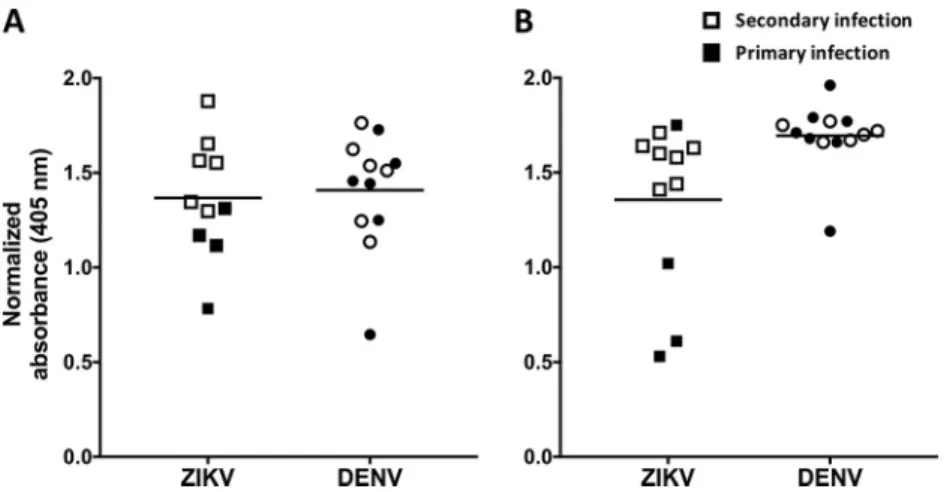
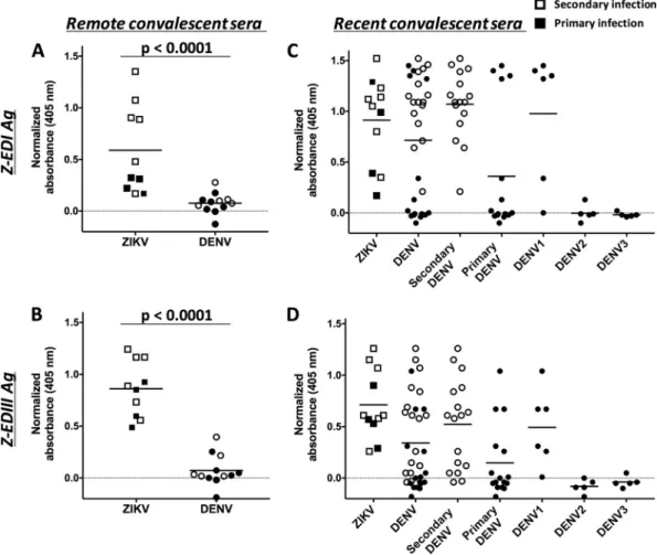
FIG3BindingofrecombinantE80antigentoserafrompatientswithremote(A)andrecent(B)ZIKV and/orDENVinfection.Serafromprimary(filledsymbols)andsecondary(unfilledsymbols)ZIKV-and DENV-infectedpatientswerediluted1:20,andtheIgGantibodiesboundtorecombinantE80antigen weremeasuredbyELISA.Seracollectedⱖ12weeksafterinfectionweredefinedasremoteinfections,and seracollectedwithinthefirst12weekswereconsideredtorepresentrecentinfections.Thehorizontal linesrepresentthemeans.
strongly with ZIKV E80, immune sera from individuals infected with DENV consistently
showed high levels of cross-reactivity with recombinant ZIKV E80 antigen in a standard
IgG
ELISA
(Fig.
3A).
Using
an
anti-MBP
monoclonal
antibody
to
capture
MBP
fusion
proteins,
we
developed
a
sandwich
ELISA
to
measure
serum
IgG
levels
to
Z-EDI
and
Z-EDIII (Fig. 4A and B). At late convalescence, ZIKV-immune sera recognized Z-EDIII and
Z-EDI antigens significantly better than DENV-immune sera (
P
⬍
0.0001 by the
Mann-Whitney
test).
Consequently,
the
Z-EDI and Z-EDIII antigens may be useful for specific
detection
of
remote
(
⬎
12
weeks)
ZIKV
infections
in
areas
with
endemic
DENV
trans-mission.
Evaluation
of
ZIKV
E80,
EDI,
and
EDIII
antigens
for
serological
detection
of
recent infections (2 to 12 weeks postinfection).
At early convalescence, immune sera
FIG 4Binding of Z-EDI and Z-EDIII with remote (A and B) and recent (C and D) convalescent-phase sera from patients infected with ZIKV and/or DENV. Primary (filled symbols) and secondary (unfilled symbols) human serum samples were diluted 1:20, and the IgG antibodies bound to Z-EDI (A and C) or Z-EDIII (B and D) were measured using a sandwich ELISA. Sera collectedⱖ12 weeks after infection were defined as remote infections, and sera collected within the first 12 weeks were considered to represent recent infections. Statistical significances are indicated at the top of the graphs (Mann-Whitney U test).Pvalues of ⬍0.0001 were considered statistically significant. The horizontal lines represent the means.
binding in our assay. Taken together, the results showed that IgG cross-reactivity with
the
Z-EDI
and
Z-EDIII
antigens
in
DENV-immune
sera
was
pronounced
in
early-convalescent-phase
samples
(
⬍
12
weeks)
from
secondary
DENV
and
primary
DENV1
infections but not
at the
late convalescent
(
⬎
12
weeks) phase.
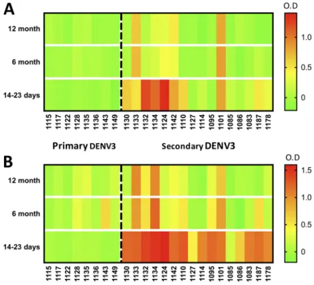
Longitudinal analysis of ZIKV EDI and EDIII cross-reactivities in DENV-immune
samples.
Next, we evaluated sequential serum samples collected as part of a
FIG5Patternsofcross-reactivityofZ-EDIII(A)andZED-I(B)antigenswithlongitudinalDENVsamples. Intensities of serum binding in sandwich ELISAs for Z-EDIII and Z-EDI are shown as heat maps. Longitudinalsamples(collected14to23days,6months,and12monthspostinfection)frompatients withprimary(leftofthedashedline)orsecondary(rightofthedashedline)DENV3infectionwerediluted 1:20,andtheIgGantibodiesboundtoZ-EDIorZ-EDIIIweremeasuredusingasandwichELISA.The resultingnormalizedODvaluesarerepresentedbyacolorscale(green,lowestvalues;yellow,middle values;andred,highestvalues).
postinfection (Fig. 5B). We concluded that among people exposed to secondary DENV
infections, cross-reactive Z-EDIII antibodies typically decline to
background levels by 6
months postinfection.
DISCUSSION
As
ZIKV
is
emerging
in
areas
with
intense
DENV
transmission
and,
more
recently,
clinical trials of DENV vaccines, there is an urgent need for simple serological assays to
distinguish
ZIKV infections
from
DENV infections.
Our
comparative
analysis
of surface
amino acid conservation among
flavivirus E proteins and homology epitope
mapping
pointed
to
three
regions
on
ZIKV
E
protein
as
potential
targets
of
ZIKV
type-specific
antibodies.
Here
we
evaluated
the
utility
of
recombinant
Z-EDI
and
Z-EDIII
antigens,
which
display
two
of
the
three
predicted
ZIKV-specific
antigenic
regions.
Our
results
demonstrate that Z-EDIII and, to a lesser extent, Z-EDI are strong candidate antigens for
serological tests to differentiate ZIKV infections from DENV infections when samples are
collected
⬎
12 weeks after infection. The recombinant antigens performed equally well
for
both primary
and secondary
infection
samples, indicating
that specificity
was
not
reduced by high levels of cross-reactive antibodies characteristic of secondary flavivirus
infection.
failed
to
eliminate the cross-reactivity, indicating
the need for additional mutations to
ablate the epitope as well as the possibility of other conserved epitopes between ZIKV
and
DENV1. In
secondary
DENV
cases, we
consistently observed
high
levels
of
cross-reactivity
at
early
convalescence,
irrespective
of
serotype
or
geographic
location
of
sample collection.
Our longitudinal analysis of Z-EDI and Z-EDIII reactivities, spanning from early to late
convalescent
phase,
showed
that
flavivirus-cross-reactive
IgG
antibodies
comprise
a
transient
population
that
is
produced
early
after
infection
and
declines
thereafter,
whereas
ZIKV-specific
responses
are
more
stable
over
time
(26).
While
the
cellular
mechanisms responsible for the differential
decline of
cross-reactive and type-specific
serum
antibodies are not known, one
possible
explanation is that
many
of
the
cross-reactive antibodies are derived from early plasmablasts or extrafollicular B cells that are
not maintained as long-lived
plasma
cells
or memory B
cells.
Development
of
serological
tests
for
diagnosing
ZIKV
infection
in
the
context
of
prior
flavivirus
infection
is
a
challenging
and
complex problem
that
remains
a
major
unmet need. To date, there are only three serological assays for ZIKV approved by the
U.S. Food and Drug Administration, under an emergency use authorization (56), and a
few other commercial
tests
are
available in
countries
outside the United States
or for
research purposes. These assays use either NS1, recombinant E, or another, unspecified
ZIKV
antigen
(57).
The
Centers
for
Disease
Control
and
Prevention
MAC
(IgM)
ELISA
exhibits well-publicized
limitations,
including
false-negative
results (58),
false-positive
results due to cross-reactive antibody from DENV infection (59), and persistence of ZIKV
IgM
beyond
the
previously
presumed
12-week
window
(60).
Our
findings
of
cross-reactive IgG binding
in early
convalescence
indicate that
this
period will be the most
challenging for optimization of assay specificity. Thus, there is roughly a 10-week period
(weeks
2
to
12) following infection
when current
and next-generation serodiagnostic
results may remain ambiguous. One important step forward is found in a recent report
evaluating an NS1-based blockade-of-binding
assay for ZIKV
diagnosis (61). This assay
leverages
a
ZIKV
type-specific
monoclonal
antibody
recognizing
a
nonconserved
epitope
on ZIKV
NS1
(62).
Again,
a certain
secondary DENV group displayed reduced
specificity
in
this
NS1-based
assay
during
early
convalescence.
It
may
be
that
a
combination
of
antigens
is
required
to
achieve
optimal
sensitivity
and
specificity
for
serum
antibody detection,
particularly during
early convalescence.
Additional
issues
preclude
optimal
implementation
of
many
currently
available
serological
assays.
First,
the
serum
panels
used
to
evaluate
these
assays
come
from
remnant clinical specimens or archived sera not collected systematically and specifically
for
analysis
of
clinical
performance
in
diagnosing
individuals
with
multiple
flavivirus
exposures.
Second,
sera from individuals
with
a
single flavivirus
infection
history and
residing in regions
where
flavivirus infection is not endemic
are
not
representative
of
the
populations
for
whom
improved
diagnostics
are
most
critical,
namely,
those
residing
in
the
tropics,
where
individuals
experience
multiple
and
frequent
flavivirus
exposures. We are involved with ongoing studies designed to address this shortcoming.
Third,
sensitivity in different IgM assays can be less than 80%, particularly
outside the
range of 6 to 60 days, when IgM assays perform best. Lastly, not only have false-positive
ZIKV test
results
been
reported due
to
current
or previous
DENV infection,
but
DENV
tests may
also be positive
following confirmed ZIKV infections. The
cumulative
expe-rience with ZIKV serodiagnosis, to date, clearly indicates that novel approaches will be
required.
SUPPLEMENTAL FILE 1,
PDF file, 0.1 MB.
SUPPLEMENTAL FILE 2,
PDF file, 0.1 MB.
Diversity
in
infecting
strains
of
ZIKV
may
elicit
antibodies
that
target
different
epitopes or different permutations of the same antigenic region of E protein. While we
evaluated only a single construct for each of the Z-EDI and Z-EDIII antigens, we believe
that
these
antigens
(from
a
ZIKV
isolate
from
French
Polynesia)
are
likely
to
be
representative of the vast majority of ZIKV strains in circulation. In fact, E protein amino
acid sequences from ZIKV isolates from several different times and places vary by only
ⱕ
1%,
and
both
African
and
Asian
lineage
strains
perform
similarly
in
binding
and
neutralization
assays,
suggesting
that ZIKV exists as a single serotype (26, 63).
While
the
present
work
provides
the
platform
for
incorporating
Z-EDI
and
Z-EDIII
into a suitable antigen-antibody binding assay for the purposes of surveillance, vaccine
efficacy studies, and risk reduction counseling, further modification of Z-EDI and Z-EDIII
may
improve
their
utility
in
the
early
convalescent
phase
of
ZIKV
infection.
Cross-reactive antibodies may be depleted using recombinant DENV antigens, but depletion
techniques
are
tedious
and
time-consuming
(26).
Introducing
amino
acid
variation
through
protein
engineering
is
an
attractive
strategy
to
eliminate
cross-reactive
antibody-binding
sites
while
preserving
unique
epitopes
within
Z-EDIII
and
Z-EDI
antigens. The
high signal we
observed
for
IgG
binding
to Z-EDIII
with
a
simple
ELISA
format is encouraging, although a combination of Z-EDI and Z-EDIII as well as fusion of
antigens to protein scaffolds
may also
be tested for improvement of the sensitivity
of
the assay. Finally, we observed
that some individuals are strongly
IgG seropositive for
only one of the Z-EDI or Z-EDIII antigen, raising the possibility that a multiplex platform
employing a panel of antigens may improve sensitivity (64). This approach also has the
advantage
of
allowing
the
design
of
expanded
antigen
panels
to
detect
antibodies
specific
for additional
pathogens that
cause
clinical
presentations
similar
to
those for
DENV
and
ZIKV.
In
conclusion,
we
have
demonstrated
that
Z-EDI
and
Z-EDIII
contain
important
epitopes that can be used to resolve current serodiagnostic limitations. Ultimately, this
work
can
lead
to
development
of
crucial
point-of-care
ZIKV
diagnostics
amenable
to
field
use
in resource-limited
settings.
In
the
process,
much can
be learned
about
the
epitopes targeted by
durable type-specific
and
cross-reactive
human
antibodies
gen-erated
upon
ZIKV
exposure,
which
is
important
for
the
design
of
highly
efficacious
DENV
and
ZIKV
vaccines.
SUPPLEMENTAL
MATERIAL
Supplemental material for this article may be found at https://doi.org/10.1128/JCM
.01504-17.
ACKNOWLEDGMENTS
These
studies
were
supported
by
NIAID,
NIH,
grants
R01AI107731
(A.M.D.S.),
R21AI134073
(A.M.D.S.
and
L.P.),
R01AI099631
(A.B.),
R21/R33AI100186
(A.B.),
P01AI106695
(E.H.),
and
U19AI118610
(E.H.)
and
by
CDC
contract
200-2017-93142
(A.M.D.S.).
Further
support
was
provided
by
ZIKAPLAN,
which
received
funding
from
the
European
Union’s
Horizon
2020
research
and
innovation
program
under
grant
agreement 734584. The Brazil pregnancy cohort was funded by FAPES grant
306/2016-74910132/16
(S.R.P.).
The
Sri
Lankan
cohort
study
was
funded
by
NIH
contracts
HHSN272200900042C
and
HHSN27220140045C
(A.S.).
The
content
of
this
study
is
solely
the
responsibility
of
the
authors
and
does
not
necessarily represent
the
official
views
of
the
National Institutes
of
Health.
Institute in Nicaragua for their dedication and high-quality work, and we are grateful to
the study participants and their families.
REFERENCES
1. Musso D, Baud D, Gubler DJ. 2016. Zika virus: what do we know? Clin Microbiol Infect 22:494 – 496.https://doi.org/10.1016/j.cmi.2016.03.032. 2. Lazear HM, Diamond MS. 2016. Zika virus: new clinical syndromes and its
emergence in the Western Hemisphere. J Virol 90:4864 – 4875.https://
doi.org/10.1128/JVI.00252-16.
3. Lazear HM, Stringer EM, de Silva AM. 2016. The emerging Zika virus epidemic in the Americas: research priorities. JAMA 315:1945–1946.
https://doi.org/10.1001/jama.2016.2899.
4. Cugola FR, Fernandes IR, Russo FB, Freitas BC, Dias JL, Guimaraes KP, Benazzato C, Almeida N, Pignatari GC, Romero S, Polonio CM, Cunha I, Freitas CL, Brandao WN, Rossato C, Andrade DG, Faria DP, Garcez AT, Buchpigel CA, Braconi CT, Mendes E, Sall AA, Zanotto PM, Peron JP, Muotri AR, Beltrao-Braga PC. 2016. The Brazilian Zika virus strain causes birth defects in experimental models. Nature 534:267–271.https://doi
.org/10.1038/nature18296.
5. Garcez PP, Loiola EC, Madeiro da Costa R, Higa LM, Trindade P, Delvec-chio R, Nascimento JM, Brindeiro R, Tanuri A, Rehen SK. 2016. Zika virus impairs growth in human neurospheres and brain organoids. Science 352:816 – 818.https://doi.org/10.1126/science.aaf6116.
6. The National Academies of Sciences, Engineering, and Medicine. 2016. Potential research priorities to inform public health and medical practice for domestic Zika virus: workshop in brief. The National Academies Press, Washington, DC.https://doi.org/10.17226/23404.
7. Bingham AM, Cone M, Mock V, Heberlein-Larson L, Stanek D, Blackmore C, Likos A. 2016. Comparison of test results for Zika virus RNA in urine, serum, and saliva specimens from persons with travel-associated Zika virus disease—Florida, 2016. MMWR Morb Mortal Wkly Rep 65:475– 478.
https://doi.org/10.15585/mmwr.mm6518e2.
8. Duffy MR, Chen T-H, Hancock WT, Powers AM, Kool JL, Lanciotti RS, Pretrick M, Marfel M, Holzbauer S, Dubray C, Guillaumot L, Griggs A, Bel M, Lambert AJ, Laven J, Kosoy O, Panella A, Biggerstaff BJ, Fischer M, Hayes EB. 2009. Zika virus outbreak on Yap Island, Federated States of Micronesia. N Engl J Med 360:2536 –2543. https://doi.org/10.1056/
NEJMoa0805715.
9. Musso D, Nhan T, Robin E, Roche C, Bierlaire D, Zisou K, Shan Yan A, Cao-Lormeau VM, Broult J. 2014. Potential for Zika virus transmission through blood transfusion demonstrated during an outbreak in French Polynesia, November 2013 to February 2014. Euro Surveill 19:20761.
https://doi.org/10.2807/1560-7917.ES2014.19.14.20761.
10. Rabe IB, Staples JE, Villanueva J, Hummel KB, Johnson JA, Rose L, MTS, Hills S, Wasley A, Fischer M, Powers AM. 2016. Interim guidance for interpretation of Zika virus antibody test results. MMWR Morb Mortal Wkly Rep 65:543–546.https://doi.org/10.15585/mmwr.mm6521e1. 11. Granger D, Hilgart H, Misner L, Christensen J, Bistodeau S, Palm J, Strain
AK, Konstantinovski M, Liu D, Tran A, Theel ES. 2017. Serologic testing for Zika virus: comparison of three Zika virus IgM-screening enzyme-linked immunosorbent assays and initial laboratory experiences. J Clin Micro-biol 55:2127–2136.https://doi.org/10.1128/JCM.00580-17.
12. Wahala WM, Silva AM. 2011. The human antibody response to dengue virus infection. Viruses 3:2374 –2395.https://doi.org/10.3390/v3122374. 13. Priyamvada L, Quicke KM, Hudson WH, Onlamoon N, Sewatanon J, Edupuganti S, Pattanapanyasat K, Chokephaibulkit K, Mulligan MJ, Wil-son PC, Ahmed R, Suthar MS, Wrammert J. 2016. Human antibody responses after dengue virus infection are highly cross-reactive to Zika virus. Proc Natl Acad Sci U S A 113:7852–7857.https://doi.org/10.1073/
pnas.1607931113.
14. Morrison AC, Minnick SL, Rocha C, Forshey BM, Stoddard ST, Getis A, Focks DA, Russell KL, Olson JG, Blair PJ, Watts DM, Sihuincha M, Scott TW, Kochel TJ. 2010. Epidemiology of dengue virus in Iquitos, Peru 1999 to 2005: interepidemic and epidemic patterns of transmission. PLoS Negl Trop Dis 4:e670.https://doi.org/10.1371/journal.pntd.0000670. 15. Crill WD, Chang GJ. 2004. Localization and characterization of flavivirus
envelope glycoprotein cross-reactive epitopes. J Virol 78:13975–13986.
https://doi.org/10.1128/JVI.78.24.13975-13986.2004.
16. Rothman AL. 2011. Immunity to dengue virus: a tale of original antigenic sin and tropical cytokine storms. Nat Rev Immunol 11:532–543.https://
doi.org/10.1038/nri3014.
17. Dai L, Song J, Lu X, Deng YQ, Musyoki AM, Cheng H, Zhang Y, Yuan Y, Song H, Haywood J, Xiao H, Yan J, Shi Y, Qin CF, Qi J, Gao GF. 2016. Structures of the Zika virus envelope protein and its complex with a flavivirus broadly protective antibody. Cell Host Microbe 19:696 –704.
https://doi.org/10.1016/j.chom.2016.04.013.
18. Kostyuchenko VA, Lim EX, Zhang S, Fibriansah G, Ng TS, Ooi JS, Shi J, Lok SM. 2016. Structure of the thermally stable Zika virus. Nature 533: 425– 428.https://doi.org/10.1038/nature17994.
19. Sirohi D, Chen Z, Sun L, Klose T, Pierson TC, Rossmann MG, Kuhn RJ. 2016. The 3.8 A resolution cryo-EM structure of Zika virus. Science 352:467– 470.https://doi.org/10.1126/science.aaf5316.
20. Kuan G, Gordon A, Aviles W, Ortega O, Hammond SN, Elizondo D, Nunez A, Coloma J, Balmaseda A, Harris E. 2009. The Nicaraguan pediatric dengue cohort study: study design, methods, use of information tech-nology, and extension to other infectious diseases. Am J Epidemiol 170:120 –129.https://doi.org/10.1093/aje/kwp092.
21. Waggoner JJ, Gresh L, Mohamed-Hadley A, Ballesteros G, Davila MJ, Tellez Y, Sahoo MK, Balmaseda A, Harris E, Pinsky BA. 2016. Single-reaction multiplex reverse transcription PCR for detection of Zika, chi-kungunya, and dengue viruses. Emerg Infect Dis 22:1295–1297.https://
doi.org/10.3201/eid2207.160326.
22. Lanciotti RS, Kosoy OL, Laven JJ, Velez JO, Lambert AJ, Johnson AJ, Stanfield SM, Duffy MR. 2008. Genetic and serologic properties of Zika virus associated with an epidemic, Yap State, Micronesia, 2007. Emerg Infect Dis 14:1232–1239.https://doi.org/10.3201/eid1408.080287. 23. Waggoner JJ, Ballesteros G, Gresh L, Mohamed-Hadley A, Tellez Y, Sahoo
MK, Abeynayake J, Balmaseda A, Harris E, Pinsky BA. 2016. Clinical evaluation of a single-reaction real-time RT-PCR for pan-dengue and chikungunya virus detection. J Clin Virol 78:57– 61.https://doi.org/10
.1016/j.jcv.2016.01.007.
24. Narvaez F, Gutierrez G, Perez MA, Elizondo D, Nunez A, Balmaseda A, Harris E. 2011. Evaluation of the traditional and revised WHO classifica-tions of dengue disease severity. PLoS Negl Trop Dis 5:e1397.https://
doi.org/10.1371/journal.pntd.0001397.
25. Weiskopf D, Angelo MA, Grifoni A, O’Rourke PH, Sidney J, Paul S, De Silva AD, Phillips E, Mallal S, Premawansa S, Premawansa G, Wijewickrama A, Peters B, Sette A. 2016. HLA-DRB1 alleles are associated with different magnitudes of dengue virus-specific CD4⫹T-cell responses. J Infect Dis 214:1117–1124.https://doi.org/10.1093/infdis/jiw309.
26. Collins MH, McGowan E, Jadi R, Young E, Lopez CA, Baric RS, Lazear HM, de Silva AM. 2017. Lack of durable cross-neutralizing antibodies against Zika virus from dengue virus infection. Emerg Infect Dis 23:773–781.
https://doi.org/10.3201/eid2305.161630.
27. Oliphant T, Nybakken GE, Engle M, Xu Q, Nelson CA, Sukupolvi-Petty S, Marri A, Lachmi BE, Olshevsky U, Fremont DH, Pierson TC, Diamond MS. 2006. Antibody recognition and neutralization determinants on domains I and II of West Nile virus envelope protein. J Virol 80:12149 –12159.
https://doi.org/10.1128/JVI.01732-06.
28. Baronti C, Piorkowski G, Charrel RN, Boubis L, Leparc-Goffart I, de Lamballerie X. 2014. Complete coding sequence of Zika virus from a French Polynesia outbreak in 2013. Genome Announc 2:e00500-14.
https://doi.org/10.1128/genomeA.00500-14.
29. Aslanidis C, de Jong PJ. 1990. Ligation-independent cloning of PCR products (LIC-PCR). Nucleic Acids Res 18:6069 – 6074.https://doi.org/10
.1093/nar/18.20.6069.
30. Metz SW, Gallichotte EN, Brackbill A, Premkumar L, Miley MJ, Baric R, de Silva AM. 2017. In vitro assembly and stabilization of dengue and Zika virus envelope protein homo-dimers. Sci Rep 7:4524.https://doi.org/10
.1038/s41598-017-04767-6.
31. Metz SW, Tian S, Hoekstra G, Yi X, Stone M, Horvath K, Miley MJ, DeSimone J, Luft CJ, de Silva AM. 2016. Precisely molded nanoparticle displaying DENV-E proteins induces robust serotype-specific neutraliz-ing antibody responses. PLoS Negl Trop Dis 10:e0005071.https://doi
.org/10.1371/journal.pntd.0005071.
epitopes on dengue virions. Proc Natl Acad Sci U S A 109:7439 –7444.
https://doi.org/10.1073/pnas.1200566109.
33. Ashkenazy H, Abadi S, Martz E, Chay O, Mayrose I, Pupko T, Ben-Tal N. 2016. ConSurf 2016: an improved methodology to estimate and visualize evolutionary conservation in macromolecules. Nucleic Acids Res 44: W344 –W350.https://doi.org/10.1093/nar/gkw408.
34. Fibriansah G, Ibarra KD, Ng TS, Smith SA, Tan JL, Lim XN, Ooi JS, Kostyuchenko VA, Wang J, de Silva AM, Harris E, Crowe JE, Jr, Lok SM. 2015. Dengue virus. Cryo-EM structure of an antibody that neutralizes dengue virus type 2 by locking E protein dimers. Science 349:88 –91.
https://doi.org/10.1126/science.aaa8651.
35. Kaufmann B, Vogt MR, Goudsmit J, Holdaway HA, Aksyuk AA, Chipman PR, Kuhn RJ, Diamond MS, Rossmann MG. 2010. Neutralization of West Nile virus by cross-linking of its surface proteins with Fab fragments of the human monoclonal antibody CR4354. Proc Natl Acad Sci U S A 107:18950 –18955.https://doi.org/10.1073/pnas.1011036107.
36. Fibriansah G, Tan JL, Smith SA, de Alwis AR, Ng TS, Kostyuchenko VA, Ibarra KD, Wang J, Harris E, de Silva A, Crowe JE, Jr, Lok SM. 2014. A potent anti-dengue human antibody preferentially recognizes the con-formation of E protein monomers assembled on the virus surface. EMBO Mol Med 6:358 –371.https://doi.org/10.1002/emmm.201303404. 37. Teoh EP, Kukkaro P, Teo EW, Lim AP, Tan TT, Yip A, Schul W, Aung M,
Kostyuchenko VA, Leo YS, Chan SH, Smith KG, Chan AH, Zou G, Ooi EE, Kemeny DM, Tan GK, Ng JK, Ng ML, Alonso S, Fisher D, Shi PY, Hanson BJ, Lok SM, MacAry PA. 2012. The structural basis for serotype-specific neutralization of dengue virus by a human antibody. Sci Transl Med 4:139ra83.https://doi.org/10.1126/scitranslmed.3003888.
38. Cockburn JJ, Navarro Sanchez ME, Goncalvez AP, Zaitseva E, Stura EA, Kikuti CM, Duquerroy S, Dussart P, Chernomordik LV, Lai CJ, Rey FA. 2012. Structural insights into the neutralization mechanism of a higher primate antibody against dengue virus. EMBO J 31:767–779.https://doi
.org/10.1038/emboj.2011.439.
39. Nybakken GE, Oliphant T, Johnson S, Burke S, Diamond MS, Fremont DH. 2005. Structural basis of West Nile virus neutralization by a therapeutic antibody. Nature 437:764 –769.https://doi.org/10.1038/nature03956. 40. Rouvinski A, Guardado-Calvo P, Barba-Spaeth G, Duquerroy S, Vaney MC,
Kikuti CM, Navarro Sanchez ME, Dejnirattisai W, Wongwiwat W, Haouz A, Girard-Blanc C, Petres S, Shepard WE, Despres P, Arenzana-Seisdedos F, Dussart P, Mongkolsapaya J, Screaton GR, Rey FA. 2015. Recognition determinants of broadly neutralizing human antibodies against dengue viruses. Nature 520:109 –113.https://doi.org/10.1038/nature14130. 41. Cherrier MV, Kaufmann B, Nybakken GE, Lok SM, Warren JT, Chen BR,
Nelson CA, Kostyuchenko VA, Holdaway HA, Chipman PR, Kuhn RJ, Diamond MS, Rossmann MG, Fremont DH. 2009. Structural basis for the preferential recognition of immature flaviviruses by a fusion-loop anti-body. EMBO J 28:3269 –3276.https://doi.org/10.1038/emboj.2009.245. 42. Lok SM, Kostyuchenko V, Nybakken GE, Holdaway HA, Battisti AJ,
Sukupolvi-Petty S, Sedlak D, Fremont DH, Chipman PR, Roehrig JT, Diamond MS, Kuhn RJ, Rossmann MG. 2008. Binding of a neutralizing antibody to dengue virus alters the arrangement of surface glycopro-teins. Nat Struct Mol Biol 15:312–317. https://doi.org/10.1038/nsmb
.1382.
43. Cockburn JJ, Navarro Sanchez ME, Fretes N, Urvoas A, Staropoli I, Kikuti CM, Coffey LL, Arenzana Seisdedos F, Bedouelle H, Rey FA. 2012. Mechanism of dengue virus broad cross-neutralization by a monoclonal antibody. Structure 20:303–314.https://doi.org/10.1016/
j.str.2012.01.001.
44. Austin SK, Dowd KA, Shrestha B, Nelson CA, Edeling MA, Johnson S, Pierson TC, Diamond MS, Fremont DH. 2012. Structural basis of differ-ential neutralization of DENV-1 genotypes by an antibody that recog-nizes a cryptic epitope. PLoS Pathog 8:e1002930. https://doi.org/10
.1371/journal.ppat.1002930.
45. Robinson LN, Tharakaraman K, Rowley KJ, Costa VV, Chan KR, Wong YH, Ong LC, Tan HC, Koch T, Cain D, Kirloskar R, Viswanathan K, Liew CW, Tissire H, Ramakrishnan B, Myette JR, Babcock GJ, Sasisekharan V, Alonso S, Chen J, Lescar J, Shriver Z, Ooi EE, Sasisekharan R. 2015. Structure-guided design of an anti-dengue antibody directed to a non-immunodominant epitope. Cell 162:493–504.https://doi.org/10.1016/j
.cell.2015.06.057.
46. Midgley CM, Flanagan A, Tran HB, Dejnirattisai W, Chawansuntati K, Jumnainsong A, Wongwiwat W, Duangchinda T, Mongkolsapaya J, Grimes JM, Screaton GR. 2012. Structural analysis of a dengue cross-reactive antibody complexed with envelope domain III reveals the
mo-lecular basis of cross-reactivity. J Immunol 188:4971– 4979.https://doi
.org/10.4049/jimmunol.1200227.
47. Barba-Spaeth G, Dejnirattisai W, Rouvinski A, Vaney MC, Medits I, Sharma A, Simon-Loriere E, Sakuntabhai A, Cao-Lormeau VM, Haouz A, England P, Stiasny K, Mongkolsapaya J, Heinz FX, Screaton GR, Rey FA. 2016. Structural basis of potent Zika-dengue virus antibody cross-neutralization. Nature 536:48 –53.https://doi.org/10.1038/nature18938.
48. Zhao H, Fernandez E, Dowd KA, Speer SD, Platt DJ, Gorman MJ, Govero J, Nelson CA, Pierson TC, Diamond MS, Fremont DH. 2016. Structural basis of Zika virus-specific antibody protection. Cell 166:1016 –1027.
https://doi.org/10.1016/j.cell.2016.07.020.
49. Wahala WM, Kraus AA, Haymore LB, Accavitti-Loper MA, de Silva AM. 2009. Dengue virus neutralization by human immune sera: role of envelope protein domain III-reactive antibody. Virology 392:103–113.
https://doi.org/10.1016/j.virol.2009.06.037.
50. Lichtarge O, Bourne HR, Cohen FE. 1996. An evolutionary trace method defines binding surfaces common to protein families. J Mol Biol 257: 342–358.https://doi.org/10.1006/jmbi.1996.0167.
51. Ashkenazy H, Erez E, Martz E, Pupko T, Ben-Tal N. 2010. ConSurf 2010: calculating evolutionary conservation in sequence and structure of pro-teins and nucleic acids. Nucleic Acids Res 38:W529 –W533.https://doi
.org/10.1093/nar/gkq399.
52. Ellman GL. 1959. Tissue sulfhydryl groups. Arch Biochem Biophys 82: 70 –77.https://doi.org/10.1016/0003-9861(59)90090-6.
53. Rogers TF, Goodwin EC, Briney B, Sok D, Beutler N, Strubel A, Nedellec R, Le K, Brown ME, Burton DR, Walker LM. 2017. Zika virus activates de novo and cross-reactive memory B cell responses in dengue-experienced donors. Sci Immunol 2:eaan6809.https://doi.org/10.1126/sciimmunol.aan6809. 54. Robbiani DF, Bozzacco L, Keeffe JR, Khouri R, Olsen PC, Gazumyan A,
Schaefer-Babajew D, Avila-Rios S, Nogueira L, Patel R, Azzopardi SA, Uhl LFK, Saeed M, Sevilla-Reyes EE, Agudelo M, Yao KH, Golijanin J, Gristick HB, Lee YE, Hurley A, Caskey M, Pai J, Oliveira T, Wunder EA, Jr, Sacra-mento G, Nery N, Jr, Orge C, Costa F, Reis MG, Thomas NM, Eisenreich T, Weinberger DM, de Almeida ARP, West AP, Jr, Rice CM, Bjorkman PJ, Reyes-Teran G, Ko AI, MacDonald MR, Nussenzweig MC. 2017. Recurrent potent human neutralizing antibodies to Zika virus in Brazil and Mexico. Cell 169:597.e11– 609.e11.https://doi.org/10.1016/j.cell.2017.04.024. 55. Bolze A, Byun M, McDonald D, Morgan NV, Abhyankar A, Premkumar L,
Puel A, Bacon CM, Rieux-Laucat F, Pang K, Britland A, Abel L, Cant A, Maher ER, Riedl SJ, Hambleton S, Casanova J-L. 2010. Whole-exome-sequencing-based discovery of human FADD deficiency. Am J Hum Genet 87:873– 881.https://doi.org/10.1016/j.ajhg.2010.10.028. 56. US FDA. 2016. Zika virus emergency use authorization.https://www.fda
.gov/MedicalDevices/Safety/EmergencySituations/ucm161496.htm#zika.
57. Safronetz D, Sloan A, Stein DR, Mendoza E, Barairo N, Ranadheera C, Scharikow L, Holloway K, Robinson A, Traykova-Andonova M, Makowski K, Dimitrova K, Giles E, Hiebert J, Mogk R, Beddome S, Drebot M. 2017. Evaluation of 5 commercially available Zika virus immunoassays. Emerg Infect Dis 23:1577–1580.https://doi.org/10.3201/eid2309.162043. 58. Davis AC. 2017. D.C.’s botched Zika testing leaves dozens of families
monitoring for symptoms. https://www.washingtonpost.com/local/dc -politics/dcs-botched-zika-testing-leaves-dozens-of-families-monitoring -for-symptoms/2017/05/09/3ab24958-34db-11e7-b373-418f6849a004_
story.html?utm_term⫽.fcd651095232.
59. Education CsDoICa. 2016. FDA warns health care providers against relying solely on Zika virus serological IgM assay results; reminds them to wait for confirmatory test results before making patient management decisions: FDA safety communication. http://www
.firstwordmedtech.com/node/992281.
60. Oduyebo T, Polen KD, Walke HT, Reagan-Steiner S, Lathrop E, Rabe IB, Kuhnert-Tallman WL, Martin SW, Walker AT, Gregory CJ, Ades EW, Carroll DS, Rivera M, Perez-Padilla J, Gould C, Nemhauser JB, Ben Beard C, Harcourt JL, Viens L, Johansson M, Ellington SR, Petersen E, Smith LA, Reichard J, Munoz-Jordan J, Beach MJ, Rose DA, Barzilay E, Noonan-Smith M, Jamieson DJ, Zaki SR, Petersen LR, Honein MA, Meaney-Delman D. 2017. Update: interim guidance for health care providers caring for pregnant women with possible Zika virus exposure—United States (in-cluding U.S. territories), July 2017. MMWR Morb Mortal Wkly Rep 66: 781–793.https://doi.org/10.15585/mmwr.mm6629e1.
flaviviruses. Proc Natl Acad Sci U S A 114:8384 – 8389.https://doi.org/
10.1073/pnas.1704984114.
62. Stettler K, Beltramello M, Espinosa DA, Graham V, Cassotta A, Bianchi S, Vanzetta F, Minola A, Jaconi S, Mele F, Foglierini M, Pedotti M, Simonelli L, Dowall S, Atkinson B, Percivalle E, Simmons CP, Varani L, Blum J, Baldanti F, Cameroni E, Hewson R, Harris E, Lanzavecchia A, Sallusto F, Corti D. 2016. Specificity, cross-reactivity, and function of antibodies elicited by Zika virus infection. Science 353:823– 826.https://doi.org/10
.1126/science.aaf8505.
63. Dowd KA, DeMaso CR, Pelc RS, Speer SD, Smith AR, Goo L, Platt DJ, Mascola JR, Graham BS, Mulligan MJ, Diamond MS, Ledgerwood JE, Pierson TC. 2016. Broadly neutralizing activity of Zika virus-immune sera identifies a single viral serotype. Cell Rep 16:1485–1491.https://doi.org/
10.1016/j.celrep.2016.07.049.