Received: 27.03.2019 Accepted: 22.05.2019
Published: 10.06.2019 http://www.polradiol.com
Original paper
Diagnostic reliability of the Thyroid Imaging Reporting and Data
System (TI-RADS) in routine practice
Allen San Shell Jabar
A,B,C,D,E,F, Prakashini Koteshwara
A,B,C,D,E,F, Jasbon Andrade
A,B,C,D,E,FDepartment of Radiodiagnosis and Imaging, Kasturba Medical College, Manipal Academy of Higher Education (MAHE), Manipal, India
Abstract
Purpose: To evaluate the diagnostic reliability of Thyroid Imaging Reporting and Data System (TI-RADS) classifica-tions described by American College of Radiology (ACR) and Kwak et al. by calculating the risk of malignancy, to assess the role of TI-RADS in reducing fine-needle aspiration cytology (FNAC) of benign lesions.
Material and methods: This was a prospective study during the period from December 2017 to August 2018. Thyroid nodules were classified using ACR TI-RADS and TI-RADS proposed by Kwak et al. The TI-RADS categorisations were compared to the final diagnosis obtained by cytopathological/histopathological analysis. The risk of malignancy for each category was calculated. Sensitivity, specificity, and positive and negative predictive values for individual suspicious ultrasound features were also assessed.
Results: We evaluated a total of 127 thyroid nodules. The risk of malignancy was 0% in ACR TR1, 0% in ACR TR2, 6.9% in ACR TR3, 29.2% in ACR TR4, and 80% in ACR TR5 categories. The risk of malignancy for TI-RADS according to Kwak et al. were 0%, 0%, 21.5%, 32.4%, 100% for TI-RADS 2, 3, 4A, 4B, and 4C categories, respectively. Kwak TI-RADS 2 and 3 had higher sensitivity in predicting benignity compared to ACR TR1 and 2 (35.4% vs. 25.9%).
Conclusions: We found TI-RADS classification to be a reliable, non-invasive, and practical method for assessing thy-roid nodules in routine practice. TI-RADS can safely avert avoidable FNACs in a significant proportion of benign thyroid lesions.
Key words: thyroid imaging reporting and data system, thyroid ultrasound, ACR-TI-RADS, Kwak TI-RADS, thyroid nodule, fine-needle aspiration cytology.
Correspondence address:
Dr. Prakashini Koteshwara, Department of Radiodiagnosis and Imaging, Kasturba Medical College, Manipal Academy of Higher Education (MAHE), Manipal, India, e-mail: [email protected]
Authors’ contribution:
Introduction
High-resolution sonography is a safe, valuable, nonionis-ing, cost-effective, readily available imaging tool for iden-tification of clinically suspected thyroid nodules. Various studies mentioned a prevalence of 2-6% of thyroid lesions on palpation, 19-35% on sonography, and 8-65% in au-topsy data [1]. Fine-needle aspiration cytology (FNAC) remains pivotal for the assessment of thyroid nodules, but it is mildly distressing, leads to additional healthcare costs, and contains rare a low risk of infection and scarring [2]. There is a growing need to craft and utilise a dependable ultrasound categorisation for evaluating thyroid nodules
and discriminating benign and malignant lesions with a reasonable level of certainty, through which we could also cut short a number of unnecessary invasive FNACs. Nu-merous studies have proposed a thyroid imaging reporting and data system (TI-RADS). These studies also brought to light a significant number of parameters for quantita-tive analysis of sonographic features [3,4]. However, the implication of these parameters differed in each study. We investigated the TIRADS classifications proposed in 2011 by Kwak et al. and the 2017 ACR-TI-RADS [5,6]. The Kwak TI-RADS is simple and similar to BI-RADS sys-tem, which has been in use for several years and is familiar to most radiologists. In ACR-TI-RADS points are given
for various ultrasound features in a thyroid nodule, with more suspicious features being awarded additional points. When assessing a nodule, its point total determines the nodule’s ACR TI-RADS stage, which varies from TR1 (be-nign lesion) to TR5 (high suspicion of malignancy) [6,7]. This study was performed to prospectively investigate the diagnostic reliability of the aforementioned TI-RADS clas-sification system in differentiating benign and malignant lesions by stratifying the risk of malignancy separately for individual TI-RADS categories and for various ultrasound features. We also wanted to estimate the decrease in the number of unnecessary FNACs.
Material and methods
This was a hospital-based, time-bound, prospective study conducted between December 2017 and August 2018 (a period of nine months) in the Department of Radio-diagnosis and Imaging. Institutional Ethics Committee approval was obtained prior to this prospective study. The study group comprised of 127 patients referred to the Department of Radiodiagnosis with clinically suspected thyroid nodules. These patients underwent convention-al high-resolution sonography, and depending on the presence or absence of various sonographic features the thyroid nodules were categorised according to TI-RADS classification. The study required an invasive investigation (FNAC) to be conducted on patients. Informed consent was obtained from every patient. All patients who did not give consent for FNAC and those with bleeding diathesis were excluded from the study. Cases with nondiagnostic or indeterminate cytology results were also excluded from our study. In cases where thyroid biopsy/surgery were per-formed, both the sonographic and cytopathology results were followed up with the final histopathology report.
Sonography technique
Ultrasound evaluation was performed on a Philips Epiq 5G ultrasound machine using a high-frequency linear L18-5 probe.
With the patient supine and neck hyper-extended, the entire gland was examined. Hyperextension of the neck was obtained by placing a pad under the shoulders. The neck was scanned in sagittal, transverse, and oblique sec-tions to optimally visualise both lobes of the thyroid and isthmus. Imaging of the lower poles of thyroid was done by making the patient swallow, because this tends to raise the thyroid gland in the neck.
Specific attention was prospectively directed to nod-ule characteristics (like composition, shape, echogenicity, margins, and echogenic foci) as those used in the ACR lexicon to describe thyroid nodules [7]. Points were as-signed to each nodule for the separate categories accord-ing to ACR-TI-RADS guidelines [6]. The sum of the points in each category determined the TI-RADS level
assigned to each nodule, with TR1 indicating 0 points; TR2 – 2 points; TR3 – 3 points; TR4 – 4-6 points; and TR5 – 7 or more points. The data obtained from the ACR point table were used to assess the Kwak TI-RADS catego-ry because the five suspicious features mentioned by Kwak et al. were a subset of the nodule characteristics described in the ACR lexicon. These suspicious features included: solid composition, marked hypoechogenicity, microcal-cification, taller than wide shape, and irregular margins. Our analysis differed slightly from the lexicon of the Kwak TI-RADS committee with respect to margin shape. In studies conducted by Srinivas et al. and Chandramohan et al., who assessed Kwak TI-RADS, ill-defined margins were included under irregular margin characteristics, and they found statistically significant correlation with malig-nancy [8,9]. In the ACR point table both ill-defined and irregular margins are categorised separately under margin characteristics, with the former lacking points and the lat-ter contributing 2 points. According to the guidelines of the ACR TI-RADS committee, if composition, echogenic-ity, or margins could not be determined for any reason like dense posterior acoustic shadowing from macrocalci-fication, they were allocated 2, 1, or 0 points, respectively.
Ultrasound-guided fine-needle aspiration cytology
technique
Ultrasound-guided FNAC of dominant or suspicious nodule was performed. The majority of FNACs were per-formed on an outpatient basis. FNAC was perper-formed us-ing a 23-gauge needle attached to a 10-ml syrus-inge. Two to three aspirations were performed on each nodule and cytology smears prepared for analysis. FNAC cases that were non-diagnostic were included in the final analysis only if surgical histopathology was available.
Statistical analysis
Collected data was entered into a Microsoft Excel data sheet and were analysed using the SPSS 25 version soft-ware. Specificity, sensitivity, and positive and negative predictive value (with 95% confidence interval [95% CI]) were calculated and used to evaluate the reliability of TI-RADS in differentiation between benign and malignant features. The odds ratio was determined to quantify how strongly the presence or absence of a particular suspicious ultrasound feature was associated with malignancy in the study population. The p values were measured using Stu-dent’s t-test. In all analyses, p < 0.05 was taken to indicate statistical significance.
Results
Of a total of 127 nodules included in this study, 110 nod-ules were from females (86.6%) and 17 from males (13.4%), with a male-to-female ratio of 1 : 6.4. We observed a slightly
higher female-to-male ratio in our study, but our study only represented the values obtained from a single hospital and was not a multicentric study. Out of 127 nodules studied 104 nodules were benign (94 nodules in females, 10 nod-ules in males) and 23 nodnod-ules were malignant (16 nodnod-ules in females, seven nodules in males). Malignancy was more common among male patients who presented with a thy-roid nodule (not statistically significant, p = 0.084). There was no significant difference in the mean age of patients with benign (mean age was 46.6 ±13.3 years) and malig-nant thyroid nodules (mean age was 47.3 ±12.6 years) (p = 0.818).
ACR-TI-RADS
We found that the risk of malignancy increased as the ACR-TI-RADS category increased. The risk of
malignan-cy in ACR TI-RADS category 1 and 2 were found to be 0%, 6.9% in category 3, 30.9% in category 4, and 77.7% in category 5.
Kwak TIRADS
None of the nodules assessed in our study could be cat-egorised into Kwak TI-RADS 5. Similarly, TI-RADS 1 refers to normal thyroid according to Kwak TI-RADS. Barring these two categories, we found in our study that the risk of malignancy significantly rose as the TI-RADS category increased. The risk of malignancy in TI-RADS categories 2 and 3 was found to be 0%, 21.5% in catego-ry 4A, 32.4% in categocatego-ry 4B, and 100.0% in categocatego-ry 4C (Tables 1 and 2).
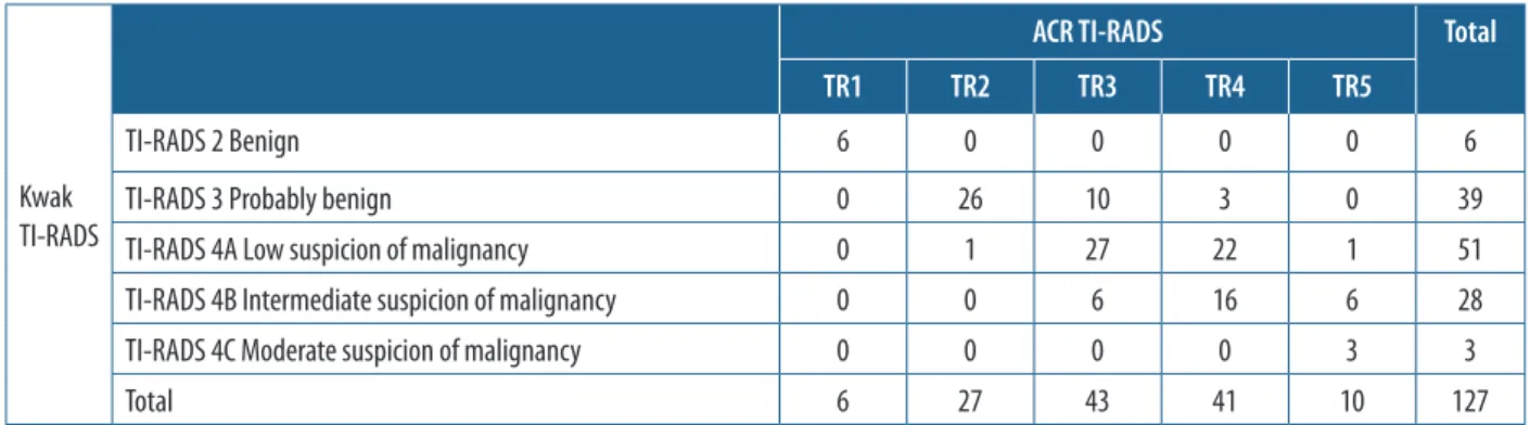
In our study sonological features that showed a signifi-cant association with malignancy were solid composition, Table 1. Cross tabulation depicting nodule distribution in Kwak and ACR TI-RADS
Kwak TI-RADS
ACR TI-RADS Total
TR1 TR2 TR3 TR4 TR5
TI-RADS 2 Benign 6 0 0 0 0 6
TI-RADS 3 Probably benign 0 26 10 3 0 39
TI-RADS 4A Low suspicion of malignancy 0 1 27 22 1 51
TI-RADS 4B Intermediate suspicion of malignancy 0 0 6 16 6 28
TI-RADS 4C Moderate suspicion of malignancy 0 0 0 0 3 3
Total 6 27 43 41 10 127
Table 2. Frequency of ultrasound features of thyroid nodules according to ACR-TI-RADS descriptors
Ultrasound feature Benign Malignant Total Risk of malignancy (%)
Composition Cystic or predominantly cystic 2 0 2 0
Spongiform 4 0 4 0
Mixed solid-cystic 44 0 44 0
Solid or predominantly solid 54 23 77 29.8
Echogenicity Anechoic 3 0 3 0
Hyperechoic or isoechoic 89 6 95 6.3
Hypoechoic 8 16 24 66.6
Very hypoechoic 0 1 1 100
Shape Wider than tall or round 98 20 118 16.9
Taller than wide 2 3 5 60.0
Margin Smooth 79 16 95 16.8
Ill-defined 16 4 20 20.0
Lobulated or irregular 4 3 7 42.8
Extra-thyroidal extension 1 0 1 0
Echogenic foci None/large comet tail artefact 66 10 76 13.1
Macrocalcifications 21 6 27 22.2
Peripheral rim calcification 11 3 14 21.4
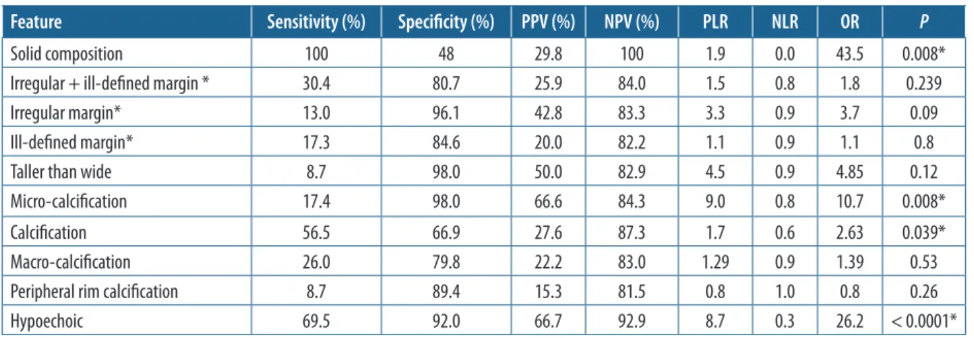
hypoechogenicity, and calcification including microcalcifi-cation. Out of the suspicious sonological features hypoecho-genicity and microcalcification showed the highest positive predictive value (PPV) (66.7%) for malignancy (Table 3).
Estimation of decrease in unnecessary fine-needle
aspiration cytologies using TIRADS
Using Kwak TI-RADS all nodules characterised un-der category 2 and 3 were benign on cyto/histopathology. Therefore, the specificity of TI-RADS categories 2 (4.7%) and 3 (30.7%) in labelling a nodule as benign was found to be 100%. The estimated decrease in unnecessary FNACs in our study was found to be 35.43%.
Specificity of ACR TI-RADS category TR1 and TR2 in labelling a nodule as benign was also found to be 100%. By avoiding these nodules the estimated decrease in un-necessary FNACs in our study would have been 25.98%.
Discussion
In 2005 the Society of Radiologists in Ultrasound (SRU) published their guidelines to help in deciding which thy-roid nodules needed FNA and which that did not [10].
Subsequently, many other studies have proven the use-fulness of ultrasound evaluation of thyroid nodules and its ability to differentiate benign from malignant nodules [3-5,11-17]. Also, professional societies brought forth their guidelines, which either focused on or included suggestions for use of ultrasonographic findings in deter-mining whether a nodule required FNA [18-22]. All these guidelines were chiefly built upon sonographic appear-ance and nodule size. The ultrasonographic appearappear-ance is important because it lends information pertaining to risk of malignancy. To predict with complete certainty if a nodule is malignant or benign is not attainable, even if strong trends exist based on ultrasonographic features. Some nodules that appear malignant will turn out to be benign, and some that appear benign will end up being malignant. To diagnose all thyroid malignancies, we will have to preform biopsy on all thyroid nodules, which is impractical and invasive. Recognising the fact that some thyroid malignancies might be missed, our goal should be in increasing the detection of significant cancers while reducing the FNA of benign nodules. Nodule size is sig-nificant because it is inversely related to prognosis [23]. Therefore, most guidelines suggest FNA from large nod-ules even if the risk of malignancy is low.
Most of the guidelines are complex, employing sev-eral sonography features and formulae that are not easy to use in daily practice. We investigated the TI-RADS classification proposed by Kwak et al., which is simple and similar to the BI-RADS system that has been in use for many years and is familiar to most radiologists. Kwak et al. found various independent ultrasonographic fea-tures that were significantly associated with malignancy. These included hypoechogenicity, marked hypoecho-genicity, taller than wide shape, micro calcifications, mi-crolobulated or irregular margins, and solid composition. We also compared our results with those obtained by Srinivas et al. [8] and Chandramohan et al. [9], who also evaluated the efficacy of Kwak TI-RADS in differentiating benign and malignant nodules (see Table 4). Our results Table 3. Statistical results of suspicious ultrasound features
Feature Sensitivity (%) Specificity (%) PPV (%) NPV (%) PLR NLR OR P
Solid composition 100 48 29.8 100 1.9 0.0 43.5 0.008*
Irregular + ill-defined margin * 30.4 80.7 25.9 84.0 1.5 0.8 1.8 0.239
Irregular margin* 13.0 96.1 42.8 83.3 3.3 0.9 3.7 0.09
Ill-defined margin* 17.3 84.6 20.0 82.2 1.1 0.9 1.1 0.8
Taller than wide 8.7 98.0 50.0 82.9 4.5 0.9 4.85 0.12
Micro-calcification 17.4 98.0 66.6 84.3 9.0 0.8 10.7 0.008*
Calcification 56.5 66.9 27.6 87.3 1.7 0.6 2.63 0.039*
Macro-calcification 26.0 79.8 22.2 83.0 1.29 0.9 1.39 0.53
Peripheral rim calcification 8.7 89.4 15.3 81.5 0.8 1.0 0.8 0.26
Hypoechoic 69.5 92.0 66.7 92.9 8.7 0.3 26.2 < 0.0001*
*In our study, while categorising lesions based on Kwak TI-RADS, we considered the margin characteristic ‘ill-defined’ along with irregular margins, similarly to studies by Srinivas et al. and Chandramohan et al., who also evaluated Kwak TI-RADS. Kwak et al. and ACR-TI-RADS do not mention ill-defined margin as suspicious feature.
Table 4. Comparison of malignancy risk according to various studies that employed Kwak TI-RADS
TI-RADS Kwak Risk of malignancy [%]
et al. Kwak et al. Chandramohan et al. Srinivaset al. studyOur
1 0 0 0 2 0 6.6 0 0 3 1.7 7.3 32 0.64 0 4A 3.3 8.3-96.6 36 4.76 21.5 4B 9.2 64 66.67 32.4 4C 44.4-72.4 59 83.33 100 5 87.5 91 100
suggest that the following independent sonological fea-tures are significantly associated with malignant cytology: solid composition, hypoechogenicity, and the presence of calcifications (including microcalcifications).
Many authors have stratified the risk of each TIRADS category separately. Though, there were differences in these values, they all followed a common pattern, with the risk of malignancy increasing from TIRADS 2 through TIRADS 5 category.
Kwak et al. had observed that irregular margins had the highest odds ratio for malignancy followed by taller than wide shape, marked hypoechogenicity, micro calci-fication, and solid composition of a thyroid nodule. We observed that solid composition had highest odds ratio, followed by hypoechogenicity and micro calcifications. Hypoechogenicity was considered one of the features of malignancy by Horvath et al. [3] and Kwak et al. [5] though the latter included only marked hypoechogenic-ity for TIRADS categorization. In our study we found hypoechogenicity to be a strong independent predictor of malignancy. Studies by Kwak et al. and Chandramo-han et al. also found statistically significant association of hypoechogenicity with malignancy. On the other hand, Srinivas et al. found no relation between hypoechogenic-ity and malignancy (Table 5).
Also, we analysed 28 4B nodules that were present in our study, of which nine were malignant. The presence of hypoechogenicity appears to be a strong predictor of malignancy in Kwak TI-RADS-4B. Six out of the nine malignant nodules in TI-RADS 4B had hypoechogenic-ity, whereas only three out of 19 benign nodules had hypoechogenicity. Sensitivity of hypoechogenicity for identifying 4B malignant nodules was 66.67%, specific-ity 84.2%, positive likelihood ratio 4.2, PPV 66.67%, and NPV 84.2%. Our sample size was only 127. Also, the per-centage of nodules that were malignant (18.1%) in our study was quiet high, probably due to our institution be-ing a tertiary care referral centre. Hence, the discrepan-cies arising due to the disparity in frequency of malignant nodules may also have contributed to hypoechogenicity being a relevant finding in our study.
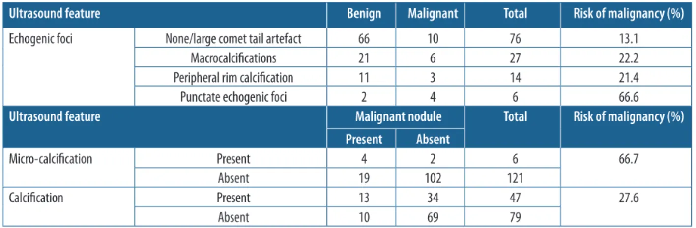
Even though microcalcification is a strong predictor of malignancy, we found the presence of calcification alone (either macrocalcification/peripheral rim calcifi-cation/microcalcification) to be an independent strong predictor of malignancy (Table 6). Vinayak and Sande in their study found that all 14 of the studied thyroid le-sions with macrocalcifications were benign [24]. A point to be noted here is that macrocalcification casts a dense posterior acoustic shadow and might impair proper eval-uation of the nodule; hence, we cannot say with certainty that other echogenic foci, especially microcalcifications, were absent.
Bonavita et al. in their study observed that the pres-ence of hyperechogenicity or the – white knight appear-ance was 100% sensitive for a nodule to be benign [25].
We found that the odds ratio for a lesion that is either isoechoic or hyperechoic being benign was 16.81 (95% CI: 5.71-49.48) (p < 0.0001).
Hoang et al. noted that although a cystic component occurs in 13-26% of all thyroid malignancies, a predom-inantly cystic appearance is uncommon [26]. Popli et al. in their study observed that all of the four cystic nodular lesions and seven lesions that showed hyperechogenic-ity in their study were found to be benign on FNAC [27]. Horvath et al. in their study found that anechoic, non-vascularised lesions with hyperechoic spots classi-fied as a colloid type 1 lesion were found to have 0% risk of malignancy. In our study all the 50 nodules that had mixed solid-cystic component or predominantly cystic component presented benign cytopathology. Malignancy was seen only among solid nodules. Hence, our study is concordant with the fact that malignancy is uncommon in predominantly cystic lesions.
We also evaluated the risk stratification system pro-posed by the American College of Radiology Thyroid Im-aging Reporting and Data System (ACR-TI-RADS) [6]. The ACR-TI-RADS committee classified thyroid nodules into five risk levels according to their ultrasonograph-ic features. The ACR-TI-RADS uses a system based on allocating points for different ultrasonographic features of nodules in five morphologic categories (composition, echogenicity, margins, echogenic foci, and shape).The point system may seem cumbersome compared with an approach based on pattern recognition, but the advantage of point systems is that they are relatively easy to use and Table 5. Comparison of statistical results analysing the significance of hy-poechogenicity between various studies
Suspicious ultrasound feature Malignant nodule Total Present Absent Hypoechogenicit y Present Kwak et al. 169 508 677 Chandra-mohan et al. 41 24 65 Srinivas et al. 5 88 93 Our study 16 8 24 Absent Kwak et al. 106 875 981 Chandra-mohan et al. 77 130 207 Srinivas et al. 20 252 272 Our study 7 96 103 Hypoechogenicit y Kwak
et al. mohan Chandra-et al. Srinivas et al. studyOur Sensitivity (%) 61.4 34.7 20.0 69.5 Specificity (%) 63.2 84.4 74.1 92.0
PPV (%) 24.9 63.0 5.3 66.7
OR 2.7 2.8 0.7 26.2
can be integrated into practices with varying volumes of patients with thyroid nodules and practices with varying levels of professional expertise. At least this approach forces the person who is obtaining and interpreting the thyroid images to focus his or her attention on each of the five important morphologic categories. Another benefit is that if future data ascribe previously unrecognised signifi-cance to sonographic findings, those findings can be easily incorporated into the ACR TI-RADS. A limitation of the point-based system is that certain findings have different implications, depending on other associated findings.
The TR1 and TR2 nodules have a risk of malignancy lower than 2%, and FNA was not recommended for these nodules. The TR3, TR4, and TR5 nodules were predicted to have a risk of malignancy of less than 5%, 5.1-20%, and greater than 20%, respectively, with FNA recommended for those nodules with threshold sizes of 2.5, 1.5, and 1.0 cm, respectively.
We did not follow ACR guidelines (neither category nor size criterion) while advising FNA correlation to ret-rospectively assess the impact in reducing unwarranted FNACs that are routinely being performed (Table 7).
Kwak vs. ACR-TI-RADS
Kwak TI-RADS 2 and 3 had higher sensitivity in predicting benignity compared to ACR TR1 and 2 (43.2% vs. 31.7%), thus being slightly better in predicting unnecessary FNAC.
PPV of Kwak TI-RADS 4 (including 4A + 4B + 4C) in assessing malignancy was 28.05%, with a sensitivity of 100% and specificity of 43.27%. PPV of TR4 and TR5 in assessing malignancy was 23.8%, with a sensitivity of 86.9% and specificity of 53.2%.
Conclusions
We found TI-RADS classification (both ACR and Kwak TI-RADS) to be a reliable, noninvasive, and practical method for assessing thyroid nodules in routine practice. Both TI-RADS classifications can safely avert avoidable FNACs in a significant proportion of benign thyroid lesions. Being a point-based system and also being amena-ble to future modifications, we prefer the use of ACR- TI-RADS.
Limitations
The sample size was limited due to the time-bound nature of the study.
The lengths of experience of the observers were not uniform. Analysis of interobserver variability was not con-ducted.
False negative and false positive cytology may have had an impact on the results obtained.
Conflict of interest
The authors report no conflict of interest. References
1. Dean DS, Gharib H. Epidemiology of thyroid nodules. Best Pract Res Clin Endocrinol Metab 2008; 22: 901-911.
2. Ardakani AA, Gharbali A, Mohammadi A. Classification of benign and malignant thyroid nodules using wavelet texture analysis of so-nograms. J Ultrasound Med 2015; 34: 1983-1989.
3. Horvath E, Majlis S, Rossi R, et al. An ultrasonogram reporting sys-tem for thyroid nodules stratifying cancer risk for clinical manage-ment. J Clin Endocrinol Metab 2009; 94: 1748-1751.
Table 6. Frequency and statistical analysis of calcification in thyroid nodules
Ultrasound feature Benign Malignant Total Risk of malignancy (%)
Echogenic foci None/large comet tail artefact 66 10 76 13.1
Macrocalcifications 21 6 27 22.2
Peripheral rim calcification 11 3 14 21.4
Punctate echogenic foci 2 4 6 66.6
Ultrasound feature Malignant nodule Total Risk of malignancy (%)
Present Absent
Micro-calcification Present 4 2 6 66.7
Absent 19 102 121
Calcification Present 13 34 47 27.6
Absent 10 69 79
Table 7. Comparison of risk of malignancy predicted in ACR TI-RADS ACR
TI-RADS ACR predicted (%)Risk of malignancy [%]Middleton et al. [21] Our study
TR1 < 2 0.3 0
TR2 1.5 0
TR3 5 4.8 6.9
TR4 5.1-20 9.1 29.2
4. Park JY, Lee HJ, Jang HW, et al. A proposal for a thyroid imaging reporting and data system for ultrasound features of thyroid carci-noma. Thyroid 2009; 19: 1257-1264.
5. Kwak JY, Han KH, Yoon JH, et al. Thyroid imaging reporting and data system for US features of nodules: a step in establishing better stratification of cancer risk. Radiology 2011; 260: 892-899. 6. Tessler FN, Middleton WD, Grant EG, et al. ACR thyroid
imag-ing, reporting and data system (TI-RADS): white paper of the ACR TI-RADS committee. J Am Coll Radiol 2017; 14: 587-595. 7. Grant EG, Tessler FN, Hoang JK, et al. Thyroid ultrasound reporting
lexicon: white paper of the ACR thyroid imaging, reporting and data system (TIRADS) committee. J Am Coll Radiol 2015; 12: 1272-1279. 8. Srinivas MNS, Amogh V, Gautam MS, et al. A prospective study to
evaluate the reliability of thyroid imaging reporting and data system in differentiation between benign and malignant thyroid lesions. J Clin Imaging Sci 2016; 6: 5.
9. Chandramohan A, Khurana A, Pushpa B, et al. Is TIRADS a prac-tical and accurate system for use in daily clinical practice? Indian J Radiol Imaging 2016; 26: 145-152.
10. Frates MC, Benson CB, Charboneau JW, et al. Management of thy-roid nodules detected at US: Society of Radiologists in Ultrasound consensus conference statement. Radiology 2005; 237: 794-800. 11. Kim D, Park J, In H, et al. Ultrasound-based diagnostic classification
for solid and partially cystic thyroid nodules. AJNR Am J Neuro-radiol 2012; 33: 1144-1149.
12. Lee MJ, Kim EK, Kwak JY, Kim MJ. Partially cystic thyroid nodules on ultrasound: probability of malignancy and sonographic differen-tiation. Thyroid 2009; 19: 341-346.
13. Moon WJ, Jung SL, Lee JH, et al. Benign and malignant thyroid nodules: US differentiation – multicenter retrospective study. Radio-logy 2008; 247: 762-770.
14. Hong YJ, Son EJ, Kim EK, et al. Positive predictive values of sonogra-phic features of solid thyroid nodule. Clin Imaging 2010; 34: 127-133. 15. Kim D, Lee E, Jung S, et al. Role of sonographic diagnosis in
man-aging Bethesda class III nodules. AJNR Am J Neuroradiol 2011; 32: 2136-2141.
16. Kim DW, Lee YJ, Eom JW, et al. Ultrasound-based diagnosis for solid thyroid nodules with the largest diameter < 5 mm. Ultrasound Med Biol 2013; 39: 1190-1196.
17. Kim EK, Park CS, Chung WY, et al. New sonographic criteria for recommending fine-needle aspiration biopsy of nonpalpable solid nodules of the thyroid. Am J Roentgenol 2002; 178: 687-691. 18. Russ G. Risk stratification of thyroid nodules on ultrasonography
with the French TI-RADS: description and reflections. Ultrasono- graphy 2016; 35: 25.
19. Haugen BR. 2015 American Thyroid Association management guide-lines for adult patients with thyroid nodules and differentiated thyroid cancer: what is new and what has changed? Cancer 2017; 123: 372-381. 20. Shin JH, Baek JH, Chung J, et al. Ultrasonography diagnosis and imaging-based management of thyroid nodules: revised Korean Society of Thyroid Radiology consensus statement and recommen-dations. Korean J Radiol 2016; 17: 370-395.
21. Perros P, Boelaert K, Colley S, et al. Guidelines for the management of thyroid cancer. Clin Endocrinol 2014; 81: 1-122.
22. Wemeau JL, Sadoul JL, d’Herbomez M, et al. Recommendations of the French Society of Endocrinology for the management of thyroid nodules. Presse Med 2011; 40 (9 Pt 1): 793-826.
23. Machens A, Holzhausen HJ, Dralle H. The prognostic value of primary tumor size in papillary and follicular thyroid carcinoma: a comparative analysis. Cancer 2005; 103: 2269-2273.
24. Vinayak S, Sande JA. Avoiding unnecessary fine-needle aspiration cytology by accuractely predicting the benign nature of thyroid nod-ules using ultrasound. J Clin Imaging Sci 2012; 2: 23.
25. Bonavita JA, Mayo J, Babb J, et al. Pattern recognition of benign nodules at ultrasound of the thyroid: which nodules can be left alone? Am J Roentgenol 2009; 193: 207-213.
26. Hoang JK, Lee WK, Lee M, et al. US Features of thyroid malignancy: pearls and pitfalls. Radiographics 2007; 27: 847-860.
27. Popli MB, Rastogi A, Bhalla P, Solanki Y. Utility of gray-scale ultra-sound to differentiate benign from malignant thyroid nodules. Indian J Radiol Imaging 2012; 22: 63-68.