Brancini, GTP, Rodrigues, GB, Rambaldi, MDSL, Izumi, C, Yatsuda, AP, Wainwright, M, Rosa, JC and Braga, GÚL
The effects of photodynamic treatment with new methylene blue N on the Candida albicans proteome.
http://researchonline.ljmu.ac.uk/4959/
Article
LJMU has developed LJMU Research Online for users to access the research output of the University more effectively. Copyright © and Moral Rights for the papers on this site are retained by the individual authors and/or other copyright owners. Users may download and/or print one copy of any article(s) in LJMU Research Online to facilitate their private study or for non-commercial research. You may not engage in further distribution of the material or use it for any profit-making activities or any commercial gain.
The version presented here may differ from the published version or from the version of the record. Please see the repository URL above for details on accessing the published version and note that access may require a subscription.
For more information please contact [email protected]
http://researchonline.ljmu.ac.uk/ Citation (please note it is advisable to refer to the publisher’s version if you intend to cite from this work)
Brancini, GTP, Rodrigues, GB, Rambaldi, MDSL, Izumi, C, Yatsuda, AP, Wainwright, M, Rosa, JC and Braga, GÚL (2016) The effects of
photodynamic treatment with new methylene blue N on the Candida albicans proteome. Photochemical and Photobiological Sciences, 12. pp.
Candida albicans proteomic alterations caused by photodynamic treatment with new methylene blue N as evaluated by two-dimensional electrophoresis and tandem mass spectrometry
Guilherme Thomaz Pereira Brancinia, Gabriela Braga Rodriguesa, Mariana de Souza Lima Rambaldia, Clarice Izumib, Ana Patrícia Yatsudaa, Mark Wainwrightc, José César Rosab and Gilberto Úbida Leite Bragaa*
aFaculdade de Ciências Farmacêuticas de Ribeirão Preto, Universidade de São Paulo, Ribeirão Preto, SP, Brazil
bFaculdade de Medicina de Ribeirão Preto, Universidade de São Paulo, Ribeirão Preto, Brazil
cSchool of Pharmacy and Biomolecular Sciences, Liverpool John Moores University, Liverpool, United Kingdom
Email: [email protected] (GTP Brancini), [email protected] (GB Rodrigues), [email protected] (MSL Rambaldi), [email protected] (C Izumi), [email protected] (AP Yatsuda), [email protected] (M Waintwright), [email protected] (JC Rosa), [email protected] (GUL Braga)
*Corresponding author: Gilberto Úbida Leite Braga
Address: Universidade de São Paulo, Faculdade de Ciências Farmacêuticas de Ribeirão Preto.
Av. do Café s/n Boco S, sala 39.
Monte Alegre, 14040-903 – Ribeirão Preto, SP – Brazil Phone: +55 16 33154723
Fax: +55 16 33154723 Email: [email protected]
ABSTRACT
Candida albicans is a human pathogenic fungus mainly affecting immunocompromised patients. Resistance to the commonly used fungicides can lead to poor treatment of mucosal infections which, in turn, can result in life-threatening systemic candidiasis. In this scenario, antimicrobial photodynamic treatment (PDT) has emerged as an effective alternative to treat superficial and localized fungal infections. Microbial death in PDT is a consequence of the oxidation of many cellular biomolecules, including proteins. Here, we report a combination of two-dimensional electrophoresis and tandem mass spectrometry to study protein damage resulting from PDT of C. albicans with new methylene blue N and red light. Two-dimensional gels of treated cells showed an increase in acidic spots in a fluence-dependent manner. Amino acid analysis revealed a decrease in histidine content after PDT which is a plausible explanation for the observed acidic shift. However, some protein spots remained unchanged. Protein identification by mass spectrometry revealed that both modified and unmodified proteins could be localized to the cytoplasm, ruling out subcellular location as the only explanation for damage selectivity. Therefore, we hypothesize that protein modification by PDT is a consequence of both photosensitizer binding affinity and the degree of exposure of photooxidizable residues on the protein surface.
1. INTRODUCTION
Candida albicans is an important opportunistic fungus responsible for a wide range of human diseases, including oral, vulvovaginal, oropharyngeal and systemic candidiasis (NUCCI et al. 2010, PFALLER and DIEKEMA 2010, KOEHLER et al. 2014, O’ROURKE β015, SOBEL 2016). Immunocompromised individuals are particularly susceptible and C. albicans infections is now a worldwide health issue
(KIM and SUDBERY 2011, PFALLER and DIEKEMA 2010, KOEHLER et al. 2014). There are several types of conventional chemotherapeutic agents available for treating fungal infections, including the polyenes, allylamines, echinocandins and azoles (MOTTA et al. 2010, RUHNKE et al. 2011, KOEHLER et al. 2014). However, given the single mode of action common to conventional antifungal agents the emergence of tolerant strains is not surprising. The development of antifungal tolerance in C. albicans
was first reported during the early 1980, mostly in severely immunocompromised patients (REX et al. 1995). Tolerance to conventional fungicides is increasing and becoming a major problem in certain groups, particularly those infected with HIV (PFALLER and DIEKEMA 2010, MARTEL et al. 2010). A further complication in the conventional chemotherapeutic approach to treat HIV-positive patients is the drug-drug interaction (PATTON et al. 2001).
Photodynamic treatment is a plausible alternative to treat superficial and localized infections caused by C. albicans and non-albicans species (GONZALES and MAISCH 2012). PDT commences with the application of a photosensitizer (PS) on the infection site. Then, light of the correct wavelength is used to irradiate the PS. Upon interaction with a photon emitted by the light source, the PS is excited to a high-energy electronic state. The decay to the ground state can take place by many processes, the most common being energy transfer to molecular oxygen (O2) to generate singlet oxygen (correctly written O2 (a1Δg), but hereinafter referred to as 1O2). Singlet oxygen is a reactive oxygen species that interacts with many biomolecules (mainly lipids and proteins) causing cell death (BERTOLONI et al. 1989, LAMBRECHTS et al. 2005). Proteins constitute almost 70% of a cell’s dry weight and are a major target for PDT-generated singlet oxygen (reviewed in DAVIES 2003). Singlet oxygen damage to proteins varies from relatively mild amino acid oxidations (mainly Trp, His, Tyr, Met
and Cys) to the more severe formation of protein aggregates (DAVIES 2003). It is important to note, however, that even a single amino acid oxidation can have consequences to cells. For example, Escherichia coli glutamine synthetase loses its activity after the oxidation of a single, catalytic-site histidine residue (RIVETT and LEVINE 1990).
The protein complement expressed by a genome is termed proteome (WILKINS
et al. 1996) and its study is generally referred to as proteomics. Despite being widely used to study fungal responses to stress (reviewed in KROLL et al. 2014), proteomics has not been employed to evaluate PDT effects on fungi proteomes. Two-dimensional electrophoresis (2-DE) was once considered the state-of-the-art technique for proteomic studies. Today it has almost completely been replaced by more refined techniques based on liquid chromatography coupled to tandem mass spectrometry (LC-MS/MS). However, 2-DE still finds its application in proteomics (ROGOWSKA-WRZESINSKA et al. 2013). Magi et al. employed 2-DE to study the apoptotic response of human leukemia cells after exposure to light with the photosensitizer Purpurin-18 (MAGI et al. 2004). Still on PDT, Dosselli et al. used 2-DE to evaluate the effects of different PS concentrations and light fluences on Staphyloccocus aureus proteome, showing a clear correlation between mortality rates and protein modification (DOSSELLI et al. 2012).
Our research group has already shown the efficiency of PDT with different phenothiazinium PS in killing C. albicans in vitro (RODRIGUES et al. 2013). High mortality rates were achieved even with low PS concentration and brief light exposure. However, a proteomic study to assess the extent of protein damage caused by PDT had not been performed. Therefore, we report here the effects of PDT with new methylene blue N (NMBN) on C. albicans proteome as evaluated by 2-DE.
2. MATERIALS AND METHODS
2.1 C. albicans and growth conditions. C. albicans ATCC 64548 was obtained from the American Type Culture Collection (Manassas, VA, USA). Cells were grown on Sabouraud dextrose agar (SDA) at 35 °C for 48 h. Isolated colonies were used to prepare a suspension (107 cells ml-1) that was then used to inoculate 50 ml of YPD medium (1% yeast extract, 2% peptone and 2% dextrose) in 125 ml erlenmeyer flasks. Incubation proceeded for six hours (35 °C, 150 rpm). Cells were then collected and washed with PBS (pH 7.4).
2.2 Photosensitizer. New methylene blue N zinc chloride double salt (NMBN) was purchased from Sigma-Aldrich, Inc. (St. Louis, MO, USA). Stock solutions (500 µM) were prepared in PBS and kept protected from light at -18 °C for a maximum of 14 days. NMBN exhibits an intense absorption at 630 nm. Molecular structure and absorption spectrum for NMBN can be seen in RODRIGUES et al. 2013.
2.3 Light source. Irradiation was performed with an array containing 96 light-emitting diodes (LED) with emission peak at 631 nm. The measured irradiance from 400 to 700 nm was 13.89 mW cm-2. Light measurement was performed with a USB4000 spectroradiometer (Ocean Optics, Dunedin, FL, USA). Emission spectrum for the light source can be seen in RODRIGUES et al. 2013.
2.4 Photodynamic treatment. Five milliliters of cell suspension (4 × 10⁷ cells ml-1) and 5 ml of PS solution were added to 15 ml tubes. Final cell and NMBN concentrations were 2 × 10⁷ cells ml-1 and 2,5 µM, respectively. Tubes were kept in the dark for an incubation period of 30 minutes. The content of each tube (10 ml) was then transferred to plastic 60-mm petri dishes which were exposed to light fluences of 0 (dark control), 3 and 10 J cm-2. During exposures, cell suspension was stirred with the help of a magnetic
stirrer. Each experiment consisted of five exposures to either 3 or 10 J cm-2 so that enough cells were available for proteomic analysis. After exposure, a 50 µ l aliquot was removed for cell survival assays. Each 10 ml cell suspension was then immediately collected by centrifugation (7,000 × g for five minutes), washed with 1 ml PBS, transferred to 2-ml tubes, frozen in liquid nitrogen and kept at -70 °C until protein extraction. Three independent experiments were performed. In addition to these experiments, 50 mM mannitol or 10 mM sodium azide were added prior to PDT with 10 J cm-2 to evaluate the effects of the hydroxyl radical (·OH) and singlet oxygen (1O2), respectively, on cell survival and C. albicans proteome. Three independent experiments were performed for both mannitol and azide additions.
2.5 Cell survival assays. After exposure to light, 50 µl were collected from the suspension, serially diluted (10-1 to 10-3) and spread on the surface of 5 ml of SDA medium in Petri dishes (60 × 15 mm). Three replicate dishes were prepared for each treatment. Cell survival to PDT was determined by counting colony forming units (CFU) and calculating the percent ratio of exposed to unexposed cells.
2.6 Protein extraction and quantification. To each tube, containing 2 × 10⁸ cells, 500 µl of extraction buffer (7 M urea, 2 M thiourea and 4% CHAPS) and 500 µl of glass beads (425-600 µm, Sigma) were added. Five microliters of Protease Inhibitor Mix (100×, GE Healthcare Life Sciences, Uppsala, Sweden) were also added. Cells were disrupted by five cycles consisting of one minute vortexing and five minutes on ice. Tubes were then centrifuged at 10,000 × g for five minutes at 4 °C and the resulting supernatant collected. Proteins in the supernatant were purified by precipitation with 6 volumes acetone overnight at -20 °C. After acetone removal, the protein pellet was resuspended in the same extraction buffer used previously and quantified by 2-D Quant Kit (GE Healthcare).
2.7 Two dimensional electrophoresis (2-DE). Protein concentrations were adjusted to 20 µg in 250 µl of rehydration buffer [7 M urea, 2 M thiourea, 4% CHAPS, 40 mM dithiothreitol (DTT), 0.5% IPG buffer 3-11 NL (GE Healthcare), 3 µl Protease Inhibitor Mix (GE Healthcare) and 0.002% bromophenol blue]. This volume was applied to 13-cm 3-11 NL Immobiline™ DryStrips (GE Healthcare) and the strips were rehydrated for 16 h. Isoelectric focusing proceeded in an IPGphor 3 system (GE Healthcare) according to the following protocol: 500 V (step and hold) to 0.5 kVh, 1000 V (gradient) to 0.8 kVh, 8000 V (gradient) to 11.3 kVh and 8000 V (step and hold) until a total of 25 kVh was reached. Strips were then equilibrated in a solution containing 75 mM Tris-HCl pH 8.8, 6 M urea, 30% glycerol, 2% sodium dodecyl sulfate (SDS) and 65 mM DTT for 15 minutes. The same procedure was repeated, but DTT was replaced by 135 mM iodoacetamide. Second dimension SDS-PAGE was carried out in 12.5% polyacrylamide gels in a SE 600 Ruby system (GE Healthcare) according to the protocol by Laemmli (LAEMMLI 1970). Gels were silver-stained following the “Short silver nitrate staining” protocol previously described (CHEVALLET et al. 2006). Stained gels were scanned on ImageScanner III and Labscan (GE Healthcare) and image analyses were performed with ImageMaster 2D Platinum 7 (GE Healthcare).
2.8 Protein identification by MS/MS. For protein identification, gels were prepared exactly as described previously, except for the higher protein load of 100 µg. Gels were then stained with Coomassie Brilliant Blue G-250 (USB Corporation, USA). Spots were excised from the gels with a sterile scalp and processed based on the protocol described in SHEVCHENKO et al. 2006. Briefly, spots were destained overnight in 0.1 M ammonium bicarbonate/50% acetonitrile and then placed in 100% acetonitrile for 10 min. After removing acetonitrile, spots were dried in a vacuum centrifuge (Speed Vac, USA) and digested with 0.5 µg of modified trypsin (Promega, USA) in 0.1 M
ammonium bicarbonate at 37 °C for 24 h. Reaction was then stopped by adding 5 µl of formic acid and the spots were kept at 4 °C for 24 h. Then, trypsin-digested peptides were passed through a C18 resin (Perseptive Biosystems, USA) to remove salt and dried in a vacuum centrifuge. Dried peptides were solubilized in 6 µl α -cyano-4-hydroxycinnaminic acid (5 mg ml-1 in 50% acetonitrile/0.1% trifluoroacetic acid, Sigma). Finally, 2 µl were spotted on a MALDI target plate. Analysis proceeded in a MALDI-TOF/TOF MS/MS (Axima Performance, Shimadzu Biotech) in positive ion mode and the ten most intense ions were selected for MS/MS. Protein identification based on mass spectra was performed with MASCOT (Matrix Science) using the following parameters: databases “Swiss-Prot” and “contaminants”, taxonomy “other fungi”, enzyme “trypsin”, up to 1 missed cleavage, fixed modification “carbamidomethyl (C)”, variable modification “oxidation (M)”, peptide tolerance “1.β Da”, MS/MS tolerance “0.8 Da”, peptide charge “+1” and “average masses”.
2.9 Amino acid analysis. Amino acid analysis was performed as described by BIDLINGMEYER et al. 1984 with slight modifications. A total of 2 × 10⁶ cells either exposed only to NMBN (control) or treated with PDT (10 J cm-2 or 20 J cm-2) were lysed with 6 N HCl at 110 °C for 22 h. This process results in both cell lysis and acid hydrolysis of proteins, yielding free amino acids. Then, free amino acids were derivatized with phenylisothiocyanate and analyzed by HPLC employing a C18 reverse-phase column. The elution process was monitored by reading the absorbance at 254 nm which corresponds to the phenylthiocarbamyl derivative of the amino acids. For each experiment, a standard (Pierce H) with known amino acids concentrations was also analyzed. Percent content of each amino acid in the samples was calculated by measuring peak area relative to the amino acid standard. Two independent experiments were performed. We then calculated percent reduction of amino acid after PDT for the
two experiments. Mean percent reduction and standard deviation was calculated by averaging the percent reduction observed in each experiment. For clarity purposes, we denoted our two experiments ‘Exp 1’ and ‘Exp β’ and report the amino acid percent content of each experiment.
3. RESULTS
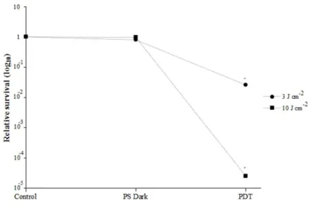
3.1 C. albicans survival after PDT. Following PDT with 2.5 µM NMBN and light fluences of 3 and 10 J cm-2, C. albicans assessed mortality was 97.4% and 99.9975%, respectively (Fig. 1). These results are in accordance with those previously published by our group and show that cell death proceeds rapidly even with low PS concentrations (RODRIGUES et al. 2013). Both treatments were used for proteomic analysis and will be hereinafter referred to as 3 J cm-2 and 10 J cm-2.
Fig. 1 – Candida albicans relative survival after PDT with light fluences of 3 or 10 J cm-2.
Control refers to cells exposed to NMBN but not irradiated. Three independent experiments were performed
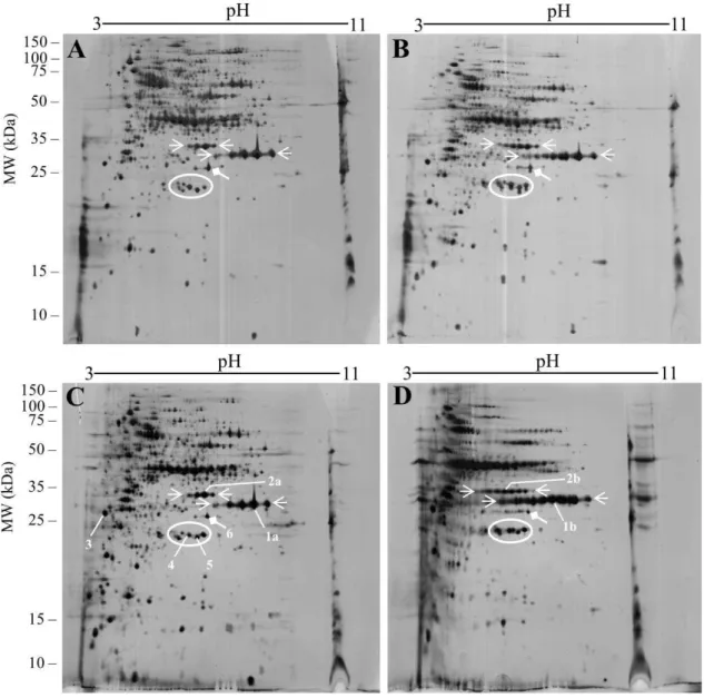
3.2 Proteomic alterations resulting from PDT. Proteomic alterations caused by PDT and assessed by 2-DE are shown in Fig. 2.
Fig. 2 – 2-D gels of control (A and C) and PDT-treated C. albicans with light fluences of 3 J cm-2 (B) and 10 J cm-2 (D). PDT resulted in the appearance of spot trains for some proteins and
this effect was observed to be fluence-dependent. Horizontal arrows delimit spots for which spot trains could be observed for both light fluences. Oblique (diamond) arrow shows a protein spot that was only modified after the 10 J cm-2 treatment. White ellipse encompasses protein
spots that were not modified by PDT. Numbers identify protein spots that were analyzed by MS/MS (see Table 1)
Treatment with the lower fluence (3 J cm-2) induced visible modifications on the
C. albicans proteome (Fig. 2A and B). These modifications consist of the appearance of new, more acidic spots when compared to unexposed control (white arrows in Figures 2A and 2B). When the light fluence was increased to 10 J cm-2, these proteome modifications were intensified, resulting in the appearance of even more acidic spots (see white arrows and compare Figures 2B and 2D). Some protein spots were found to remain after the lower fluence treatment, undergoing modification when treatment intensity was increased (oblique diamond arrow in Figure 2), a further indication of a fluence-dependent mechanism. At least a group of protein spots (white ellipse, Fig. 2) suffered no modification even after the high intensity treatment. Taken together, these results suggest that PDT-induced protein modification on C. albicans is both fluence-dependent and protein-specific, with some proteins being less susceptible to modification than others.
The addition of 50 mM mannitol prior to PDT could not prevent cell death nor proteomic alteration to any extent (Fig. 3), indicating that the hydroxyl radical (·OH) is not the reactive species responsible for the observed damage.
Fig. 3 – Effects of PDT in the presence of 50 mM mannitol. (A) Relative survival after PDT with 10 J cm-2 in the presence of mannitol. 2-D gels before (B) and after (C) PDT in the
presence of mannitol. Overall, mannitol did not improve cell survival or reduce protein modification, indicating that the hydroxyl radical is not responsible for either cell mortality or protein damage
On the other hand, addition of 10 mM sodium azide reduced cell death from 99.9975% to around 31% for the 10 J cm-2 treatment (Fig 4A) and also could reduce some of the proteome damage (Fig 4 B and C), indicating that 1O2 is at least partially responsible for both cell death and proteomic alteration in C. albicans.
Fig. 4 – Effects of PDT in the presence of 10 mM sodium azide. (A) Relative survival after PDT with 10 J cm-2 in the presence and absence of azide. 2-D gels before (B) and after (C) PDT
in the presence of azide. Overall, azide greatly reduced cell mortality and also prevented some protein modification, implicating singlet oxygen in both cell mortality and protein damage
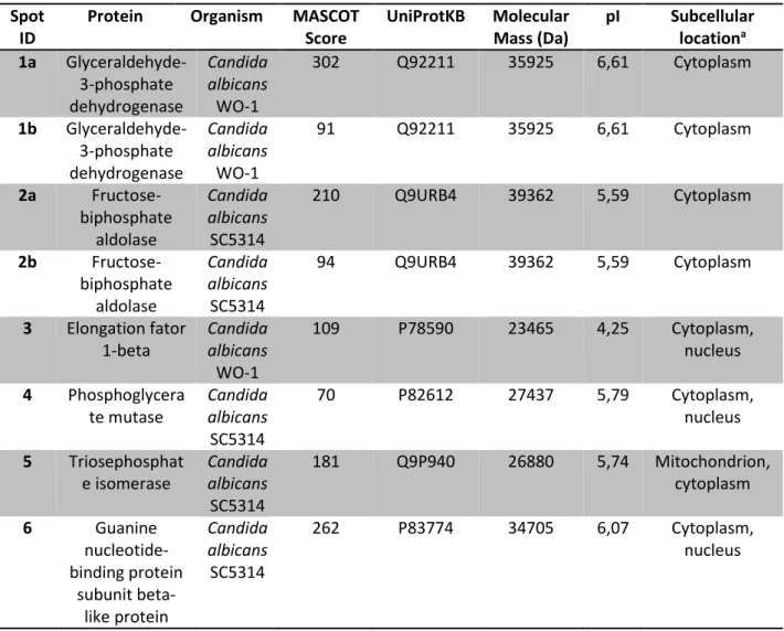
3.3 Protein identification by MS/MS. A total of eight spots were analyzed by MALDI-TOF/TOF MS/MS for protein identification. Four (spots 1a, 1b, 2a and 2b, Fig 2) represented modified proteins while three (spots 3, 4 and 5) represented unmodified proteins. Also, spot 6 represented a protein that was only modified by the high-fluence treatment. All protein spots were identified and corresponded to six different proteins. The identified proteins’ subcellular locations were predicted by using WoLF PSORT (HORTON et al. 2007). Results are shown in Table 1.
Table 1 – Protein identification by MALDI-TOF/TOF MS/MS. Spot ID can be visualized in Figure 2
aSubcellular location predicted using WoLF PSORT (HORTON et al. 2007)
Spot ID
Protein Organism MASCOT Score UniProtKB Molecular Mass (Da) pI Subcellular locationa 1a Glyceraldehyde-3-phosphate dehydrogenase Candida albicans WO-1 302 Q92211 35925 6,61 Cytoplasm 1b Glyceraldehyde-3-phosphate dehydrogenase Candida albicans WO-1 91 Q92211 35925 6,61 Cytoplasm 2a Fructose-biphosphate aldolase Candida albicans SC5314 210 Q9URB4 39362 5,59 Cytoplasm 2b Fructose-biphosphate aldolase Candida albicans SC5314 94 Q9URB4 39362 5,59 Cytoplasm 3 Elongation fator 1-beta Candida albicans WO-1 109 P78590 23465 4,25 Cytoplasm, nucleus 4 Phosphoglycera te mutase Candida albicans SC5314 70 P82612 27437 5,79 Cytoplasm, nucleus 5 Triosephosphat e isomerase Candida albicans SC5314 181 Q9P940 26880 5,74 Mitochondrion, cytoplasm 6 Guanine nucleotide-binding protein subunit beta-like protein Candida albicans SC5314 262 P83774 34705 6,07 Cytoplasm, nucleus
Glyceraldehyde 3-phosphate dehydrogenase, fructose biphosphate aldolase, phosphoglycerate mutase and triosephosphate isomerase (spots 1, 2, 4 and 5, respectively) are all carbohydrate metabolism enzymes and can be localized to the cytoplasm. The fact that the former two are extensively modified while the latter two are not further suggests that PDT-induced modification is protein-specific and not necessarily related to protein subcellular location.
Elongation factor 1-beta (spot 3) was also localized to the cytoplasm and its corresponding spot was chosen for its acidic nature. A low isoelectric point protein
(negatively charged at physiological pH) would be expected to have a greater interaction with the positively charged NMBN and hence be readily modified. However, elongation factor 1-beta also appears to be resistant to PDT-induced modification.
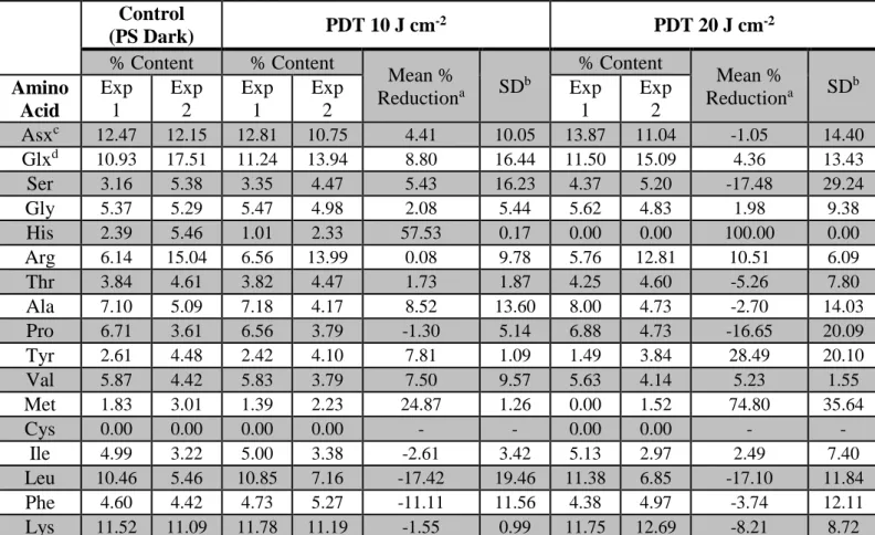
3.4 Amino acid analysis. Analysis of amino acids showed that His content had a reduction of 57.53% (SD = 0.17) for the 10 J cm-2 treatment when compared to unexposed control (Table 2). To verify whether a fluence increase would result in greater amino acid loss, we performed amino acid analysis after PDT with a light fluence of 20 J cm-2. This increased intensity reduced the His content by 100% relative to control. This result is consistent with His being one the most reactive amino acids towards singlet oxygen (WEIL et al. 1951). Other amino acids reactive to singlet oxygen are Trp, Tyr, Met, and Cys (WEIL et al. 1951). Trp cannot be evaluated by our technique because it is destroyed during sample preparation. Cys content was already zero even in the control group. Tyr residues underwent a 7.81% (SD = 1.09) reduction after PDT with 10 J cm-2. This number increased to 28.49% (SD = 20.10) when light fluence reached 20 J cm-2. A fluence-dependent amino acid loss was also observed for Met, with content reduction going from 24.87% (SD = 1.26) at 10 J cm-2 to 74.80% (SD = 35.64) at 20 J cm-2.
Table 2 – Amino acid percent content in untreated (PS Dark) and PDT-treated C. albicans with light fluences of 10 or 20 J cm-2. Two independent experiments were performed and are denoted Exp 1 and Exp 2. For clarity purposes, we
report the amino acid percent content observed in each experiment separately. Note the reduction in His content and also losses of Tyr and Met
aMean % reduction was calculated by averaging the percent reduction observed in each experiment (see section 2.9) bStandard deviation
cAspartic acid or asparagine dGlutamic acid or glutamine
Control (PS Dark) PDT 10 J cm -2 PDT 20 J cm-2 % Content % Content Mean % Reductiona SD b % Content Mean % Reductiona SD b Amino Acid Exp 1 Exp 2 Exp 1 Exp 2 Exp 1 Exp 2 Asxc 12.47 12.15 12.81 10.75 4.41 10.05 13.87 11.04 -1.05 14.40 Glxd 10.93 17.51 11.24 13.94 8.80 16.44 11.50 15.09 4.36 13.43 Ser 3.16 5.38 3.35 4.47 5.43 16.23 4.37 5.20 -17.48 29.24 Gly 5.37 5.29 5.47 4.98 2.08 5.44 5.62 4.83 1.98 9.38 His 2.39 5.46 1.01 2.33 57.53 0.17 0.00 0.00 100.00 0.00 Arg 6.14 15.04 6.56 13.99 0.08 9.78 5.76 12.81 10.51 6.09 Thr 3.84 4.61 3.82 4.47 1.73 1.87 4.25 4.60 -5.26 7.80 Ala 7.10 5.09 7.18 4.17 8.52 13.60 8.00 4.73 -2.70 14.03 Pro 6.71 3.61 6.56 3.79 -1.30 5.14 6.88 4.73 -16.65 20.09 Tyr 2.61 4.48 2.42 4.10 7.81 1.09 1.49 3.84 28.49 20.10 Val 5.87 4.42 5.83 3.79 7.50 9.57 5.63 4.14 5.23 1.55 Met 1.83 3.01 1.39 2.23 24.87 1.26 0.00 1.52 74.80 35.64 Cys 0.00 0.00 0.00 0.00 - - 0.00 0.00 - - Ile 4.99 3.22 5.00 3.38 -2.61 3.42 5.13 2.97 2.49 7.40 Leu 10.46 5.46 10.85 7.16 -17.42 19.46 11.38 6.85 -17.10 11.84 Phe 4.60 4.42 4.73 5.27 -11.11 11.56 4.38 4.97 -3.74 12.11 Lys 11.52 11.09 11.78 11.19 -1.55 0.99 11.75 12.69 -8.21 8.72 4. DISCUSSION
C. albicans had previously been shown to be effectively photoinactivated in vitro using NMBN and red light. Also, mortality rates well over 99% could be achieved even with low PS concentrations (RODRIGUES et al. 2013). However, a more in-depth study at the molecular level was missing. Even though lipids – especially those of the membrane – are considered major targets during PDT (BÖCKING et al. 2000, LAMBRECHTS et al. 2005), proteins also play a crucial role, owing to their high abundance within cells and their reactivity towards singlet oxygen (DAVIES 2003).
Therefore, we employed a combination of 2-DE and mass spectrometry to evaluate protein damage on C. albicans after PDT.
Our results show that, for some proteins, PDT induced an acidic shift, that is, the appearance of a series of new protein spots with lower isoelectric point (pI) (Fig 2). This proteomic alteration was shown to be fluence-dependent, with a higher number of modified spots appearing as treatment intensity increased from 3 to 10 J cm-2 (Fig 2B and 2D).
The acidic shift observed has been previously shown following protein oxidation employing -radiolysis (DAVIES 1987). However, this system generates ·OH, which reacts with lysine and arginine residues, causing their conversion to aminoadipic and glutamic semialdehydes, respectively (REQUENA et al. 2001). Since the formation of these semialdehydes involves the elimination of the amino and guanidine groups, the oxidized proteins become more acid. However, photosensitization with NMBN generates mostly 1O2 (WAINWRIGHT et al. 1998), which preferentially reacts with Trp, His, Tyr, Met, and Cys residues (WEIL et al. 1951). Of these, only His would be expected to influence the isoelectric point of proteins. Indeed, amino acid analysis of photosensitized cells showed that the Arg and Lys contents were unchanged, while the His content decreased by almost 60% after PDT with 10 J cm-2 and by 100% after PDT with 20 J cm-2 (Table 2). Rivett and Levine have shown that the metal-catalyzed oxidation of glutamine synthetase from E. coli results in the specific loss of two His residues per enzyme subunit. Concomitant with His oxidation, new acidic forms of the enzyme were seen upon isoelectric focusing (RIVETT and LEVINE 1990). Although the employed system generates mostly ·OH, the authors could not demonstrate the loss of amino acids other than His. This result shows that His oxidation alone can considerably change the pI of a protein.
Some protein spots in our gels remained unmodified even after the higher intensity treatment, suggesting that PDT might be protein-specific. It is important to note, however, that we evaluated protein damage on the basis of pI change. Tryptophan, a readily oxidized amino acid, would not contribute to pI change and its oxidation cannot be assessed by standard amino acid analysis because Trp is destroyed upon sample preparation. The fact that most of the identified proteins are cytoplasmic rules out different subcellular location as the only explanation for the selectivity observed. Instead, we hypothesize that protein-selective oxidation depends mostly on two factors: first, the interaction between protein and photosensitizer; and second, whether photooxidizable residues are present at or near the interaction site. Indeed, a series of studies by Jori et al. have shown the selective photooxidation of those residues involved in binding of the photosensitizer to the protein (JORI et al. 1970a, 1970b, 1971a, 1971b). Also, others have shown that histidine residues buried deep inside the protein structure are not photooxidazible (KENKARE and RICHARDS 1966). Different photosensitizer binding affinities have been shown for human and bovine serum albumins (CHAKRABARTY et al. 2007). Given that these proteins share around 80% homology, it is evident that different proteins could greatly diverge in their ability to bind the photosensitizer. As an example of the importance of protein and PS affinity, the photooxidation of glyceraldehyde-3-phosphate dehydrogenase (GAPDH, spots 1a and 1b, Table 1) from rabbit muscle in the presence of Rose Bengal and light results in specific oxidation of His-38 and subsequent enzyme activity loss. Both NAD+ and substrate (glyceraldehyde-3-phosphate) provide partial protection by competing with Rose Bengal for the binding site (FRANCIS et al. 1973). Even though the authors did not perform isoelectric focusing of the oxidized GAPDH, it is reasonable to assume that the protein became more acid after His oxidation. On the other hand, triosephosphate
isomerase (spot 5, Table 1) from Brewer’s yeast was found to be considerably resistant to photooxidation, undergoing no enzyme activity loss even after 300 minutes of illumination in the presence of 10 µM Rose Bengal (KRIETSCH et al. 1970). Interestingly, the authors have also shown that the same enzyme from rabbit muscle was much more susceptible to photooxidation under the same conditions (KRIETSCH et al. 1970). On our gels, triosephosphate isomerase was found to be unmodified after the high-intensity treatment, probably indicating a closer proximity in this aspect to the yeast’s enzyme.
In a recent work, Dosselli et al. studied the effect of PDT with a porphyrin derivative on membrane-associated proteins of the Gram-positive bacterium
Staphylococcus aureus by using 2-DE. Their results showed that, with increasing treatment intensity, proteins would form aggregates, resulting in spots disappearing from 2-D gels. Because of the high molecular weight of these protein aggregates, they were only seen in the stacking gel of one-dimensional SDS-PAGE (DOSSELLI et al. 2012). Even though we did not perform 1-D SDS-PAGE, it is reasonable to assume that some protein aggregation took place during PDT. That is because protein cross-linking mediated by 1O2 starts with His oxidation and later proceeds to covalent binding of amino groups from other amino acids to oxidized histidine, forming both intra- and intermolecular His-His and His-Lys bonds (SHEN et al. 2000, LIU et al. 2014). Also, photooxidation of myoglobin leads almost exclusively to the formation of intramolecular cross-linking and results in loss of protein free amino groups (VAN STEVENINCK and DUBBELMAN 1984). In turn, this loss of free amino groups changes the isoelectric focusing of myoglobin, resulting in the appearance of more acidic forms (VAN STEVENINCK and DUBBELMAN 1984). On a side note, it has been shown that these cross-links are lost during sample preparation prior to 1-D
SDS-PAGE (i. e. boiling in the presence of SDS/dithiothreitol) (SHEN et al. 1996), so that some protein aggregation may have been overlooked during the work of Dosselli (DOSSELLI et al. 2012).
Also employing 2-DE, Magi et al. studied the proteome of human leukemia cells (HL-60) after PDT-induced apoptosis. They also reported the appearance of acidic isoforms for some structural proteins (e. g. -actin and tubulin-α-1-chain) and associated this event with protein carbonylation assessed by western blotting with 2,4-dinitrophenylhydrazine (DNPH) (MAGI et al. 2004). The authors argued that PDT could result in the side-chain carbonylation of Lys, Arg, Pro, and Thr residues. However, 1O2 generated by PDT reacts mostly with Trp, His, Tyr, Met, and Cys residues (WEIL et al. 1951). It is more likely that the protein carbonylation observed by the authors is the result of, for instance, Trp oxidation, as the end-product kynurenine is expected to give a positive result with DNPH (SILVESTER et al. 1998). The same is valid for His oxidation, which also results in carbonyl-containing products (TOMITA et al. 1969).
Recently, Alves et al. studied the photoinactivation of the bacteria E. coli and
Staphylococcus warneri by using two different cationic porphyrins as photosensitizers (ALVES et al. 2015). The authors observed that, after PDT, the SDS-PAGE protein profile of both E. coli and S. warneri was altered. Overall, PDT caused both the disappearance and the appearance of protein bands on SDS-PAGE gels and this modification was found to be different for each of the porphyrins tested. Unfortunately, the authors did not perform mass spectrometry analyses for protein identification, relying instead on assignments based on molecular weight (ALVES et al. 2015). Based on these results, it would indeed be interesting to study the effect of a different photosensitizer on C. albicans proteome.
In our experiments, the addition of 10 mM sodium azide pior to PDT greatly reduced cell mortality and could prevent some protein damage (Fig. 4). Knowing that azide is a well-known 1O2 scavenger, we can say that singlet oxygen is at least partially responsible for the photosensitizing process. It is interesting to note that not all protein damage was prevented even with an azide:NMBN molar ratio of 4000:1. Indeed, Kim et al. showed that the enzyme activity of isocitrate dehydrogenase decreased to 20% after treatment with 2 µM Rose Bengal (RB) and light, but was restored to only about 60% when the enzyme was photosensitized in the presence of 50 mM sodium azide, an azide:RB molar ratio of 25000:1 (KIM et al. 2004). Even if the authors did not discuss this result, it is logical to suggest that azide cannot scavenge 1O2 generated by a tightly protein-bound photosensitizer. As expected, the addition of 50 mM mannitol did not prevent either cell death or protein damage (Fig. 3), further indicating that ·OH is not the reactive species responsible for the photosensitizing effect of NMBN.
As a final remark, it should always be borne in mind that PDT-induced cell death is a complex process, also involving the oxidation of biomolecules other than proteins. As an example, peroxidation of membrane lipids by singlet oxygen produces 4-hydroxynonenal (HNE) (NAGAOKA et al. 2005). HNE covalently attaches itself to proteins by reacting with Cys, His and Lys residues (NAGAOKA et al. 2005, NADKARNI and SAYRE 1995), further complicating proteomic analyses. For instance, treatment of GAPDH (Fig 2, spot 1a and 1b) with HNE results in decreased enzyme activity and loss of Cys, His, and Lys residues upon amino acid analysis (UCHIDA and STADTMAN 1993). Given the loss of His and Lys residues, it would be expected that HNE-modified proteins would become more acidic. Indeed, incubation of low-density lipoprotein (LDL) with HNE causes an increased negative charge of the
LDL molecule resulting mainly from Lys modification by the peroxidation product (JÜRGENS et al. 1986).
5. CONCLUSION
Photodynamic treatment with NMBN has great impact on the C. albicans
proteome. Some proteins display an acidic shift upon isoelectric focusing that is fluence-dependent while others show no pI change at all. We conclude that protein damage during PDT is protein-specific and speculate that it might arise from different binding affinities for the photosensitizer and from the degree of exposure of photooxidizable residues on the protein surface.
ACKNOWLEDGMENTS
This research was supported by the State of São Paulo Research Foundation (FAPESP) (12/15204-8) and by the Brazilian National Council for Scientific and Technological Development (CNPq) (PQ 304192/2012-0) to G.U.L.B. We sincerely thank FAPESP for a MS scholarship to G.T.P.B. (2013/0248-8).
REFERENCES
ALVES, E.; ESTEVES, A. C.; CORREIA, A.; CUNHA, Â.; FAUSTINO, M. A. F.; NEVES, M. G. P. M. S.; ALMEIDA, A. Protein profiles of Escherichia coli and
Staphylococcus warneri are altered by photosensitization with cationic porphyrins. Photochemical and Photobiological Sciences 14, 1169-1178, doi: 10.1039/C4PP00194J, 2015.
BERTOLONI, G.; REDDI, E.; GATTA, M.; BURLINI, C.; JORI, G. Factors influencing the haematoporphyrin-sensitized photoinactivation of Candida albicans. Journal of General Microbiology 135, 957-966, doi: 10.1099/00221287-135-4-957,
1989.
BIDLINGMEYER, B. A.; COHEN, S. A.; TARVIN, T. L. Rapid analysis of amino acids using pre-column derivatization. Journal of Chromatography 336, 93-104, doi: 10.1016/S0378-4347(00)85133-6, 1984.
BÖCKING, T.; BARROW, K. D.; NETTING, A. G.; CHILCOTT, T. C.; COSTER, H. G. L.; HÖFER, M. Effects of singlet oxygen on membrane sterols in the yeast
Saccharomyces cerevisiae. European Journal of Biochemistry 267, 1607-1618, doi: 10.1046/j.1432-1327.2000.01179.x, 2000.
CHAKRABARTY, A.; MALLICK, A.; HALDAR, B.; DAS, P.; CHATTOPADHYAY, N. Binding interaction of a biological photosensitizer with serum albumins: a biophysical study. Biomacromolecules 8, 920-927, doi: 10.1021/bm061084s, 2007.
CHEVALLET, M.; LUCHE, S.; RABILLOUD, T. Silver staining of proteins in polyacrylamide gels. Nature Protocols 1 (4), 1852-1858, doi: 10.1038/nprot.2006.288, 2006.
DAVIES, K. J. A. Protein damage and degradation by oxygen radicals, I. General aspects. The Journal of Biological Chemistry 262 (20), 9895-9901, 1987.
DAVIES, M. J. Singlet oxygen-mediated damage to proteins and its consequences. Biochemical and Biophysical Research Communications 305, 761-770, doi: 10.1016/S0006-291X(03)00817-9, 2003.
DOSSELLI, R.; MILLIONI, R.; PURICELLI, L.; TESSARI, P.; ARRIGONI, G.; FRANCHIN, C.; SEGALLA, A.; TEARDO, E.; REDDI, E. Molecular targets of antimicrobial photodynamic therapy identified by a proteomic approach. Journal of Proteomics 77, 329-243, doi: 10.1016/j.jprot.2012.09.007, 2012.
FRANCIS, S. H.; MERIWETHER, B. P.; PARK, J. H. Effects of photooxidation of histidine-38 on the various catalytic activities of glyceraldehyde-3-phosphate dehydrogenase. Biochemistry 12 (2), 346-355, doi: 10.1021/bi00726a026, 1973.
GONZALES, F. P.; MAISCH, T. Photodynamic inactivation for controlling Candida albicans infections. Fungal Biology 116, 1-10, doi: 10.1016/j.funbio.2011.10.001, 2012.
HORTON, P.; PARK, K-J.; OBAYASHI, T.; FUJITA, N.; HARADA, H.; ADAMS-COLLIERS, C. J.; NAKAI, K. WoLF PSORT: protein localization predictor. Nucleic Acids Research 35, 585-587, doi: 10.1093/nar/gkm259, 2007.
JORI, G.; GENNARI, G.; GALIAZZO, G.; SCOFFONE, E. Photo-oxidation of horse heart cytochrome C. Evidence for methionine-80 as a heme ligand. FEBS Letters 6 (3), 267-269, doi: 10.1016/0014-5793(70)80074-6, 1970a.
JORI, G.; GALIAZZO, G.; TAMBURRO, A. M.; SCOFFONE, E. Dye-sensitized photooxidation as a tool for determining the degree of exposure of amino acids residues in proteins. The methionyl residues in ribonuclease A. The Journal of Biological Chemistry 245 (13), 3375, 3383, 1970b.
JORI, G.; GENNARI, G.; FOLIN, M.; GALIAZZO, G. Probing the topography of proteins in solution by photosensitized oxidation. The heme environment in horse heart
ferrocytochrome c. Biochimica et Biophysica Acta 229, 525-528, doi: 10.1016/0005-2795(71)90215-7, 1971a.
JORI, G.; GENNARI, G.; TONIOLO, C.; SCOFFONE E. Probing the topography of proteins in solution by photosensitized oxidation. The catalytic region of papain. Journal of Molecular Biology 59, 151-168, doi: 10.1016/0022-2836(71)90418-9, 1971b.
JÜRGENS, G.; LANG, J.; ESTERBAUER, H. Modification of human low-density lipoprotein by the lipid peroxidation product 4-hydroxynonenal. Biochimica et Biophysica Acta 875, 103-114, doi: 10.1016/0005-2760(86)90016-0, 1986.
KENKARE, U. W.; RICHARDS, F. M. The histidyl residues in ribonuclease-S. Photooxidation in solution and in single crystals; the iodination of hisitine-12. The Journal of Biological Chemistry 241 (13), 3197-3206, 1966.
KIM, J.; SUDBERY, P. Candida albicans, a major human fungal pathogen. The Journal of Microbiology 49, 171-177, doi: 10.1007/s12275-011-1064-7, 2011.
KIM, S. Y.; TAK, J. K.; PARK, J-W. Inactivation of NAD+-dependent isocitrate dehydrogenase by singlet oxygen derived from photoactivated rose bengal. Biochimie 86, 501-507, doi: 10.1016/j.biochi.2004.08.001, 2004.
KOEHLER, P.; TACKE, D.; CORNELY, O. A. Our 2014 approach to candidaemia. Mycoses 57 (10), 581-583, doi: 10.1111/myc.12207, 2014.
KRIETSCH, W. K. G.; PENTCHEV, P. G.; KLINGENBÜRG, H.; HOFSTÄTTER, T.; BÜCHER, T. The isolation and crystallization of yeast and rabbit liver triose phosphate isomerase and a comparative characterization with the rabbit muscle enzyme.
European Journal of Biochemistry 14 (2), 289-300, doi: 10.1111/j.1432-1033.1970.tb00289.x, 1970.
KROLL, K. PÄHTZ, V.; KNIEMEYER, O. Elucidating the fungal stress response by proteomics. Journal of Proteomics 97, 151-163, doi: 10.1016/j.jprot.2013.06.001, 2014.
LAEMMLI, U. K. Cleavage of structural proteins during the assembly of the head of bacteriophage T4. Nature 227, 680-686, doi: 10.1038/227680a0, 1970.
LAMBRECHTS, S. A. G.; AALDERS, M. C. G.; VAN MARLE, J. Mechanistic study of the photodynamic inactivation of Candida albicans by a cationic porphyrin. Antimicrobial Agents and Chemotherapy 49 (5), 2026-2034, doi: 10.1128/AAC.49.5.2026-2034.2005, 2005.
LIU, F.; LU, W.; FANG, Y.; LIU, J. Evolution of oxidation dynamics of histidine: non-reactivity in the gas phase, peroxides in hydrated clusters, and pH dependence in
solution. Physical Chemistry Chemical Physics 16, 22179-22191, doi:
10.1039/C4CP03550J, 2014.
MAGI, B.; ETTORRE, A.; LIBERATORI, S.; BINI L.; FROSALI, S.; NERI, P.; PALLINI, V.; DI STEFANO, A. Selectivity of protein carbonylation in the apoptotic response to oxidative stress associated with photodynamic therapy: a cell biochemical and proteomic investigation. Cell Death and Differentiation 11, 842-852, doi: 10.1038/sj.cdd.4401427, 2004.
MARTEL, C. M.; PARKER, J. E.; BADER, O.; WEIG, M.; GROSS, U.; WARRILOW A. G. S.; KELLY, D. E.; KELLY, S. L. A clinical isolate of Candida albicans with mutations in ERG11 (encoding sterol 14α-demethylase) and ERG5 (encoding C22
desaturase) is cross resistant to azoles and amphotericin B. Antimicrobial Agents and Chemotherapy 54, 3578-3583, doi: 10.1128/AAC.00303-10, 2010.
MOTTA, A. L.; de ALMEIDA, G. M. D.; de ALMEIDA JR., J. N.; NASCIMENTO BURATTINI, M.; ROSSI, F. Candidemia epidemiology and susceptibility profile in the largest Brazilian teaching hospital complex. Brazilian Journal of Infectious Diseases 14, 441-448, doi: 10.1590/S1413-86702010000500004, 2010.
NADKARNI, D.; SAYRE, L. M. Structural definition of early lysine and histidine adduction chemistry of 4-hydroxynonenal. Chemical Research in Toxicology 8, 284-291, doi: 10.1021/tx00044a014, 1995.
NAGAOKA, Y.; OTSU, K.; OKADA, F.; SATO, K.; OHBA, Y.; KOTANI, N.; FUJII, J. Specific inactivation of cysteine protease-like cathepsin by singlet oxygen generated
from naphthalene endoperoxides. Biochemical and Biophysical Research
Communications 331, 215-223, doi: 10.1016/j.bbrc.2005.03.146, 2005.
NUCCI, M.; QUEIROZ-TELLES, F.; TOBÓN A. M.; RESTREPO, A.; COLOMBO A. L. Epidemiology of opportunistic fungal infections in Latin America. Clinical Infectious Diseases 51, 561-570, doi: 10.1086/655683, 2010.
O’ROURKE A. Infective oesophagitis: epidemiology, cause, diagnosis and treatment options. Current Opinion in Otolaryngology & Head & Neck Surgery 23 (6), 459-463, doi: 10.1097/MOO.0000000000000199, 2015.
PATTON, L. L.; BONITO, A. J.; SHUGARS, D. A. A systematic review of the effectiveness of antifungal drugs for the prevention and treatment of oropharyngeal candidiasis in HIV-positive patients. Oral Surgery, Oral Medicine, Oral Pathology,
Oral Radiology, and Endodontology 92 (2), 170-179, doi:10.1067/moe.2001.116600, 2001.
PFALLER, M. A.; DIEKEMA, D. J. Epidemiology of invasive mycoses in North
America. Critical Reviews in Microbiology 36, 1-53, doi:
10.3109/10408410903241444 2010.
REQUENA, J.; CHAO, C-C.; LEVINE, R. L.; STADTMAN, E. R. Glutamic and aminoadipic semialdehydes are the main carbonyl products of metal-catalyzed oxidation of proteins. Proceedings of the National Academy of Science of the United States of America 98 (1), 69-74, doi: 10.1073/pnas.98.1.69, 2001.
REX, J. H.; RINALDI, M. G.; PFALLER, M. A. Resistance of Candida species to fluconazole. Antimicrobial Agents and Chemotherapy 39 (1), 1-8, 1995.
RIVETT, A. J.; LEVINE, R. L. Metal-catalyzed oxidation of Escherichia coli glutamine synthetase: correlation of structural and functional changes. Archives of Biochemistry and Biophysics 278 (1), 26-34, doi: 10.1016/0003-9861(90)90226-O, 1990.
RODRIGUES, G. B.; DIAS-BARUFFI, M.; HOLMAN, N.; WAINWRIGHT, M. BRAGA, G. U. L. In vitro photodynamic inactivation of Candida species and mouse fibroblasts with phenothiazinium photosensitisers and red light. Photodiagnosis and Photodynamic Therapy 10, 141-149, doi: 10.1016/j.pdpdt.2012.11.004, 2013.
ROGOWSKA-WRZESINSKA, A.; LE BIHAN, M-C.; THAYSEN-ANDERSEN, M.; ROEPSTORFF, P. 2D gels still have a niche in proteomics. Journal of Proteomics 88, 4-13, doi: 10.1016/j.jprot.2013.01.010, 2013.
RUHNKE M.; RICKERTS V.; CORNELY, O. A.; BUCHHEIDT D.; GLOCKNER A.; HEINZ, W.; HÖHL, R.; HORRÉ, R.; KARTHAUS, M.; KUJATH, P.; WILLINGER,
B.; PRESTERL, E.; RATH, P.; RITTER, J.; GLASMACHER, A.; LASS-FLÖRL, C.; GROLL, A. H. Diagnosis and therapy of Candida infections: joint recommendations of the German speaking mycological society and the Paul-Ehrlich-Society for Chemotherapy. Mycoses 54 (4), 279-310, doi: 10.1111/j.1439-0507.2011.02040.x, 2011.
SHEN, H-R.; SPIKES, J. D.; KOPEČKOVÁ, P.; KOPEČEK, J. Photodynamic crosslinking of proteins, II. Photocrosslinking of a model protein–ribonuclease A. Journal of Photochemistry and Photobiology B: Biology 35, 213-219, doi: 10.1016/S1011-1344(96)07300-9, 1996.
SHEN, H-R.; SPIKES, J. D.; SMITH, C. J.; KOPEČEK, J. Photodynamic cross-linking of proteins, IV. Nature of the His-His bond(s) formed in the rose bengal-photosensitized cross-linking of N-benzoyl-L-histidine. Journal of Photochemistry and Photobiology A: Chemistry 130, 1-6, doi: 10.1016/S1010-6030(99)00200-2, 2000.
SHEVCHENKO, A.; TOMAS, H.; HAVLIS, J.; OLSEN, J. V.; MANN, M. In-gel digestion for mass spectrometry characterization of proteins and proteome. Nature Protocols 1 (6), 2856-2860, doi: 10.1038/nprot.2006.468, 2006.
SILVESTER, J. A.; TIMMINS, G. S.; DAVIES, M. J. Protein hydroperoxides and carbonyl groups generated by porphyrin-induced photo-oxidation of bovine serum albumin. Archives of Biochemistry and Biophysics 350 (2), 249-258, doi: 10.1006/abbi.1997.0495, 1998.
SOBEL J. D. Recurrent vulvovaginal candidiasis. American Journal of Obstetrics and Gynecology 214 (1), 15-21, doi: 10.1016/j.ajog.2015.06.067, 2016.
TOMITA, M.; MASACHIKA, I.; UKITA, T. Sensitized photooxidation of histidine and its derivatives. Products and mechanism of the reaction. Biochemistry 8 (12), 5149-5160, doi:10.1021/bi00840a069, 1969.
UCHIDA, K.; STADTMAN, E. R. Covalent attachment of 4-hydroxynonenal to glyceraldehyde-3-phosphate dehydrogenase. A possible involvement of intra - and intermolecular cross-linking reaction. The Journal of Biological Chemistry 268 (9), 6388-6393, 1993.
VAN STEVENINCK, J.; DUBBELMAN, T. M. A. R. Photodynamic intramolecular crosslinking of myoglobin. Biochimica et Biophysica Acta 791, 98-101, doi: 10.1016/0167-4838(84)90286-3, 1984.
WAINWRIGHT, M.; PHOENIX, D. A.; LAYCOCK, S. L.; WAREING, D. R. A.; WRIGHT, P. A. Photobactericidal activity of phenothiazinium dyes against methicillin-resistant strains of Staphylococcus aureus. FEMS Microbiology Letters 160, 177-181, doi: 10.1111/j.1574-6968.1998.tb12908.x, 1998.
WEIL, L.; GORDON, W. G.; BUCHERT, A. R. Photoöxidation of amino acids in the presence of methylene blue. Archives of Biochemistry and Biophysics 33, 90-109, doi: 10.1016/0003-9861(51)90084-7, 1951.
WILKINS, M. R.; PASQUALI, C.; APPEL, R. D.; OU, K.; GOLAZ, O.; SANCHEZ, J-C.; YAN, J. X.; GOOLEY A. A.; HUGHES, G.; HUMPHERY-SMITH, I.; WILLIAMS, K. L. HOCHSTRASSER, D. From proteins to proteomes: large scale protein identification by two-dimensional electrophoresis and amino acid analysis. Biotechnology 14, 61-65, doi: 10.1038/nbt0196-61, 1996.