Review
Structural and stoichiometric determinants of Ca
2 +
release-activated
Ca
2 +
(CRAC) channel Ca
2 +
-dependent inactivation
Nathan R. Scrimgeour
a, David P. Wilson
a, Greg J. Barritt
b, Grigori Y. Rychkov
a,⁎
aSchool of Medical Sciences, University of Adelaide, Adelaide, South Australia 5005, Australia
bDepartment of Medical Biochemistry, School of Medicine, Flinders University, Adelaide, South Australia 5001, Australia
a b s t r a c t
a r t i c l e i n f o
Article history:
Received 18 October 2013
Received in revised form 3 January 2014 Accepted 14 January 2014
Available online 26 January 2014 Keywords: STIM1 Orai1 Ca2+ channel CRAC Gating Ca2+ -dependent inactivation
Depletion of intracellular Ca2+stores in mammalian cells results in Ca2+entry across the plasma membrane
me-diated primarily by Ca2+release-activated Ca2+(CRAC) channels. Ca2+influx through these channels is required
for the maintenance of homeostasis and Ca2+signaling in most cell types. One of the main features of native
CRAC channels is fast Ca2+-dependent inactivation (FCDI), where Ca2+entering through the channel binds to
a site near its intracellular mouth and causes a conformational change, closing the channel and limiting further Ca2+entry. Early studies suggested that FCDI of CRAC channels was mediated by calmodulin. However, since
the discovery of STIM1 and Orai1 proteins as the basic molecular components of the CRAC channel, it has become apparent that FCDI is a more complex phenomenon. Data obtained using heterologous overexpression of STIM1 and Orai1 suggest that, in addition to calmodulin, several cytoplasmic domains of STIM1 and Orai1 and the selec-tivityfilter within the channel pore are required for FCDI. The stoichiometry of STIM1 binding to Orai1 also has emerged as an important determinant of FCDI. Consequently, STIM1 protein expression levels have the potential to be an endogenous regulator of CRAC channel Ca2+influx. This review discusses the current understanding of
the molecular mechanisms governing the FCDI of CRAC channels, including an evaluation of further experiments that may delineate whether STIM1 and/or Orai1 protein expression is endogenously regulated to modulate CRAC channel function, or may be dysregulated in some pathophysiological states.
© 2014 Elsevier B.V. All rights reserved.
Contents
1. Introduction . . . 1282
2. Molecular components of CRAC channels . . . 1282
3. FCDI of CRAC channels . . . 1282
4. Channel subunit stoichiometry . . . 1283
5. Key domains and residues within STIM1 . . . 1283
6. Key domains and residues of Orai1 . . . 1283
6.1. N-terminus . . . 1283
6.2. Intracellular loop . . . 1284
6.3. C terminus . . . 1284
6.4. Orai1 pore residues . . . 1284
7. Proposed mechanism for FCDI . . . 1285
8. Orai1/STIM1stoichiometry in light of new crystal structure data . . . 1285
9. Future perspectives . . . 1285
10. Conclusions . . . 1286
Conflicts of interest . . . 1286
References . . . 1286
Abbreviations:BAPTA, 1,2-bis(o-aminophenoxy)ethane-N,N,N′,N′-tetraacetic acid; CAD, CRAC activation domain; CaM, calmodulin; CMD, CRAC modulation domain; CRAC, Ca2+ release-activated Ca2+
; EGTA, ethylene glycol tetraacetic acid; ER, endoplasmic reticulum; FCDI, Fast Ca2+
-dependent inactivation; ICRAC, Ca2+
release-activated Ca2+
; SOC, store-operated Ca2+
channel; STIM1, Stromal Interacting Molecule 1
⁎ Corresponding author. Tel.: +61 8 8313 3979; fax: +61 8 8313 3356. E-mail address:[email protected](G.Y. Rychkov). 0005-2736/$–see front matter © 2014 Elsevier B.V. All rights reserved. http://dx.doi.org/10.1016/j.bbamem.2014.01.019
Contents lists available atScienceDirect
Biochimica et Biophysica Acta
j o u r n a l h o m e p a g e : w w w . e l s e v i e r . c o m / l o c a t e / b b a m e m1. Introduction
Store-operated Ca2+channels (SOCs) are a class of plasma membrane
channels activated upon depletion of intracellular endo/sarcoplasmic reticulum Ca2+stores. The most extensively characterised SOC is the
Ca2+release-activated Ca2+(CRAC) channel. The importance of CRAC
channels has been demonstrated in many cell types. Physiologically, CRAC current (ICRAC) is the critical Ca2+entry mechanism that leads to
activation of T lymphocytes, and genetic loss of function mutations manifest as severe combined immunodeficiency[7,13,43]. Given the ubiquitous importance of Ca2+as a signalling molecule, I
CRAChas been
identified as vital in many other processes, including maintenance of skeletal muscle tone[31,58], ectodermal development[13,31,43], and tumourigenesis[5,9,67]. Three defining characteristics of native CRAC channels include: exclusive activation by Ca2+store depletion, high
selectivity for Ca2+over monovalent cations, and fast Ca2+-dependent
inactivation (FCDI). FCDI is typically studied in 10 mM or higher external Ca2+and at hyperpolarised membrane potentials; however, it remains
prominent at physiologically relevant concentrations and membrane potentials in both overexpressed channels and in native CRAC chan-nels of Jurkat T cells and RBL-1 cells[8,21,51,52,75]. FCDI may play an important role in shaping Ca2 +signals and in limiting Ca2 +entry but due to its complex nature, involving numerous domains and residues in both STIM1 and Orai1, remains a less well-understood feature of the CRAC channel. This review discusses the structural elements of STIM1 and Orai1 relevant to FCDI and outlines the current understanding of the molecular mechanisms underlying Ca2+-dependent gating of the
CRAC channel.
2. Molecular components of CRAC channels
Although CRAC channels were first biophysically characterised twenty years ago[6,17,46,74], investigations into the molecular basis of CRAC channel activity were limited until the discovery of the two pro-teins that form the functional channel. Stromal Interacting Molecule 1 (STIM1) acts as the endoplasmic/sarcoplasmic reticulum (ER/SR) Ca2+
sensor[26,48], while Orai1 forms the Ca2+permeable pore on the
plas-ma membrane[34,44,63]. Following depletion of Ca2 +from ER/SR,
STIM1 oligomerises, migrates towards regions of the ER/SR membrane in direct apposition to the plasma membrane, and interacts with Orai1. The STIM1/Orai1 complexes form functional CRAC channels that mediate ICRAC. Ectopic co-expression of STIM1 and Orai1, or its
pore-forming homologues Orai2/3, results in large ICRACactivated by
store depletion[27,34,53,62]. For a detailed review of the pathway of CRAC channel activation, see[23].
3. FCDI of CRAC channels
Fast Ca2 +-dependent inactivation (FCDI) is a negative feedback
mechanism that wasfirst described in voltage-gated Ca2 +channels
[3]. In CRAC channels, FCDI can be observed during whole cell patch clamp recording by applying voltage steps to negative potentials from a holding potential of 0 mV after full activation of ICRACby store
deple-tion[8,18,28,75]. The negative voltage step results in an instant increase in current through the open CRAC channels, but as Ca2+passes through
the channel pore, the current inactivates from its peak to a steady state with a biexponential time course, with time constants of ~ 10 ms and 100 ms (e.g.Fig. 1A)[8,18,28,75]. The evidence that this type of inacti-vation gating in the CRAC channel is a Ca2+-dependent process comes
from the extensive testing of the effects of different extracellular and in-tracellular [Ca2+], and Ca2+buffers on ICRACkinetics. FCDI is completely
lost when current is carried by monovalent cations in the absence of external divalent cations[28,75], while increasing the external Ca2 +
concentration results in an accelerated and increased extent of inactiva-tion[8,75]. Furthermore, the fast Ca2+buffer 1,2-bis(o-aminophenoxy)
ethane-N,N,N′,N′-tetraacetic acid (BAPTA) in the patch pipette reduces the extent of inactivation compared to the slower Ca2+buffer ethylene
glycol tetraacetic acid (EGTA)[8,18,75].
While FCDI has been consistently observed in recordings of endoge-nous ICRAC, it has been reported to be reduced or absent in
over-expressed STIM1/Orai1 ICRAC [27,50,55,65]. Original investigations
using overexpressed STIM1/Orai1 described ICRACCa2+-dependent
in-activation as a complex behavior that included a phase in which current increased with time[22,27,50]. It was speculated that there may be three Ca2+-dependent processes occurring: fast and slow exponential
phases of FCDI and a third“reactivation”phase[27]. Several contempo-rary studies failed to identify a reactivation phase, with FCDI similar or only slightly weaker than that seen in endogenous ICRAC[55,65].
Two mammalian homologues of Orai1, called Orai2, and Orai3 [7,63], co-expressed with STIM1 are able to reconstitute CRAC-like currents, albeit with some notable differences in their biophysical properties[22,27]. The Ca2+-dependent kinetics of Orai2 and
Orai3-mediated currents have been reasonably consistent, with Orai2 displaying moderate FCDI and no Ca2+-dependent reactivation[22,27], and Orai3 showing prominent and rapid FCDI and no Ca2+-dependent
reactiva-tion[22,27,50]. To date, the Ca2+binding site for FCDI and the domain
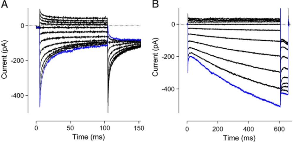
Fig. 1.Examples of FCDI and Ca2+-dependent reactivation. Example ICRACfrom HEK293 cells with exogenously expressed STIM1 and Orai1. Traces are recorded in response to the initial steps ranging from +62 mV to−138 mV in 20-mV increments, the most negative step shown in blue, followed by a second step to−118 mV. (A) Strongly inactivating currents, similar to that which would be expected where an excess of STIM1 relative to Orai1 exists. (B) Strongly reactivating currents, similar to that which would be expected where an excess of Orai1 rel-ative to STIM1 exists. The example here shows a longer initial step, demonstrating that the reactivation fails to approach a steady state even after 600 ms.
containing the fast inactivation gate have not been conclusively identi-fied. Some likely candidates are discussed in this review.
4. Channel subunit stoichiometry
The disparity in the kinetics of Ca2 +-dependent gating of STIM1/ Orai1-mediated current has led to the suggestion that Orai1 and STIM1 may form channels with variable stoichiometry. This idea was supported by experiments in which the expression levels of Orai1 and STIM1 were varied. Increasing the ratio of STIM1 to Orai1 produced stronger FCDI (Fig. 1A). In contrast, decreasing the ratio of STIM1 relative to Orai1 caused a loss of FCDI and introduction of Ca2+-dependent reac-tivation of ICRAC[51](Fig. 1B). Associated with the change in the STIM1/
Orai1 ratio were changes in apparent selectivity for Ba2+and Sr2+over
Ca2+and responsiveness to the nonspecific I
CRACblocker 2-APB[51].
Overexpression of Orai1 without STIM1 overexpression does not result in any increase in ICRACand has in some instances been shown
to reduce endogenous ICRAC[34,55,63]. Thesefindings led to the
sugges-tion that a large excess of Orai1 results in an insufficient pool of STIM1 being able to bind to and activate each Orai1 channel pore, with the con-sequence of a reduced whole cell current.
This idea has been supported and extended through the use offl uo-rescence imaging techniques and concatemers of Orai1 and STIM1 to di-rectly control or quantify the relative amounts of Orai1 and STIM1 expressed in an individual cell[15,24]. It was initially proposed that the Orai1 pore functions as a tetramer[20,36,42]. However, recent data from the crystal structure of theDrosophila melanogaster homo-logue of Orai suggest that Orai1 forms a hexameric pore[19]. Each Orai1 subunit can bind two STIM1 subunits as shown by yeast two-hybrid screens and coimmunoprecipitation experiments where the activation domain of STIM1 was capable of binding at both C- and N-termini of Orai1[41]. The 1 Orai1:2 STIM1 stoichiometry produces an ICRACthat is maximally activated and shows the greatest degree of
FCDI. In contrast, in the 1 Orai1:1 STIM1 stoichiometry, whole cell cur-rents are smaller, and Ca2+-dependent reactivation of ICRACis exhibited
[15,24].
5. Key domains and residues within STIM1
STIM1 was identified using high throughput RNAi screens in con-junction with Ca2+imaging assays tofind genes that regulate
intracel-lular Ca2 +concentration[26,70]. STIM1 is a ubiquitously expressed
685 amino acid, single transmembrane domain, protein. Cell surface bi-otinylation shows that as much as 25% of immunoprecipitatable STIM1 is located on the plasma membrane[30]. However, STIM1 required for CRAC channel activation is located on the endoplasmic reticulum mem-brane[34].Fig. 2shows the schematic structure of STIM1, with key do-mains marked. The mature STIM1 protein is cleaved of its N-terminal 22 amino acid signal peptide during translation. The regions required for Ca2 +sensing and oligomerisation of STIM1 are found within the
re-maining ER/SR luminal N-terminal region (aa. 23–200). This region contains an ER/SR targeting peptide, a pair of EF-hand domains for ER/ SR luminal Ca2+sensing, and a sterileα-motif (SAM) domain required
for oligomerisation of STIM1 (Fig. 2)[30,57,64]. Within the SAM domain are two N-glycosylation sites that modulate the rate of STIM1 transloca-tion to junctransloca-tional ER upon store depletransloca-tion[21].
The cytoplasmic region of STIM1 contains regions critical for interac-tion with Orai1. The minimum region of STIM1 necessary for CRAC channel activation is a ~ 100 amino acid long region known as the CRAC activation domain (CAD; aa. 342–448) (Fig. 2)[37,41,69]. Ectopic expression of a construct containing CAD but lacking all other regions of STIM1 (STIM1-CAD) with any of the Orai proteins is sufficient to cause a constitutive activation of CRAC current. In contrast, STIM1 constructs lacking the CAD are unable to activate current following store depletion [37,41,69]. An interesting property of ICRACactivated by STIM1-CAD is
that it fails to exhibit FCDI[40,69]. The CRAC modulatory domain
(CMD; aa. 474–485), C-terminal to CAD, is required for FCDI (Fig. 2) [4,22,40]. Expression of Orai1 with truncated STIM1 containing CAD and CMD produces current which retains FCDI. The CMD is rich in neg-atively charged glutamate and aspartate residues. The role of each of these negatively charged residues appears to be complex. Alanine neutralisation of D475/6A and D478/81A in the CMD reduces FCDI, while E482/3A causes increased and accelerated FCDI[40]. Neutralisation of six of these residues, including both E482 and E483, completely abolished FCDI of Orai1[40].
6. Key domains and residues of Orai1
Orai1 is a 301 amino acid protein that is expressed on the plasma membrane of most cell types. Orai1 is predicted to have four transmem-brane domains, with N and C termini as well as a single loop within the cytoplasmic space (Fig. 2).
6.1. N-terminus
The N-terminal residues 74–90 of Orai1 are highly conserved amongst species and are essential for channel activation; N-terminal
Fig. 2.Schematic diagram of Orai1/STIM1 organisation and key domains. Single Orai1 and STIM1 subunits are labelled, with associated subunits and relevant membranes unlabelled and faded for context. Intermolecular interactions are represented by dashed lines. STIM1 is a single transmembrane domain protein located on the endoplasmic reticulum mem-brane, and during store depletion migrates to regions of the membrane closely apposed to the plasma membrane. Key domains shown are the Ca2+
binding EF hand domain pair (green boxes), the SAM domain required for STIM1 oligomerisation (cyan box), CAD (red box), and CMD (black). Orai1 is located on the plasma membrane with 4 trans-membrane domains and the N-terminus and C-terminus within in the cytoplasm. Trans-membrane residues discussed are shown in red. TransTrans-membrane domain 1 lines the pore, with the selectivityfilter formed by a ring of six E106 residues in the functional hexameric channel. Key domains marked are CBDOrai1 (cyan), the intracellular loop (yellow) and the C terminal STIM1 binding region (green). Sequences for highlighted domains are given with specific residues discussed in this paper marked in red.
truncations of Orai1 up to aa. 74 allow for current to be activated upon store depletion[25]. The N-terminal Calmodulin (CaM) Binding Domain of Orai1 (CBDOrai1, aa. 68–91) binds CaM in a Ca2+-dependent manner
[40]. Mutations of the CBDOrai1that prevent CaM binding, e.g. A73E,
W76A, W76E, W76S and Y80E, result in currents with reduced FCDI and prominent reactivation in response to hyperpolarising steps[40]. In contrast, mutations of the CBDOrai1that retained CaM binding, Y80A
and Y80S, enhance FCDI[40]. The notion of CaM involvement in FCDI is consistent with earlier observations that expression of a Ca2+ -insensitive CaM mutant in hepatocytes reduced FCDI of native ICRAC. In
this case, it was hypothesised that the mutant CaM would compete with wild-type CaM for Ca2+-independent tethering sites on the chan-nel, exerting a dominant negative effect on FCDI[28]. Consistent with immunoprecipitation and yeast two-hybrid data[41], theDrosophila Orai crystal structure reveals that the N-terminus forms a helix that extends into the cytoplasmic space and is also spatially available to bind STIM1 [19,49].The N-terminus of Orai1 also contains one arginine-rich and two proline-rich regions (aa. 1–47), which are either minimally conserved or completely absent in Orai2 and Orai3. The func-tion of these regions remains poorly defined. Substitution of the Orai1 N-terminus into Orai3 causes a change in Ca2 +-dependent kinetics,
introducing a reactivation phase similar to that of Orai1, but not associ-ated with the usual strong inactivation of Orai3. While only a single pro-line-rich region is required for reactivation of Orai1, all three proline/ arginine-rich regions are required for Orai3 reactivation, making it diffi -cult to define an essential domain for reactivation and suggesting that other regions may be involved[11].
6.2. Intracellular loop
The Orai1 intracellular loop between transmembrane domains 2 and 3 is highly conserved between Orai1, 2 and 3, and between species, sug-gesting that it has an important function[10,56]. Consistent with this notion, co-transfection of STIM1 and Orai1 with residues 151–154 with-in the loop mutated to alanwith-ine results with-in an with-increase with-in whole cell cur-rent amplitude in response to store depletion[56]. Associated with this increase is a complete loss of FCDI. Interestingly, the transfection of a short peptide derived from the intracellular loop was able to restore FCDI, leading the investigators to the suggestion that the intracellular loop of Orai1 may form the inactivation gate[56]. Similarly, using chimeric Orai constructs, it has been demonstrated that the intracellular loop makes a substantial contribution to determining the inactivation profile of the construct. For example, an Orai1 intracellular loop substituted into an Orai3 construct displays inactivation similar to Orai1[10].
6.3. C terminus
A region of the Orai1 C terminus, identified as aa. 268–291, contain-ing a series of charged residues has shown by immunoprecipitation and yeast two-hybrid assays to interact with STIM1-CAD[39,41]. Deletion of this region results in a complete loss of ICRAC[25,39,41]. TheDrosophila
Orai crystal structure confirmed the availability of this region to bind STIM1[19,49].
The C-terminus of Orai1-3 is also likely to regulate FCDI[11,22]. Chimeric Orai constructs where the C-terminus of Orai1, 2 and 3 were interchanged demonstrated that the characteristic inactivation proper-ties of the different Orai homologues are influenced by the C-terminus. For example, a chimera of Orai1 with an Orai3 intracellular C-terminus displays robust FCDI similar to wild-type Orai3[22]. The stronger FCDI associated with the Orai2 and Orai3 C-termini compared to Orai1 has led to the hypothesis that a conserved series of three glutamates found in Orai2 and Orai3, compared to the two present in Orai1, may be responsible for this phenomenon. Consistent with this, alanine neutralisation of all three glutamates strongly reduced the extent of FCDI[22]. In contrast, one report did notfind any change in the
inactivation profile following the swap of only C-termini between Orai1 and Orai3 but instead suggested that the C-terminus acts in coop-eration together with the other intracellular domains of Orai3. For ex-ample, swapping both N- and C-termini had a greater effect than would be expected from the sum of single N- or C-terminus swaps[11]. 6.4. Orai1 pore residues
The transmembrane domain 1 (TM1) of each of the six Orai1 sub-units that constitute a CRAC channel is understood to line the channel pore. Mutation analysis has identified several residues within or near the TM1 as being important for permeation and therefore likely to be positioned within the domain's helical structure facing the centre of the pore[31]. In addition, E190Q mutation in TM3 has also been report-ed to interfere with permeation [44,62,65]. The transmembrane residues discussed below have also been reported to modulate Ca2+
-dependent kinetics of ICRAC, suggesting that CRAC channel gating and
permeation may be coupled processes.
Based on the understanding of voltage-gated Ca2+channels, where transmembrane glutamates form the selectivity centre[59,66], E106 of Orai1 in TM1 was identified as the CRAC channel selectivity centre. Mutations of E106 of Orai1 to glutamine (E106Q) or alanine (E106A) have a dominant negative effect, suppressing any WT CRAC current [12,44,62]. A conservative mutation to aspartate (E106D)[44,62,65], or of the homologous residue inDrosophila Orai (E180D)[44,68], produces a channel that retains activation by store depletion when co-expressed with STIM1 but has altered permeation properties. E106D Orai1 shifts the reversal potential of STIM1/Orai1-mediated current towards 0 mV, indicating a loss of selectivity for Ca2+over monovalent
cations[44,62,68]. These data conclusively demonstrate that E106 of Orai1 is critical in determining the selectivity of CRAC channels.
In addition to its altered selectivity profile, E106D Orai1 has also been reported to display altered fast Ca2 +-dependent kinetics
com-pared to WT Orai1[52,65]. Initial investigations of E106D Orai1 showed that in response to steps to negative membrane potentials, the current undergoes a very rapid phase of current decay to a steady state in a bath solution containing 20 mM Ca2 +and 130 mM Na+. This decay
can befitted with a single exponential with a time constant of approxi-mately 1.5 ms, which is significantly faster than what is normally seen for FCDI[65]. Since E106D Orai1 is permeable to Na+, and replacement
of all Na+in the extracellular solution with 110 mM Ca2+eliminated
this current decay, the investigators reasonably concluded that the ob-served phenomenon was due to the block of Na+current by Ca2 +
[65]. Consistent with these data, a reduction of extracellular [Ca2+] to
micromolar levels slowed down the kinetics of the block of Na+current, while progressive increases of extracellular [Ca2+] accelerated the
ki-netics of the block of Na+current[65]. The loss of current inactivation
in 110 mM Ca2+was taken as evidence that the E106D mutant lacks FCDI and that the mechanism of FCDI may be linked to the selectivity filter[65]. Using a different approach, a subsequent study utilised im-permeable NMDG+in place of Na+in the extracellular solution and
found that at lower Ca2+concentrations, in response to steps to
nega-tive membrane potentials, inactivation of E106D Orai1 current could be observed and that this inactivation was dependent on extracellular Ca2+[52]. The most striking difference between this inactivation and
typical FCDI of WT Orai1 was that it was greatly accelerated and able to befitted with a single exponential with a time constant of approxi-mately 1 ms in 10 mM extracellular Ca2+. The inactivation was found to be independent of relative STIM1 expression levels and maintained even if expressed with STIM1-CAD, indicating that the inactivation was independent of STIM1[52]. Taken together with data showing that, unlike FCDI of WT Orai1, the inactivation of the E106D Orai1 could be supported by permeating Sr2+and that changing the
intracel-lular Ca2+buffer from EGTA to BAPTA had no effect on the kinetics of the current, it was concluded that the binding site regulating inactiva-tion of E106D mutant was different from that of typical FCDI[52]. Two
possible reasons for this are that E106D mutation affects either the in-teraction between Orai1 and STIM1, or the mechanism of inactivation within the pore. The clear effects of this mutation strongly suggest that Ca2 +-dependent inactivation is likely to be associated with the
selectivityfilter[52,65].
A V102I mutation of Orai1 was reported to potentiate ICRAC in
response to hyperpolarising steps both in the presence and absence of Ca2+[54]. Since this potentiation was independent from the
extracellu-lar ion composition, it was suggested that this mutation introduces a slow voltage gate due to interaction of the V102I residue with a gating charge at the selectivityfilter, presumably at E106[54]. In contrast, wild-type STIM1/Orai1 channels have been found to show neither inac-tivation nor potentiation in the absence of external Ca2+, no matter how
strong inactivation or potentiation is with external Ca2+[51,65]. It
re-mains unclear as to whether a purely voltage-dependent gating process exists for this mutant, or if external Ca2+is required for potentiation of
ICRAC. A later study found that current mediated by V102I Orai1 is infl
u-enced by the relative expression of STIM1/Orai1, and at high STIM1/ Orai1 relative expression displays strong FCDI[52]. The discrepancies in thesefindings relating to the kinetics of V102I Orai1 remain unresolved.
A second study also suggests that V102I Orai1 is unlikely to be func-tionally different from WT Orai1; however, other mutations of this residue suggest that the V102 residue may influence gating. When mu-tated from hydrophobic valine to mildly hydrophobic or polar residues, for example V102C, Orai1 becomes constitutively active, independent of STIM1 or store depletion, poorly selective for Ca2+and displays no FCDI
[33]. However, when co-expressed with STIM1, upon store depletion V102C Orai1 associates with STIM1 and displays high Ca2+selectivity
and FCDI. These data confirm that interaction with STIM1 changes the properties of the Orai1 pore and that FCDI is not an intrinsic property of Orai1 alone[32].
Mutation of E190 in transmembrane domain 3 of Orai1 to aspartate or alanine has no influence on Orai1 current. Mutation of the same res-idue to glutamine (E190Q) has been reported to result in a channel which is activated by store depletion but displays reduced selectivity for Ca2 +[44,62,65]. The degree of loss of selectivity associated with
E190Q has been inconsistent between studies[45,62,65]and evidence that E190C cannot be chemically induced to form disulfide bonds, nor bind permeating Cd2+, implies that E190 is unlikely to line the pore
[33,73]. It has also been suggested that E190Q Orai1 kinetics is characterised by a Ca2+-dependent reactivation phase[65]. However,
similar to early studies of V102I Orai1 that predated the understanding of the fundamental importance of the stoichiometry of Orai1/STIM1, it now seems unlikely that this residue is involved in regulating Ca2+ -dependent kinetics of ICRAC[52].
7. Proposed mechanism for FCDI
The results discussed in this review indicate that FCDI is a complex process influenced by 1) the stoichiometry of binding between STIM1 and Orai1, 2) cytoplasmic domains of STIM1 and Orai1, and 3) the selec-tivity centre within the Orai1 pore. A plausible working model bringing together many of these concepts to explain the regulation of FCDI of CRAC channels is similar to that proposed for CaV1.2, where Ca2 +
-dependent CaM binding to the cytoplasmic site of the channel increases the affinity of the selectivityfilter for Ca2+, causing Ca2+to“stick”in the
pore and prevent further ion permeation[1]. Although this exact model may not necessarily directly apply to FCDI in CRAC channels, it high-lights the possibility of the interaction between the selectivity centre in the pore and the CaM binding site in the cytoplasmic domain of the channel, where Ca2+binding to one these sites causes a conformational
change at another. Such interaction could explain why E106D mutation in the selectivity centre causes very similar changes in ICRACkinetics[52]
as Y80A (or Y80S) mutation in CBDOrai1[40]. Both mutants, E106D and
Y80A, exhibited greatly accelerated FCDI, compared to WT Orai1,
which followed a single exponential time course. It may be that the E106D mutation allosterically alters the Orai1 CaM binding domain, or that both the E106D pore mutant and the Y80A/Y80S mutants cause a similar change in the affinity or efficacy of Ca2 +binding to a site in
the pore, which regulates FCDI.
8. Orai1/STIM1stoichiometry in light of new crystal structure data The recent discovery of the hexameric crystal structure ofDrosophila Orai allows the electrophysiological and molecular data regarding STIM1/Orai1 stoichiometry and FCDI to be analysed in a new light. Inter-estingly, while the transmembrane region of the channel has a six-fold symmetry, the cytoplasmic domains adopt two alternating conforma-tions, conferring a three-fold symmetry where pairs of Orai subunits have closely interacting C-termini[19]. If a single STIM1 subunit bridges the N- and C-termini STIM1 binding sites, then this would only allow three binding sites per channel. STIM1 has been suggested to function as a dimer[38,69,72]. If this is the case, this would contradict the model in which two STIM1 subunits bind to each Orai1 subunit for max-imal ICRACactivation[49]. However, the level of oligomerisation of
STIM1 subunits remains undefined, and it has also been suggested that STIM1 may function as a tetramer, given that STIM1-CAD forms tet-ramers in solution[41].
The conformation of the Orai1 C-termini in activated Orai1-STIM1 complexes remains unknown. It has been speculated that perhaps in the activated state, the initial STIM1 interaction may disrupt the Orai pairing, and instead the C-termini extend directly down into the cyto-plasm to reveal six binding sites[19]. While these models can possibly explain how STIM1 can bind Orai1 in a 2:1 ratio, they do not explicitly account for how other possible stoichiometries may activate the chan-nel and display weaker FCDI or reactivation.
One possibility is that in a hypothetical extended conformation, there exists enough space for an STIM1 subunit each to bind to both the N- and C-termini, giving twelve binding sites. As demonstrated by Parket al.(2009), STIM1-CAD binds to both the C-terminus and N-terminus of Orai1, with a higher affinity for the C-terminus[41]. It would therefore be reasonable to suggest that when the ER/SR is nearly full and STIM1 concentration at the junctional ER is low, available Ca2+
binding sites associated with STIM1-CAD would only be present at the C-terminus which would favour Ca2+-dependent reactivation. In
con-trast, when ER/SR Ca2 +is fully depleted and the amounts of STIM1
are saturating, STIM1-CAD occupies both N- and C-terminal binding sites on Orai1, which may create a completely different set of Ca2+
bind-ing sites, and/or different type of conformational changes in response to Ca2+binding, favouring FCDI.
In a model where a single STIM1 subunit within a dimer bridges the N- and C-termini Orai1 binding sites, this gives six STIM1 binding sites. In this case, when two or fewer sites are occupied, the channel is closed, as at least one STIM1 subunit per Orai subunit is required for channel activation[15]. Once three sites are occupied, the channel opens, and with each subsequent STIM1 dimer binding, the open probability or sin-gle channel conductance increases, resulting in increased whole cell ICRAC. In a CRAC channel where only a minimum required number, for
a functional channel, of STIM1 binding sites on Orai1 are occupied, Ca2 +entering through the pore promotes reactivation, whereas in a
channel with all or almost all STIM1 binding sites occupied, entering Ca2+causes FCDI.
9. Future perspectives
There are currently no physiological examples of ICRACdisplaying
FCDI associated with low STIM1 expression relative to Orai1, such as Ca2+-dependent reactivation. One possible example in an experimental
setting is the distinct ICRACBa2+conductances of mast cells, RBL and Jurkat
cells[16]. We have previously found that differences in STIM1:Orai1 ex-pression were also associated with differences in Ba2+conductance[51].
It is plausible that variable stoichiometry of STIM1:Orai1 and subse-quent variable ICRACproperties may have functional significance. The
activation of store-operated channels is graded depending on the level of intracellular store depletion[2,14,29], presumably by recruitment of additional activated STIM1 as stores are progressively depleted. There-fore, FCDI would be graded in a similar manner to ICRACamplitude,
where during partial depletion with a relatively small amount of acti-vated STIM1, small currents with little or no FCDI would be likely and during full depletion large currents with strong FCDI would be expected. The kinetics of ICRACactivated in response to partial store depletion has
not been thoroughly investigated, though one study did not report any changes in kinetics under these conditions[29]. In addition, little is known about the physiological regulation of Orai1 and STIM1 expres-sion in many cell types, or if expresexpres-sion changes during development, or during stressful or pathophysiological conditions. There is some evidence that these may be the case, for example in the dynamic expres-sion patterns of STIM1 throughout muscle development[58], oncogene or tumour suppressor-dependent expression of STIM1 in a model can-cer cell line[47], and altered expression of STIM1 and Orai1 during T cell activation[60], or following vascular injury[71]. Future work is likely to focus on endogenous regulation mechanisms of STIM and Orai protein expression, given that these data have provided the frame-work to linkin vivoexamples of altered relative STIM1/Orai1 expression with the functional consequences of altered Ca2+signaling. In addition,
the formation of heteromeric STIM1/Orai channels is an intriguing con-cept that remains largely unexplored. There is conclusive evidence that heteromeric Orai channels are able to be formed[27,35,50,62]and, given the functional differences between Orai subunits, may be able to display a wide range of characteristics and be tuned for specific roles in certain tissues or under certain conditions.
10. Conclusions
The discovery of STIM1 and Orai1 has underpinned rapid advance-ments in the understanding of CRAC channel function over the past eight years. In that time, the pathway from store depletion to channel activation has been well defined, and the functional significance of many individual domains of Orai1 and STIM1 has been described, if not yet synthesised into a“whole channel”model. The recent publica-tion of crystal structure data forDrosophilaOrai offers a new set of chal-lenges and opportunities. Thefinding of a hexameric pore may force a reevaluation of many results that presumed a tetrameric pore. However, recent electrophysiological data obtained using Orai1 concatamers sug-gest that classical ICRACis mediated by Orai1 tetramer, not hexamer[61].
Given the importance of the stoichiometry of STIM1:Orai1 binding and the functional significance of the cytoplasmic domains of both STIM1 and Orai1 on ICRACproperties as discussed in this review, a full
under-standing of CRAC channel Ca2+-dependent gating will require the addi-tional knowledge of how Orai1 and STIM1 subunits are organised in the functional channel.
Conflicts of interest
The authors declare that they have no conflict of interest. References
[1] O. Babich, V. Matveev, A.L. Harris, R. Shirokov, Ca2+
-dependent inactivation of CaV1.2 channels prevents Gd3+
block: does Ca2+
block the pore of inactivated chan-nels? J. Gen. Physiol. 129 (2007) 477–483.
[2] O. Brandman, J. Liou, W.S. Park, T. Meyer, STIM2 is a feedback regulator that stabilizes basal cytosolic and endoplasmic reticulum Ca2+
levels, Cell 131 (2007) 1327–1339. [3] P. Brehm, R. Eckert, Calcium entry leads to inactivation of calcium channel in
Para-mecium, Science 202 (1978) 1203–1206.
[4] I. Derler, M. Fahrner, M. Muik, B. Lackner, R. Schindl, K. Groschner, C. Romanin, A Ca2+
release-activated Ca2+
(CRAC) modulatory domain (CMD) within STIM1 me-diates fast Ca2+
-dependent inactivation of ORAI1 channels, J. Biol. Chem. 284 (2009) 24933–24938.
[5] M. Faouzi, F. Hague, M. Potier, A. Ahidouch, H. Sevestre, H. Ouadid-Ahidouch, Down-regulation of Orai3 arrests cell-cycle progression and induces apoptosis in breast cancer cells but not in normal breast epithelial cells, J. Cell. Physiol. 226 (2011) 542–551.
[6] C. Fasolato, M. Hoth, R. Penner, A GTP-dependent step in the activation mechanism of capacitative calcium influx, J. Biol. Chem. 268 (1993) 20737–20740.
[7] S. Feske, Y. Gwack, M. Prakriya, S. Srikanth, S.H. Puppel, B. Tanasa, P.G. Hogan, R.S. Lewis, M. Daly, A. Rao, A mutation in Orai1 causes immune deficiency by abrogating CRAC channel function, Nature 441 (2006) 179–185.
[8] L. Fierro, A.B. Parekh, Fast calcium-dependent inactivation of calcium release-activated calcium current (CRAC) in RBL-1 cells, J. Membr. Biol. 168 (1999) 9–17.
[9] M. Flourakis, V. Lehen'kyi, B. Beck, M. Raphael, M. Vandenberghe, F.V. Abeele, M. Roudbaraki, G. Lepage, B. Mauroy, C. Romanin, Y. Shuba, R. Skryma, N. Prevarskaya, Orai1 contributes to the establishment of an apoptosis-resistant phenotype in pros-tate cancer cells, Cell Death Dis. 1 (2010) e75.
[10] I. Frischauf, M. Muik, I. Derler, J. Bergsmann, M. Fahrner, R. Schindl, K. Groschner, C. Romanin, Molecular determinants of the coupling between STIM1 and Orai channels: differential activation of Orai1-3 channels by a STIM1 coiled-coil mutant, J. Biol. Chem. 284 (2009) 21696–21706.
[11]I. Frischauf, R. Schindl, J. Bergsmann, I. Derler, M. Fahrner, M. Muik, R. Fritsch, B. Lackner, K. Groschner, C. Romanin, Cooperativeness of Orai cytosolic domains tunes subtype-specific gating, J. Biol. Chem. 286 (2011) 8577–8584.
[12] Y. Gwack, S. Srikanth, S. Feske, F. Cruz-Guilloty, M. Oh-hora, D.S. Neems, P.G. Hogan, A. Rao, Biochemical and functional characterization of Orai proteins, J. Biol. Chem. 282 (2007) 16232–16243.
[13] Y. Gwack, S. Srikanth, M. Oh-Hora, P.G. Hogan, E.D. Lamperti, M. Yamashita, C. Gelinas, D.S. Neems, Y. Sasaki, S. Feske, M. Prakriya, K. Rajewsky, A. Rao, Hair loss and defective T- and B-cell function in mice lacking ORAI1, Mol. Cell. Biol. 28 (2008) 5209–5222.
[14] A.M. Hofer, C. Fasolato, T. Pozzan, Capacitative Ca2+
entry is closely linked to the fill-ing state of internal Ca2+
stores: a study using simultaneous measurements of ICRAC and intraluminal [Ca2+
], J. Cell Biol. 140 (1998) 325–334.
[15] P.J. Hoover, R.S. Lewis, Stoichiometric requirements for trapping and gating of Ca2+ release-activated Ca2+
(CRAC) channels by stromal interaction molecule 1 (STIM1), Proc. Natl. Acad. Sci. U. S. A. 108 (2011) 13299–13304.
[16] M. Hoth, Calcium and barium permeation through calcium release-activated calcium (CRAC) channels, Pflugers Arch. 430 (1995) 315–322.
[17] M. Hoth, R. Penner, Depletion of intracellular calcium stores activates a calcium cur-rent in mast cells, Nature 355 (1992) 353–356.
[18]M. Hoth, R. Penner, Calcium release-activated calcium current in rat mast cells, J. Physiol. 465 (1993) 359–386.
[19] X. Hou, L. Pedi, M.M. Diver, S.B. Long, Crystal structure of the calcium release-activated calcium channel Orai, Science 338 (2012) 1308–1313.
[20] W. Ji, P. Xu, Z. Li, J. Lu, L. Liu, Y. Zhan, Y. Chen, B. Hille, T. Xu, L. Chen, Functional stoi-chiometry of the unitary calcium-release-activated calcium channel, Proc. Natl. Acad. Sci. U. S. A. 105 (2008) 13668–13673.
[21] T. Kilch, D. Alansary, M. Peglow, K. Dorr, G. Rychkov, H. Rieger, C. Peinelt, B.A. Niemeyer, Mutations of the Ca2+
-sensing stromal interaction molecule STIM1 reg-ulate Ca2+
influx by altered oligomerization of STIM1 and by destabilization of the Ca2+
channel Orai1, J. Biol. Chem. 288 (2013) 1653–1664.
[22] K.P. Lee, J.P. Yuan, W. Zeng, I. So, P.F. Worley, S. Muallem, Molecular determinants of fast Ca2+
-dependent inactivation and gating of the Orai channels, Proc. Natl. Acad. Sci. U. S. A. 106 (2009) 14687–14692.
[23] R.S. Lewis, Store-operated calcium channels: new perspectives on mechanism and function, Cold Spring Harb. Perspect. Biol. 3 (2011).
[24]Z. Li, L. Liu, Y. Deng, W. Ji, W. Du, P. Xu, L. Chen, T. Xu, Graded activation of CRAC channel by binding of different numbers of STIM1 to Orai1 subunits, Cell Res. 21 (2011) 305–315.
[25] Z. Li, J. Lu, P. Xu, X. Xie, L. Chen, T. Xu, Mapping the interacting domains of STIM1 and Orai1 in Ca2+
release-activated Ca2+
channel activation, J. Biol. Chem. 282 (2007) 29448–29456.
[26] J. Liou, M.L. Kim, W.D. Heo, J.T. Jones, J.W. Myers, J.E. Ferrell Jr., T. Meyer, STIM is a Ca2+sensor essential for Ca2+-store-depletion-triggered Ca2+influx, Curr. Biol. 15 (2005) 1235–1241.
[27] A. Lis, C. Peinelt, A. Beck, S. Parvez, M. Monteilh-Zoller, A. Fleig, R. Penner, CRACM1, CRACM2, and CRACM3 are store-operated Ca2+channels with distinct functional properties, Curr. Biol. 17 (2007) 794–800.
[28]T. Litjens, T. Nguyen, J. Castro, E.C. Aromataris, L. Jones, G.J. Barritt, G.Y. Rychkov, Phospholipase C-gamma1 is required for the activation of store-operated Ca2+ channels in liver cells, Biochem. J. 405 (2007) 269–276.
[29] R.M. Luik, B. Wang, M. Prakriya, M.M. Wu, R.S. Lewis, Oligomerization of STIM1 cou-ples ER calcium depletion to CRAC channel activation, Nature 454 (2008) 538–542. [30]S.S. Manji, N.J. Parker, R.T. Williams, L. van Stekelenburg, R.B. Pearson, M. Dziadek, P.J. Smith, STIM1: a novel phosphoprotein located at the cell surface, Biochim. Biophys. Acta 1481 (2000) 147–155.
[31] C.A. McCarl, C. Picard, S. Khalil, T. Kawasaki, J. Rother, A. Papolos, J. Kutok, C. Hivroz, F. Ledeist, K. Plogmann, S. Ehl, G. Notheis, M.H. Albert, B.H. Belohradsky, J. Kirschner, A. Rao, A. Fischer, S. Feske, ORAI1 deficiency and lack of store-operated Ca2+
entry cause immunodeficiency, myopathy, and ectodermal dysplasia, J. Allergy Clin. Immunol. 124 (2009)(1311–8 e7).
[32]B.A. McNally, A. Somasundaram, M. Yamashita, M. Prakriya, Gated regulation of CRAC channel ion selectivity by STIM1, Nature 482 (2012) 241–245.
[33] B.A. McNally, M. Yamashita, A. Engh, M. Prakriya, Structural determinants of ion per-meation in CRAC channels, Proc. Natl. Acad. Sci. U. S. A. 106 (2009) 22516–22521. [34] J.C. Mercer, W.I. Dehaven, J.T. Smyth, B. Wedel, R.R. Boyles, G.S. Bird, J.W. Putney Jr.,
Orai2 with the intracellular calcium sensor, Stim1, J. Biol. Chem. 281 (2006) 24979–24990.
[35] O. Mignen, J.L. Thompson, T.J. Shuttleworth, Both Orai1 and Orai3 are essential com-ponents of the arachidonate-regulated Ca2+
-selective ARC channels, J. Physiol. 586 (2007) 185–195.
[36] O. Mignen, J.L. Thompson, T.J. Shuttleworth, Orai1 subunit stoichiometry of the mammalian CRAC channel pore, J. Physiol. 586 (2008) 419–425.
[37] M. Muik, M. Fahrner, I. Derler, R. Schindl, J. Bergsmann, I. Frischauf, K. Groschner, C. Romanin, A cytosolic homomerization and a modulatory domain within STIM1 C terminus determine coupling to ORAI1 channels, J. Biol. Chem. 284 (2009) 8421–8426.
[38] M. Muik, M. Fahrner, R. Schindl, P. Stathopulos, I. Frischauf, I. Derler, P. Plenk, B. Lackner, K. Groschner, M. Ikura, C. Romanin, STIM1 couples to ORAI1 via an intra-molecular transition into an extended conformation, EMBO J. 30 (2011) 1678–1689. [39] M. Muik, I. Frischauf, I. Derler, M. Fahrner, J. Bergsmann, P. Eder, R. Schindl, C. Hesch, B. Polzinger, R. Fritsch, H. Kahr, J. Madl, H. Gruber, K. Groschner, C. Romanin, Dynamic coupling of the putative coiled-coil domain of ORAI1 with STIM1 mediates ORAI1 channel activation, J. Biol. Chem. 283 (2008) 8014–8022.
[40]F.M. Mullins, C.Y. Park, R.E. Dolmetsch, R.S. Lewis, STIM1 and calmodulin interact with Orai1 to induce Ca2+-dependent inactivation of CRAC channels, Proc. Natl. Acad. Sci. U. S. A. 106 (2009) 15495–15500.
[41] C.Y. Park, P.J. Hoover, F.M. Mullins, P. Bachhawat, E.D. Covington, S. Raunser, T. Walz, K.C. Garcia, R.E. Dolmetsch, R.S. Lewis, STIM1 clusters and activates CRAC channels via direct binding of a cytosolic domain to Orai1, Cell 136 (2009) 876–890. [42] A. Penna, A. Demuro, A.V. Yeromin, S.L. Zhang, O. Safrina, I. Parker, M.D. Cahalan, The
CRAC channel consists of a tetramer formed by Stim-induced dimerization of Orai dimers, Nature 456 (2008) 116–120.
[43] C. Picard, C.A. McCarl, A. Papolos, S. Khalil, K. Luthy, C. Hivroz, F. LeDeist, F. Rieux-Laucat, G. Rechavi, A. Rao, A. Fischer, S. Feske, STIM1 mutation associated with a syndrome of immunodeficiency and autoimmunity, N. Engl. J. Med. 360 (2009) 1971–1980.
[44] M. Prakriya, S. Feske, Y. Gwack, S. Srikanth, A. Rao, P.G. Hogan, Orai1 is an essential pore subunit of the CRAC channel, Nature 443 (2006) 230–233.
[45] M. Prakriya, R.S. Lewis, Regulation of CRAC channel activity by recruitment of silent channels to a high open-probability gating mode, J. Gen. Physiol. 128 (2006) 373–386.
[46] C. Randriamampita, R.Y. Tsien, Emptying of intracellular Ca2+
stores releases a novel small messenger that stimulates Ca2+influx, Nature 364 (1993) 809–
814. [47]M.F. Ritchie, C. Yue, Y. Zhou, P.J. Houghton, J. Soboloff, Wilms tumor suppressor 1
(WT1) and early growth response 1 (EGR1) are regulators of STIM1 expression, J. Biol. Chem. 285 (2010) 10591–10596.
[48] J. Roos, P.J. DiGregorio, A.V. Yeromin, K. Ohlsen, M. Lioudyno, S. Zhang, O. Safrina, J.A. Kozak, S.L. Wagner, M.D. Cahalan, G. Velicelebi, K.A. Stauderman, STIM1, an essential and conserved component of store-operated Ca2+channel function, J. Cell Biol. 169 (2005) 435–445.
[49]B.S. Rothberg, Y. Wang, D.L. Gill, Orai channel pore properties and gating by STIM: implications from the Orai crystal structure, Sci. Signal. 6 (2013) pe9.
[50] R. Schindl, I. Frischauf, J. Bergsmann, M. Muik, I. Derler, B. Lackner, K. Groschner, C. Romanin, Plasticity in Ca2+
selectivity of Orai1/Orai3 heteromeric channel, Proc. Natl. Acad. Sci. U. S. A. 106 (2009) 19623–19628.
[51] N. Scrimgeour, T. Litjens, L. Ma, G.J. Barritt, G.Y. Rychkov, Properties of Orai1 medi-ated store-opermedi-ated current depend on the expression levels of STIM1 and Orai1 proteins, J. Physiol. 587 (2009) 2903–2918.
[52] N.R. Scrimgeour, D.P. Wilson, G.Y. Rychkov, Glutamate 106 in the Orai1 pore contributes to fast Ca2+
-dependent inactivation and pH dependence of Ca2+
release-activated Ca2+
(CRAC) current, Biochem. J. 441 (2012) 743–753.
[53]J. Soboloff, M.A. Spassova, X.D. Tang, T. Hewavitharana, W. Xu, D.L. Gill, Orai1 and STIM reconstitute store-operated calcium channel function, J. Biol. Chem. 281 (2006) 20661–20665.
[54] M.A. Spassova, T. Hewavitharana, R.A. Fandino, A. Kaya, J. Tanaka, D.L. Gill, Voltage gating at the selectivityfilter of the Ca2+
release-activated Ca2+
channel induced by mutation of the Orai1 protein, J. Biol. Chem. 283 (2008) 14938–14945.
[55] M.A. Spassova, J. Soboloff, L.P. He, W. Xu, M.A. Dziadek, D.L. Gill, STIM1 has a plasma membrane role in the activation of store-operated Ca(2+
) channels, Proc. Natl. Acad. Sci. U. S. A. 103 (2006) 4040–4045.
[56] S. Srikanth, H.J. Jung, B. Ribalet, Y. Gwack, The intracellular loop of Orai1 plays a cen-tral role in fast inactivation of Ca2+
release-activated Ca2+
channels, J. Biol. Chem. 285 (2009) 5066–5075.
[57]P.B. Stathopulos, L. Zheng, G.Y. Li, M.J. Plevin, M. Ikura, Structural and mechanistic insights into STIM1-mediated initiation of store-operated calcium entry, Cell 135 (2008) 110–122.
[58] J. Stiber, A. Hawkins, Z.S. Zhang, S. Wang, J. Burch, V. Graham, C.C. Ward, M. Seth, E. Finch, N. Malouf, R.S. Williams, J.P. Eu, P. Rosenberg, STIM1 signalling controls store-operated calcium entry required for development and contractile function in skeletal muscle, Nat. Cell Biol. 10 (2008) 688–697.
[59] S. Tang, G. Mikala, A. Bahinski, A. Yatani, G. Varadi, A. Schwartz, Molecular localiza-tion of ion selectivity sites within the pore of a human L-type cardiac calcium chan-nel, J. Biol. Chem. 268 (1993) 13026–13029.
[60]P. Thakur, A.F. Fomina, Density of functional Ca2+release-activated Ca2+(CRAC) channels declines after T-cell activation, Channels 5 (2011) 510–517.
[61] J.L. Thompson, T.J. Shuttleworth, How many Orai's does it take to make a CRAC channel? Sci. Rep. 3 (2013) 1961.
[62] M. Vig, A. Beck, J.M. Billingsley, A. Lis, S. Parvez, C. Peinelt, D.L. Koomoa, J. Soboloff, D.L. Gill, A. Fleig, J.P. Kinet, R. Penner, CRACM1 multimers form the ion-selective pore of the CRAC channel, Curr. Biol. 16 (2006) 2073–2079.
[63] M. Vig, C. Peinelt, A. Beck, D.L. Koomoa, D. Rabah, M. Koblan-Huberson, S. Kraft, H. Turner, A. Fleig, R. Penner, J.P. Kinet, CRACM1 is a plasma membrane protein essen-tial for store-operated Ca2+
entry, Science 312 (2006) 1220–1223.
[64] R.T. Williams, S.S. Manji, N.J. Parker, M.S. Hancock, L. Van Stekelenburg, J.P. Eid, P.V. Senior, J.S. Kazenwadel, T. Shandala, R. Saint, P.J. Smith, M.A. Dziadek, Identification and characterization of the STIM (stromal interaction molecule) gene family: coding for a novel class of transmembrane proteins, Biochem. J. 357 (2001) 673–685. [65]M. Yamashita, L. Navarro-Borelly, B.A. McNally, M. Prakriya, Orai1 mutations alter
ion permeation and Ca2+
-dependent fast inactivation of CRAC channels: evidence for coupling of permeation and gating, J. Gen. Physiol. 130 (2007) 525–540. [66]J. Yang, P.T. Ellinor, W.A. Sather, J.F. Zhang, R.W. Tsien, Molecular determinants of
Ca2+
selectivity and ion permeation in L-type Ca2+
channels, Nature 366 (1993) 158–161.
[67] S. Yang, J.J. Zhang, X.Y. Huang, Orai1 and STIM1 are critical for breast tumor cell mi-gration and metastasis, Cancer Cell 15 (2009) 124–134.
[68] A.V. Yeromin, S.L. Zhang, W. Jiang, Y. Yu, O. Safrina, M.D. Cahalan, Molecular identi-fication of the CRAC channel by altered ion selectivity in a mutant of Orai, Nature 443 (2006) 226–229.
[69]J.P. Yuan, W. Zeng, M.R. Dorwart, Y.J. Choi, P.F. Worley, S. Muallem, SOAR and the polybasic STIM1 domains gate and regulate Orai channels, Nat. Cell Biol. 11 (2009) 337–343.
[70]S.L. Zhang, Y. Yu, J. Roos, J.A. Kozak, T.J. Deerinck, M.H. Ellisman, K.A. Stauderman, M.D. Cahalan, STIM1 is a Ca2+
sensor that activates CRAC channels and migrates from the Ca2+
store to the plasma membrane, Nature 437 (2005) 902–905. [71] W. Zhang, K.E. Halligan, X. Zhang, J.M. Bisaillon, J.C. Gonzalez-Cobos, R.K. Motiani, G.
Hu, P.A. Vincent, J. Zhou, M. Barroso, H.A. Singer, K. Matrougui, M. Trebak, Orai1-mediated I (CRAC) is essential for neointima formation after vascular injury, Circ. Res. 109 (2011) 534–542.
[72]Y. Zhou, P. Meraner, H.T. Kwon, D. Machnes, M. Oh-hora, J. Zimmer, Y. Huang, A. Stura, A. Rao, P.G. Hogan, STIM1 gates the store-operated calcium channel ORAI1 in vitro, Nat. Struct. Mol. Biol. 17 (2010) 112–116.
[73] Y. Zhou, S. Ramachandran, M. Oh-Hora, A. Rao, P.G. Hogan, Pore architecture of the ORAI1 store-operated calcium channel, Proc. Natl. Acad. Sci. U. S. A. 107 (2010) 4896–4901.
[74] A. Zweifach, R.S. Lewis, Mitogen-regulated Ca2+
current of T lymphocytes is activat-ed by depletion of intracellular Ca2+
stores, Proc. Natl. Acad. Sci. U. S. A. 90 (1993) 6295–6299.
[75]A. Zweifach, R.S. Lewis, Rapid inactivation of depletion-activated calcium current (ICRAC) due to local calcium feedback, J. Gen. Physiol. 105 (1995) 209–226.