R E S E A R C H A R T I C L E
Open Access
Quantifying the impact of inhalational
burns: a prospective study
Si Jack Chong
1, Yee Onn Kok
1*, Rosanna Xiang Ying Tay
1, Desai Suneel Ramesh
2, Kok Chai Tan
1and Bien Keem Tan
1Abstract
Background:Inhalational injury is a major cause of morbidity and mortality in burns patients. This study aims to analyse the clinical outcomes, complications and bacteriology of inhalational burn patients.
Methods:A prospective study was done on consecutive admissions to Burn Department, Singapore General Hospital over 15 months from January 2015 to March 2016. Presence of inhalational injury, demographics, complications and outcomes was recorded. Diagnosis of inhalational injury was based on history, symptoms and nasoendoscopy. Diagnosis of acute respiratory distress syndrome (ARDS), acute kidney injury (AKI) and infective complications were according to the Berlin criteria, acute kidney injury network (AKIN) classification stage 2 and above and the American Burns Association guidelines.
Results:Thirty-five patients (17.3%) had inhalational burns out of 202 patients (63.4% male, 57.4% Chinese population). The average age was 43 ± 16.7 years (range 16–86), and percentage of total body surface area (%TBSA) was 12.1 ± 18.0 (range 0–88). In patients with inhalational injury, age was 38.9 ± 17.2 years and %TBSA was 30.3 ± 32.3. In patients without inhalational injury, age was 44.1 ± 12.8 years and %TBSA was 8.3 ± 9.59. Compared to patients with cutaneous injury alone, patients with inhalational burns had more surgeries (3 ± 7.07 vs 1 ± 1.54,p= 0.003), increased length of stay (21 days vs 8 days,p= 0.004) and higher in-hospital mortality rate (17.1% vs 0.6%,p< 0.001). Incidence of ARDS and AKI was 48.6% and 37.1%, respectively, compared to 0.6% and 1.2% in the patients without inhalational injury (p< 0.001). Patients with inhalational injury had increased incidence of bacteraemia (31.4% vs 2.4%,p< 0.001), pneumonia (37.1% vs 1.2%, p< 0.001) and burn wound infection (51.4% vs 25.1%,p= 0.004). Inhalational injury predicted AKI with an adjusted odds ratio (OR) of 17.43 (95% confidence interval (CI) 3.07–98.87,p< 0.001); ARDS, OR = 106.71 (95% CI 12.73–894.53,p< 0.001) and pneumonia, OR = 13.87 (95% CI 2.32–82.94,p= 0.004).Acinetobacter baumannii was the most frequently cultured bacteria in sputum, blood and tissue cultures with inhalational injury. Gram-negative bacteria were predominantly cultured from tissue in patients with inhalational injury, whereas gram-positive bacteria were predominantly cultured from tissue in patients without inhalational injury.
Conclusions:Inhalational injury accompanying burns significantly increases the length of stay, mortality and complications including AKI, ARDS, infection and sepsis.
Keywords:Inhalational, Burns, Injury, Acute kidney injury, Acute respiratory distress syndrome, Pneumonia
* Correspondence:[email protected]
1Department of Plastic, Reconstructive and Aesthetic Surgery, Singapore
General Hospital, Level 5, Academia, Singapore 169865, Singapore Full list of author information is available at the end of the article
Background
The importance of inhalational injury as a predictor of mortality is widely recognised [1–4]. Few prospective studies, especially in a tropical burns setting, compared clinical outcomes in patients with inhalational burns and those with cutaneous burns only [5]. This study aims to analyse the effect of inhalational injury on inpatient mortality, length of stay and complications.
Methods
A prospective observational study was done on consecutive admissions to Burn Department, Singapore General Hospital from January 2015 to March 2016. Patients were excluded if they are deceased within 24 h of admission or transferred (Fig.1). Data was collected on our Research Electronic Data Capture (REDCap) database. The study was approved by the Singapore General Hospital Institutional Review Board.
In-hospital mortality, number of surgeries, length of stay and percentage of total body surface area (%TBSA) were recorded. Complications analysed were acute kid-ney injury (AKI), acute respiratory distress syndrome (ARDS), pneumonia and culture-positive burns wound infection and bacteraemia.
Statistical analysis
Data was analysed using IBM SPSS Statistics with insti-tutional statistician’s help.χ2and Mann-WhitneyUtests
were used. Binary logistic regression was applied to de-termine if inhalational injury was an independent pre-dictor of complications, removing effect of %TBSA and age. p< 0.05 was considered to indicate a statistically significant result. Dependent variables were the out-come/complication, e.g., AKI. Covariates were inhala-tional injury, age and %TBSA.
Inhalational injury
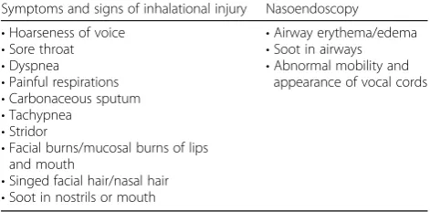
Inhalational injury was diagnosed by attending doctors in patients with history of smoke/fumes inhalation/fires in an enclosed space and three or more symptoms or signs and one positive finding on nasoendoscopy (Table1).
Bronchoscopy was performed only in patients with mucus plugging and was not part of diagnostic criteria. Patients referred from overseas with prior diagnosis were included.
Treatment principles
Patients were treated according to Advanced Burn Life Support (ABLS) [6] principles and modified Parkland’s regime (2–3 ml/kg/TBSA). In particular, if there was in-halational injury based on clinical findings or nasoendo-scopy, patients with inhalational burns were monitored with hourly oxygen saturations and given a heparinised saline nebuliser (50 U heparin in 5 ml normal saline). Patients were intubated if findings on nasoendoscopy
suggested severe airway injury with airway or vocal cord edema and if they had stridor or had an in-appropriate PaO2 /FiO2 (P/F) ratio. Other factors taken into account include age—patients more than 60 years old with risk factors for poor cough reflex were intubated.
Ventilation was interchanged between conventional ventilation and airway pressure release ventilation (APRV) depending on patient response. Tidal volume was 6–8 ml/kg and positive end expiratory pressure (PEEP) was titrated to P/F ratio as in the ARDSNet study/ protocol [7]. Prone positioning and neuromuscular par-alysis are weak recommendations in the Surviving Sepsis Guidelines [8] and also not practical (in cases of facial swelling and abdominal distention). In refractory hypox-aemia, APRV is used as recruitment and haemodynamics are superior [9–11] with improved compliance, reduced duration of ventilation and lower mortality.
Indications for antibiotic therapy was for prophylaxis for all major burns > 20%, infected burns and 5 days post graft or biobrane. Intravenous cefazolin is the usual first line.
Surgical treatment strategy was for early biobrane (epi-dermal skin dressing) or excision within 48 h. Biobrane as the first surgery has dramatically reduced the need for subsequent excision in partial thickness burns by reducing burns conversion and providing ‘coverage’. Subsequent surgeries were for further excision and grafting if burns conversions, infection or biobrane non-take occurred.
Non-infective complications
Berlin definition for ARDS was used [12]. Arterial blood gas was obtained in patients with inhalational injury, respiratory distress or abnormal chest radiographs. AKI
was defined as ≥ acute kidney injury network (AKIN)
classification stage 2 [13].
Infective complications
The American Burns Association recommendations for diagnosis of infectious complications in patients with
thermal trauma were followed [14]. In patients with
clinical suspicion of pneumonia/infection, appropriate investigations were ordered and bacteria cultures were recorded.
Sputum cultures in patients with pneumonia were obtained via induced sputum with nebulised saline. Bronchoalveolar lavage (BAL) in patients with bronchos-copy was done in patients with desaturations refractory to mechanical ventilation.
Results
Characteristics of patients included in this study were
listed in Table 2. The most common mechanisms of
injury were fire and scalds. Thirty-five (17.3%) patients had inhalational burns out of 202 patients (63.4% male, 57.4% Chinese population). The average age was 43 ± 16.7 years (range 16–86), and %TBSA was 12.1 ± 18.0 (range 0–88). In patients with inhalational injury, age was 38.9 ± 17.2 years and %TBSA was 30.3 ± 32.3. In patients without inhalational injury, age was 44.1 ± 12.8 years and %TBSA was 8.3 ± 9.59.
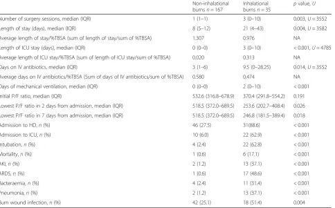
Compared to patients with cutaneous injury alone, patients with inhalational burns had more surgeries (3 ± 7.07 vs 1 ± 1.54,p= 0.003), increased length of stay
(21 days vs 8 days, p= 0.004) and higher in-hospital
mortality rate (17.1% vs 0.6%,p< 0.001) (Table3). Incidence of ARDS and AKI was 48.6% and 37.1%, re-spectively, compared to 0.6% and 1.2% in the patients without inhalational injury (p< 0.001). Patients with inha-lational injury had increased incidence of bacteraemia (31.4% vs 2.4%, p< 0.001), pneumonia (37.1% vs 1.2%,
p< 0.001) and burn wound infection (51.4% vs 25.1%,
p= 0.004). Inhalational injury predicted AKI with an ad-justed odds ratio (OR) of 17.43 (95% confidence interval (CI) 3.07–98.87,p< 0.001); ARDS, OR of 106.71 (95% CI 12.73–894.53, p< 0.001) and pneumonia, OR of 13.87 (%95 CI 2.32–82.94,p= 0.004) (Table4).
Patients with inhalational burns had a higher %TBSA. Seventeen out of 35 patients fitted the Berlin criteria (P/F ratio < 300 and within 7 days). This is reflected in Table4
(lowest P/F ratio < 300). Within the group of PIB, those with P/F ratios < 300 in the first week had a higher in-hospital mortality (29.4%) compared to those with P/F ratios > 300 in the first week (5.56%) (p= 0.08). We could not get statistical significance when adjusted for %TBSA and for both age and %TBSA (Table5).
Bacteriology
In patients with infective complications, cultures were taken and the number of positive cultures for a microbe was expressed as a percentage of all positive cultures (Fig.2).
In patients with inhalational injury, Acinetobacter
baumannii (A. baumannii) was the most frequently cul-tured in sputum, tissue and blood cultures.
Pneumonia was most frequently caused byA.
bauman-nii followed by Pseudomonas aeruginosa (P. aeruginosa)
andKlebsiella pneumoniae (K. pneumoniae). Table 1Symptoms and signs suggestive of inhalational injury
Symptoms and signs of inhalational injury Nasoendoscopy
•Hoarseness of voice
•Sore throat
•Dyspnea
•Painful respirations
•Carbonaceous sputum
•Tachypnea
•Stridor
•Facial burns/mucosal burns of lips and mouth
•Singed facial hair/nasal hair
•Soot in nostrils or mouth
•Airway erythema/edema
•Soot in airways
Table 3Clinical outcomes, complications and mortality of the burn patients
Non-inhalational burnsn= 167
Inhalational
burnsn= 35 p
value,U
Number of surgery sessions, median (IQR) 1 (1–1) 3 (0–10) 0.003,U= 3552
Length of stay (days), median (IQR) 8 (5–12) 21 (4–43) 0.004,U= 3582
Average length of stay/%TBSA (sum of length of stay/sum of %TBSA) 1.307 0.976 NA
Length of ICU stay (days), median (IQR) 0 (0–0) 3 (0–10) < 0.001,U= 4785
Average length of ICU stay/%TBSA (sum of length of ICU stay/sum of %TBSA) 0.020 0.313 NA
Days on IV antibiotics, median (IQR) 3 (1–6) 9.5 (0–28.25) 0.014,U= 3552
Average days on IV antibiotics/%TBSA (Sum of days of IV antibiotics/sum of %TBSA) 0.580 0.474 NA
Days of mechanical ventilation, median (IQR) 0 (0–0) 2 (0–10) < 0.001
Initial P/F ratio, median (IQR) 532.6 (316.8–678.9) 370.4 (291.8–554.2) 0.191
Lowest P/F ratio in 2 days from admission, median (IQR) 518.5 (372.0–689.5) 253.6 (202.7–408.4) 0.026
Lowest P/F ratio in 7 days from admission, median (IQR) 518.5 (372.0–689.5) 246.8 (181.5–389.4) 0.018
Admission to HD,n(%) 46 (27.5) 31(88.6) < 0.001
Admission to ICU,n(%) 10 (6.0) 22 (62.9) < 0.001
Intubation,n(%) 4 (2.4) 22 (62.8) < 0.001
Mortality,n(%) 1 (0.6) 6 (17.1) < 0.001
AKI,n(%) 2 (1.2) 13 (37.1) < 0.001
ARDS,n(%) 1 (0.6) 17 (48.6) < 0.001
Bacteraemia,n(%) 4 (2.4) 11 (31.4) < 0.001
Pneumonia,n(%) 2 (1.2) 13 (37.1) < 0.001
Burn wound infection,n(%) 42 (25.1) 18 (51.4) 0.004
%TBSApercentage of total body surface area,ARDSacute respiratory distress syndrome,AKIacute kidney injury,IVintravenous,ICUintensive care unit, NAnot applicable,HDhigh dependency unit,IQRinterquartile range,P/FPaO2 /FiO2
Table 2Patient characteristics included in the study
Demographics All patients
n(%)
Patients with inhalational injuryn(%)
Patients without inhalational
injuryn(%) p
value,U
Number 202 (100) 35 (17.3) 167 (82.7)
Age (years)
Mean (SD) 43.1 (16.7) 38.8 (17.25) 44.1 (12.78)
Range 16–86 18–69 16–86
Median (IQR) 42 (29–56) 43(29–58) 35 (30–48) 0.176,U= 2451
%TBSA, median (IQR) 5 (2.38–13.0) 5 (2.5–12) 9 (0–66) 0.036,U= 3582
Facial burns 72 (35.6) 18 (51.4) 54 (32.3) 0.032
Upper body burns 162 (80.2) 21 (60.0) 141 (84.5) 0.001
Sex
Male 128 (63.4) 26 (74.2) 102 (61.1)
Female 74 (36.6) 9 (25.7) 65 (38.9)
Race
Chinese 116 (57.4) 15 (42.8) 101(60.5)
Indian 19 (9.4) 3 (8.6) 16 (9.6)
Malay 22 (10.9) 4 (11.4) 18 (10.8)
Others 45 (22.2) 13 (37.1) 32 (19.2)
Table 4Adjusted odds ratios (OR) of getting poorer clinical outcomes compared to the median, adjusted for age and %TBSA (except age—adjusted for %TBSA, %TBSA—adjusted for age only)
Non-inhalational burnsn(%)
Inhalational burnsn(%) pvalue Adjusted OR* 95% CI for OR pvalue
%TBSA > median,5 92 (55.1%) 24 (68.6%) 0.142 1.66 0.76–3.64 0.204
Age > median, 42 (years) 88 (52.7%) 14 (40.0%) 0.172 0.71 0.31–1.62 0.414
Number of surgery sessions > median, 1 131 (78.4%) 23 (65.7%) 0.108 0.21 0.07–0.62 0.004
Length of stay > median, 8.5 (days) 79 (47.3%) 22 (62.9%) 0.094 1.12 0.47–2.67 0.797
Days on IV antibiotics > median, 4 83 (49.7%) 22 (62.9%) 0.157 0.86 0.36–2.11 0.749
Days of mechanical ventilation > median, 2.5 3 (1.8%) 15 (42.9%) < 0.001 18.38 2.97–113.68 0.002
Initial P/F ratio < median, 394.02 3 (1.8%) 14 (40.0%) < 0.001 44.85 9.10–221.13 < 0.001
Lowest P/F ratio < median, 343.88 1 (0.6%) 15 (42.9%) < 0.001 140.58 13.14–1504.25 < 0.001
Lowest P/F ratio < 300 1 (0.6%) 17 (48.6%) < 0.001 480.70 28.47–8115.89 < 0.001
Mortality 1 (0.6%) 6 (17.1%) P/F<300:5/17(29.4%)
P/F>300:1/18 (5.6%)
< 0.001 0.604 0.01–44.53 0.818
*Odds ratio adjusted for %TBSA and age
%TBSApercentage of total body surface area,CIconfidence interval,P/FPaO2 /FiO2,IVintravenous
Table 5Adjusted odds ratios of complications in patients with inhalational injury using logistic regression analysis
Inhalational injury Inhalational injury, adjusted for %TBSA
Inhalational injury, adjusted for %TBSA and age (except %TBSA, adjusted for age only)
OR 95% CI pvalue Adjusted OR 95% CI pvalue Adjusted OR 95% CI pvalue
%TBSA > median, 5 1.78 0.82–3.87 0.146 – – – 1.66 0.76–3.64 0.204
Age > median, 42 (years) 0.60 0.29–1.26 0.175 0.71 0.31–1.62 0.414 – – –
All complications 8.33 3.62–19.17 < 0.001 5.31 2.17–12.98 < 0.001 6.27 2.49–15.77 < 0.001
Intubation 68.96 20.65–230.3 < 0.001 67.19 12.47–361.89 < 0.001 86.99 14.22–544.43 < 0.001
Pneumonia 48.75 10.30–230.55 < 0.001 13.87 2.32–82.94 0.004 35.51 3.90–323.07 0.002
ARDS 156.78 19.69–1248.17 < 0.001 106.71 12.73–894.53 < 0.001 480.70 28.47–8115.89 < 0.001
AKI 48.75 10.31–230.55 < 0.001 17.43 3.07–98.87 < 0.001 31.71 4.38–229.7 0.001
Bacteraemia 18.68 5.50–63.39 < 0.001 2.39 0.38–15.04 NS 2.56 0.40–16.57 NS
Burn wound infection 3.15 1.49–6.67 0.003 1.28 0.49–3.35 NS 1.35 0.52–3.54 NS
Mortality 34.35 3.99–295.86 0.001 0.38 0.01–11.29 NS 0.60 0.01–44.53 NS
HD admission 20.39 6.82–60.95 < 0.001 15.70 4.87–50.62 < 0.001 15.55 4.82–50.20 < 0.001
ICU admission 26.57 10.41–67.90 < 0.001 16.26 5.23–50.57 < 0.001 16.63 5.28–52.34 < 0.001
Number of surgery sessions > median, 1
0.53 0.24–1.16 0.111 0.22 0.08–0.64 0.005 0.21 0.07–0.62 0.004
Length of stay > median, 8.5 1.89 0.89–3.99 0.098 1.05 0.44–2.47 0.915 1.12 0.47–2.67 0.797
Days on IV antibiotics > median, 4 1.71 0.81–3.63 0.160 0.84 0.35–2.04 0.699 0.86 0.36–2.11 0.749
Days of mechanical ventilation > median 2.5
41.00 10.91–154.04 < 0.001 11.94 2.34–60.8 0.003 18.38 2.97–113.682 0.002
Initial P/F ratio < median 394.02 36.45 9.67–137.40 < 0.001 28.89 7.02–118.89 < 0.001 44.85 9.10–221.13 < 0.001
Lowest P/F ratio in 2 days < median, 343.88
124.50 15.61–993.29 < 0.001 78.88 9.28–670.76 < 0.001 140.58 13.14–1504.25 < 0.001
These three species were also the most common in
the patients with bacteraemia together with Candida
tropicalis,EnterobacterandEnterococcus faecium. In tis-sue cultures,A. baumanniiandP. aeruginosawere most common followed byStaphylococcus aureus (S. aureus).
A. baumannii was less common in patients without inhalational injury.
In patients without inhalational injury, burn wound
in-fections were most commonly due to S. aureus followed
byP. aeruginosaandA. baumannii. In those with inhala-tional injury, A. baumanniiandP. aeruginosa were more common.
Discussion
Burn management has improved over the past decades with advances in fluid resuscitation, nutritional support, pulmonary care and infection control practices. This includes improvements in wound care/dressing, such as our previously described enhanced total body wrap tech-nique using negative pressure wound therapy, which protects the wound from micro-organisms and provides an efficient channel to clear excessive exudate while
keeping the wounds moist [15]. This reduces frequency of dressing changes and lowers infection rates through prevention of strikethrough [15]. Of note, incidence of burn wound infections in our centre have been reducing over the years, from 50.2% in a 2003–2005 study [5] to 29.7% in our study. Currently, infectious and respiratory complications remain a leading cause of morbidity. In
burns cases with %TBSA of 40–75%, almost all deaths
are due to sepsis from infectious complications and/or inhalation injury [16].
It is well-reported that inhalational injury results in lon-ger stays and higher mortality which results in significant economic burden. In the current study, we prospectively followed patients in a burn unit and quantified the impact of inhalational injury on clinical outcomes and complica-tions. All patients were managed uniformly using our in-stitution’s burns protocol.
Mortality
Our study found that presence of inhalational injury was associated with increased mortality. The overall in-hospital mortality was 3.5%. In-hospital mortality in patients with inhalational injury was 17.1%, compared to those without inhalational injury of 0.6%.
The benchmark inhalational mortality is high and ranges from 9.5 to 46.6% internationally. Typically, pa-tients with respiratory failure from inhalational burns have up to 50% risk of mortality. Our mortality rate of 17.1% (6 patients) was likely due to concomitant respira-tory failure, large %TBSA burns (> 40%) and accompany-ing renal failure, accountaccompany-ing for their high mortality rate. The 6 patients were 23–48 years old, typically with se-vere Endorf-Gameli type 3 or 4 bronchoscopy findings with delayed resuscitation and debridement (72 h). The crux in further reduction of inhalational mortality we believe lies in early aggressive burns resuscitation, cover-age and aggressive intensive care unit (ICU) respiratory care to modulate the systemic inflammatory response syndrome (SIRS) inflammatory response.
Statistical significance was not reached for mortality when adjusting for %TBSA, possibly due to the low mor-tality in the group without inhalational injury.
Inhalational injury has been shown to be a signifi-cant predictor of mortality in various multivariable retrospective studies on the prognostic factors in burn patients (Table6).
Shirani et al. estimated that the burn-related death rate is 20% higher in patients with combined inhalation injury and cutaneous burns than in those with cutaneous burns alone [17]. In a 2014 Taiwanese study, 21,791 burns pa-tients from 44 hospitals were retrospectively reviewed. The overall mortality rate was 2.1%, and inhalation injur-ies were found in 7.9% of the patients. The mortality rate of inhalation and non-inhalation injury group was 17.9%
Table 6 Retrospective studies on inhalational injury, mortality, length of stay and other clinical outcomes O ur study SGH Chong Texas Shi rani 19 87 [ 17 ] Uta h Ho llingsed 1993 [ 18 ] North Carol ina Smith 1994 [ 19 ] Toky o Suz uki 2005 [ 2 ] Egyp t El-He lbawy 20 11 [ 4 ] Taiwan Chen 2014 [ 1 ] USA Anand 2015 [ 3 ] n 20 2 10 58 52 9 1447 5560 281 21,791 506,628 Type of study Pros pective st udy Re trospective sing le centr e Re trospe ctive sing le centr e Retrospe ctive single cen tre Retros pective mu lticentre Retros pective sing le centre Retros pective mult icentre Retros pective multic entre PIB (%) 17 .3 35 5. 7 19.6 30.4 46.3 7.9 3.47 Age (years) (m edian) O verall: 42 PIB: 43 PCB : 35 PCB : 27 Abnorm al xenog raphy : 37 Abno rmal bron choscop e: 39 (me an) PIB: 20.2 RF : 36.6 (mean ) Overall: 30 (3 mon ths – 93 years ) (me an (SD)) Overall: 40.1 (26.2) PIB: 49 .0 (20. 5) PCB: 36.2 (27.4) – (me an(SD)) Overall: 30 .9 (22. 6) (me an) Overall: 30 %TB SA (m edian) O verall: 5 PIB: 5 PCB : 9 PCB : 23 Abno rmal xenog raphy : 37 Abno rmal bron choscop e:50 (me an) PIB: 16.2 RF : 40.4 (mean )18% (0.4 – 100%) me an (SD )) Overall: PIB: 29.9 (30.3 ) PCB: 16.1 (17.3) – mean Overall: 12.2% – Mortality (%) O verall 3.5 PIB: 17.1 PCB : 0.6 O verall: 22.7 PIB: 46.6 PCB : 9.6 O verall: 6.2 PIB -witho ut RF:0 -with RF: 27 PCB : -witho ut RF: 3 -with RF: 50 Overall 9.5 PIB 31 PCB 4.3 Overall: 15.8 PIB: 33 .6 PCB: 8.1 Overall: 23 .1 PIB: 41 .5 PCB: 7. 2 Overall 2.1 PIB: 17.9 PCB: 0. 76 Overall 3.73 PIB: 4× increase in mort ality vs PCB Leng th of st ay (days) PIB: 21 PCB : 8 –
PIB -witho
ut RF: 17.8 -with RF: 42 .6
PCB -witho
and 0.7%, respectively, similar to our results [1]. In Suzuki et al.’s 17-year study, the mortality rate was 15.8% and multivariable analysis revealed inhalational injury to be the most important predictor of death [2]. In Smith et al's study, inhalational injury was a significant predictor for mortality, but the most important predictor of mortal-ity was still %TBSA [18].
As in Table6, the mortality ranges from 17% to 46.6% in inhalational burns. The reasons for high mortality in inhalational burns despite low %TBSA cutaneous burns is because of associated pneumonia, SIRS, sepsis and associated organ failure [1–3,17].
ARDS
Patients with inhalational injury had > 100 times increase in developing ARDS. Incidence of ARDS in patients with inhalational injury was 48.6%, compared to 0.6% in the other group. A 4-year review of 529 burns patients re-ported a 20% incidence of ARDS, compared to 2% in patients without inhalational injury (p< 0.001) [19].
During inhalational injury, the inflammatory response forms airway casts and cause V/Q mismatch, pulmonary edema and cell death, promoting airway occlusion and pulmonary dysfunction [1]. In patients with large cuta-neous burns, capillary hyper-permeability occurs not only at the injured site, but also in regions distant from the injury [20, 21] resulting in worsening of pulmonary edema. This systemic response is more severe when the thermal injury is associated with smoke inhalation [22].
AKI
Inhalational injury was also a predictor of AKI. In Coca’s retrospective study, inhalational injury was an independent predictor of AKI (OR = 3.6 (95% CI 1.76–7.34), p< 0.001) [23]. Other significant factors in multivariable analysis were catheter infections and sepsis. Both inhalational injury and AKI were independent predictors of mortality.
Pneumonia
In this study, inhalational injury was an independent risk factor for pneumonia. It was associated with 13 times higher likelihood of pneumonia, independent of %TBSA. Shirani et al.’s hallmark study quantitated that both inha-lational injury and pneumonia have contributions to mortality that are independent and additive [17]. In that study, expected mortality increased by a maximum of 20% in the presence of inhalation injury alone, 40% in the presence of pneumonia alone and 60% when both inhalation injury and pneumonia were present.
Length of stay
After adjusting for %TBSA (Table 3), ICU stay and
length of stay was longer for inhalational burns due to respiratory complications. Days of intravenous (IV)
antibiotics were comparable. This is a result of more op-erative sessions, complications and longer duration of IV antibiotics. We postulate the increase in operative ses-sions to be due to increased wound infections requiring debridement and wound conversion due to systemic sepsis, cytokine alteration and inflammatory response. A study using the National Inpatient Sample database of 506,628 admissions found inhalational injury was a factor for length of stay (OR = 2.01 (95% CI 1.84–2.21),p< 0.001) [3].
Bacteriology
Patients with inhalational burns were infected with gram-negative organisms, common agents for invasive infection. The prevalence ofA. baumanni in our tropical burns unit has been previously reported [5, 24]. A. bau-manni is endemic to the hospital because of the tropical climates and the particular high survivability of Acineto-bacter even in adverse conditions [24] The most common agents wereA. baumannifollowed by methicillin-resistant
Staphylococcus aureus (MRSA) then P. aeruginosa [24].
Interestingly, A. baumanni was the most common
aetiologic agent in our patients with inhalational burns, but not in patients without inhalational burns.
Patients with burns are exposed to infections from loss of the skin barrier. In patients with inhalational burns, the respiratory tract is also damaged. The longer length of stay in hospital and duration of IV antibiotics may account for prevalence of resistant gram-negative noso-comial infections in inhalational burns patients. In an Indian study, gram-positive organisms were predomin-antly cultured during the first week of admission whereas gram-negative organisms with increasing levels of
resist-ance were common from the second week onwards [16].
In patients with major burns, we may occasionally find multi-resistance organisms developing at 1–2 weeks of stay, which may be caused by antibiotic use.
Limitations
criteria. We have optimised the variability by ensuring final diagnostic assessments are by attendings.
Bronchoscopy is an objective means of diagnosing and grading inhalational injury—with several grading systems proposed. This can potentially allow inter-centre com-parisons should a standardised and validated grading system be accepted [25]. However, if bronchoscopy were to be done for diagnostic purposes in all patients in our study, some patients would be subjected to a bronchos-copy not otherwise indicated for diagnostic/therapeutic purposes. In patients with unlikely lower airway injury, it may not be necessary to pass the endoscope below the true vocal folds for negative diagnosis.
Bronchoscopy may be uncomfortable for the patient and requires careful anaesthesia of the larynx to avoid obstruc-tion due to laryngospasm in a potentially inflamed airway. Furthermore, some institutions may not have the equip-ment or expertise for bronchoscopy. Fiberoptic laryngos-copy is better tolerated by the patients, requires minimal medication, and is often more readily available [26]. One study has addressed this with an objective decision tree that can be readily replicated in different centres, including bi-level endoscopic examination of the oro-rhino-pharynx and tracheobronchial tree and also clinical criteria [25]. Studies have discussed using bronchoscopy to evaluate se-verity of inhalational burns [27,28]. Several scales of sever-ity have been proposed; these have not been able to predict clinical outcomes [25,27].
Further bacteriology studies can include analysis of time from injury to the date of culture for bacteriology and resistance of bacteria cultured.
Conclusion
Inhalational injury is associated with increased length of stay, morbidity and complications such as ARDS, AKI, pneumonia and infection. Mortality is higher (29.4% to 5.6%) if P/F is < 300. Counselling patients and their fam-ily regarding the associated morbidity is essential. Statis-tical significance was not reached when adjusted for %TBSA and age. Larger sample sizes are needed. Inhala-tional burn care is an important step towards advancing burn management, given its association with morbidity. Bronchoscopy classification is also encouraged as this was lacking in this study. This requires early recognition of inhalational burns, early lung recruitment protocol, early intubation, early ICU care, earlier lavage and earlier burns debridement to reduce the SIRS response.
Abbreviations
AKI:Acute kidney injury; APRV: Airway pressure release ventilation; ARDS: Acute respiratory distress syndrome; CI: Confidence interval; ICU: Intensive care unit; IV: Intravenous; MRSA: Methicillin-resistant
Staphylococcus aureus; NS: Not significant; OR: Odds ratio; PCB: Patients with only cutaneous burns; PEEP: Peak end expiratory pressure; PIB: Patients with inhalational burns; REDCap: Research Electronic Data Capture; RF: Respiratory failure; %TBSA: Percentage of total body surface area
Acknowledgements
The authors would like to thank Professor Tai Bee Choo for her statistical guidance.
Availability of data and materials
Database used during the current study is available from the corresponding author upon reasonable request.
Authors’contributions
CSJ designed the research. KYO and RT carried out analysis of the data for this study with the database provided by KYO and CSJ. CSJ, KYO and RT participated in drafting the manuscript. All the authors read and approved the final manuscript. DSR participated in writing about the treatment principles—particularly the intubation and ventilation principles.
Ethics approval and consent to participate
Our research was done with the ethics approval from the SingHealth Centralised Institutional Review Board (CIRB) with CIRB reference number: 2014/2130D.
Consent for publication
Not applicable
Competing interests
The authors declare that they have no competing interests.
Author details
1Department of Plastic, Reconstructive and Aesthetic Surgery, Singapore
General Hospital, Level 5, Academia, Singapore 169865, Singapore. 2
Department of Anaesthesiology, Singapore General Hospital, Level 5, Academia, Singapore 169865, Singapore.
Received: 17 August 2017 Accepted: 16 July 2018
References
1. Chen MC, Chen MH, Wen BS, Lee MH, Ma H. The impact of inhalation injury in patients with small and moderate burns. Burns. 2014;40(8):1481–6. 2. Suzuki M, Aikawa N, Kobayashi K, Higuchi R. Prognostic implications of
inhalation injury in burn patients in Tokyo. Burns. 2005;31(3):331–6. 3. Veeravagu A, Yoon BC, Jiang B, Carvalho CM, Rincon F, Maltenfort M, et al.
National trends in burn and inhalation injury in burn patients: results of analysis of the nationwide inpatient sample database. J Burn Care Res. 2015; 36(2):258–65.
4. El-Helbawy RH, Ghareeb FM. Inhalation injury as a prognostic factor for mortality in burn patients. Ann Burns Fire Disasters. 2011;24(2):82–8. 5. Chong SJ, Song C, Tan TW, Kusumawijaja G, Chew KY. Multi-variate analysis
of burns patients in the Singapore General Hospital Burns Centre (2003-2005). Burns. 2009;35(2):215–20.
6. American Burn Association. Advance Burn Life Support provider manual. 2011. 7. Acute Respiratory Distress Syndrome Network, Brower RG, Matthay MA,
Morris A, Schoenfeld D, Thompson BT, Wheeler A. Ventilation with lower tidal volumes as compared with traditional tidal volumes for acute lung injury and the acute respiratory distress syndrome. N Engl J Med. 2000; 342(18):1301–8.
8. Society of Critical Care Medicine. Surviving Sepsis Campaign: International Guidelines for Management of Sepsis and Septic Shock: 2016.
9. Zhou Y, Jin X, Lv Y, Wang P, Yang Y, Liang G, et al. Early application of airway pressure release ventilation may reduce the duration of mechanical ventilation in acute respiratory distress syndrome. Intensive Care Med. 2017; 43(11):1648–59.
10. Andrews PL, Shiber JR, Jaruga-Killeen E, Roy S, Sadowitz B, O’Toole RV, et al. Early application of airway pressure release ventilation may reduce mortality in high-risk trauma patients: a systematic review of observational trauma ARDS literature. J Trauma Acute Care Surg. 2013;75(4):635–41.
11. Kaplan LJ, Bailey H, Formosa V. Airway pressure release ventilation increases cardiac performance in patients with acute lung injury/adult respiratory distress syndrome. Crit Care. 2001;5(4):221–6. Epub 2001 Jul 2 12. ARDS Definition Task Force, Ranieri VM, Rubenfeld GD, Thompson BT,
13. Mehta RL, Kellum JA, Shah SV, Molitoris BA, Ronco C, Warnock DG, Levin A. Acute Kidney Injury Network: report of an initiative to improve outcomes in acute kidney injury. Crit Care. 2007;11(2):R31.
14. Greenhalgh DG, Saffle JR, Holmes JH 4th, Gamelli RL, Palmieri TL, Horton JW, et al. American Burn Association consensus conference to define sepsis and infection in burns. J Burn Care Res. 2007;28(6):776–90.
15. Low OW, Chong SJ, Tan BK. The enhanced total body wrap—the new frontier in dressing care for burns. Burns. 2013;39(7):1420–2. 16. Ghai S, Sachdeva A, Mahajan R, Dogra S, Soodan S, Mahajan B.
Bacteriological and antibiotic susceptibility profile of aerobic burn wound isolates at a tertiary care institute in Northern India. J Appl Environ Microbiol. 2015;3(4):95–100.
17. Shirani KZ, Pruitt BA Jr, Mason AD Jr. The influence of inhalation injury and pneumonia on burn mortality. Ann Surg. 1987;205(1):82–7.
18. Smith DL, Cairns BA, Ramadan F, Dalston JS, Fakhry SM, Rutledge R, et al. Effect of inhalation injury, burn size, and age on mortality: a study of 1447 consecutive burn patients. J Trauma. 1994;37(4):655–9.
19. Hollingsed TC, Saffle JR, Barton RG, Craft WB, Morris SE. Etiology and consequences of respiratory failure in thermally injured patients. Am J Surg. 1993;166(6):592–6.
20. Traber D, Herndon D, Enkhbaatar P, et al. The pathophysiology of inhalation injury. D. Herndon (Ed.), Total burn care. 4th ed. Edinburgh: Saunders Elsevier; 2012. p. 219–28.
21. Jones SW, Williams FN, Cairns BA, Cartotto R. Inhalation injury:
pathophysiology, diagnosis, and treatment. Clin Plast Surg. 2017;44:505–11. 22. Soejima K, Schmalstieg FC, Sakurai H, Traber LD, Traber DL.
Pathophysiological analysis of combined burn and smoke inhalation injuries in sheep. Am J Physiol Lung Cell Mol Physiol. 2001;280(6):L1233–41. 23. Coca SG, Bauling P, Schifftner T, Howard CS, Teitelbaum I, Parikh CR. Contribution of acute kidney injury toward morbidity and mortality in burns: a contemporary analysis. Am J Kidney Dis. 2007;49(4):517–23. 24. Chim H, Tan BH, Song C. Five-year review of infections in a burn intensive
care unit: high incidence of acinetobacter baumannii in a tropical climate. Burns. 2007;33(8):1008–14.
25. Ikonomidis C, Lang F, Radu A, Berger MM. Standardizing the diagnosis of inhalation injury using a descriptibe score and mucosal injury criteria. Burns. 2011;38:513–9.
26. Muehlberger T, Kunar D, Munster A, Couch M. Efficacy of fiberoptic laryngoscopy in the diagnosis of inhalation injuries. Arch Otolaryngol Head Neck Surg. 1998;124(9):1003–7.
27. Spano S, Hanna S, Li Z, Wood D, Cartotto R. Does bronchoscopic evaluation of inhalation injury severity predict outcome? J Burn Care Res. 2016;37:1–11. 28. Endorf FW, Gamelli RL. Inhalation injury, pulmonary perturbations, and fluid