Open Access
Software
Integration and acceleration of virtual microscopy as the key to
successful implementation into the routine diagnostic process
Stephan Wienert
1, Michael Beil
2, Kai Saeger
1, Peter Hufnagl
3and
Thomas Schrader*
3Address: 1VMscope GmbH, Chariteplatz 1, 10117 Berlin, Germany, 2Lausitz University of Applied Sciences, Großenhainer Str. 57, 01968 Senftenberg, Germany and 3Institue of Pathology, Charité – University Hospital Berlin, Chariteplatz 1, 10117 Berlin, Germany
Email: Stephan Wienert - [email protected]; Michael Beil - [email protected]; Kai Saeger - [email protected]; Peter Hufnagl - [email protected]; Thomas Schrader* - [email protected]
* Corresponding author
Abstract
Background: The virtual microscopy is widely accepted in Pathology for educational purposes and teleconsultation but is far from the routine use in surgical pathology due to the technical requirements and some limitations. A technical problem is the limited bandwidth of a usual network and the delayed transmission rate and presentation time on the screen.
Methods: In this study the process of secondary diagnostic was evaluated using the "T.Konsult Pathologie" service of the Professional Association of German Pathologists within the German breast cancer screening program. The characteristics of the access to the WSI (Whole Slide Images) have been analyzed to explore the possibilities of prefetching and caching to reduce the presentation and transfer time with the goal to increase user acceptance. The log files of the web server were analyzed to reconstruct the movements of the pathologist on the WSI and to create the observation path. Using a specialized tool the observation paths were extracted automatically from the log files. The attributes linearity, 3-point-linearity, changes per request, and number of consecutive requests were calculated to design, develop and evaluate different caching and prefetching strategies.
Results: The analysis of the observation paths showed that a complete accordance of two image requests is a very rare event. But more frequently a partial covering of two requested image areas can be found. In total 257 diagnostic paths from 131 WSI have been extracted and analysed. On average a diagnostic path consists of 16 image requests and takes 189 seconds between first and last image request. The mean linearity was 0,41 and the mean 3-point-linearity 0,85. Three different caching algorithms have been compared with respect to hit rate and additional image requests on the WSI server. Tests demonstrated that 95% of the diagnostic paths could be loaded without any deletion of entries in the cache (cache size 12,2 Megapixel). If the image parts are stored after JPEG compression this complies with less than 2 MB.
Discussion: WSI telepathology is a technology which offers the possibility to break the limitations of conventional static telepathology. The complete histological slide may be investigated instead of sets of images of lesions sampled by the presenting pathologist. The benefit is demonstrated by the high diagnostic security of 95% accordance between first and second diagnosis.
Published: 9 January 2009
Diagnostic Pathology 2009, 4:3 doi:10.1186/1746-1596-4-3
Received: 19 March 2008 Accepted: 9 January 2009
This article is available from: http://www.diagnosticpathology.org/content/4/1/3 © 2009 Wienert et al; licensee BioMed Central Ltd.
1) Costs: The financial expanses for scanning and storing are currently very high. A slide scanner costs between 60 and 120 TE. A scanned slide has a file size between hun-dreds of megabytes and several gigabytes. Thus virtual microscopy is used almost by university institutes only.
2) Scanning time: Despite several technical improve-ments, the scanning time needed for a set of slides does not yet satisfy the requirements of daily routine work. The scanning time lies between 1 and 5 minutes for a biopsy and between 5 and 20 minutes for a surgical specimen
3) Speed of virtual microscopes: With respect to image transfer and display times the working speed of virtual microscopy is lower in comparison to conventional microscopy. Another critical point is the lack of appropri-ate user interface devices. Conventional computer mouse is inadequate.
Caching and prefetching may speed up image loading, the bottle neck in virtual microscopy. Nevertheless the posi-tive effects of different prefetching and caching technolo-gies depend on the user's behavior. To our knowledge no reports on such user data have been ever published. Nobody knows the diagnostic paths of pathologists exploring histological slides.
From the technical point of view prefetching is useful to load expected areas of a WSI before the user asks for them. Caching holds the information of areas already seen in the fast memory of the computer (cache). The effectiveness depends directly on the behavior of the pathologists. A prefetch is preferred in case of a high probability of fore-cast the next region of WSI. The caching mechanism is val-uable if the user goes several times through the same area or compares current areas with areas already seen.
The telepathological diagnostic process differs between the three application cases primary, secondary and tertiary diagnosis. In case of the primary telepathology the pathol-ogist has to scan all slides and the complete area of each WSI to find the right diagnosis because he or she sees every case the first time and has to work like under the same conditions as in conventional microscopy.
microscopy offers the possibility to access every part of the slide in highest magnification and best quality.
In this study the process of secondary diagnostic was eval-uated using the "T.Konsult Pathologie" service of the Pro-fessional Association of German Pathologists within the German breast cancer screening program [16]. The charac-teristics of the access to the virtual slides was analyzed to explore the possibilities of prefetching and caching to reduce the presentation and transfer time with the goal to increase user acceptance.
Methods
All individual slides of 149 cases from the routine diag-nostic process were scanned with three scanners (Olym-pus Slide, Zeiss Mirax, Hamamatsu Nanozoomer). 247 WSI were created and stored on the image server. Only biopsies and excisions on standard glass slides with 25 mm × 75 mm × 1 mm were included in this study.
The cases were prepared for the telepathology consulta-tion process in the T.Konsult – Server of the Professional Association of German Pathologists and assigned ran-domly to 37 pathologists for a confirmation of the pri-mary diagnosis (second opinion). Following clinical data were added to each case for secondary diagnostic process: local case number, gender, age, localization of the lesion, working diagnosis and tumor code expressed in ICD-O. The access to the WSI was realized with "Slide Link List" – a database tool for administration (VMscope GmbH Ber-lin). After registration and login pathologists reviewed the case information and the WSI's of his or her assigned cases (fig. 1).
The German teleconsultation service T. Konsult Patholo-gie runs on a server with an Intel Xeon processor, 512 MB RAM and 4 hard drives with 80 GB in a RAID system. Win-dows 2003 Server system was used as operating system and Microsoft SQL-Server 2000 worked as database engine. Microsoft Internet Information Services operated as web server.
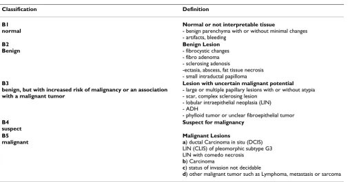
classification scheme is used to unify diagnoses and to offer comparability. The main decision criterion is the relation to the malignant potential (see table 1).
The log files of the web server were analyzed to reconstruct the movements of the pathologist on the WSI and to cre-ate the observation path [5]. Using a specialized tool the observation paths were extracted automatically from the log files (fig. 2). The following attributes were calculated
- Linearity – the Euclidean distance between the starting and the end point of the observation path divided by the length of the way between these points
- 3-point-linearity – linearity of all sections of the observa-tion paths, consisting of three following image requests.
- Changes per request concerning the position in the WSI, the magnification, the size of the requested field of view
- Number of the consecutive requests with the same mag-nification
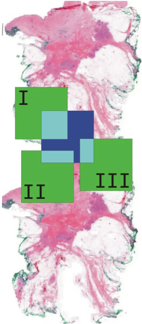
The analysis of the observation paths showed that a com-plete accordance of two image requests is a very rare event. But more frequently a partial covering of two requested image areas can be found. This is the reason for the success of caching algorithms which hold parts of images in the fast memory to load only the missing image parts from the image server. Three different caching algorithms have been developed (fig. 3):
A1: every covering image part in the cache is used
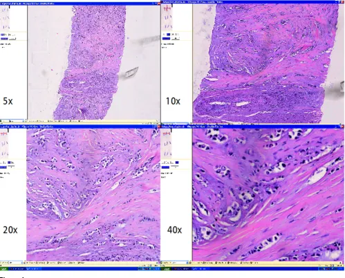
WSI-Viewer with different magnifications
Figure 1
A2: only the largest covering image part in the cache is used
A3: limitation on a certain number of arising parts of images per image request.
The algorithms have been evaluated by their hit rate (amount of pixels from the cache on the number of all requested pixels) and the number of additional requests on the WSI server. Such additional requests appear if only
a part of the image request can be satisfied by the cache content. The remaining area must be requested from the WSI server in form of separate rectangles. In this case latency periods and protocol overhead (i.e. HTTP, TCP/ IP) of the network is multiple, which is a direct drawback of caching.
As a result of the analysis of the diagnostic paths we found that between two requests often only the position within the WSI is changed but not the resolution (fig 4). The probability of a change of the resolution between two image requests is about 20%. Consequently it makes sense to load an image of identical resolution after two requests with the same resolution have been already placed (prefetch). The linearity of three consecutive image requests was high in the all diagnostic paths which have been analysed. That is why it makes sense to load an image in the same moving direction β and step width as in the image requests before.
Results
We compared conventional original diagnosis with telepathological diagnosis on the basis of WSI. There was a accordance of 94,56% between conventional and WSI diagnosis with respect to the B-classification. The image quality was good or excellent for 79,84% of the images. The time needed for the diagnostic process of one case was lower than 20 minutes.
with a malignant tumor - scar, complex sclerosing lesion - lobular intraepithelial neoplasia (LIN) - ADH
- phylloid tumor or unclear fibroepithelial tumor
B4 suspect
Suspect for malignancy
B5 malignant
Malignant Lesions
a) ductal Carcinoma in situ (DCIS) LIN (CLIS) of pleomorphic subtype G3 LIN with comedo necrosis
b) Carcinoma
c) status of invasion not decidable
d) other malignant tumor such as Lymphoma, metastasis or sarcoma
Demonstration and analysis tool
Figure 2
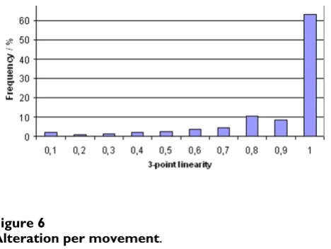
In total 257 diagnostic paths from 131 WSI have been extracted and analysed. On average a diagnostic path con-sists of 16 image requests and takes 189 seconds between first and last image request. The results of the features which have been calculated for each path are shown in fig-ures 5, 6, 7. The mean linearity was 0,41 and the mean 3-point-linearity 0,85.
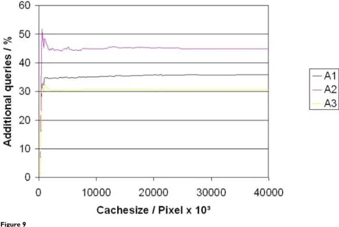
The three different caching algorithms have been com-pared with respect to hit rate and additional image requests on the WSI server (fig. 8, 9). The algorithm A3 was limited to a maximum of 5 image parts for one image request. Tests demonstrated that 95% of the diagnostic paths could be loaded without any delete of entries in the cache (cache size 12,2 Megapixel). If the image parts are stored after JPEG compression this complies with less than 2 MB.
About 22% of the total image area of a diagnostic path may be loaded already by prefetching. An amount of 25,89% of the prefetched image area is needed and
ana-Caching Algorithms (Green area I, II, III are viewed areas and stored at the cache
Figure 3
Caching Algorithms (Green area I, II, III are viewed areas and stored at the cache. Blue area is the new requested view. Turquoise areas are the overlaps of viewed areas and requested area.) A1 uses the overlaps of I and II, A2 uses only the overlap I and in A3 only the overlaps I and II are used.
Linearity
Figure 4 Linearity.
3-point linearity
Figure 5
nostic reliability of 95% accordance between first and sec-ond diagnosis. The remaining amount of 5% goes back to the diagnostic problem itself and has no correlation to the technology of WSI telepathology. Also conventional stud-ies show a discrepancy amount of that level [16].
The characteristics of the diagnostic path for the different diagnostic procedures were not evaluated yet. We assume that there are differences due to the fact that different questions should be solved by the pathologists. In case of primary diagnosis the pathologist has to classify the tumour and to describe the in-situ-parts of the lesions as well as the surgical margin. Different views with different magnifications have to be used and the approach should be more systematically.
In the secondary and tertiary diagnostic process the first diagnosis should be confirmed by the consultant. Some diagnostic aspects are not so important now. The main question for the consultant is whether he can accept the proposed diagnosis.
We recorded some special features. Mostly short diagnos-tic paths have been registered. There may be two reasons for that. These are firstly the bias with respect to the study circumstances and secondly the characteristic of the
diag-nostic process himself, if the primary diagnosis is already known. The diagnostic process may be very fast and based on low magnifications, if in the second opinion only a carcinoma diagnosis has to be confirmed. Therefore pathologist use only one or two magnifications and move the slide systematically in few directions. That is the rea-son for the high values in linearity and 3-point-linearity. This underlines the possibilities of virtual microscopy where fields of view may be several times larger than under the conventional microscope. This results in a lim-ited number of changes in position and resolution. The movements with high linearity indicate the meander like investigation of the slide as a preferred searching method in secondary diagnostic.
The possibilities to speed up the viewing process by cach-ing and prefetchcach-ing are immense. Up to 25% of the over-all investigated area may be presented without long Internet dependent loading times only by use of appropri-ate cache memory. This value may be increased up to 33% by combination of caching and prefetching. The real
pos-Alteration per movement
Figure 6
Alteration per movement.
Number of consecutive movements in the same magnifica-tion
Figure 7
Number of consecutive movements in the same magnification.
Cache hit ratio
sible time win depends on the characteristic of the Inter-net connection to the WSI server and is not simple to specify. The following figure 9 illustrates the reduction of the loading time in dependence of the Internet connec-tion quality under the assumpconnec-tion that 25% of the pixels may be taken from the cache. The table was calculated for image size of 743 × 685 pixel of 66,4 Kbyte size. This image size corresponds to the working area of the used vir-tual microscope and a monitor resolution of 1024 × 768 pixels.
For the calculation the transfer rate was taken as net usea-ble capacity. This is not attainausea-ble reachausea-ble in practice with respect to protocol overhead and latency periods. That means that the time savings in practical use would be higher.
Conclusion
The general procedure of investigating a histological slide with respect to the type of diagnosis – second opinion – was in the focus of our paper. We could show that the safety of diagnostic on WSI is comparable to the conven-tional diagnostic based on glass slides. Discrepancies go
back to problems with the difficulty of the diagnosis itself and not to technical problems with virtual microscopy.
The specific behaviour in the diagnostic process may be used for the improvement of the user acceptance of virtual microscopy. Strong reductions of loading times are possi-ble by using caching and prefetching.
Within further investigations we will analyse the user behaviour for other diagnostic procedures (primary and tertiary diagnostic) based on the investigation of diagnos-tic paths. Our goal is the development of special strategies for caching and prefetching with respect to the type of diagnosis and the diagnostic question.
Competing interests
The authors declare that they have no competing interests.
Authors' contributions
SW developed the algorithms and evaluated them. MB participated in the design of the study and evaluated the algorithms. KS developed the WSI interface and the server technology. PH designed the telepathology study and
Additional queries
Figure 9
Publish with BioMed Central and every scientist can read your work free of charge "BioMed Central will be the most significant development for disseminating the results of biomedical researc h in our lifetime."
Sir Paul Nurse, Cancer Research UK
Your research papers will be:
available free of charge to the entire biomedical community
peer reviewed and published immediately upon acceptance
cited in PubMed and archived on PubMed Central
yours — you keep the copyright
Submit your manuscript here:
http://www.biomedcentral.com/info/publishing_adv.asp
BioMedcentral
3. Ficsor L, Varga V, Berczi L, Miheller P, Tagscherer A, Wu ML, Tulassay Z, Molnar B: Automated virtual microscopy of gastric biop-sies. Cytometry B Clin Cytom 2006, 70(6):423-31.
4. Schrader T, Beil M, Schmidt D, Dietel M, Lindemann G: Virtual microscopy in medical research: Open European Nephrol-ogy Science Center (OpEN.SC). In SPIE: Medical Imaging 2007
San Diego, USA: SPIE; 2007.
5. Schrader T, Niepage S, Leuthold T, Saeger K, Schluns K, Hufnagl P, Kayser K, Dietel M: The diagnostic path, a useful visualisation tool in virtual microscopy. Diagn Pathol 2006, 1:40.
6. DICOM, Supplement 122: Specimen Identification and
Revised Pathology SOP Classes, W.G. 26, Editor. Digital Imaging and Communications in Medicine (DICOM). . 7. Bloodgood RA, Ogilvie RW: Trends in histology laboratory
teaching in United States medical schools. Anat Rec B New Anat
2006, 289(5):169-75.
8. Boutonnat J, Paulin C, Faure C, Colle PE, Ronot X, Seigneurin D: A pilot study in two French medical schools for teaching histol-ogy using virtual microscopy. Morphologie 2006, 90(288):21-5. 9. Fujita K, Crowley RS: The Virtual Slide Set – a curriculum
development system for digital microscopy. AMIA Annu Symp Proc 2003:846.
10. Glatz-Krieger K, Glatz D, Mihatsch MJ: Virtual slides: high-quality demand, physical limitations, and affordability. Hum Pathol
2003, 34(10):968-74.
11. Glatz-Krieger K, Spornitz U, Spatz A, Mihatsch MJ, Glatz D: Factors to keep in mind when introducing virtual microscopy. Vir-chows Arch 2006, 448(3):248-55.
12. Kayser K, Gortler J, Goldmann T, Vollmer E, Hufnagl P, Kayser G:
Image standards in Tissue-Based Diagnosis (Diagnostic Sur-gical Pathology). Diagn Pathol 2008, 3:17.
13. Kayser K, Gortler J, Metze K, Goldmann T, Vollmer E, Mireskandari M, Kosjerina Z, Kayser G: How to measure image quality in tis-sue-based diagnosis (diagnostic surgical pathology). Diagn Pathol 2008, 3(Suppl 1):S11.
14. Kayser K, Radziszowski D, Bzdyl P, Sommer R, Kayser G: Towards an automated virtual slide screening: theoretical considera-tions and practical experiences of automated tissue-based virtual diagnosis to be implemented in the Internet. Diagn Pathol 2006, 1:10.
15. Saeger K, Schmidt D: [Digital slide training portal: Training slides available on the Internet from the German division of the IAP.]. Pathologe 2006, 27(6):477-480.
16. Schrader T, Hufnagl P, Schlake W, Dietel M: Study of Efficiency of Teleconsultation: The Telepathology Consultation Service of the Professional Association of German Pathologists for the Screening Program of Breast. Verh Dtsch Ges Path 2005,
89:211-218.