SALIVARY STATUS OF DIABETIC CHILDREN
Vanya K. Veleganova1, Veselina K. Kondeva1, Yordanka I. Uzunova2, Kiril K. Simitchiev3 1
Dept. of Pediatric Dentisry, Faculty of Dental Medicine, Medical University, Plovdiv 2
Dept. of Chemistry, Facuity of Pharmacy, Medical University, Plovdiv 3
Dept. of Analytical Chemistry and Computer Chemisty, Paisii Hilendarski University, Plovdiv
Abstract
Saliva may provide us with clues for the evaluation of dental caries susceptibility and dental caries risk. The aim of this study was to compare salivary parameters: flow rate, pH, buffering capacity, glucose in saliva and bacterial count of Streptococcus Mutans and Lactobacillus in healthy and suffering from DM1 children and adolescents in Plovdiv region. The study was conducted on 128 children, including 68 children suffering from diabetes mellitus type 1 5-18 years of age,that were refered for treatment of dibetes mellitus to the Paediatric Clinic at Medical University in Plovdiv (groupII) and 60 controlsI (group I) aged 5 to 18 years. Metabolic control was determined for each diabetic child by percentage of glucosilated hemoglobin (HbA1C) 29 children were judged to be well controlled (HbA1C≤ 8%) 38 tric children (group IIA) were poorly controlled (HbA1C≥ 8%) (group IIB).The following salivary parameters are determined: salivary flow rate, salivary buffer capacity, salivary pH salivary glucose, microbial counts of Streptococcus mutans and Lactobacillus. The statistical analysis was performed using χ2
-test and Fisher’s exact test. Saliva is an easy to collect and study fluid that could help diagnose and monitor systematic diseases, such as diabetes.
Key words: children, diabetes mellitus type 1, saliva
1.1. Introduction
Diabetes mellitus (DM) is a group of metabolic diseases characterized by hyperglycemia due to insulin deficiency, a resistance to its peripheral action or both. This disease is classified as type 1 diabetes or type 2. Type 1 diabetes accounts for 10% of cases and is caused by the autoimmune destruction of the beta cells of the islets of Langerhans of the pancreas, which leads to a total lack of insulin (World Health Organization, 2011). Cases of type 1 diabetes also increased significantly across countries (Soltèsz G et al., 2003).
Advanced epidemiological study in Bulgaria in 2006 shows that the incidence of DM in the adult population is 8.5%, and increased incidence of diabetes in children. During the period 2000-2002, this incidence reaches 9.3/100 000 children, the incidence is the same for girls and boys. In Plovdiv region the average annual incidence of DM among children (0-18 years) amounted to 6.7/100 000 (Борисова М. 2006, Дамянова М. 2000).
Common oral complications of DM are periodontal diseases, hyposalivation, dry mouth, glossodynia, infenctions and ulcers of the mouth, tooth decay, and lichen planus (Albrecht M. et al. 1988, Alemzadeh R. et al. 2004, Miralles Jordá L. et al. 2006, Moura S.A.B. et al. 2007, Orbak R. et al. 2008, Siudikiene J. et al. 2008, Siudikiene J. et al. 2006, Tenovuo J. et al 1986.). Assuming first that diabetics are susceptible to dental caries due to the higher glucose concentration in the saliva, an increased acidity of the oral cavity, decreased salivation, diseases of the salivary glands, the other factors such as a reduced intake of sugars and adequate insulin treatment may contribute to reducing caries in these patients (Miralles Jordá L. et al. 2006, Siudikiene J. et al. 2008).
Saliva is a complex body fluid that provides a general protective function of the exposed oral tissues. Saliva is a fluid mixture of separate components from a number of sources. Major and minor salivary glands have the highest contribution to the overall saliva. The physiological resistance of the hard
tooth tissue and periodontal and oral mucosa, is largely determined by the protective properties of saliva (Edgar WM. et al. 2004).
The amount and composition of the saliva depend on many factors, as well as on the type of salivary glands. The output of saliva in healthy subjects ranges from 0.5 to 1.5 liters per day. Saliva is of two kinds - unstimulated (resting) and stimulated. Unstimulated saliva is primarily the result of the secretion of the submandibular salivary gland (65 %) and counts with hydratating and antibacterial function. Its average rate is 0,5 ml/min, but varies widely (Edgar WM. et al. 2004). As risk values are adopted less than 1 ml/min. Factors affect ing the salivary secretion are the hydration, time of the day and season of the year (Bergdah M. et al. 2000).
Both in children and adults, decreased salivation, and the lowering of the pH of the saliva is associated with a low pH of the dental plaque, an increased number of Lactobacilli and Candida, and respectively, high risk for the onset of caries (Aframian DJ. et al. 2006, Dawes C. 2003).
Clinically low flow rate of saliva (less than 1 ml/min stimulated saliva) is considered to be the strongest indicator of increased risk of tooth decay. Such a change is considered to be abnormal and is manifested in certain medical conditions and diseases that result in impaired function of the salivary glands (Bergdah M. et al. 2000, Edgar WM. et al. 2004, Gitny J. 1997, Von Bültzingslöwen I. et al. 2007). Diabetes mellitus type 1 and 2 refer to the diseases that are a potential cause of the decreased saliva of children (Джемилева, Т. 1998, Рашкова М., и кол. 2009, Рашкова М., и кол. 2009, Albrecht M. et al. 1988, Karjalainen KM. et al. 1996).
Saliva as a test fluid offers significant advantages to serum, due to its non-invasive access. It could act as a prognostic indicator on susceptibility and risk to dental caries. The protective function of the saliva is determined, first, by activating its buffer systems at lowering of pH, and on the other hand, a greater quantity of released saliva washes out food residues and dilutes acids in the mouth (Rios D. et al. 2006, Van Nieuw Amerongen A. et al. 2004). Low values of pH of saliva and dental plaque hinder the development of some oral bacteria, while the number of Streptococcus mutans, Lactobacillus acidophillus and other acidifying microorganisms increases (Harper D.S. et al. 1984). It has been found that a combination of Lactobacillus and Streptococcus mutans is important, particularly in saliva, in which protective factors are reduced. This increases the risk of developing dental caries (Loesche W.J. 1986).
The flow of unstimulated saliva in the healthy child is greater than 1,0 ml/min, somewhat higher than in the adult (0,7 ml/min). Factors contributing to the salivary dysfunction in children generally do not differ from those of adults. Any factor which reduces of body fluids influences parasympathic innervation of the salivary glands or is directly damaging them, thus leading to a decrease of salivary flow. The consequences are dental caries and dental erosion. The normal amount of separated saliva by its buffer capacity and the elution of the acids contributes to the formation of pellikles and also supplies the ions, which are necessary for the remineralization of demineralised enamel. (Edgar WM. et al. 2004). The clinical picture in a child with salivary hypofunction can be expressed by the symptomatology of the dry mouth ,or development of oral pathology, such as candida infection in the mouth and tooth decay multiple (Джемилева, Т. 1998, Leone C. W. et al. 2001).
There is ample evidence of the role of the saliva in the pathogenesis of dental caries (Krase B. 1988, Leone C. W. et al. 2001, Loesche W.J. 1986, Tanzer J.M. 1997, Tenovuo J. 1991, Van Houte J. 1993, Van Nieuw Amerongen A. et al. 2004). In a state of diseases, a number of changes in saliva are established.Being a factor of the metabolism and protection of the oral cavity, in case of disturbed reactivity of the body, the saliva accepts pathogenic properties (Bergdah M. et al. 2000). Diabetes mellitus is a disease accompanied by severe abnormalities in metabolism, and leading to large variations in the physical and biochemical properties of body fluids. Saliva as a biological fluid is subject to these patterns, and depending on the state of the organism, undergoes compensatory changes of its biological properties. The main changes found in patients with type 1 diabetes were hyposalivation and change in the composition of saliva. Mostly these changes are related to the levels of glucose, proteins, alpha amylase and peroxidase activity (Alves C. et al. 2006, Aydin S. 2007). Many reports have announced for qualitative and quantitative effect on diabetes mellitus in saliva
(Василева и кол., Карова 2011, Рашкова М. и кол. 2009, 2010, Alemzadeh R. et al. 2004, Edblad E. et al. 2001, Karjalainen KM. et al. 1996, Lopez MM. et al., Miralles Jordá L. et al. 2006, Moore PA. et al. 2001, Moreira AR. et al. 2009, Reuterving CO. et al. 1987, Siudikiene J. et al. 2006, Sreebny LM. et al.1992, Swanljung O. et al. 1992, Twetman S. et al. 2002). There is ample evidence of the role of the saliva in the pathogenesis of dental caries (Krase B. 1988, Leone C. W. et al. 2001, Loesche W.J. 1986, Tanzer J.M. 1997, Tenovuo J. et al. 1991, Van Houte J. 1993, Van Nieuw Amerongen A. et al. 2004). Diabetes mellitus is a disease accompanied by severe abnormalities in metabolism, and leads to large changes in the physical and biochemical properties of body fluids. The saliva as a biological fluid is subject to these patterns, and depending on the state of the organism, it undergoes compensatory changes of its biological properties. The main changes found in patients with type 1 diabetes were hyposalivation and changes in the composition of saliva. Mostly these changes are related to the levels of glucose, proteins, alpha amylase and peroxidase activity (Alves C. et al. 2006, Aydin S. 2007).
In children are encountered both type 1 and type 2 diabetes. In these patients, xerostomia is frequently due to polyuria and associated fluid loss, which is a protective response of the body - an attempt to reduce the increase in blood glucose level. The dry mouth and thirst can be considered as the initial symptoms of the diabetes mellitus (Guggenheimer J. & Moore PA. (2003, Moore PA. et al. 2001, Sreebny LM. et at. 1992, Toors FA. 1992, Von Bültzingslöwen I. et al. 2007).
Xerostomia is the result of decreased salivary secretion, but changed salivary composition may play an important role in the strengthening of xerostomia (Anttila SS. 1998). Patients with DM 1, mainly those with weak glycemic control may have reduced salivation (Guggenheimer J. & Moore PA. 2003). The relationship between DM 1, salivary composition and xerostomia is widely studied. It has been found that most patients with DM 1 have salivary dysfunction and differences in biochemical salivary composition as compared to healthy patients (Lopez MM., Moore PA. et al. 2001, Orbak R. et al. 2008, Siudikiene J. et al. 2006, Swanljung O. et al. 1992).
Oral complications occur primarily in patients with uncontrolled diabetes. They are associated with a change in the response to infection, microvascular changes, and probably- increased glucose concentration in the saliva and crevicular fluid. This leads to a decrease in pH of the saliva, the development of acidogeninc flora and plaque formation (Twetman S. et al. 2002). In poor glycemic control are influenced some key parameters of saliva as salivary flow buffering capacity, glucose concentration and the acidogenic bacteria count, namely that of Lactobacillus and Streptococcus Mutans (SM). In sound individuals the bacteria count in the saliva is 10 bacterial cells in 1ml (Kneist S. 1998). Saliva is an antibacterial environment, which prevents invasion of microorganisms in the mouth, so that insufficient salivary secretion leads to increase the bacterial count. SM are the main cause of oral diseases and and in their progression participate also Lactobacilli and other species ( Tanzer 1992).
There is evidence that most children have specific immune factors shortly after birth and levels of antibodies against SM tend to increase with age (Tenovuo J. et al. 1987, Smith DJ. Et al. 1993). Specific immune factors of saliva sIgA and gingival crevicular fluid (IgG, IgA and IgM) affect the colonization of pathogenic activity SM. Both sIgA and IgG inhibit the adherence of bacteria to saliva, coated hydroxylapatite and epithelial surfaces and neutralize bacterial toxins (Aaltonen AS. Et al. 1985, 1990, Tenovuo J. et al. 1990). Children with affected secretion of the salivary glands, due to sickness or certain medications are at higher risk of early colonization of SM (Koga-Ito C. et al. 2004, Toors FA. 1992). Saliva contains several antimicrobial substances which mediate the selective adherence and colonization on tooth surfaces. Agglutinins, including mucins, glycoproteins, fibronectin, lysozyme and salivary IgA (sIgA) promote) agglutination of SM and enhance the removal of bacteria (Tenovuo J. et al. 1987, 1991, 1997).
Blood sugar level of diabetics is higher, and this entails an increase of carbohydrates in the saliva and the formation of lactic acid therein. Functioning in conditions of hyperglycemia, salivary glands probably change their glucose tolerance and participate in normalizing blood sugar (Василева С. и кол.).
Patients with type 1 diabetes mellitus (DM1) and poor metabolic control show elevated levels of glucose in the saliva as a result of hyperglycemia, decreased clearance of salivary glucose, impaired neuro-regulatory mechanisms of the salivary gland, and increased permeability of the basement membrane of the parotid gland (Jurysta C. et al 2009, Siudikiene J. et al. 2008).
Increased levels of glucose in the saliva stimulate bacterial growth and the production of lactic acid, leading to a decrease of the pH and buffering capacity of the saliva, and converts them to risk factors for the development of dental caries. Increased oral acidity resulting from the higher level of glucose in the saliva causes a change in the dental biofilm and facilitates the colonization of Streptococcus Mutans and Lactobacillus (Siudikiene J. et al. 2006, 2008).
1.2. Aim
The aim of this study was to compare salivary parameters: flow rate, pH, buffering capacity, glucose in saliva and bacterial count of Streptococcus Mutans and Lactobacillus in healthy and suffering from DM1 children and adolescents in Plovdiv region.
1.3. Material and methods
The study was conducted on 128 children, including 68 children suffering from diabetes mellitus type 1 5-18 years of age,that were refered for treatment of dibetes mellitus to the Paediatric Clinic at Medical University in Plovdiv (groupII) and 60 controlsI (group I) aged 5 to 18 years. Metabolic control was determined for each diabetic child by percentage of glucosilated hemoglobin (HbA1C) 29 children were judged to be well controlled (HbA1C≤ 8%) 38 tric children (group IIA) were poorly controlled (HbA1C≥ 8%) (group IIB). All diabetic children were treated with insulin.The control group consisted 60 clinically healthy children met the following requirements: absence of active disease, no history of drug therapy within previous months.
The results of the examination of the saliva were compared with a control group of healthy children, corresponding in number and age of children studied diabetics. Metabolic control was determined for each diabetic child by percentage of glucosilated hemoglobin (HbA1C) 29 children were judged to be well controlled (HbA1C≤ 8%) 38 children (IIA group) were poorly controlled (HbA1C≥ 8%) (IIB group).
Tests for evaluation of saliva - evaluated were certain properties of saliva: degree of hydration, viscosity and unstimulated saliva amount, pH and buffering capacity, and Str. Mutans and Lactobacillus - count in stimulated saliva.
Degree of hydration was estimated visually on the labial glands of the lower lip.The lip was avertet, bllotet with gauze, and was observed the formation of droplets of smaller saliva labial salivary glands. If they are formed for a time longer than 1 min has a low degree of hydration. If formed in less than 1 minute, there is a normal degree of hydration (fig. 1).
Fig 1. Sampling of unstimulated saliva
Visually was evaluated the consistency of unstimulated saliva in the oral cavity. Sticky frothy saliva indicates an increased viscosity. Watery clear saliva indicates normal viscosity. The viscosity and foaming properties of the saliva depend on higher level of protein. The haze of the saliva is related to the presence of mucus and epithelial cells, in particular of bacteria in the oral cavity.
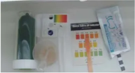
The amount of stimulated saliva, pH and buffering capacity were measured by test GC Saliva-Check BUFFER, GC EUROPE NV in the morning between 8:00 and 11:00, an hour after eating, in an isolated and quiet room. Saliva was collected for 5 min in a graduated measuring cup and reported the amount her.
The pH of the saliva was determined by test strips immersed in the container with the saliva for 10 seconds, and their color is compared with a pre-prepared scale. The values are defined as: high acidity - 5.0-5.8; moderate acidity 6.0-6.6; healthy saliva - 6.8-7.8 (fig.2).
Fig 2. Recording of saliva pH and buffer capacity
To determine the buffering capacity of saliva were used strips provided in kits for colorimetric determination of buffer capacity of saliva - CRT ® buffer. Determination for each patient was performed with about 1 ml undiluted mixed saliva. Pipetted stimulated saliva from the container was dropped in each of the three fields of the test band.The color of the field changed immediately, but the results were assessed after the expiration of the manufacturer's reaction time (2 minutes) in color scale.
- Green - 4 points
- Green / Blue - 3 points
- Blue - 2 points
- Red / Blue - 1 point
- Red - 0 points
A total sum of points:
- 0-5 - reported very low buffering capacity
- 6-9 - low buffering capacity
- 10-12 - normal buffering capacity
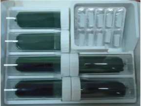
Used were commercially available microbiological tests Vivacare Line - CRT ® - bacteria, (Ivoclar - Vivadent ®; Liechtenstein), based on "selective medium" and known in Anglo-Saxon literature as "test to the chair of the patient" ("chair-side test "). "Chair-side test" is used for semi quantitative detection of Streptococcus mutans, Lactobacillus acidophillus saliva. Blue agar containing bacitracin and methylene blue is intended to Streptococcus mutans, while the transparent agar - Rogosa, is used for Lactobacillus (Kneist S. 1998, Tanzer J.M. 1997).
The required volume of undiluted saliva for agar plating is about 0,5 ml (Vivadent, CRT ® - bacteria: "Focus on caries risk test"). Seeded material is placed in an incubator with a thermostat for 48 hours at 37 ˚ C (CULTURA Incubator Brutschrank, Temp 25 ˚- 45 ˚ C, Ivoclar - Vivadent®) (fig. 3).
Fig 3. Samples ready for incubation
After expiry of the incubation period, the streptococci are visualized as small blue colonies of diameter < 1 mm onto the blue agar. On the back of the holder lactobacilli are visualized as white colonies on transparant agar. The bacteria grout rate was determined semi-quantitatively with the naked eye, according to the manufacturer's instructions. Comparing with the standard provided by the manufacturer reference scale allows to evaluate the risk of dental caries. Determination of microbial numbers greater than 105 CFU / ml, indicates a high risk of being infected by caries (fig.4, fig 5).
- Low risk for the development of dental caries in ≤ 105 CFU / ml for Streptococcus mutans, Lactobacillus acidophillus.
- High risk for the development of dental caries at ≥ 105 CFU / ml for Streptococcus mutans, Lactobacillus acidophillus.
Fig 4. Selective media covered with protective foil
Fig 5. Scale for recording the microbial count
For identification of microbial count was used saliva instead of dental plaque because the saliva is sufficiently representative for the available microflora in the oral cavity (22, 57).
Used was a standard kit for determining glucose (AMS, Italy).Saliva samples were thawed and centrifuged, and 10 μL of supernatant was transferred to an Eppendorf tube type mixed with 1 μL reagent and homogenized.Similarly prepared standard solution and a blank. After mixing, all the solutions were incubated for 10 minutes in a water bath at 37 ° C. The absorbance of the solutions was measured after incubation at 505 nm.
For the purposes of statistical analysis of the results were used the following methods: χ2 -test, Fisher’s exact test.
1.4. Results and Discussion
Between the control group (group I) and the group of children with diabetes mellitus (group II) there is a significant difference in unstimulated salivary flow rate (p = 0.016), while the diference between the subgroups of diabetics (group IA and IB) is not significant (p = 0.0804). Data obtained in the study of unstimulated salivary flow rate show higher shares of a normal amount of saliva formed to 1 minute in healthy children (77%) compared to children with diabetes mellitus (56%) (Diagr. 1).
Siudikiene et al. and Moreira et al. reported that the stimulated and unstimulated saliva flow rate is reduced in diabetic patients, whereas Lopez et al. described that only unstimulated flow rate is reduced. Reduced salivary flow, one of the most important factors associated with caries prevalence, could potentially explain the cariogenic changes in the oral environment of diabetics. The degree of body dehydration may also be related to decreased unstimulated flow rate caused by renal water loss (osmotic diurese and polyuria) in patients with diabetes (Edgar WM. Et al. 2004). Reduced salivary flow rate caused by hyperglycemia is haracteristic mainly for period of poor metabolic control of diabetes. During this period glucose leakage into the oral cavity may occur, thereby facilitating the grouth of aciduric bacteria and caries lesion development (Karjalainen KM. et al. 1996, Reuterving CO. et al. 1987). In contrast, Belazi et al., Swanljung et al. and Edblad et al. did not find significant differences in the salivary flow rates between diabetics and nondiabetic.
The results from Diagram 2 show that the percentage of stimulated salivary flow rate in children in the control group was higher (57%), as compared with children with diabetes (15%). There is a statistical significance of differences in group I and II (p = 0.000003).There was no significant difference between the relative shares of the diabetics subgroups in terms of reduced stimulated saliva flow rate (p = 0,840).
77%
56% 59% 53%
23%
44% 41% 47%
0% 20% 40% 60% 80% 100%
group I (N = 60)
group II (N = 68)
group IIA (N = 29)
group IIB (N = 38)
Diagram 1. Unstimulated salivary flow rate
after 1 min
before 1 min
Rashkova et al. reported the following results: reduced salivary flow rate (stimulated and unstimulated), increasead saliva viscosity, significantly more acid saliva and decreased buffer capacity. They also report of a correlation between dental caries, viscosity and pH of the saliva in diabetic. These two indicators are the most significant factors for assessment of the risk for caries development.
There were found statistically significant differences between group I and II with respect to the pH of saliva (p = 0.003). Only in the group of children with diabetes, 16% have an acidic pH in the range of 5 - 5.8. Among children in the control group are not recorded values of salivary pH in this range. Shares of the diabetics with respect to salivary pH in the sub groups with good and poor metabolic control are very close and comparing the results did not show statistical significance at p = 0.362 (Diagram3).
There are reports indicating that diabetic patients have more acidic saliva (Василева С. и кол., Рашкова М. и кол. 2010, Aframian DJ. Et al. 2006, Alemzadeh R. et al. 2004), whereas other autors have not reported this difference (Aaltonen AS. Et al. 1990, Albrecht M. et al. 1988).
Studies on the consistency of the saliva showed that the significant difference was observed in the saliva bubbles - 40% of children in the control group and 9% in the experimental group of children. Children with diabetes have more fluid saliva 69% compared to 45% healthy children. A comparison
22%
44% 45% 45% 22%
41% 45% 39% 57%
15% 10% 16%
0% 20% 40% 60% 80% 100%
group I (N = 60)
group II (N = 68)
group IIA (N = 29)
group IIB (N = 38)
Diagrama 2. Stimulated salivary flow rate
Normal (> 5 ml)
Between low (3.5 - 5 ml)
Very low (< 3.5 ml)
16% 21% 11% 23%
16% 10% 21% 77% 68% 69% 68%
0% 20% 40% 60% 80% 100%
group I (N = 60)
group II (N = 68)
group IIA (N = 29)
group IIB (N = 38) Diagram 3. Saliva pH
6.8 - 7.8 pH 6 - 6.6 pH 5 - 5.8 pH
of the results for consistency of saliva in healthy children and children with diabetes showed statistically significant differences p = 0.00014 ( Diagrama 4 ).
Lopez et al. reported flow rate, wich was significantly diminished in diabetics, was associated to salivary viscosity and foam.
There is not statistically significant difference in buffer capacity between children from both treatment groups and the two subgroups (p =0,325; p =0,514) (Diagrama 5).
In the study of Moreira et al. patients with DM 1 presented a significant decrease in unstimulated salivary flow. These observations are similar to those reported by Siudikiene et al. and Lopez et al. Dry mouth wos a major complaint of many diabetic patients. The low unstimulated salivary flow in yoyng patients with DM 1 is particularly relevant since this condition is usually more frequent among old adults and is an indication of a high caries risk in DM 1 children (Moore PA. et al. 2001, Peldyak J. et al. 2002). Early detection of salivary hypofunction in children is important for preventing the deleterious oral afetets which follow the absence of salivary protection in oral cavity.
45%
69% 66% 71%
40%
9% 10% 8%
15% 22%
24% 21%
0% 20% 40% 60% 80% 100%
group I (N = 60)
group II (N = 68)
group IIA (N = 29)
group IIB (N = 38) Diagram 4. Saliva consistency
Sticky frothy saliva Frothy bubbly saliva Water clear saliva
2% 7% 7% 8%
33% 34% 28%
39% 65% 59% 66% 53%
0% 20% 40% 60% 80% 100%
group I (N = 60)
group II (N = 68)
group IIA (N = 29)
group IIB (N = 38)
Diagrama 5. Saliva buffer capacity
Normal (10 - 12)
Low (6 - 9)
Very low (0 - 5)
In our studies no statistical difference was found in the levels of Steptococcus Mutans between children from group I and II, as well as within the group of diabetics (p =0,797; p =0,346). It is noteworthy higher titer Steptococcus Mutans in children in the control group 33% (diagr.6).
In this study titer of Lactobacillus acidophillus in the experimental group was higher than that of the children in the control group. When comparing the results, no statistically significant difference, p = 0.007, but this value is very close to the limit p = 0.05. Data for higher values of microbial including Lactobacillus acidophillus in our study are confirmed by our findings values of acidic saliva in children with diabetes mellitus.Mutans streptococci are considered to be the main aetiological microorganism in most of the oral diseases,with Lactobacillus and other microorganisms participating in the disease progression (Krase B. 1988, Tanzer 1992). Twestman et al. found an assootian of metabolic control in children with diabetes mellitus type 1, whith elevated rate of dental caries. He observed that uncontrolled levels of blood glucose levels affect salivary factors such as flow rate, buffer capacity, glucose content and levels of acidogenic bacteria (Lactobacilli and Streptococcus Mutans) (Twetman S. et al. 2002).
85%
65% 65% 67% 15%
35% 35% 33%
0% 20% 40% 60% 80% 100%
group I (N = 27)
group II (N = 54)
group IIA (N = 23)
group IIB (N = 30) Diagram 7. Lactobacillus counts in saliva
≥ 10⁵CFU⁄ml
≤ 10⁵CFU⁄ml 67% 72% 65% 80%
33% 28% 35% 20%
0% 20% 40% 60% 80% 100%
group I (N = 27)
group II (N = 54)
group IIA (N = 23)
group IIB (N = 30)
Diagram 6. Streptococcus Mutans counts in saliva
≥ 10⁵CFU⁄ml ≤ 10⁵CFU⁄ml
According to Twetman et al. the cut-off value of stimulated resting salivary glucose concentraction for poor metabolic control and caries associated risk factor is > 12,5 mg/l. The limit of quantification of the applied method for analysis of the salivary glucose in the current work was 8,1 mg/l which allowed us also to differenciatethe patients in respect to the cited cut-off value. Classifying our study patients based on of the same cut-off value 12.5 mg/l it appears that 50% of diabetics and 30% of non-diabetic children showed values exceeding it.
Jurysta et al, Lopez et al. reported increased glucose concentration and exretion in both unstimulated and stimulated saliva in the diabetic children compared to control subjects. These results are in agreement with ours results for salivary clucose and with those of Karjalainen et al. who reported that salivary level decreased after beginning insulin treatment.
1.5. Conclusion
The result of our study are:
1. The reduced flow of both unstimulated and stimulated saliva is the most important risk factor for caries evelopment in children with DM type 1
2. The low Ph of the saliva in diabetic children is a result of disturbed buffer capacity and leads to colonization with Streptococcus Mutans and Lactobacillus
3. The metabolic control of diabetes has influence on the salivary parameters
Our data support the view that appropriate evaluation of salivary clinical parameters, such as salivary flow rate and buffer capacity, is recommended when assisting diabetic children. These data can be obtained by simple techniques. Early detection of salivary hypofunction in children is important for preventing the deleterious oral afetets which follow the absence of salivary protection in oral cavity.
References
Борисова М., Р. Ковачева. Кръгла маса: „Епидемиологично проучване на някои ендокринни заболявания в България”, Пролетен учебен симпозиум, 18-20 май, 2006
Василева С., Л. Христов, Реакция и електролитен състав на слюнката при деца, болни от захарен диабет. Стоматология, LVIII, №5
68%
50% 60% 43%
32%
50% 40% 57%
0% 20% 40% 60% 80% 100%
group I (N = 60)
group II (N = 44)
group IIA (N = 20)
group IIB (N = 23) Diagram 8. Salivary glucose (mg/l)
> 12.5 mg/l
<= 12.5 mg/l
Дамянова М., Захарен диабет у деца и юноши. Теоретически и практически основи. София, 2000
Джемилева, Т. Ксеростомия, Ацер, София, 1998
Карова Е. Влияние на инхалаторните кортикостероиди върху количеството и pH на нестимулирана слюнка при астматици. Дентална медицина, 1/2011
Рашкова М., К. Коприварова, Н. Тонева, М. Белчева, М. Константинова, Г. Жегова. Оценка на течната орална среда и орална Candida при деца с диабет. Проблеми на денталната медицина, том XXXV/2009 – част II. МУ – Факултет по дентална медицина – София
Рашкова М., К. Коприварова, Н. Тонева, М. Константинова. Промени в качествата на слюнка при деца с диабет. Дентална медицина
Рашкова М., Н. Тонева, С. Търгова, К. Коприварова, М. Константинова, Ю. Стайкова. Изследване на кортизол в слюнката и оценка на оралната среда при деца с диабет. Проблеми на денталната медицина, том XXXVI/2010 – част II. МУ – Факултет по дентална медицина – София
Aaltonen AS, Tenovuo J, Lehtonen OP, Saksala R, Meurman O. Serum antibodies against oral Streptococcus mutans in young children in relation to dental caries and maternal close-contacts. Arch Oral Biol 1985;30:331-335
Aaltonen AS, Tenovuo J, Lehtonen OP, Saksala R. Maternal caries incidence and salivary close-contacts with children affect antibody levels to Streptococcus mutans in children. Oral Microbiol Immunol 1990;5:12-18
Aframian DJ, Davidowitz T, Benoliel R. The distribution of oral mucosal pH values in healthy saliva secretors. Oral Dis. 2006; 12(4):420-3
Albrecht M, Banoczy J, Tamas GJR Dental and oral symptoms of diabetes mellitus. Community Dent Oral Epidemiol 16, 378-380, 1988
Alemzadeh R, Wyatt DT. Diabetes mellitus. In: Behrman RE, Kliegman RM, Jenson HB, ed. Nelson textbook of pediatrics. 17th ed. Philadelphia: Saunders; 2004. p.1960-63
Alves C., Brandão M., Andion J. et al.: Atendimento odontológico do paciente com diabetes melito: recomendações para a prática clínica. R. Ci. Méd. Biol., 2006, 5, 97 -110
Anttila SS., Knuuttila ML. & Sakki TK. (1998). Depressive symptoms as an underlying factor of the sensation of dry mouth. Psychosom Med, Vol. 60, No.2, (apr), pp. (215-218), ISSN 1534-7796
Aydin S.: A comparison of ghrelin, glucose, alpha -amylase and protein levels in saliva from diabetics. J. Biochem. Mol. Biol., 2007, 40, 29 -35
Belazi MA.; Galli-Tsinopoulou A., Drakoulakos D., Fleva A. & Papanayiotou PH. (1998). Salivary alterations in insulin-dependent diabetes mellitus. Int J Paediatr Dent, Vol. 8, No.1, (mar), pp. (29-33), ISSN 1365-263X
Bergdah M, J. Bergdahl. Low unstimulated salivary flow and subjective oral dryness: association with medication, anxiety, depression and stress. J Dent Res, 2000, 79:1652-1658
Dawes C.: What is the critical pH and why does a tooth dissolve in acid? J Can Dent Assoc, 2003, 69:722-724
Edblad E, Lundin SA, Sjodin B, Aman J, Caries and salivary status in young adults with type 1 diabetes. Swed Dent J 2001; 25:53-60
Edgar WM, Dawes C, O’Mullane. Saliva and dental health3 ed..London. Br Dent J, 2004; 32-49
Emilson C.G.: Prevalence of Streptococcus mutans with different colonial morpfologies in human plaque and saliva. Scand. J. Dent. Res., 1983; 91(1):26-32
Gitny J. Astma medication and caries, Br Dent J, 1997, 182, 3:8
Guggenheimer J. & Moore PA. (2003). Xerostomia: etiology, recognition and 1. treatment. J Am Dent Assoc, Vol. 134, No.1, (jan), pp. (61-69), ISSN1943-4723
Harper D.S., W. J. Loeshe. Growth and acid tolerance of human dental plaque bacteria, Archs. Oral. Biol.,1984; 29:843-848
Jurysta C., Bulur N., Oguzhan B. et al.: Salivary glucose concentration and excretion in nor- mal and diabetic subjects. J. Biomed. Biotechnol., 2009, 426 -430
Karjalainen KM, Knuuttila MLE, Kaar ML Salivary factors in children and adolescents with insulin-dependent diabetes mellitus. Pediatr Dent 18, 306-311, 1996
Kneist S.: Begleiphenomene in der Mikrobiologischen Speicheldiagnostik, Oralprophylaxe, 1998; 20:208-217
Koga-Ito C, Martins C, Balducci I, Jorge A. Correlation among mutans streptococci counts, dental caries, and IgA to Streptococcus mutans in saliva. Pesqui Odontol Bras 2004;18: 350-355
Krase B: Biological factors as indicators of future caries. Int Dent J, 1988, 38:219-255
Leone C. W., F. G. Oppenheim. Physical and Chemical Aspects of Saliva as Indicators of Risk for Dental Caries in Humans. J Dent Educ; 2001:65:1054-1062
Loesche W.J.: Role of Streptococcus mutans in human dental decay. Microbiol. Rev., 1986; 50:353-380
Lopez MM., Colloca ME., Pбez RG., Schallmach JN., Koss MA. & Chervonagura A. (2003). Salivary characteristics of diabetic children. Braz Dent J, Vol. 14, No.1, (jun), pp. (26-31), ISSN 0103-6440
Miralles Jordá L., Silvestre Donat F.J., Hernandez -Mijares A. et al.: Dental caries in type 1 diabetics: influence of systemic factors of the disease upon the development of dental car- ies. Med. Oral. Patol. Oral. Cir. Bucal., 2006, 11, E256-E260
Moore PA., Guggenheimer J., Etzel KR., Weyant RJ. & Orchard T. (2001). Type 1 diabetes mellitus, xerostomia, and salivary flow rates. Oral Surg Oral Med Oral Pathol Oral Radiol Endod, Vol. 92, No.3, (sep), pp. (281-291), ISSN 1528-395X
Moreira AR., Passos LA., Sampaio FC., Soares MSM. & Oliveira RJ. (2009). Flow rate, pH and calcium concentration of saliva of children and adolescents with type 1 diabetes mellitus. Braz J Med Biol Res, Vol. 42, No.8, (aug), pp. (707-711), ISSN 1414 431X
Moura S.A.B., Medeiros A.M.C., Costa F.R.H. et al.: Diagnostic value of saliva in oral and systemic diseases: a literature review. Pesq. Bras. Odontop. Clin. Integr., 2007, 7, 187 -194
Orbak R., Simsek S., Orbak Z., Kavrut F. & Colak M. (2008). The influence of type-1 diabetes mellitus on dentition and oral health in children and adolescents. Yonsei Med J, Vol. 49, No. 3, (jun), pp. (357-765), ISSN 1976-2437
Peldyak J, Makinen KK. Xylitol for caries prevention. J Dent Hyg. 2002; 76(4):276-85
Reuterving CO, Reuterving G, Hдgg E, Ericson T. Salivary flow rate and salivary glucose concentration in patients with diabetes mellitus: Influence of severity of diabetes. Diabete Metab 1987; 13(4):457-62
Rios D., H.M. Honorio, A.C. Magalhres, A.C.B. Delbem, M.A.A.M Machado, S.M.B. Silva, M.A.R. Buzalaf: Effect of salivary stimulatiom on erosion of human and bovine enamel subjected or not tosubsequent abrasion: an in situ/ex vivo stude, Car. Res., 2006; 40:218-22
Siudikiene J., Machiulskiene V., Nyvad B. et al.: Dental caries increments and related factors in children with type 1 diabetes mellitus. Caries Res., 2008, 42, 354 -362
Siudikiene J., Machiulskiene V., Nyvad B., Tenovuo J. & Nedzelskiene I. (2006). Dental caries and salivary status in children with type 1 diabetes mellitus, related to metabolic control of the disease. Eur J Oral Sci, Vol. 114, No.1, (feb), pp. (8-14), ISSN 1600-0722
Smith DJ, Taubman MA. Emergence of immune competence in saliva. Crit Rev Oral Biol Med 1993;4:335-341
Soltèsz G., Patterson C., Dahlquist G.: Diabetes in the young: a global perspective. [in:] Diabetes atlas. (ed.) D. Gan, 2nd ed. International Diabetes Federation, Gent, 2003, 113 -134
Sreebny LM., Yu A., Green A. & Valdini A. (1992). Xerostomia in diabetes mellitus. Diabetes Care, Vol. 15, No.5, (jul), pp. (900-904), ISSN 1935-5548
Swanljung O., Meurman JH., Torkko H., Sandholm L., Kaprio E. & Maenpaa J. (1992). Caries and saliva in 12-18-year-old diabetics and controls. Scand J Dent Res, Vol. 100, No.6, (dec), pp. (310-313), ISSN 0029-845X
Tanzer J.M.: Salivary and plaque microbiological test and the management of dental caries. J Dent Educ, 1997, 61(11):866-875
Tanzer, (1992), Ooshima et al, (1994), Van Houte et al, Clinical Illness Conn Mutans Streptococci.
Dental Caries - a Multifactorial Disease 1991;Website link:
http://ethesis.helsinki.fi/julkaisut/laa/hamma/vk/gronroos/ch2.htm
Tenovuo J, Alanen P, Larjava H, Viikari J, Lehtonen Op. Oral health of patients with insulindependent diabetes mellitus. Scand J Dent Res 1986: 94: 338-46
Tenovuo J, Lehtonen OP, Aaltonen AS. Caries development in children in relation to the presence of mutans streptococci in dental plaque and of serum antibodies against whole cells and protein antigen I/II of Streptococcus mutans. Caries Res 1990;24:59-64
Tenovuo J, Lehtonen OP, Aaltonen AS. Serum and salivary antibodies against Streptococcus mutans in young children with and without detectable oral S. mutans. Caries Res 1987;21:289-296
Tenovuo J, Lumikari M, Soukka T. Salivary lysozyme, lactoferrin and peroxidases: antibacterial effects on cariogenic bacteria and clinical applications in preventive dentistry. Proc Finn Dent Soc 1991;87:197-208
Tenovuo J. Salivary parameters of relevance for assessing caries activity in individuals and populations. Community Dent Oral pidemiol 1997;25:82-86.
Toors FA. Chewing gum and dental health. Literature review. Rev Belge Med Dent. 1992;47(3):67-92
Twetman S, I.Johansson,D.Birkhed,T.Nederfors.Caries incidence in young type diabetes mellitus patients in relation to metabolic kontol and caries- associated risk factors.Caries res.2002;36:31-35
Van Houte J.: Microbiological predictors of caries risk, Adv. Dent. Res. 1993; 7(2):87-96
Van Nieuw Amerongen A., J.G. Bolscher, E. C. I. Veerman: Salivary proteins: protective and diagnostic value in cariology? Car. Res., 2004; 38:247-253
Von Bültzingslöwen I., Sollecito TP., Fox PC., Daniels T., Jonsson R., Lockhart PB., Wray D., Brennan MT., Carrozzo M., Gandera B., Fujibayashi T., Navazesh M., Rhodus NL. & Schiødt M. (2007). Salivary dysfunction associated with systemic diseases: systematic review and clinical management recommendations. Oral Surg Oral Med Oral Pathol Oral Radiol Endod, Vol. 103, No.suppl 1, (mar), pp. (S57.e1-15), ISSN 1528-395X
World Health Organization: Prevalence of Diabetes. http://www.who.int/diabetes/facts/ world_fi gures/en/ (2011.08.26). Global status report on noncommunicable diseases 2010. Geneva, World Health Organization, 2011.