Endonuclease-Mediated Long PCR
and Its Application to Restriction
Mapping
Chengtao Her and Richard M. Weinshilboum
Department of Pharmacology, Mayo Medical School, Mayo Clinic, Mayo Foundation, Rochester MN 55905, USA
The polymerase chain reaction (PCR) is the most widely used technique for the study of DNA. Applications for PCR have been extended significantly by the development of “long” PCR, a technique that makes it possible to amplify DNA fragments up to 40 kb in length. This article describes two novel applications of the long PCR technique, one which simplifies restriction mapping and another which enhances amplification specificity and yield. The same primers used to perform the long PCR amplification can be used as probes to perform restriction mapping of the DNA fragment amplified. Restriction digestion performed prior to long PCR amplification can be used to selectively suppress the amplification of members of families of closely related DNA sequences, thereby making it possible to selectively amplify one of a group of highly homologous sequences. These two complimentary techniques, both involving use of the long PCR paired with restriction digestion, have potential application in any laboratory in which PCR is performed.
Introduction
The polymerase chain reaction (PCR) has become the most widely used technique for thestudy of DNA; with applications pertaining to cDNA cloning, gene cloning, polymorphismdetection, mutagenesis and allele-specific diagnosis among many others (1, 20).Each of theseapplications has been extended by the development of “long” PCR (2, 3), a technique whichmakes it possible to amplify DNA fragments up to 40 kb in length.This article will describetwo applications in which long PCR has been paired with DNA restriction endonuclease digestion. In one case, the approach serves to simplify restriction mapping, a technique central to DNA characterization.The second approach involves performing restriction digestion prior to PCR amplification to “selectively suppress” the amplification of highly homologous DNAsequences.
Restriction mapping is a very useful step in the characterization of a large DNA molecule.DNA mapping by restriction enzyme digestion has been used routinely in preparation for subcloning, in the analysis of restriction fragment length polymorphisms, for comparing different genomic DNA clones, and in the construction of physical maps of chromosomes (4).Several different approaches have been used to perform restriction mapping (5-8). The “classical” approach involved digestion of the DNA fragment, initially with a single restrictionenzyme and, subsequently, with a combination of enzymes, followed by gel electrophoresis (5).
A significant improvement was made by Smith et al. when they suggested that the DNAfragment of interest be radioactively labeled at both ends with 32P using polynucleotidekinase and 32P-γ-ATP (6).The end-labeled DNA was then digested with an enzyme for whichthere was only a single restriction site on the target sequence, and the two resulting fragmentswere separated by gel electrophoresis. These two DNA fragments, each labeled at one end,were then subjected to partial restriction digestion with different restriction enzymes.Theresultant overlapping restriction fragments, all with a common radioactively labeled terminus, were separated by gel electrophoresis and detected by autoradiography.
Another restriction mapping technique involved partial digestion of genomic DNA that had been subcloned intolambda phage or a cosmid vector, followed by Southern analysis performed with radioactivelylabeled oligonucleotide probes which had been designed on the basis of known vector sequence.Generation of this type
of restriction map often began with lambda-terminase digestion of acosmid that contained the insert of interest (7).Lambda-terminase cleavage was used tolinearize the cosmid at a unique sequence and to generate protruding ends.The linearized cosmid was then subjected to partial restriction digestion, followed by hybridization withradioactively labeled cos site-specific oligonucleotides that hybridized to the 12 bp 5'-protrudingends created by lambda-terminase digestion (7).
A conceptually similar method utilizedradioactively labeled T3- and T7-specific oligonucleotides that were hybridized to the ends ofan insert that had been excised from a cosmid vector by NotI digestion (8).After the DNAinsert had been separated from the vector by complete digestion with NotI, it was then subjectedto partial digestion with other restriction enzymes.The resulting restriction fragments were separated by gel electrophoresis, followed by transfer to a membrane. The immobilizedrestriction fragments could be detected by the use of radioactively labeled end-specific T3 or T7oligonucleotides (8).
Unfortunately, the lambda-terminase technique is not suitable for mapping cosmids that contain multiple cos sites, and the T3-T7-specific technique requires that the insertcontain no NotI sites.Development of a vector-independent procedure for restriction mappingwould make it possible to obtain restriction maps without the requirement that vector sequencebe known, and, of more importance, without the requirement for subcloning.Development ofthe long PCR now makes such an approach feasible (2, 3).
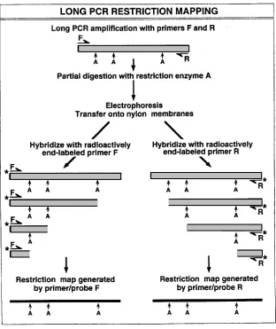
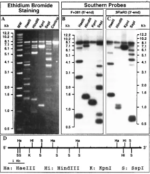
Long PCR-based restriction mapping can potentially be used to generate restriction mapsquickly and simply for any DNA sequence that can be amplified (9), which, for the long PCR,includes regions up to 40 kb in length (2, 3).The technique utilizes the same general principles used in the 3'- and 5'-labeling of restriction fragments generated from either purified DNAmolecules or sequences which have been cloned into a vector.However, the long PCR approach does not require purification or cloning of the DNA molecule to be characterized, and it does not require knowledge of a vector sequence.The technique is illustrated schematically in Figure1.The same primers used to perform the PCR amplification are radioactively labeled so theycan also serve as probes for the detection of DNA fragments generated by partial restrictionenzyme digestion of the long PCR amplification product.The method has been used successfully to generate restriction maps for the human estrogen sulfotransferase gene (9, 10) andthe human thiopurine methyltransferase gene (11).Application of this long PCR-based techniquefor restriction mapping of the human estrogen sulfotransferase gene is shown in Figure 2 (9, 10).A different, but related application of long PCR also pairs the technique with restrictiondigestion, but in this case restriction digestion is performed before the amplification reaction tomake possible the selective amplification of a single member of a family of highly homologousDNA sequences (12).
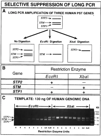
High specificity and adequate yield of the desired amplification product are importantissues in all PCR reactions, including long PCR amplifications.Many different strategies have been used in attempts to increase reaction yield and specificity.For example, considerable efforthas been expended on the optimal design of PCR primers and on the modification of reactionconditions, (1, 13, 14).“Selective suppression” by restriction endonuclease digestion is a longPCR technique that is intended to enhance both specificity and yield when the long PCR is usedto amplify individual members of families of closely related DNA sequences.We tested the feasibility of this approach by using it to amplify individual members of a group of three highlyhomologous human chromosome 16 phenol sulfotransferase genes,
also promotes increased yields of the desiredamplification product.This approach is especially useful when it is not possible to designoptimal primers, either as a result of limited knowledge of the DNA sequence to be amplifiedor in situations in which multiple copies of very similar sequences are present in the templateDNA. Restriction digestion has been used previously as a way to remove contaminating DNAprior to the addition of template (17) or as a method to enhance the amplification of relativelyshort, known sequences (18). However, in selective suppression of long PCR, the amplificationof entire genes or large portions of genes is often the goal, and much of the amplified sequencemay be unknown.The most appropriate endonuclease for a given application can be selectedon the basis of either the results of pilot experiments or knowledge of restriction patterns for one or more of the highly related sequences to be amplified.This strategy reduces the effortrequired to design specific PCR primers for the amplification of very similar sequences, and itis particularly useful when limited sequence information is available for primer design.
We haveillustrated the selective suppression technique in Figure 3 by showing its use for the specific amplification of two of the three phenol sulfotransferase genes found on human chromosome 16(15, 16).The figure shows both a schematic outline of the technique (Figure 3A) and actual datafor an amplification performed after selective suppression by EcoRI or XbaI digestion (Figure3C).Because of the very high degree of sequence identity present among these genes, a singleprimer pair can be used to amplify STP1, STP2 and STM.The particular pair of primers usedto obtain the data shown in Figure 3 amplifies the coding exons of all three genes in fragments7.5, 7.0 and 6.9 kb in length for STP2, STM and STP1, respectively (12, 15, 16; Figure 3). However, STP2 is not cleaved by XbaI restriction digstion, while both STP1 and STM are cut by this enzyme (see the table in Figure 3B). Conversely, STM has no restriction sites for EcoRI, while STP1 and STP2 both contain EcoRI restriction sites. The enhanced specificity and yield of STM
amplification after prior restriction digestion with EcoRI, and the enhanced specificity and yieldof the STP2 amplification product following incubation with XbaI are shown in Figure 3C.Inessence, this approach converts the high degree of sequence similarity present within membersof some gene families from a disadvantage into an advantage.Use of selective suppressionmakes it possible to study individual members of gene families with highly similar sequencesby “spotlighting”, in turn, each member of the family (Figure 3).Therefore, selectivesuppression, a technique that involves restriction endonuclease digestion performed in the samereaction tube prior to long PCR amplification, like the use of the long PCR to simplify andspeed restriction mapping, should have potential application in any laboratory in which the PCRis performed.
Protocols
Required Reagents and Equipment
Figure 3.Selective suppression of the long PCR by restriction endonuclease digestion.(A) Aschematic diagram of selective suppression of the long PCR as applied to the human phenolsulfotransferase genes
STP1, STP2 and STM is shown.These three human chromosome 16genes can each be amplified by use of the primers F2 and R2 (12, 15, 16).The effect ofdigestion of human genomic DNA with either EcoRI or XbaI prior to long PCR amplificationof these genes is depicted schematically.(B)Susceptibility of human phenol sulfotransferasegenes STP2, STM and STP1 to restriction digestion with EcoRI and
Protocol 1: Long PCR-Based Restriction Mapping
Long PCR reaction
All reagents for long PCR except primers and template are supplied with the GeneAmp XL PCR Kit or similar kits that can be obtained from other suppliers. Please referto the GeneAmp XL PCR Kit instructions for additional instructions.
1. Prepare master mixtures of the lower and upper PCR buffers in two sterile microcentrifuge tubes.Quantities of reagents for a single reaction are listed below.Multiply by the desired number of reactions.Mix well by vortexing.
Lower buffer Upper buffer
1.6 µl Forward primer (10 pmol/µl) 18 µl 3.3x XL buffer
1.6 µl Reverse primer (10 pmol/µl) 1 µl DNA template (80-160 ng/µl) 12.0 µl 3.3x XL buffer 2 µl rTth DNA polymerase, XL
4.4 µl 25 mM Mg(CH3CO2)2 39 µl ddH2O 8.0µl dNTPs (2.5 mM each)
12.4 µl ddH2O
2. Program the thermal cycler as follows:
File 1 80˚C for 5 min; 25˚C for at least 2 min; 1 cycle
File 2 94˚C for 1 min; 1 cycle
File 3 94˚C for 30 s, 66˚C for 10 min; 16 cycles File 4 94˚C for 30 s, 66˚C for 10 min, with 15 s 12 cycles
incremental lengthening step for each cycle
File 5 72˚C for 10 min; 1 cycle
File 6 4˚C hold
The temperature during the denature/annealing step must be adjusted on the basis ofthe properties of the primers used.
3. Pipette 40 µl of the lower PCR buffer into the bottom of a 0.5 ml thin-walled PCRtube.
4. Add one AmpliWax PCR Gem 100 bead to the PCR tube.
5. Place the PCR tubes in the thermal cycler.Heat at 80˚ C for 5 min and then cool to25˚C (i.e. initiate thermal cycler file 1, as outlined in step 2).
6. Pipette 60 µl of the upper PCR buffer into the PCR tubes at 25˚C. 7. Proceed with the remainder of the PCR cycle as outlined in step 2.
Partial Restriction Enzyme Digestion
1. Decide how many restriction enzymes are to be used for restriction mapping. Preparetwo sets of microcentrifuge tubes for each enzyme and pipette 40 µl of the PCRreaction mixture into one set of microcentrifuge tubes on ice. 2. Pipette 3 µl of 0.5 M EDTA, pH 8.0, into the other set of microcentrifuge tubes.
These tubes will be used to terminate the restriction digestion reaction. 3. Add 2 µl of the restriction enzyme to be tested to the microcentrifuge tubes
with thePCR mixture.Mix well, centrifuge briefly and then incubate at the appropriatetemperature.(See Notes and Tips 2).
Electrophoresis
1. Add an appropriate quantity of gel-loading buffer to the partially digested DNA sample.Place 20 µl of each sample in a 0.8% agarose gel cast in 1x TAE.A duplicate series of samples should be analyzed on the other side of the gel to be probed with the alternative primer.
2. Perform electrophoresis overnight in 1x TAE buffer at 1 volt/cm.
Southern Blot
1. Stain the gel with ethidium bromide and photograph under UV light with a fluorescentruler placed beside the DNA marker lane.
2. Prepare the gel for capillary transfer after trimming as follows:
(i) Soak the gel in 500 ml 0.25 M HCl for 15 min and then rinse with ddH2O. (ii) Soak the gel in 500 ml 0.5 M NaOH in 1.5 M NaCl for 15 min, and then
rinsewith ddH2O. (iii) Repeat step (ii) two times.
(iv) Soak the gel in 500 ml 0.5 M Tris-HCl, pH 7.5 in 1.5 M NaCl for 15 min, andthen rinse with ddH2O.
(v) Repeat step (iv).
3. Cut an MSI nylon membrane to a suitable size, and soak the membrane for 15 minin 5x SSPE .
4. Transfer DNA from the gel to the nylon membrane overnight by capillary action in20x SSPE.
Radioactive Labelling of Primers
1. Prepare reagents for T4 polynucleotide kinase reactions for the two long PCR primersin two microcentrifuge tubes as listed, and then incubate at 37˚C for 30 min.
3 µl oligonucleotide primer (10 pmol/µl) 2 µl 10x kinase reaction buffer
2 µl 32P-γ-ATP (7000 Ci/mmol)
2 µl T4 polynucleotide kinase 11 µl ddH2O
2. Inactivate the T4 polynucleotide kinase by incubation at 97˚C for 2 min. 3. Purify the labeled oligonucleotides with Sephadex G-25 Quick Spin Columns
according to the manufacturers directions.
4. Use 1 µl of the purified primer to determine the specific activity.
Hybridization
1. Rinse the nylon membrane from “Southern Blot” step 4 in 5x SSPE.Air dry the membrane onWhatman paper, and then vacuum-dry the membrane at 80˚C for 1.5 hr.
2. Prehybridize the membrane at 37˚C for 1 hr in ExpressHyb hybridization solution (approx. 0.2 ml/cm2).
3. Discard the original ExpressHyb solution and add an identical volume of fresh ExpressHyb solution and a suitable quantity of the probe (1-2 x 10⁶ cpm/ml). Mixwell and incubate at 37˚C for 1 hr.
4. Wash the membrane three or more times at room temperature in 2x SSC with 0.05%SDS.If there is more than one membrane, each membrane should be washedseparately.
Protocol 2: Selective Suppression of Long PCR
Restriction Digestion and Long PCR Reactions
1. Prepare a “master mixture” of the lower PCR buffer in a sterile microcentrifuge tube.Quantities of reagents required to perform a single reaction are listed below. Multiplyby the desired number of reactions and mix well by vortexing.
Lower buffer
1.6 µl Forward primer (10 pmol/µl) 1.6 µl Reverse primer (10 pmol/µl) 23.4 µl 3.3x PCR buffer
8.0µl dNTPs (2.5 mM each) 43.4 µl ddH2O
2. Program the thermal cycler as follows:
File 1 80˚C, 5 min 25˚C, at least 2 min 37˚C, 20 min 1 cycle
File 2 94˚C, 1 min 1 cycle
File 3 94˚C, 30 s 66˚C, 10 min 16 cycles
File 4 94˚C, 30 s 66˚C, 10 min with 15 s 12 cycles incremental lengthening
step for each cycle
File 5 72˚C for 10 min 1 cycle
File 6 4˚C hold
3. Pipette 78 µl of the lower PCR buffer into a 0.5 ml thin-walled PCR tube. 4. Add one AmpliWax PCR Gem 100 bead to each PCR tube.
5. Place the reaction tubes in the thermal cycler. 6. Prepare the upper PCR buffer as listed below:
Upper buffer
6.6 µl 3.3x XL buffer 4.4 µl 25 mM Mg(CH3CO2)2 2.0 µl rTth DNA polymerase, XL
2.0µl Restriction enzyme at an appropriate concentration 1.0 µl DNA template (80 - 160 ng/µl)
6.0 µl ddH2O
Mix well by pipeting up and down. Restriction enzymes are diluted in 1x restrictionenzyme buffer and should be added to the upper buffer mixture immediately beforethe PCR cycle is initiated.
7. Initiate the PCR cycle outlined in step 2.
8. During the thermal cycler File 1 25˚C incubation, add 22 µl of the upper PCR bufferover the wax barrier.
Electrophoresis
1. Analyze 10 µl of each amplification product by agarose gel electrophoresis.
Notes and Tips
2. It is recommended that a pilot experiment be performed to determine the appropriateamount of a particular restriction enzyme that is required to obtain partial digestion.Many restriction enzymes possess variable activities when restriction digestion isperformed in the PCR reaction mixture (19).We have found it convenient to performa series of restriction digestion reactions with different concentrations of a particular restriction enzyme while holding restriction digestion time constant.
3. Eighty to 160 ng of human genomic DNA should be adequate for the long PCR amplification of a 10 kb target sequence.
4. Restriction maps generated from either end of a target DNA fragment with a particularrestriction enzyme should agree (Figure 1), especially within the central portion of thetarget sequence.Lack of consistency between these results usually reflects nonspecifichybridization of the probes to the partially digested DNA fragments. This problem canbe addressed by: (a) increasing hybridization temperature; (b) using a highly stringentwash after the hybridization; or (c) using a probe that is located internal to the PCRamplification primer, to achieve greater specificity.
References
1. Mullis, K.B., Ferré, F. and Gibbs, R.A. 1994. The Polymerase Chain Reaction. Birkhäuser, Boston, Massachusetts.
2. Barnes, W.M. 1994. PCR amplification of up to 35 kb DNA with high fidelity and highyield from lambda bacteriophage templates. Proc. Natl. Acad. Sci. USA 91: 2216-2220.
3. Cheng, S., Fockler, C., Barnes, W.M. and Higuchi, R. 1994. Effective amplificationof long targets from cloned inserts and human genomic DNA. Proc. Natl. Acad. Sci.USA 91: 5695-5699.
4. Garcia, E., Elliott, J., Gorvad, A., Brandriff, B., Gordon, L., Soliman, K.M., Ashworth, L.K., Lennon, G., Burgin, M., Lamerdin, J. and Carrano, A.V. 1995. Acontinuous high-resolution physical map spanning 17 megabases of the q12, q13.1, andq13.2 cytogenetic bands of human chromosome 19. Genomics 27: 52-66.
5. Lewin, B. 1994. Isolating the gene. In: Genes V.Oxford University Press. New York.p. 129-134.
6. Smith, H.O. and Birnstiel, M.L. 1976. A simple method for DNA restriction sitemapping. Nucleic Acids Res. 3: 2387-2398.
7. Rackwitz, H.R., Zehetner, G., Murialdo, H., Delius, H., Chai, J.H., Poustka, A., Frischauf, A. and Lehrach, H. 1985. Analysis of cosmids using linearization by phage lambda terminase. Gene 40: 259-266.
8. Evans, G.A. and Wahl, G.M. 1987. Cosmid vectors for genomic walking and rapidrestriction mapping. Meth. Enzymol. 152: 604-610.
9. Her, C. and Weinshilboum, R.M. 1995. Rapid restriction mapping by use of long PCR.BioTechniques 19: 530-532.
10. Her, C., Aksoy, I.A., Kimura, S., Brandriff, B.F., Wasmuth, J.J. and Weinshilboum,R.M. 1995. Human estrogen sulfotransferase gene (STE): cloning, structure andchromosomal localization.Genomics 29: 16-23. 11. Szumlanski, C., Otterness, D., Her, C., Lee, D., Brandriff, B., Kelsell, D., Spurr,
N., Lennard, L., Wieben, E. and Weinshilboum, R.M. 1996. Thiopurine methyltransferasepharmacogenetics: human gene cloning and characterization of a common polymorphism.DNA and Cell Biol. 15: 17-30.
12. Her, C. and Weinshilboum, R.M. 1996. Long PCR: selective suppression by restrictionendonuclease digestion. BioTechniques. 21: 764-766.
for PCRprimer design. PCR Methods and Applications. 3: S30-37.
14. Chou, Q., Russell, M., Birch, D., Raymond, J. and Bloch, W. 1992. Prevention of pre-PCR mis-priming and primer dimerization improves low-copy-number amplifications.Nucleic Acids Res. 20: 1717-1723.
15. Aksoy, I.A. and Weinshilboum, R.M. 1995. Human thermolabile phenol sulfotransferasegene (STM):molecular cloning and structural characterization. Biochem. Biophys. Res.Commun. 208: 786-795.
16. Her, C., Raftogianis, R. and Weinshilboum, R.M. 1996. Human phenol sulfotransferase STP2 gene: molecular cloning, structural characterization and chromosomal localization.Genomics 33: 409-420.
17. Furrer, B., Candrian, U., Wieland, P. and Lüthy, J. 1990. Improving PCR efficiency.Nature 346: 324.
18. Sharma, J.K., Gopalkrishna, V. and Das, B.C. 1992. A simple method for eliminationof unspecific amplifications in polymerase chain reaction.Nucleic Acids Res. 20: 6117- 6118.
19. Blanck, A., Gluck, B., Wartbichler, R., Bender, S., Poll, M. and Brandl, A. 1995. Activity of restriction enzymes in a PCR mix. Biochemica 2: 14.
• MALDI-TOF Mass Spectrometry in Microbiology
Edited by: M Kostrzewa, S Schubert (2016)
www.caister.com/malditof
• Aspergillus and Penicillium in the Post-genomic Era
Edited by: RP Vries, IB Gelber, MR Andersen (2016)
www.caister.com/aspergillus2
• The Bacteriocins: Current Knowledge and Future Prospects
Edited by: RL Dorit, SM Roy, MA Riley (2016)
www.caister.com/bacteriocins
• Omics in Plant Disease Resistance
Edited by: V Bhadauria (2016)
www.caister.com/opdr
• Acidophiles: Life in Extremely Acidic Environments
Edited by: R Quatrini, DB Johnson (2016)
www.caister.com/acidophiles
• Climate Change and Microbial Ecology: Current Research and Future Trends
Edited by: J Marxsen (2016)
www.caister.com/climate
• Biofilms in Bioremediation: Current Research and Emerging Technologies
Edited by: G Lear (2016)
www.caister.com/biorem
• Microalgae: Current Research and Applications
Edited by: MN Tsaloglou (2016)
www.caister.com/microalgae
• Gas Plasma Sterilization in Microbiology: Theory, Applications, Pitfalls and New Perspectives
Edited by: H Shintani, A Sakudo (2016)
www.caister.com/gasplasma
• Virus Evolution: Current Research and Future Directions
Edited by: SC Weaver, M Denison, M Roossinck, et al. (2016)
www.caister.com/virusevol
• Arboviruses: Molecular Biology, Evolution and Control
Edited by: N Vasilakis, DJ Gubler (2016)
www.caister.com/arbo
• Shigella: Molecular and Cellular Biology
Edited by: WD Picking, WL Picking (2016)
www.caister.com/shigella
• Aquatic Biofilms: Ecology, Water Quality and Wastewater Treatment
Edited by: AM Romaní, H Guasch, MD Balaguer (2016)
www.caister.com/aquaticbiofilms
• Alphaviruses: Current Biology
Edited by: S Mahalingam, L Herrero, B Herring (2016)
www.caister.com/alpha
• Thermophilic Microorganisms
Edited by: F Li (2015)
www.caister.com/thermophile
• Flow Cytometry in Microbiology: Technology and Applications
Edited by: MG Wilkinson (2015)
www.caister.com/flow
• Probiotics and Prebiotics: Current Research and Future Trends
Edited by: K Venema, AP Carmo (2015)
www.caister.com/probiotics
• Epigenetics: Current Research and Emerging Trends
Edited by: BP Chadwick (2015)
www.caister.com/epigenetics2015
• Corynebacterium glutamicum: From Systems Biology to Biotechnological Applications
Edited by: A Burkovski (2015)
www.caister.com/cory2
• Advanced Vaccine Research Methods for the Decade of Vaccines
Edited by: F Bagnoli, R Rappuoli (2015)
www.caister.com/vaccines
• Antifungals: From Genomics to Resistance and the Development of Novel Agents
Edited by: AT Coste, P Vandeputte (2015)
www.caister.com/antifungals
• Bacteria-Plant Interactions: Advanced Research and Future Trends
Edited by: J Murillo, BA Vinatzer, RW Jackson, et al. (2015)
www.caister.com/bacteria-plant
• Aeromonas
Edited by: J Graf (2015)
www.caister.com/aeromonas
• Antibiotics: Current Innovations and Future Trends
Edited by: S Sánchez, AL Demain (2015)
www.caister.com/antibiotics
• Leishmania: Current Biology and Control
Edited by: S Adak, R Datta (2015)
www.caister.com/leish2
• Acanthamoeba: Biology and Pathogenesis (2nd edition)
Author: NA Khan (2015)
www.caister.com/acanthamoeba2
• Microarrays: Current Technology, Innovations and Applications
Edited by: Z He (2014)
www.caister.com/microarrays2
• Metagenomics of the Microbial Nitrogen Cycle: Theory, Methods and Applications
Edited by: D Marco (2014)
www.caister.com/n2 Caister Academic Press is a leading academic publisher of
advanced texts in microbiology, molecular biology and medical research. Full details of all our publications at caister.com