Cracking the Ubiquitin Code:
The Ubiquitin Toolbox
Monique P.C. Mulder*, Katharina F. Witting and Huib Ovaa*
Oncode Institute and Department of Cell and Chemical Biology, Chemical Immunology, Leiden University Medical Centre, Leiden, the Netherlands.
*Correspondence: [email protected] and [email protected] https://doi.org/10.21775/cimb.037.001
Abstract
Ubiquitination, a post-translational modification, regulates a vast array of fundamental biological pro-cesses with dysregulation of the dedicated enzymes giving rise to pathologies such as cancer and neu-rodegenerative diseases. Assembly and its ensuing removal of this post-translational modification, determining a large variety of biological functions, is executed by a number of enzymes sequentially activating, conjugating, ligating, as well as deu-biquitinating. Considering the vast impact of ubiquitination on regulating cellular homeostasis, understanding the function of these vast enzyme networks merits the development and innovation of tools. Thus, advances in synthetic strategies for generating ubiquitin, permitted the develop-ment of a plethora of ubiquitin assay reagents and numerous activity-based probes (ABPs) enable the study of enzymes involved in the complex system of ubiquitination. With ubiquitination playing such a pivotal role in the pathogenesis of a multitude of diseases, the identification of inhibitors for ubiq-uitin enzymes as well as the development of ABPs and high-throughput assay reagents is of utmost importance. Accordingly, this article will review the current state-of-the-art activity-based probes, reporter substrates, and other relevant tools based on Ub as a recognition element while highlighting the need of innovative technologies and unique concepts to study emerging facets of ubiquitin biology.
Introduction
and the RING-in-between-RING E3 (RBR) E3 enzymes (Vittal et al., 2015). In contrast to HECT E3 ligases, which utilize a direct transfer mechanism to relay activated ubiquitin to its substrate lysines, and RING E3s that employ an indirect scaffolding mechanism, RBR (RING-between-RING) ligases possess a trilateral domain architecture consisting of three zinc-binding domains – a RING1 domain flanked by an in-between-RING (IBR) domain, adjacent to a RING2 domain (Walden and Rit-tinger, 2018). On E2 Ub thioester recognition by RING1, it is transferred to the catalytic cysteine of RING2, which then facilitates transfer to the lysine of the substrate (Spratt et al., 2014; Walden and Rit-tinger, 2018).
Importantly, ubiquitination is a reversible pro-cess. The ubiquitination status of a protein can be regulated by removal or editing of ubiquitin chains, which is carried out by a group of approximately 100 deubiquitinating enzymes (DUBs) (Fig. 2.1A). Several categories of human DUBs have been identified to date; including the subfamilies of ubiquitin-specific proteases (USPs), ubiquitin C-terminal hydrolases (UCHs), Machado-Joseph disease proteases (MJD), ovarian tumour domain proteases (OTUs), motif interacting with Ub-containing novel DUB family (MINDYs) and zinc finger with UFM1-specific peptidase domain protein (ZUFSPs) cleaving Ub linkages through a cysteine protease mechanism whereas JAB1/
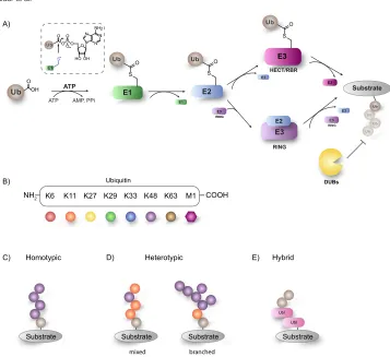
Figure 2.1 The complexity of ubiquitination. (A) The ubiquitination cascade, an orchestrated interplay of
enzymes. (B) Self-modification of ubiquitin on one of its seven lysine residues results in a variety of different
linkage types. Additionally, Ub can modify itself using the N-terminal methionine residue. (C) Increased
complexity can be achieved by linking the Ub-modules in various manners leading to homotypic Ub-chains,
in which the same type of Ub linkage is found or as (D) heterotypic linkages, which can either be mixed or
branched. (E) Modification by a Ubl yielding hybrid chains.
A)
B) DUBs
NH2 K6 K11 K27 K29 K33 K48 K63 M1 COOH
Ubiquitin
C)
Substrate Substrate Substrate
D)
Homotypic Heterotypic E) Hybrid
Ubl
Substrate Ubl
E3
ATP AMP, PPi
ATP
HECT/RBR
RING
E3
RING
E2
Substrate
Ub
Ub
Ub Ub E2
E1
E2
E3
E1
E3 RING
Ub OH O
E3
E2
mixed branched
O O
PO O
O O
HO OH N N
N N NH2
S Ub
E1
S O
S O
S
O Ub
Ub
MPN/MOV34 proteases (JAMMs) are zinc dependent metallo-proteases (Komander et al., 2009; Abdul Rehman et al., 2016; Hermanns et al., 2018). For some of these DUBs, linkage specificity has also been observed, further modulating the cellular response to ubiquitination (Komander and Rape, 2012; Harrigan et al., 2018). Intricate coordi-nation of substrate ubiquiticoordi-nation by E3 ligases and DUBs is integral to maintain cellular homeostasis with deregulation leading to the onset and progres-sion of numerous pathologies including cancer, neurodegenerative diseases, inflammatory, and infectious diseases arising from their deregulation (Scheffner and Kumar, 2014; Harrigan et al., 2018). This complex interplay is perhaps best exemplified by the ubiquitination of the tumour suppressor p53 by the E3 ligase MDM2 which is counterbalanced by USP7 deubiquitination thereby preventing proteasomal degradation but also regulating its expression levels (Nag et al., 2013).
To further modulate the biological conse-quence of Ubiquitination, Ub can undergo self-modification by forming isopeptide bonds between the N-terminal methionine (Met1-linked ubiquitination) or any of the internal seven lysine (Lys6, Lys11, Lys27, Lys29, Lys33, Lys48, Lys63) ε-amines (Lys-linked ubiquitination) of one Ub molecule and the C-terminal carboxylic acid of another Ub molecule (Fig. 2.1B). In this manner, homotypic poly Ub chains of a single linkage type consisting of M1, K6, K11, K27, K29, K33, K48 or K63 can be formed (Fig. 2.1C), each having unique structural features creating distinct signal-ling events (Komander, 2009). While K48-Ub, one of the most abundant linkage type (Michel et al., 2017), destines substrates for proteasomal degrada-tion, K33-linked Ubiquitin chains mediate protein trafficking (Yuan et al., 2014).
All of these linkages have been detected in cells and their abundance changes during specific cellular events, indicative of their various functions (Xu et al., 2009). In addition, heterotypic chains of multiple ubiquitin linkage types adopting mixed or branched topology can be formed (Fig. 2.1D), opening up an even more complex layer of post-translational modification (Kim et al., 2007). The increased regulation of cellular processes especially by heterotypic ubiquitin chains is underscored by the observation that branched K11/K48 Ubiquitin chains promote proteasomal degradation in vitro
(Meyer and Rape, 2014), while mixed K11/K63 linked Ubiquitin chains regulate the endocytic internalization of the major histocompatibility complex class 1 (MHC1) (Boname et al., 2010).
Additionally, Ub itself can be post translationally modified to further modulate the biological fate, most prominently by acetylation, phosphorylation, and more recently ribosylation (Yang et al., 2017). The consequences of such an additional modifi ca-tion is best exemplified by the phosphorylation of Ub by PINK1 resulting in Parkin recruitment and activation (Herhaus and Dikic, 2015). Further-more, this additional layer of complexity can be expanded to include modification with Ubiquitin-like modifiers (Ubls) – a class of proteins that share high structural similarity and a common β-grasp fold with Ub such as SUMO, NEDD8 and ISG15 (Fig. 2.1E) (Kwon and Ciechanover, 2017). These UBL modifiers are attached to the target protein via their own dedicated E1, E2 and E3 enzymes and deconjugated with dedicated proteases.
Discovery of Ub and its role in proteasome mediated protein degradation was awarded with the Nobel Prize in Chemistry in 2004 (Giles, 2004). However, the complexity of the ubiquit-ination network and its cellular roles are far more diverse than just being a degradation signal. In the past years an enormous biochemical effort has been made in developing reagents and tools to study this complex enzyme cascade. Here, we will discuss the advances made in the chemical toolbox to study a broad range of biochemical and biological aspects of ubiquitin.
Chemical approaches to ubiquitination
remains enzymatically unattainable. On top of this some of the enzyme combinations reported are not linkage specific and further sample processing using DUBs (with their own specificity issues) is needed. Therefore, much effort has been put into making differentially linked ubiquitin derivatives or ubiquitinated proteins through semi-synthetic and synthetic strategies to circumvent traces of other linkages and assure homogenous prepara-tion. Moreover, for study of the (de)ubiquitination network, modifying Ub derivatives with a specific handle to generate a particular Ub-based probe or enzyme substrate makes it even more challenging to prepare such a modified Ub conjugate enzymati-cally.
Semi-synthetic strategies
One of the most powerful semi-synthetic approaches for the production of large peptides and small proteins has been intein-based chemistry. This methodology relies on protein trans-splicing (PTS) which through a series of acyl shifts forms a thioester that can react with thiol or amine nucleo-philes (Mootz, 2009). Expansion of the genetic code with unnatural amino acids (UAAs) has fur-ther aided the field of protein semi-synthesis and permitted the incorporation of unnatural amino acids facilitating the production of ubiquitin-based reagents (Trang et al., 2012; Wals and Ovaa, 2014; Rösner et al., 2015). While genetic code expansion-based methods are clearly useful, most do require certain expertise that can only be found in special-ized labs and often require specific E. coli strains and tRNA pairs that might not be widely accessible. Another semi-synthetic strategy to generate fl uoro-genic ubiquitin and diubiquitin substrates exploits the E1-enzyme mediated C-terminal amidation reaction to equip the ubiquitin C-terminus with several reactive groups (Wang et al., 2014).
Synthetic strategies
Although efforts to synthesize ubiquitin have been pioneered by Briand et al. (1989) and Ramage et al. (1994) in the late 1980s, the chemical synthesis of natively linked ubiquitinated peptide conjugates was first established by Muir and co-workers (Chat-terjee et al., 2007). Their photo cleavable auxiliary (Aux) mediated ligation approach has paved the way for several chemical strategies for ubiquit-ination. Recently, two Aux mediated chemical
ubiquitination methods have been reported. In the first approach Chatterjee and co-workers used a 2-aminooxyethanethiol Aux to mediate chemical ubiquitination (Weller et al., 2014). Their meth-odology enabled the preparation of the native isopeptide linkage by mild reductive removal of the Aux or alternatively, retention of the ligation Aux yielded protease-resistant non-native analogues of ubiquitinated peptides. Secondly, Liu and co-workers used the trifluoroacetic acid (TFA)-labile 1-(2,4-dimethoxyphenyl)-2-mercaptoethyl Aux to assist the synthesis of K27-linked di- and tri-Ub chains (Pan et al., 2016).
The native chemical ligation (NCL) reaction, an important extension of the chemical ligation field, is widely used to construct large poly peptides or proteins by reacting an N-terminal cysteine residue to C-terminal thioester peptide followed by trans-thiolation and S-to-N-acyl migration giving an amide bond as final product (Dawson et al., 1994). This powerful technique, has been employed by Brik and co-workers and Ovaa and co-workers to synthesize Ub dimers of defined linkage by the incorporation of a δ- or γ-thiolysine moiety at a des-ignated lysine residue to allow NCL with a thioester moiety, which had previously been introduced by Yang et al. (2009) (El Oualid et al., 2010; Kumar et al., 2010). Recently, this methodology was adapted to create Ub mutants containing both a thiolysine- and a thioester entity, allowing polymerization under NCL conditions (van der Heden van Noort et al., 2017). The development of γ-thionorleucine (ThioNle) as handle for native chemical ligation-desulfurization has expanded the thiolated amino acid toolbox further and serves as a methionine substitute in NCL, making the N-terminal ubiquit-ination towards full synthetic linear M1 diubiquitin possible for the first time (Xin et al., 2018).
Liu and co-workers describe an alternative NCL strategy that does not require the use of the δ- or γ-thiolysine moieties. Here a premade isopeptide-linked Ub isomer, which has an N-terminal Cys and a C-terminal hydrazide, is the key building block to assemble atypical Ub chains in a modular fashion resulting in the synthesis of several linkage- and length-defined atypical Ub chains, including K27-linked tetra-Ub and K11/K48-branched tri-, tetra-, penta-, and hexa-Ubs (Tang et al., 2017).
of Ub unlocked the potential of the above described methodologies. The ubiquitin module can be synthesized with total linear synthesis, or from frag-ments. During the total linear Fmoc-based SPPS approach, the growing peptide chain is stabilized by the incorporation of special building blocks, that prevent the formation of aggregates as the Ub chain grows (El Oualid et al., 2010). These SPPS strategies have allowed for the site-specific installa-tion of a wide variety of reactive groups, unnatural amino acids, fluorescent labels, or pull-down han-dles (Hameed et al., 2017). Recently, a microwave assisted SPPS methodology for ubiquitin was reported that avoids the use of aggregation breakers and allows synthesis of isoUb in just one day. Here a four segment three step ligation method is used to synthesize K33/K11 mixed triUb (Qu et al., 2018). Another study, exploits an intermolecular side reac-tion, observed while synthesizing Ub on a trityl resin, occurring between the N-terminal amine of one Ub molecule and the activated C-terminus of another Ub molecule to obtain natively M1-linked polymeric ubiquitin chains (van der Heden van Noort et al., 2018). The length of these M1-linked poly Ub chains (up to ten Ub-residues) is unprec-edented in a single chemical reaction, giving easy access towards bona fide M1 poly Ub chains shown to be fully recognized by the enzymatic ubiquitina-tion cascade, as exemplified by DUB (OTULIN) cleavage and E1 activation (Uba1). This research not only provides a platform for the development of novel tools based on polymeric Ub in the near future, but also highlights new insights important to consider in experimental design for the construc-tion of large peptides (van der Heden van Noort et al., 2018).
Despite these technological advances, numer-ous aspects of Ub signalling are difficult to study with a native isopeptide bond. Since the proteolytic activity of DUBs degrades the poly-Ub chain, crys-tallization or pulldown experiments are rendered impossible. In order to study stable complexes between poly Ub chains and DUBs, catalytically inactive DUBs are typically used. Yet, this approach yields numerous drawbacks, especially in biological settings necessitating the use of proteolysis-resist-ant Ub-chains. Utilizing a variety of chemistries, a broad range of poly-Ubiquitin chains of all linkage types can be generated giving access to studying mechanistic aspects of DUB cleavage as well as
elucidating the role of the Ub-chains in a cellular environment.
In the field of Ub-chemistry, examples of non-hydrolyzable Ub conjugates generating strategies include the oxime-based ligation (Shanmugham et al., 2010), Huisgen cycloaddition reaction between an alkyne and azide (Flierman et al., 2016) or thiol-ene chemistry leading to a forged thioether bridge (Valkevich et al., 2012). Of note is that the thus generated linkage between two following Ub-modules is not the native isopeptide bond. Some of these unnatural linkages are gener-ally accepted to be adequate amide-bond mimics and several examples show that poly Ub material containing this linkage is tolerated and advanta-geous in biological settings (Flierman et al., 2016; Zhang et al., 2017). It has however also been shown that slight modifications in this isopeptide linker region can have a dramatic effect on bio-logical function (Haj-Yahya et al., 2012). Although synthetic strategies allow complete control over modifications, the experimental design needs to be carefully evaluated when using these reagents in biological settings to further the understanding of Ubiquitination.
Advantage of the chemical approaches described above over biochemical methods is the complete control over regioselectivity in the reaction and thus formation of only the desired (poly-)Ub chain. Another superiority is the potential ease of introducing modifications to the chain such as for instance incorporation of reactive groups on the C-terminal side converting the chains into an activity-based probe.
Beyond ubiquitin – crosstalk with
other post-translational modifications
cells Ub pool, preventing the processing of exist-ing Ub chains by host DUBs, and use it to its own advantage. These SidE effectors are the first reported class of enzymes that are able to ubiquitinate target proteins independent of the normally employed enzymatic cascade of E1, E2, and E3 enzymes (Bhogaraju et al., 2016; Puvar et al., 2017). In a recent study, the design and synthesis of propargylated ADP-ribose building block is presented employing a copper-catalysed cycloaddition reaction in which an Ub azide (Arg42 replaced by azido-homoalanine) an ana-logue of Ub-ADPr, was prepared. Subsequently, this triazole-containing Ub-ADPr was shown to be recognized in western blot and accepted by SdeA in an auto-ubiquitination assay, instigating a useful platform for the biological interrogation of Ub-ADPr biology (Liu et al., 2018).
Additionally, there is a growing evidence implying crosstalk between ubiquitin and ubiqui-tin-like (UbL) proteins, increasing the complexity and fine-tuning cellular responses further. Best studied is the crosstalk between ubiquitin and SUMO (Nie and Boddy, 2016), but ubiquit-inated-NEDD8 chains and crosstalk between Ub and Nedd8 signalling pathways have also been reported (Leidecker et al., 2012; Singh et al., 2014), as well as the existence of ubiquitinated FAT10 (Buchsbaum et al., 2012) and ISGylated ubiquitin (Fan et al., 2015). To address these unmet needs on hybrid chains, (semi-)synthetic strategies for obtaining ubiquitinated Rub1, the yeast NEDD8 homologue (Singh et al., 2014) and SUMO-2–K63diUb hybrid chains (Bonda-lapati et al., 2017) have already been reported. Despite these advancements, synthetic strategies for obtaining full-length Ubl proteins have long been neglected. Only recently, efforts to devise synthetic strategies for Ubl proteins such as Nedd8 (Ekkebus et al., 2013), SUMO (Dobrotă et al., 2012; Wucherpfennig et al., 2014; Boll et al., 2015; Mulder et al., 2018) and Ufm1 (Ogunkoya et al., 2012; Witting et al., 2018) have been under-taken not only providing access to Ubl reagents allowing research on their respective enzymatic cascades, but also enabling future developments on hybrid chains enabling in depth studies on their crosstalk with ubiquitin.
Visualizing ubiquitin in action – Ub reagents targeting DUBs and ligases
Activity-based probes (ABPs) are powerful tools to study enzyme activities in vitro and in vivo and have been helpful for studying the activity of enzymes. They typically consist of three elements – a reactive group, a recognition element and a reporter tag and have been instrumental in not only identify-ing but also studyidentify-ing DUBs and more recently the conjugating and ligating enzymes of the Ub cascade (Hewings et al., 2017). Additionally, the introduc-tion of a facile linear solid phase peptide synthesis method for ubiquitin, permitted the development of a plethora of ubiquitin assay reagents, such as fluorogenic assays, native and non-hydrolyzable ubiquitin-linkages, and even poly-ubiquitin chains thereby enabling the characterization of these enzymes.
Taking a snapshot of DUB activity – ABPs targeting the deconjugation machinery
Ubiquitin O H N O G76 OH
Native isopeptide linked diUb
1st Generation 2nd Generation 3rd Generation
O N H H N O N H O G75
G76 δ β
γ ε
Native isopeptide linked diUb O N H H N O N H O G75
G76 δ β
γ ε Distal Proximal Distal Proximal Ub-VS
Borodovsky et al. , 2001
O O
H
N OS
O H N OMe O O H N Ub-VME
Borodovsky et al., 2002
Ub-PA
Ekkebus et al., 2013
O N H N N H O
G75 N N
β δ
γ
Triazole-linked diUb - proximal C-terminal PA warhead (all seven linkages) Flierman et al., 2016
Triazole-linked VA-diUb (all seven linkages) McGouran et al., 2013
O O N H H N N H O G75 O N H H N N H O G75 O O S H N N H O G75 O N N N β γ β O N H H N O N H O G75 β δ γ ε K48C- and K63C- linked VA-diUb Li et al., 2014
Amide-linked VA-diUb (all seven linkages) Mulder et al., 2014
K48- and K63- linked Dha-diUb
Haj-Yahya et al., 2014
catalytic site warhead S1 S1’ S2 DUB S1 S1’ S2 DUB S1 S1’ S2 DUB
A) B) C)
O H N
inactive. In a recent study, a novel type of ABP containing a methyldisulfide warhead that captures DUBs reversibly, by means of active-site-specific disulphide exchange, allowing the release of an active enzyme was presented (de Jong et al., 2017). The significance of this probe lies in its ability to isolate active DUBs from their cellular environment retaining present cell-specific post-translational modifications that might regulate DUB activity. Although only proof of principal studies have been performed, this novel technology holds great promise for the future capture, release, and follow up investigations of native active cysteine DUBs in cellular contexts.
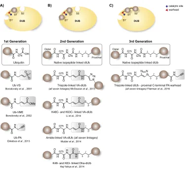
However, while activity-based probes have greatly increased our understanding of DUB reactivity and have enabled the discovery of new DUBs such as the OTU (Balakirev et al., 2003) and MINDY (MIU-containing novel DUB) classes (Abdul Rehman et al., 2016), these ABPs offer limited information on poly-Ubiquitin chain recognition and processing, since the existing di-Ubiquitin reagents contained isopeptide-linked Ubiquitin modules. While this characteristic allows the profiling of recombinant deubiquitinat-ing enzymes towards their linkage specificity and kinetics (Mevissen et al., 2013), a major limitation is its incompatibility with the cellular environment
which modulates DUB activity, thus necessitat-ing innovative tools specifically addressing these questions. With the advent of synthetic strategies, a 2nd generation of probes has emerged where, between two ubiquitin modules, a reactive group is positioned at the site of proteolytic action of the DUB allowing its covalent capture (Fig. 2.2B). An initial report by Iphofer et al. (2012) show a Michael acceptor linking the C-terminus of a distal Ub and short peptides representing K48 or K63 diUb. Later reports include the entire palette of Ub-chains allowing access to all seven lysine linked diubiquitin probes with a warhead in-between the distal and proximal ubiquitin module. Numerous research groups have independently reported ABPs utilizing a vinyl amide electrophilic trap between non-natively linked Ub moieties linked through a triazole, thiol ether (McGouran et al., 2013; Li et al., 2014) or an amide bond closely resembling the native isopeptide in both length as structure (Mulder et al., 2014). An alternative warhead is described by Haj-Yahya et al., here thiol elimination of Ub(G76C)-Ub results in dehydroalanine (Dha) as an electrophilic trap between two Ub modules (Haj-Yahya et al., 2014).
Although these covalent vinyl amide probes have allowed more detailed structural investigation of diubiquitin-specific DUB recognition (Mevis-sen et al., 2016), they do not allow investigation of additional Ubiquitin-binding sites, referred to as the S1’ (proximal), S1 (middle), and S2 (distal) binding sites (Kulathu, 2016). To investigate the contribution of the Ubiquitin binding sites to polyubiquitin chain processing by DUBs, a third generation of probes (Fig. 2.2C) generated by click chemistry and C-terminally modified with propargyl (PA) were devised (Flierman et al., 2016). Utility of this reagent enabled the struc-tural characterization of the K48 polyubiquitin cleaving mechanism of the SARS DUB PLpro, revealing that the S1-S1’ binding mode of K48-linked ubiquitin dictates the enzyme specificity for K48-Ubiquitin over ISG15, which binds only in the S1 site (Békés et al., 2016).
Despite the variety of di-ubiquitin-specific ABPs, designing effective tools to study the M1-linked chain type has posed a challenge primarily due to differences in chemistry imposed by the ‘linear’ peptide linkage. In attempts to create an linear diUb ABP, the methionine 1 (M1) of the proximal Ub
was replaced by the electrophilic dehydroalanine (Dha) residue. However, this probe was cleaved by OTULIN and USP2 rather than reacting cova-lently with the active site cysteine residues. A more recent design addressed this issue by replacing the Gly76 of the distal Ub by Dha (Weber et al., 2017). Although the UbG76Dha-Ub probe showed high selectivity for OTULIN, it did not label other M1-cleaving DUBs, indicating that Gly76 of the distal Ub is essential for recognition and cleav-age of linear diUb by other M1 cleaving DUBs. Interestingly, the first report on the fully synthetic preparation of linear diubiquitin reveals that the methionine to norleucine substitution of the proxi-mal Ub affects the hydrolysis rate of DUBs towards the linear diUb chain (Xin et al., 2018). Assessment of DUB-mediated cleavage of the synthetic (NLE1-linked) and expressed (M1-(NLE1-linked) linear diUb was assed using OTULIN, USP16 and USP21, known to specifically cleave the linear Ub linkage, dem-onstrated that synthetic NLE1-linked linear diUb was processed less efficiently than M1-linked linear diUb (Xin et al., 2018).
Collectively, these observations indicate a more profound role for methionine and Gly76 in the interaction between M1-linked diubiquitin and DUBs, complicating the way for the design of linear diUb-based activity-based probes and assay reagents. Furthermore, these ABPs together with the insights gained from both structural and biochemical studies underscore that the interaction dynamics of di-Ubiquitin chains are far more com-plex than previously assumed.
The numerous activity-based probes have furthered our mechanistic, kinetic, and biological understanding of DUBs as well as enabled the dis-covery of new DUB classes, yet these reagents do not target the JAMM/MPN and Machado-Jacob-Disease protein (MJD) metalloprotease DUBs. Developing such reagents akin to those for the other DUB families is urgently needed in order to dissect the role of these proteases in diseases.
Relaying ubiquitin to its substrate – ABPs targeting the ubiquitin conjugation machinery
Whereas DUBs have been extensively profiled using ABPs, the Ub-conjugating and ligating enzymes have only recently become the focus of ABP devel-opment. The delay in developing suitable reagents to profile the E1-E2-E3 enzymes is largely due to the challenges attributed with targeting a sequential enzymatic cascade rather than a single enzyme.
While ABPs originally designed to specifically target DUBs, such as HA-Ub-VME and Ub-VS, display cross-reactivity with HECT E3 ligases, they are not designed for monitoring Ub-conjugating and ligating enzyme activity concurrently (Boro-dovsky et al., 2001; Love et al., 2009), necessitating the development of ABPs and reagents specifically devised for the Ub conjugation machinery.
At the apex of the ubiquitination cascade, the E1 enzyme activates the C-terminal carboxylate of ubiquitin in an ATP-dependent manner. In this initial step, the Ub-AMP adenylate is formed under the consumption of ATP and magnesium. Subsequently, the intermediate undergoes nucleo-philic attack by the adjacent catalytic E1 active site cysteine resulting thioester bond and the simul-taneous release of AMP (Olsen and Lima, 2013). Early efforts towards developing Ub-based probes targeting the E1-enzyme were pioneered by Lu et al. (2010), who used a C-terminal 5′ -sulfonyladeno-sine modified Ub or Ubl. This design [Fig. 2.3A(I)] permitted the mechanistic study of the E1-catalysed adenylation and thioesterification by crosslinking it with the Ub/Ubl probe. A major drawback of the semisynthetic approach taken by Lu et al. (2010) is the alteration of the Ub/Ubl sequence. An and Statsyuk (2016) later published a method to efficiently generate the ABPs reported by Lu et al. (2010) while retaining the ‘native’ sequence, utiliz-ing a native chemical ligation strategy followed by the conversion of cysteine to Dha, permitting the trapping of the ‘tetrahedral E1-Ubl-AMP interme-diate’. Owing to the mechanism-based approach of these Ub/Ubl-AMP probes, it reacts directly with the E1-Ub/Ubl thioester intermediate resulting in the formation of the covalent Ub/Ubl–ABP1 conjugate structurally mimicking the Ub-AMP intermediate. Other advancements by Statsyuk and co-workers employed a mechanism-based approach [Fig. 2.3A(II)] using an AMP-derived
compound (ABP1), which due to its structural resemblance of the Ub/Ubl-adenylate reacts with the Ub/Ubl substrates rather than the respective E1 enzymes (An and Statsyuk, 2013). However, while this ABP has the advantage of being cell-permeable, cross-reactivity issues limits its utility to monitoring ubiquitination of substrates in vitro. Together, these approaches all mimic the Ub-Ubl-adenylate intermediate restricting these ABPs to the E1, enabling them to be processed downstream the cascade towards E2 and HECT- and RBR-E3 enzymes.
A) E1 probes Ub71 Ub73 H N N H O HO OH N N N N HNR O O N H H N O O R74 G75 R Ub-AVSN Lu et al., 2010
UbDha-AMP An et al., 2016
(I) Mimicking the tetrahedral intermediate of the E1–Ub–AMP complex.
(II) Mechanism based probe consuming free Ub
(III) ABPP of E1 enzyme tranthiolation activity O O HO OH N N N N HN SO O
H2N O
O HO OH N N N N HN SO O HN O ABP1 An et al., 2013
Ub76
E1
E2 SH TDAE E2 S EWG S EWG
S
E2 E2-AVS
Stanley et al., 2015
B) E2-E3 probes
N H
O NN
N HN S
O
O O
N H
O NN
N HN S
O N H O N N N H N O S S ∆Ub E2 E2 E3 ∆Ub Ub73 TDAE
Pao et al., 2016
Ub74 TDAE Pao et al., 2018
C) E1-E2-E3 cascading probe
pTolSO2H
Ub-AVS E2 N H OH O O Ub75 Ub75 E1 H N O S O Ub75 H N O S O Ub75 H N O S O Ub75 E1
Ub75 Ub75
H N O HO O S H N O HO O S H N O HO O S E2 E2 E3 E3 ATP AMP, PPi ATP ∆Ub Ub-Dha Mulder et al., 2016
E1 -S E1 N H O O O
Ub75 AMP
(I) (II) S H N N H H N N H O O O HO OH N N N N NH2 O O HS
Figure 2.3 Current activity-based probes targeting the ubiquitin cascade. (A) Targeting the E1 enzyme in a mechanism-based manner by (I) mimicking the tetrahedral intermediate of the E1–Ub–AMP complex or (II) using an AMP-derived compound (ABP1) or (III) utilizing the E1-transthiolation activity. (B) Capturing Ub–E2–E3 interactions by a modular approach, where Ub-TDAE reacts with an E2 generating an ABP reactive towards HECT- and RBR- E3 ligases. (C) Cascading E1–E2–E3 ABP sequentially reacting with the E1, E2 and E3
enzymes by either forming (i) the thioester yielding the transferable Ub-probe or as (ii) a thioether, which allows
exemplifies the utility and potential of ABPs and foreshadows the extent of future possibilities for these chemical tools (Pao et al., 2018).
In order to address the shortcomings of existing Ub-ABPs for studying multiple types of enzymes from the UPS simultaneously, Mulder et al. (2016) developed a mechanistically engaged ABP (Fig. 2.3C). Here the C-terminal Gly76 is replaced by Dha, thereby retaining a native car-boxy terminus thus allowing it to be processed by the native Ub conjugation machinery in the same ATP-dependent manner with E1–Ub-based and E2–Ub-based probes transiently formed in situ allowing relay to the E2 and E3 enzymes. Most notably, at each transthiolation step, the probe also has the option of reacting covalently with the active site Cys. However, in contrast to native ubiquitin, this cascading probe is inert towards lysine residues in target proteins, making it applicable to chemo-proteomics approaches. Additionally, its ATP-dependant reactivity is advantageous for proteome‐wide profiling experiments, as ATP-depletion permits facile background subtraction. Beyond its application for chemoproteomics, the utility of this unique cascading ABP has been showcased using living cells, where the effects of E1 enzyme inhibition on ubiquitination were visualized (Mulder et al., 2016). These experiments highlight the power of in-cell enzymology of the entire Ub cascade over-coming the limitation of labelling experiments in lysates, which are devoid of the organization and interaction of cellular structures.
The recent emergence of E2–Ub-ABPs and the novel Ub-ABP Ub-Dha greatly expand the Ub toolbox and provide new ways to decipher the cellular functions and structural/biochemical properties of HECT ligases in specific cellular contexts as well as potentially in normal and dis-ease state. However, of the three major classes of E3s, the current probes are only reactive towards HECT/RBR ligases, as these E3 ligases mechanis-tically rely on an active-site cysteine. RING E3s do not possess such an active site cysteine and merely serve as platforms to bring Ub charged E2’s and substrates together, thereby making them unsuited for direct probing using ABPs.
Assay reagents – real time monitoring of activity
Measuring catalytic activity of (de)ubiquitinating enzymes is key not only to understand their bio-logical function but also to inhibitor development efforts. In contrast to the probes described above these reagents lack a Michael acceptor element and thus do not form a covalent complex with their target enzymes, but instead rely on a fluorescent reporter tag allowing correlation of the enzymes native activity and/or specificity.
An important class of Ub based assay rea-gents are the fluorogenic assay reagents where a quenched fluorophore is conjugated via an amide bond at the C-terminal end of Ub. DUB activity and recognition will hydrolyse the amide bond at the C-terminus of Ub, releasing the fluorophore and simultaneously start to fluoresce. Hence the increase in fluorescence is a direct measure of DUB activity. One of the first fluorogenic reagents to measure the catalytic activity of DUBs is Ub ami-nomethyl coumarin (UbAMC) (Dang et al., 1998). Hassiepen et al. (2007) later report on a substituted rhodamine-110 (Rho110) scaffold with favourable fluorescent properties, making Ub-Rho110 a more preferred reagent in high throughput screening assays due to its non-overlapping spectrum with many small molecule inhibitors). In a similar set up, DUB mediated amino-luciferin release can be assayed in a bioluminescence approach using a luciferase assay, allowing the study of DUBs at lower concentrations (Orcutt et al., 2012). Another striking example illustrating the utility of fl uores-cent ubiquitin reagents are the non-hydrolyzable di-ubiquitin AMC reagents, which allow the moni-toring of chain specific proteolysis mediated by S1–S2 interactions on the DUB. In analogy to the diUb-PRG covalent probes, these substrates allowed mechanistic dissection of DUB specificity and cleavage rate, exemplified by the finding that the S2 ubiquitin binding pocket of OTUD3 confers its preference for K11 Ub-linkages as well as accel-erating Ub hydrolysis (Flierman et al., 2016).
DUBs would. Given that Ub-linkages govern a plethora of biological processes finetuning the cel-lular responses to a variety of stimuli, assessing the dynamics of Ubiquitin chain processing by DUBs is critical. Therefore, fluorescent polarization (FP) reagents were developed where Ub is conjugated via a native isopeptide linkage to a fluorophore car-rying substrate derived peptide (Tirat et al., 2005; Geurink et al., 2012). Assays with these reagents are based on a change in fluorescence polarization on cleavage of the isopeptide bond between Ub and a fluorophore labelled peptide. While the unpro-cessed large Ub-FP reagents tumble slowly giving high fluorescence polarization, the processed small fluorophore containing peptide tumbles faster and hence the polarization of light decreases. The synthetic advancements enabled the generation of a palette of FP reagents as well as the generation of FP reagents based on UBLs like the three SUMO isoforms, NEDD8 and ISG15 (Geurink et al., 2012).
Another class of reagents are Fluorescent Reso-nance Energy Transfer (FRET)-based reagents that make use of a fluorophore and quenching moiety in close proximity of each other. On DUB proteolysis, the FRET signal decreases over time, which can be measured in a fluorescence spectrometer enabling the study of enzyme linkage specific kinetics in real time. Geurink et al. (2016) prepared all seven iso-peptide-linked diUb FRET assay reagents by native chemical ligation using Rhodamine-Ub as the FRET-donor and TAMRA-Ub as the FRET-accep-tor, permitting insights into the catalytic efficiency of vOTU. From the kinetic measurements it became apparent that the preference for K6-linked di-Ubiquitin chains over K48 chains resulted from an increased catalytic turnover rate kcat and not Ub-binding (KM) (Geurink et al., 2016).
Using a similar technology, a high-throughput screening (HTS) assay for the E2 enzyme UBC13 was developed by combining a fluorochrome (Fl)-conjugated ubiquitin (fluorescence acceptor) with terbium (Tb)-conjugated ubiquitin (fluorescence donor) in a TR-FRET assay, such that the assembly of mixed chains of Fl- and Tb-ubiquitin creates a robust TR-FRET signal. In this particular study, this reagent enabled the identification of E2 inhibi-tors (Madiraju et al., 2012).
While numerous reagents to assay the cata-lytic activity of DUBs have been reported, the
development of reagents enabling the monitoring of Ubiquitin ligase activity has been lagging behind due to the complexity of these enzymes. An elegant attempt to generate reagents to efficiently monitor the transthiolation activity of HECT- and RBR-E3 ligases is the development of the ‘Bypassing System’ (ByS) by Park et al. (2015). This approach exploits a simple design – a Ub thioester mimic in the form of UbMES (mercaptoethanesulfonate), permitting the direct transthiolation of the catalytic cysteine of the E3 ligase while eliminating the need for the E1 and E2 enzymes as well as ATP. Further devel-opment of this concept led to the generation of a fluorescent Ub thioester permitting the detection of both transthiolation and ligation activities of HECT E3 ligases (Krist et al., 2016). Given the facile detection method and the requirement for only the E3 enzyme and UbFluor, this mechanism-based reagent is well suited for high throughput screens (HTS) for Ub ligase inhibitors (Foote et al., 2017).
What does the future hold?
Unravelling the complexity of the highly sophis-ticated ubiquitination system is aided greatly by the development of numerous ABPs and reagents reporting on the dynamics and structural mecha-nisms of (de)ubiquitinating enzymes involved. Given the intrinsic role of Ub in the pathogenesis of a variety of diseases, most notably cancer and neurodegenerative diseases, enzymes involved in this system are emerging drug targets. The utility of these activity-based probes and reagents has been showcased by the discovery and validation of a USP7 inhibitor utilizing both Ub-AMC in the ini-tial high-throughput screen and later Ub-VS in the validation studies (Reverdy et al., 2012; Lamberto et al., 2017). Without a doubt the next generation of Ub based tools will help increase our knowledge, ultimately leading to new diagnostic tools or thera-peutics making it to the clinic.
of heterotypic and hybrid Ub chains; developing ABPs capable of capturing metalloprotease DUBs; advancing tools for specifically targeting distinct HECT and RBR-E3 ligases; and lastly, optimizing cell delivery methodologies for ABPs to enable in-cell enzymology.
Customized tools – warranting study on a new complex layer of DUB recognition
The advent of numerous ABPs and reagents for interrogating the different aspects of deubiquitinat-ing enzymes, have enabled profound insights into the structural, biochemical and biological role of these ‘erasers’. More recently, the generation of tools specifically designed for dissecting the proteolytic processing of ubiquitin chains by DUBs have revealed profound differences among these pro-teases in their specificity. Adding to this complexity, the discovery of heterotypic and hybrid Ubiquitin chains warrants the development of customized tools in order to understand the regulatory roles of DUBs in this context.
Given the recent insights that heterotypic Ubiq-uitin chains play a profound role in fine-tuning cellular responses (Xu et al., 2009), investigations into its biological and structural role need to be undertaken. To propel the study of their role, innovative ABPs recapitulating the structural and functional aspects of these mixed and branched Ubiquitin-chains need to be generated. Fur-thermore, the recent advances in synthetically obtaining Ubl proteins, permits the development of hybrid Ub/Ubl chains. Generation of such probes, especially for in-cell enzymology or proteomics context would be particularly conducive as the E3 ligases and DUBs regulating these heterotypic and hybrid Ub-chains are unknown (Xu et al., 2009). Furthermore, generating such complex linkages is a challenging feat as the E2/E3 enzymes generating these linkages in vitro are largely unknown and the known ones produce a mixture of linkage types that are difficult to separate by chromatography (Fag-giano et al., 2016). Moreover, the modification of Ubiquitin or its Ubiquitin linkage by another PTM complicates the deciphering of the temporal order of events, which underlies the biological role of this modification. The urgent need for such ABPs and assay reagents is illustrated by the recently discov-ered MINDY DUBs, which preferentially cleave
K48 and K63 tetra-Ub linkages, raising the ques-tion whether they might display reactivity towards K48/K63 linkages (Xu et al., 2009; Ohtake and Tsuchiya, 2017). Since there are currently no ABPs recapitulating the mixed K48/K63 Ubiquitin link-age available, investigating this aspect is hampered.
Currently, the metalloprotease DUBs have been neglected in the development of ABPs and reagents partly due to the difficulty of designing these tools. Unlike other deubiquitinating enzymes, metallo-protease DUBs do not have an active-site cysteine, but instead hydrolyse the isopeptide bonds of ubiquitinated substrates with a water-coordinated zinc ion. Designing chemical probes with potent and specific zinc-ion chelating reactive groups is prerequisite to generating an innovative toolkit for metalloprotease DUBs. Generally, metallopro-teases are typically expressed as an inactive form (zymogen) inhibited by additional proteins and require proteolytic processing before rendering the active enzyme (Saghatelian et al., 2004). This additional layer of regulation, however, introduces another layer of complexity that must be taken into account when designing such reagents (Saghat-elian et al., 2004). Introducing such innovative chemical probes would propel the study of these understudied deubiquitinating enzymes and enable the development of therapeutics.
The quest for E3 ligase inhibitors – challenges and opportunities
reproducible high-throughput inhibitor screening, was developed (De Cesare et al., 2018). Yet, one of the most challenging aspects to consider in such an undertaking is the lack of comprehensive prereq-uisite knowledge of the interacting E2–E3 enzyme pairs, which substantially modulate the biological outcome (De Cesare et al., 2018).
Despite this progress, the current ABPs target-ing the ubiquitin conjugattarget-ing cascade utilize either a modular approach (e.g. E2–Ub probe conjugates) or are mechanistic-based relying on the active-site cysteine (Mulder et al., 2016) or the ATP-binding pocket (An and Statsyuk, 2013, 2016) thereby being limited to indiscriminately detecting HECT- and RBR- E3-ligases. This limitation could potentially be overcome by designing ABPs featur-ing increased selectivity for HECT/RBR-E3 ligases by utilizing specific Ub-variants generated by phage display (Zhang et al., 2016). Since some mechanis-tic aspects of E3 ligase-mediated catalysis is intrinsic to most E3 ligase probe designs, it excludes direct labelling of the scaffolding RING E3-ligases, which ironically comprise the vast majority of ligases that are pivotal in cancer development and progression (Wang et al., 2017). Yet, prerequisite for devising ABPs capable of selectively labelling RING E3 ligases is a priori knowledge of the specific inter-faces between E2 and RING-E3 enzyme amenable to protein–protein interaction disruption.
Probing ubiquitination in living cells Most ABP profiling experiments are performed using either recombinant enzymes or cell lysates, yet this does not recapitulate the activity of the enzymes in a cellular context. Since lysing cells results in disruption of the cellular compartmental-ization as well as in dilution of the enzymes which might affect enzyme reactivity, delivery of DUB and ubiquitin ligase ABPs into intact cells is of critical importance. However, to achieve this, several meth-ods including electroporation (Mulder et al., 2016) or the use of cell-penetrating peptides attached to the Ub-ABP (Gui et al., 2018; Hameed et al., 2018) have been reported. Additionally, the introduction of ABPs into living cells permit the visualization and in-cell enzymology of the ubiquitin cascade enzymes in a spatial and temporal context. The critical need for an intact cellular environment for proper enzymatic function of Ubiquitin enzymes arises from the interaction with protein complexes
as well as their substrates, but also the intrinsic regulation by cellular signalling events such as phosphorylation (Sowa et al., 2009; Heideker and Wertz, 2015). The significance of additional post-translational modification, e.g. phosphorylation, of DUBs to enhance their proteolytic activity is highlighted by the necessity of serine phospho-rylation of OTUD5/DUBA (Huang et al., 2012). Furthermore, cross-regulation of DUBs with E2 enzymes (Wiener et al., 2012) and E3-ligases (Heideker and Wertz, 2015) underscore the sig-nificance of studying the ubiquitin cascade in living cells. One notable example of aforementioned interactions is the well characterized deubiquitinat-ing enzyme USP7, which binds to the E3 ligase MDM2 and its substrate tumour suppressor p53 through its TRAF-domain (Sheng et al., 2006). Considering the significance of an functional cel-lular environment for the enzymatic function of the ubiquitin enzymes, their biochemical study should be conducted in living cells thus meriting ABPs compatible with in-cell enzymology. Another facet necessitating in-cell enzymology using ABPs is the application in proteomics to access not only the functional consequence of these interactions, particularly in the context of pharmacological inhi-bition (De Cesare et al., 2018).
Conclusion
platform of reagents and ABPs have the potential to accelerate drug discovery efforts targeting all aspects of the ubiquitin cascade. Yet, the frontier of Ubiquitin activity-based probe and reagent development lies in the introduction of innovative technologies and unique concepts enabling the dis-section of many enigmatic aspects of ubiquitination as well as accessing enzymes previously not targeted by conventional ABP designs.
References
Abdul Rehman, S.A., Kristariyanto, Y.A., Choi, S.Y., Nkosi, P.J., Weidlich, S., Labib, K., Hofmann, K., and Kulathu, Y. (2016). MINDY-1 is a member of an evolutionarily conserved and structurally distinct new family of deubiquitinating enzymes. Mol. Cell 63, 146–155. https://doi.org/10.1016/j.molcel.2016.05.009. An, H., and Statsyuk, A.V. (2013). Development of
activity-based probes for ubiquitin and ubiquitin-like protein signaling pathways. J. Am. Chem. Soc. 135, 16948– 16962. https://doi.org/10.1021/ja4099643.
An, H., and Statsyuk, A.V. (2016). Facile synthesis of covalent probes to capture enzymatic intermediates during E1 enzyme catalysis. Chem. Commun. 52, 2477–2480. https://doi.org/10.1039/c5cc08592f. Balakirev, M.Y., Tcherniuk, S.O., Jaquinod, M., and
Chroboczek, J. (2003). Otubains: a new family of cysteine proteases in the ubiquitin pathway. EMBO Rep. 4, 517–522. https://doi.org/10.1038/sj.embor. embor824.
Békés, M., van der Heden van Noort, G.J., Ekkebus, R., Ovaa, H., Huang, T.T., and Lima, C.D. (2016). Recognition of Lys48-linked di-ubiquitin and deubiquitinating activities of the SARS coronavirus papain-like protease. Mol. Cell 62, 572–585. https://doi.org/10.1016/j. molcel.2016.04.016.
Bhogaraju, S., Kalayil, S., Liu, Y., Bonn, F., Colby, T., Matic, I., and Dikic, I. (2016). Phosphoribosylation of ubiquitin promotes serine ubiquitination and impairs conventional ubiquitination. Cell 167, 1636–1649.e13. Boll, E., Drobecq, H., Ollivier, N., Blanpain, A., Raibaut, L.,
Desmet, R., Vicogne, J., and Melnyk, O. (2015). One-pot chemical synthesis of small ubiquitin-like modifier protein-peptide conjugates using bis(2-sulfanylethyl) amido peptide latent thioester surrogates. Nat. Protoc.
10, 269–292. https://doi.org/10.1038/nprot.2015.013. Boname, J.M., Thomas, M., Stagg, H.R., Xu, P., Peng, J.,
and Lehner, P.J. (2010). Efficient internalization of MHC I requires lysine-11 and lysine-63 mixed linkage polyubiquitin chains. Traffic 11, 210–220. https://doi. org/10.1111/j.1600-0854.2009.01011.x.
Bondalapati, S., Eid, E., Mali, S.M., Wolberger, C., and Brik, A. (2017). Total chemical synthesis of SUMO-2-Lys63-linked diubiquitin hybrid chains assisted by removable solubilizing tags. Chem. Sci. 8, 4027–4034. https://doi. org/10.1039/c7sc00488e.
Borodovsky, A., Kessler, B.M., Casagrande, R., Overkleeft, H.S., Wilkinson, K.D., and Ploegh, H.L. (2001). A novel active site-directed probe specific for deubiquitylating enzymes reveals proteasome association of USP14.
EMBO J. 20, 5187–5196. https://doi.org/10.1093/ emboj/20.18.5187.
Borodovsky, A., Ovaa, H., Kolli, N., Gan-Erdene, T., Wilkinson, K.D., Ploegh, H.L., and Kessler, B.M. (2002). Chemistry-based functional proteomics reveals novel members of the deubiquitinating enzyme family. Chem. Biol. 9, 1149–1159.
Bremm, A., Freund, S.M., and Komander, D. (2010). Lys11-linked ubiquitin chains adopt compact conformations and are preferentially hydrolyzed by the deubiquitinase Cezanne. Nat. Struct. Mol. Biol. 17, 939–947. https:// doi.org/10.1038/nsmb.1873.
Briand, J.P., Van Dorsselaer, A., Raboy, B., and Muller, S. (1989). Total chemical synthesis of ubiquitin using BOP reagent: biochemical and immunochemical properties of the purified synthetic product. Pept. Res. 2, 381–388. Buchsbaum, S., Bercovich, B., and Ciechanover, A. (2012). FAT10 is a proteasomal degradation signal that is itself regulated by ubiquitination. Mol. Biol. Cell 23, 225–232. https://doi.org/10.1091/mbc.E11-07-0609.
Chatterjee, C., McGinty, R.K., Pellois, J.P., and Muir, T.W. (2007). Auxiliary-mediated site-specific peptide ubiquitylation. Angew. Chem. Int. Ed. Engl. 46, 2814– 2818. https://doi.org/10.1002/anie.200605155. Dang, L.C., Melandri, F.D., and Stein, R.L. (1998).
Kinetic and mechanistic studies on the hydrolysis of ubiquitin C-terminal 7-amido-4-methylcoumarin by deubiquitinating enzymes. Biochemistry 37, 1868– 1879. https://doi.org/10.1021/bi9723360.
Dawson, P.E., Muir, T.W., Clark-Lewis, I., and Kent, S.B. (1994). Synthesis of proteins by native chemical ligation. Science 266, 776–779.
De Cesare, V., Johnson, C., Barlow, V., Hastie, J., Knebel, A., and Trost, M. (2018). The MALDI-TOF E2/E3 ligase assay as universal tool for drug discovery in the ubiquitin pathway. Cell Chem. Biol. 25, 1117–1127.e4.
de Jong, A., Witting, K., Kooij, R., Flierman, D., and Ovaa, H. (2017). Release of enzymatically active deubiquitinating enzymes upon reversible capture by disulfide ubiquitin reagents. Angew. Chem. Int. Ed. Engl. 56, 12967–12970. https://doi.org/10.1002/anie.201706738.
Dobrotă, C., Fasci, D., Hădade, N.D., Roiban, G.D., Pop, C., Meier, V.M., Dumitru, I., Matache, M., Salvesen, G.S., and Funeriu, D.P. (2012). Glycine fluoromethylketones as SENP-specific activity based probes. Chembiochem
13, 80–84. https://doi.org/10.1002/cbic.201100645. Ekkebus, R., van Kasteren, S.I., Kulathu, Y., Scholten, A.,
Berlin, I., Geurink, P.P., de Jong, A., Goerdayal, S., Neefjes, J., Heck, A.J., et al. (2013). On terminal alkynes that can react with active-site cysteine nucleophiles in proteases. J. Am. Chem. Soc. 135, 2867–2870. https:// doi.org/10.1021/ja309802n.
El Oualid, F., Merkx, R., Ekkebus, R., Hameed, D.S., Smit, J.J., de Jong, A., Hilkmann, H., Sixma, T.K., and Ovaa, H. (2010). Chemical synthesis of ubiquitin, ubiquitin-based probes, and diubiquitin. Angew. Chem. Int. Ed. Engl. 49, 10149–10153. https://doi.org/10.1002/ anie.201005995.
Fan, J.B., Arimoto, K., Motamedchaboki, K., Yan, M., Wolf, D.A., and Zhang, D.E. (2015). Identification and characterization of a novel ISG15-ubiquitin mixed chain and its role in regulating protein homeostasis. Sci. Rep.
5, 12704. https://doi.org/10.1038/srep12704. Flierman, D., van der Heden van Noort, G.J., Ekkebus,
R., Geurink, P.P., Mevissen, T.E., Hospenthal, M.K., Komander, D., and Ovaa, H. (2016). Non-hydrolyzable diubiquitin probes reveal linkage-specific reactivity of deubiquitylating enzymes mediated by S2 pockets. Cell Chem. Biol. 23, 472–482. https://doi.org/10.1016/j. chembiol.2016.03.009.
Foote, P.K., Krist, D.T., and Statsyuk, A.V. (2017). High-throughput screening of HECT E3 ubiquitin ligases using UbFluor. Curr. Protoc. Chem. Biol. 9, 174–195. https://doi.org/10.1002/cpch.24.
Geurink, P.P., El Oualid, F., Jonker, A., Hameed, D.S., and Ovaa, H. (2012). A general chemical ligation approach towards isopeptide-linked ubiquitin and ubiquitin-like assay reagents. Chembiochem 13, 293–297. https://doi. org/10.1002/cbic.201100706.
Geurink, P.P., van Tol, B.D., van Dalen, D., Brundel, P.J., Mevissen, T.E., Pruneda, J.N., Elliott, P.R., van Tilburg, G.B., Komander, D., and Ovaa, H. (2016). Development of diubiquitin-based FRET probes to quantify ubiquitin linkage specificity of deubiquitinating enzymes. Chembiochem 17, 816–820. https://doi.org/10.1002/ cbic.201600017.
Giles, J. (2004). Chemistry Nobel for trio who revealed molecular death-tag. Nature 431, 729.
Goldenberg, S.J., Marblestone, J.G., Mattern, M.R., and Nicholson, B. (2010). Strategies for the identification of ubiquitin ligase inhibitors. Biochem Soc Trans 38, 132–136. https://doi.org/10.1042/BST0380132. Goru, S.K., Pandey, A., and Gaikwad, A.B. (2016). E3
ubiquitin ligases as novel targets for inflammatory diseases. Pharmacol. Res. 106, 1–9.
Grabe, G.J., Zhang, Y., Przydacz, M., Rolhion, N., Yang, Y., Pruneda, J.N., Komander, D., Holden, D.W., and Hare, S.A. (2016). The Salmonella effector SpvD is a cysteine hydrolase with a serovar-specific polymorphism influencing catalytic activity, suppression of immune responses, and bacterial virulence. J. Biol. Chem. 291, 25853–25863.
Gui, W., Ott, C.A., Yang, K., Chung, J.S., Shen, S., and Zhuang, Z. (2018). Cell-permeable activity-based ubiquitin probes enable intracellular profiling of human deubiquitinases. J. Am. Chem. Soc. 140, 12424–12433. https://doi.org/10.1021/jacs.8b05147.
Haj-Yahya, M., Eltarteer, N., Ohayon, S., Shema, E., Kotler, E., Oren, M., and Brik, A. (2012). N-methylation of isopeptide bond as a strategy to resist deubiquitinases. Angew. Chem. Int. Ed. Engl. 51, 11535–11539. https:// doi.org/10.1002/anie.201205771.
Haj-Yahya, N., Hemantha, H.P., Meledin, R., Bondalapati, S., Seenaiah, M., and Brik, A. (2014). Dehydroalanine-based diubiquitin activity probes. Org. Lett. 16, 540–543. https://doi.org/10.1021/ol403416w. Hameed, D.S., Sapmaz, A., and Ovaa, H. (2017). How
chemical synthesis of ubiquitin conjugates helps to understand ubiquitin signal transduction. Bioconjug. Chem. 28, 805–815. https://doi.org/10.1021/acs. bioconjchem.6b00140.
Hameed, D.S., Sapmaz, A., Gjonaj, L., Merkx, R., and Ovaa, H. (2018). Enhanced Delivery of Synthetic Labelled Ubiquitin into Live Cells by Using Next-Generation Ub-TAT Conjugates. Chembiochem. 19, 2553–2557. Harrigan, J.A., Jacq, X., Martin, N.M., and Jackson, S.P.
(2018). Deubiquitylating enzymes and drug discovery: emerging opportunities. Nat. Rev. Drug Discov. 17, 57–78. https://doi.org/10.1038/nrd.2017.152. Hassiepen, U., Eidhoff, U., Meder, G., Bulber, J.F., Hein, A.,
Bodendorf, U., Lorthiois, E., and Martoglio, B. (2007). A sensitive fluorescence intensity assay for deubiquitinating proteases using ubiquitin-rhodamine110-glycine as substrate. Anal. Biochem. 371, 201–207.
Heap, R.E., Gant, M.S., Lamoliatte, F., Peltier, J., and Trost, M. (2017). Mass spectrometry techniques for studying the ubiquitin system. Biochem. Soc. Trans. 45, 1137– 1148. https://doi.org/10.1042/BST20170091. Heideker, J., and Wertz, I.E. (2015). DUBs, the regulation of
cell identity and disease. Biochem. J. 467, 191. Herhaus, L., and Dikic, I. (2015). Expanding the ubiquitin
code through post-translational modification. EMBO Rep. 16, 1071–1083. https://doi.org/10.15252/ embr.201540891.
Hermanns, T., Pichlo, C., Woiwode, I., Klopffleisch, K., Witting, K.F., Ovaa, H., Baumann, U., and Hofmann, K. (2018). A family of unconventional deubiquitinases with modular chain specificity determinants. Nat. Commun.
9, 799. https://doi.org/10.1038/s41467-018-03148-5. Hewings, D.S., Flygare, J.A., Bogyo, M., and Wertz, I.E.
(2017). Activity-based probes for the ubiquitin conjugation-deconjugation machinery: new chemistries, new tools, and new insights. FEBS J. 284, 1555–1576. https://doi.org/10.1111/febs.14039.
Hicke, L. (2001). Protein regulation by monoubiquitin. Nat. Rev. Mol. Cell Biol. 2, 195–201. https://doi. org/10.1038/35056583.
Hospenthal, M.K., Freund, S.M., and Komander, D. (2013). Assembly, analysis and architecture of atypical ubiquitin chains. Nat. Struct. Mol. Biol. 20, 555–565. https://doi. org/10.1038/nsmb.2547.
Huang, O.W., Ma, X., Yin, J., Flinders, J., Maurer, T., Kayagaki, N., Phung, Q., Bosanac, I., Arnott, D., Dixit, V.M., et al. (2012). Phosphorylation-dependent activity of the deubiquitinase DUBA. Nat. Struct. Mol. Biol. 19, 171–175. https://doi.org/10.1038/nsmb.2206. Huguenin-Dezot, N., De Cesare, V., Peltier, J., Knebel,
A., Kristaryianto, Y.A., Rogerson, D.T., Kulathu, Y., Trost, M., and Chin, J.W. (2016). Synthesis of isomeric phosphoubiquitin chains reveals that phosphorylation controls deubiquitinase activity and specificity. Cell Rep. 16, 1180–1193.
Iphofer, A., Kummer, A., Nimtz, M., Ritter, A., Arnold, T., Frank, R., van den Heuvel, J., Kessler, B.M., Jansch, L., and Franke, R. (2012). Profiling ubiquitin linkage specificities of deubiquitinating enzymes with branched ubiquitin isopeptide probes. Chembiochem. 13, 1416– 1420.
Jackson, S.P., and Durocher, D. (2013). Regulation of DNA damage responses by ubiquitin and SUMO. Mol. Cell 49, 795–807. https://doi.org/10.1016/j. molcel.2013.01.017.
during the cell cycle. Biochim. Biophys. Acta Mol. Cell Res. 1865, 1924–1933.
Kim, H.T., Kim, K.P., Lledias, F., Kisselev, A.F., Scaglione, K.M., Skowyra, D., Gygi, S.P., and Goldberg, A.L. (2007). Certain pairs of ubiquitin-conjugating enzymes (E2s) and ubiquitin-protein ligases (E3s) synthesize nondegradable forked ubiquitin chains containing all possible isopeptide linkages. J. Biol. Chem. 282, 17375–17386.
Komander, D. (2009). The emerging complexity of protein ubiquitination. Biochem Soc Trans 37, 937-953.10.1042/BST0370937.
Komander, D., and Rape, M. (2012). The ubiquitin code. Annu. Rev. Biochem. 81, 203–229. https://doi. org/10.1146/annurev-biochem-060310-170328. Komander, D., Clague, M.J., and Urbé, S. (2009). Breaking
the chains: structure and function of the deubiquitinases. Nat. Rev. Mol. Cell Biol. 10, 550–563. https://doi. org/10.1038/nrm2731.
Krist, D.T., Park, S., Boneh, G.H., Rice, S.E., and Statsyuk, A.V. (2016). UbFluor: a mechanism-based probe for HECT E3 ligases. Chem. Sci. 7, 5587–5595. https:// doi.org/10.1039/C6SC01167E.
Krist, D.T., Foote, P.K., and Statsyuk, A.V. (2017). UbFluor: A fluorescent thioester to monitor HECT E3 ligase catalysis. Curr. Protoc. Chem. Biol. 9, 11–37. https:// doi.org/10.1002/cpch.17.
Kulathu, Y. (2016). Novel diubiquitin probes expand the chemical toolkit to study DUBs. Cell Chem. Biol. 23, 432–434. https://doi.org/10.1016/j. chembiol.2016.04.001.
Kumar, K.S., Spasser, L., Erlich, L.A., Bavikar, S.N., and Brik, A. (2010). Total chemical synthesis of di-ubiquitin chains. Angew. Chem. Int. Ed. Engl. 49, 9126–9131. https://doi.org/10.1002/anie.201003763.
Kwon, Y.T., and Ciechanover, A. (2017). The ubiquitin code in the ubiquitin-proteasome system and autophagy. Trends Biochem. Sci. 42, 873–886.
Lam, Y.A., Xu, W., DeMartino, G.N., and Cohen, R.E. (1997). Editing of ubiquitin conjugates by an isopeptidase in the 26S proteasome. Nature 385, 737– 740. https://doi.org/10.1038/385737a0.
Lamberto, I., Liu, X., Seo, H.S., Schauer, N.J., Iacob, R.E., Hu, W., Das, D., Mikhailova, T., Weisberg, E.L., Engen, J.R., et al. (2017). Structure-guided development of a potent and selective non-covalent active-site inhibitor of USP7. Cell Chem. Biol. 24, 1490–1500.e11.
Leidecker, O., Matic, I., Mahata, B., Pion, E., and Xirodimas, D.P. (2012). The ubiquitin E1 enzyme Ube1 mediates NEDD8 activation under diverse stress conditions. Cell Cycle 11, 1142–1150. https://doi.org/10.4161/ cc.11.6.19559.
Levin-Kravets, O., Tanner, N., Shohat, N., Attali, I., Keren-Kaplan, T., Shusterman, A., Artzi, S., Varvak, A., Reshef, Y., Shi, X., et al. (2016). A bacterial genetic selection system for ubiquitylation cascade discovery. Nat. Methods 13, 945–952. https://doi.org/10.1038/ nmeth.4003.
Li, G., Liang, Q., Gong, P., Tencer, A.H., and Zhuang, Z. (2014). Activity-based diubiquitin probes for elucidating the linkage specificity of deubiquitinating enzymes. Chem. Commun. 50, 216–218. https://doi. org/10.1039/c3cc47382a.
Liu, Q., Kistemaker, H.A.V., Bhogaraju, S., Dikic, I., Overkleeft, H.S., van der Marel, G.A., Ovaa, H., van der Heden van Noort, G.J., and Filippov, D.V. (2018). A general approach towards triazole-linked adenosine diphosphate ribosylated peptides and proteins. Angew. Chem. Int. Ed. Engl. 57, 1659–1662. https://doi. org/10.1002/anie.201710527.
Love, K.R., Pandya, R.K., Spooner, E., and Ploegh, H.L. (2009). Ubiquitin C-terminal electrophiles are activity-based probes for identification and mechanistic study of ubiquitin conjugating machinery. ACS Chem. Biol. 4, 275–287. https://doi.org/10.1021/cb9000348. Lu, X., Olsen, S.K., Capili, A.D., Cisar, J.S., Lima, C.D.,
and Tan, D.S. (2010). Designed semisynthetic protein inhibitors of Ub/Ubl E1 activating enzymes. J. Am. Chem. Soc. 132, 1748–1749. https://doi.org/10.1021/ ja9088549.
Maculins, T., Carter, N., Dorval, T., Hudson, K., Nissink, J.W., Hay, R.T., and Alwan, H. (2016). A generic platform for cellular screening against ubiquitin ligases. Sci. Rep.
6, 18940. https://doi.org/10.1038/srep18940. Madiraju, C., Welsh, K., Cuddy, M.P., Godoi, P.H.,
Pass, I., Ngo, T., Vasile, S., Sergienko, E.A., Diaz, P., Matsuzawa, S., et al. (2012). TR-FRET-based high-throughput screening assay for identification of UBC13 inhibitors. J. Biomol. Screen. 17, 163–176. https://doi. org/10.1177/1087057111423417.
Marblestone, J.G., Larocque, J.P., Mattern, M.R., and Leach, C.A. (2012). Analysis of ubiquitin E3 ligase activity using selective polyubiquitin binding proteins. Biochim. Biophys. Acta 1823, 2094–2097. https://doi. org/10.1016/j.bbamcr.2012.06.013.
McCann, A.P., Scott, C.J., Van Schaeybroeck, S., and Burrows, J.F. (2016). Deubiquitylating enzymes in receptor endocytosis and trafficking. Biochem. J. 473, 4507–4525.
McGouran, J.F., Gaertner, S.R., Altun, M., Kramer, H.B., and Kessler, B.M. (2013). Deubiquitinating enzyme specificity for ubiquitin chain topology profiled by di-ubiquitin activity probes. Chem. Biol. 20, 1447–1455. https://doi.org/10.1016/j.chembiol.2013.10.012. Mevissen, T.E., Hospenthal, M.K., Geurink, P.P., Elliott,
P.R., Akutsu, M., Arnaudo, N., Ekkebus, R., Kulathu, Y., Wauer, T., El Oualid, F., et al. (2013). OTU deubiquitinases reveal mechanisms of linkage specificity and enable ubiquitin chain restriction analysis. Cell 154, 169–184. https://doi.org/10.1016/j.cell.2013.05.046. Mevissen, T.E.T., Kulathu, Y., Mulder, M.P.C., Geurink,
P.P., Maslen, S.L., Gersch, M., Elliott, P.R., Burke, J.E., van Tol, B.D.M., Akutsu, M., et al. (2016). Molecular basis of Lys11-polyubiquitin specificity in the deubiquitinase Cezanne. Nature 538, 402–405. https:// doi.org/10.1038/nature19836.
Meyer, H.J., and Rape, M. (2014). Enhanced protein degradation by branched ubiquitin chains. Cell 157, 910–921. https://doi.org/10.1016/j.cell.2014.03.037. Michel, M.A., Elliott, P.R., Swatek, K.N., Simicek,