R E S E A R C H
Open Access
Tracking sex-dependent differences in a
mouse model of CLN6-Batten disease
McKayla J. Poppens
1†, Jacob T. Cain
1†, Tyler B. Johnson
1, Katherine A. White
1, Samantha S. Davis
1,
Rachel Laufmann
1, Alexander D. Kloth
2and Jill M. Weimer
1,3*Abstract
Background:CLN6-Batten disease is a rare neurodevelopmental disorder characterized pathologically by the accumulation of lysosomal storage material, glial activation and neurodegeneration, and phenotypically by loss of vision, motor coordination, and cognitive ability, with premature death occurring in the second decade of life. In this study, we investigate whether sex differences in a mouse model of CLN6-Batten disease impact disease onset and progression.
Results:A number of noteworthy differences were observed including elevated accumulation of mitochondrial ATP synthase subunit C in the thalamus and cortex of femaleCln6mutant mice at 2 months of age. Moreover, female mutant mice showed more severe behavioral deficits. Beginning at 9 months of age, female mice demonstrated learning and memory deficits and suffered a more severe decline in motor coordination. Further, compared to their male counterparts, female animals succumbed to the disease at a slightly younger age, indicating an accelerated disease progression. Conversely, males showed a marked increase in microglial activation at 6 months of age in the cortex relative to females.
Conclusions:Thus, as femaleCln6mutant mice exhibit cellular and behavioral deficits that precede similar
pathologies in male mutant mice, our findings suggest the need for consideration of sex-based differences in CLN6 disease progression during development of preclinical and clinical studies.
Keywords:Neuronal ceroid lipofuscinoses, Rare disease, Lysosomal storage disorder, Neurodegenerative disease, Pediatric disease
Background
Batten disease (neuronal ceroid lipofuscinoses) comprises a family of autosomal-recessive neurodegenerative dis-eases characterized by lysosomal accumulation of auto-fluorescent lipopigment [1–4]. Pathological hallmarks of Batten disease include neuronal death in cortical and thal-amic regions of the brain and massive gliosis throughout the CNS, presenting functionally as degeneration of vi-sion, psychomotor delay, and premature death [5–7]. CLN6-Batten disease, resulting from mutations inCLN6, constitutes two distinct diseases: a pediatric form, also referred to as variant late infantile neuronal ceroid
lipofuscinoses, and a rare, less-severe adult-onset form re-ferred to as Kufs type A disease [8,9]. The pediatric vari-ant of CLN6 disease begins between the ages of 18 months and 8 years, presenting with language impair-ment, motor deterioration and cognitive deficiencies, followed by vision loss, seizures, and ultimately premature death during the second decade of life [10]. There are a number of naturally occurring CLN6 animal models used in therapeutic development, including theCln6nclfmouse model that contains a similar point mutation as found in human patients and develops the classical pathophysio-logical hallmarks of Batten disease, such as intracellular inclusion, retinal degeneration, hind-limb paralysis, and premature death [11–13]. Many of the past Batten disease studies have excluded female mice from therapeutic stud-ies to avoid confounding variables related to sex hormones and chromosomal differences [14, 15]. However, these biological disease modifiers can potentially limit the
* Correspondence:[email protected]
†McKayla J. Poppens and Jacob T. Cain contributed equally to this work.
1Pediatrics and Rare Diseases Group, Sanford Research, Sioux Falls, SD, USA 3Department of Pediatrics, Sanford School of Medicine, University of South
Dakota, Sioux Falls, SD, USA
Full list of author information is available at the end of the article
translatability of mouse findings to female patients, as sex-based differences that may affect disease susceptibility, disease severity, and therapeutic efficacy [14]. For ex-ample, sex specific symptomatic differences have been reported in other neurodegenerative diseases including Alzheimer’s disease, Parkinson’s disease, multiple sclerosis, and autism spectrum disorders [14, 16–19]. In addition, patient response to therapeutics has varied by sex, as estrogen, testosterone, and other sex-linked genes may affect drug effectiveness [20].
In the Batten disease field, studies on CLN3-Batten disease, a genetically distinct subtype of Batten disease, have shown differences in disease progression in patients depending on sex [8, 9, 21, 22]. On average, female CLN3-Batten disease patients present with symptoms 1 year later than their male counterparts, have accelerated disease progression following symptom onset, and die 1 year earlier than males [21]. Initial characterization of the Cln3Δ7/8 murine model did not consider sex-based differences, however, recent work with this model has demonstrated that females exhibit poorer performance in behavioral tests [23, 24]. Additionally, the naturally occurring mouse model of CLN8-Batten disease has shown sex differences in femaleCln8mndmice, where fe-male retinas exhibited higher levels of retinal oxidative stress and caspase-3 activity compared to males [25]. These findings prompted us to investigate sex discrepan-cies in outcomes associated with CLN6 disease, explor-ing differences in disease onset and progression between male and femaleCln6nclfmice. We describe subtle histo-pathological differences between the sexes and a more rapid disease progression in femaleCln6nclfmice. Conse-quently, including sex as a factor during studies and sub-sequent analyses can ensure proper development of therapeutic treatments for patients with CLN6 disease.
Results
Cln6nclfmice have sex and age dependent pathological differences in the brain
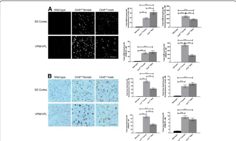
Sex dependent differences in the classic pathological hallmarks of Batten disease were examined in the thal-amus and somatosensory cortex of wild-type and
Cln6nclf mice, two areas of the brain that are affected early in Batten disease. Accumulation of autofluorescent storage material (ASM) in the brain is a manifestation common to all variants of Batten disease. At two and 6 months of age,Cln6nclf mice of both sexes had accumu-lation of ASM within the ventral posteromedial and ven-tral posterolateral (VPM/VPL) nuclei of the thalamus and somatosensory cortex relative to wild-type mice (Fig.1a, b). At all-time points examined and within each brain region examined, wild-type males versus females showed no difference from one another (data not shown) and, therefore, are represented as a single
combined sample. Male Cln6nclf mice had significantly more ASM in the somatosensory cortex at 2 months than their female counterparts (30 fold increase), how-ever at 6 months the females had increased levels ASM in both regions (300 to 450 fold increase). This possibly reflects the observation reported in CLN3-Batten disease patients females present with a faster disease progression [21]. As an additional measure of cellular accumulation, mitochondrial ATP synthase subunit C, a constituent of ASM, was examined as well. WhileCln6nclf mice exhib-ited greater accumulation of subunit C in the VPM/VPL and somatosensory cortex at both time points, female
Cln6nclf mice showed greater subunit C burden than male Cln6nclfmice at 2 months of age (80 fold increase) (Fig. 2c, d). By 6 months of age, this difference had leveled off between the sexes (10 fold increase). Consid-ering 6 month female Cln6nclf mice accumulate greater amount of total ASM, it’s possible that this accumulation is made up of constituents other than subunit C.
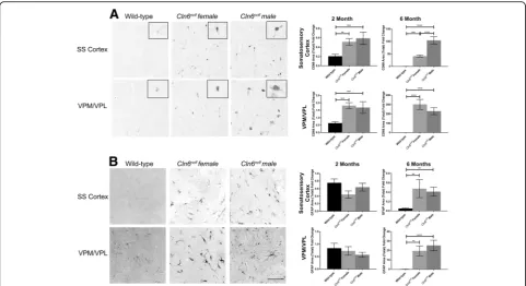
Reactive gliosis is another marker of Batten disease that can be used to measure disease severity as the mice age. At 6 months of age, whileCln6nclfmice collectively had elevated astrocyte activation (GFAP+) and micro-gliosis (CD68+) in the VPM/VPL and somatosensory cortex, there were no differences between the sexes (0.5 to 20 fold increase) (Fig. 2a-d). Interestingly, male
Cln6nclfmice had heightened microgliosis in the somato-sensory cortex compared to their female Cln6nclf coun-terparts at 6 months of age (100 fold increase) (Fig. 2b). As glial activation can contribute to or ward off neuron loss, we also assessed whether there was any gross neur-onal loss between the sexes at these time points. When measuring the thickness of the cortical plate in sev-eral regions, there were no differences between wild-type and Cln6nclf mice at any time point in any region (Additional file 1: Figure S1). Thus, any changes in classic Batten disease pathology did not provoke a gross loss or stabilization of neurons in either sex up to 6 months of age.
Cln6nclfmice have genetic, sex, and age dependent differences in behavioral tests
apparent in male mice until after 10 months of age (Fig. 3a). Deficits became more prominent in both sexes over time.
In the Morris water maze, a navigation test of spatial learning and memory,Cln6nclf mice required more time than wild-type mice to navigate to the platform begin-ning at 11 months of age, while female Cln6nclf mice specifically showed performance regression as early as 9 months of age (Fig. 3b). Importantly, at 11 months, female Cln6nclf mice reached the platform later than male Cln6nclf mice, indicating that female Cln6nclf mice have more prominent learning and memory deficits than male mice of the same age. FemaleCln6nclfmice contin-ued to perform poorly at 12 months of age, but could not be compared toCln6nclfmale mice at this age as the male mice were unable swim, due to physical conditions. Swim speed is shown as a control, and it should be noted that 11- and 12-month-old female Cln6nclf mice were slower than their wild-type counterparts in reach-ing the platform. A slower swim speed may have been a confounding variable in the analysis of navigation times of the animals at 11 and 12 months.
Lastly, we plotted a Kaplan-Meier survival curve to es-timate the fraction of living Cln6nclf animals over time.
Diseased animals’ premature death occurred 1 month earlier, on average, for femaleCln6nclfmice compared to their male counterparts (14 and 15 months, respectively), while wild-type mice lived to ~ 28 months (Fig.3c).
Discussion
In this study, we show differences in the progression CLN6 disease between sexes in a Cln6nclf mouse model. Female Cln6nclf mice presented with an earlier increase subunit C accumulation, and subsequently performed more poorly on behavioral tests and perished earlier than their male counterparts. MaleCln6nclfmice, on the other hand, showed an increase in ASM at an earlier time point, as well as an increase in microglial activity at 6 months of age. While there have been previous com-prehensive natural history studies of CLN6 disease pa-tients, interpretation of sex-based outcomes is limited due to varyingCLN6mutations [9, 26,27]. In the CLN3 variant of Batten disease, where the delta 7/8 mutation is common and affects ~ 75% of patients, male patients display symptoms before female patients, though females ultimately progress more quickly and die prior to males [21, 28]. It’s possible that the momentary increase in ASM seen in male mice reflects an early disease
Fig. 1Sex differences evident inCln6nclfaccumulation of autofluorescent storage material (ASM) and mitochondrial ATP synthase subunit C in brain.
presentation, though this doesn’t translate into earlier func-tional difficulties. As the molecular underpinnings of CLN6 disease are not well understood, the extent to which these pathological changes translate to behavior changes inCln6
mice will need to be the subject of future study.
The observed sex driven differences in disease progres-sion are not unique to theCln6nclfmice or Batten disease. While the biological basis for variance in disease progres-sion between sexes is unknown, hormonal factors may contribute to the observed differences. Among adults with neurodegenerative diseases, estradiol appears to play a protective role in females [29,30]. However, in adolescent females with juvenile Batten disease, estrogen may be
doing the exact opposite: CLN3-Batten disease females of post-pubertal age, when estrogen levels are elevated, dem-onstrated earlier loss of independence, and thus, estrogen may be contributing to the rapid disease progression [21].
Batten disease is an immune-mediated disease charac-terized by chronic neuroinflammation that is sustained by persistent glial activation in the brain, leading to damage and death of neighboring neurons and glial cells. Gonadal hormones support coordination of neuron-glia interactions and regulate reactive gliosis and neuroin-flammation [31–33]. Control of reactive gliosis by progesterone and estradiol is well documented, yet glio-sis regulation by androgens has not been extensively
Fig. 3Sex differences evident inCln6nclfbehavior and survival outcomes.aFemaleCln6nclfmice perform more poorly at the rotarod motor task beginning at six months of age. MaleCln6nclfmice do not begin to perform poorly until 10 months of age.bFemaleCln6nclfmice perform more poorly at the Morris water maze task beginning at nine months of age. MaleCln6nclfmice do not begin to perform poorly until 11 months of age.
cSwim speed shown as a control for the Morris water maze task.dFemaleCln6nclfmice perish one month earlier than maleCln6nclfmice. Asterisks (*) show comparisons between wild-type andCln6nclfanimals, with light blue for female comparisons and dark blue for male comparisons. Hash signs (#) show comparisons between male and femaleCln6nclfanimals.N= 3–10, *p< 0.05, **p< 0.01, ***p< 0.001, ****p< 0.0001
explored. In general, estradiol appears to reduce astro-cyte activity in the cerebral cortex, though, this contra-dicts the femaleCln6 mouse presentation of heightened astrocytosis. However, evidence suggests that testoster-one decreases reactive astroglia and microglia after neur-onal damage [31,32,34]. Further, female microglia have been shown to exhibit higher phagocytic capacity than males and proinflammatory conditions, while male micro-glia have more efficient migratory response [35, 36]. Because glial cells become cytotoxic when chronically activated, female reduction in microglial activity may be a sign of advanced disease progression as these glial cells may have become inactive [37]. Furthermore, sex differ-ences in microglial number may result from differdiffer-ences in chemotactic signaling and consequent microglial recruit-ment in males and females. Indeed, males exhibit higher levels of chemokines CCL20 and CCL4 in the hippocam-pus and cortex during critical periods of development, while females have elevated levels of proinflammatory cytokine interleukin (IL)-1β [38]. However, chemokines have not been studied in great detail in a healthy brain or in Batten disease, and the extent to which they may play a part in neurodegeneration remains unclear. Additionally, females are more vulnerable than males to develop Alzheimer’s disease, and although androgens have been studied less extensively than estrogens, androgens exert anti-inflammatory effects on microglia in AD models [39, 40].
Sex-based differences in Batten disease may also be re-lated to the rise of autoantibodies in females. Estrogen has been shown to increase autoantibodies in systemic lupus erythematosus, accelerating disease progression [41]. In Batten disease, a similar autoimmune response in the CNS of both animal and patient populations contributes to disease progression [21]. Overall, hormo-nal differences in males and females likely explain some sex-specific immune responses, modifying disease course. As suppression of the immune system has been used in preclinical and clinical Batten disease studies, the extent to which these therapies are beneficial in both sexes should be a point of focus in the future [42,43].
Conclusions
Here, we provide the first sex comparison of patho-logical and behavioral differences in Cln6nclfmice, find-ing notable differences between the sexes. Moreover, our findings are identical to observed by another laboratory working with the same CLN6 mutant strain (personal communication, Drs. Stephanie Hughes and Hannah Best). TheCln6nclf mouse model echoes the accelerated disease progression reported of females with Batten disease, and this information will be instrumental in pro-viding appropriate treatments to female Batten disease patients in the future [21]. Currently, no definitive
treatments or cures exist for CLN6 disease, a rapidly-progressing neurodevelopmental disease. However, as potential therapeutics are being investigated, sex-related differences must be taken in to account to target trans-latability to women.
Methods
Ethics statement/animals
All animal studies were performed in an AAALAC accredited facility in strict accordance with National Institutes of Health guidelines and were approved by the Sanford Institutional Animal Care and Use Committee (USDA License 46-R-0009). Wild-type and homozygous
Cln6nclf mutant mice (Jackson Laboratory, Bar Harbor, ME) on C57BL/6 J backgrounds were used for all studies and were housed under identical conditions. For the immunohistochemistry experiments, 3–6 mice were used per group. For the behavior studies, 10 mice were used per group. As the mice aged and perished, this reduced the N in some groups to 3 at the last few time points.
Neurobehavior testing
Rotarod
Beginning at 3 months of age, mice were tested monthly (up to 12 months of age) on a Rotamex-5 Rotarod (Columbus Instruments, Columbus, OH, USA) to assess motor abilities. The machine’s initial speed was set to 0.3 rpm (rpm) and accelerated at 0.3 rpm every two sec-onds until maximum speed (36 rpm) was reached. Mice were trained over 9 trials: 3 consecutive trials, followed by a 30-mintue resting period, 3 more consecutive trials, followed by another 30-min resting period, and a final 3 consecutive trials. Subsequent testing after a four-hour resting period modeled the training session. Latency time to fall from the rod was recorded and averaged for each of a mouse’s nine testing trials to give one value per mouse.
Morris water maze
then tested in opaque water colored with white non-toxic tempura paint and an unflagged platform. On each test day, the mice were given 60 s per trial for eight trials to locate the platform; a training session consisting of four trials was implemented in the morning followed by a three-hour resting period, followed by a testing session of four trials in the afternoon. Mice were tested on four consecutive days, starting at a different visual cue each day. Any-maze software (Stoelting Co., Wood Dale, IL, USA) tracked test duration and swim speed for each mouse. Quantifications of each recording were averaged from the sixteen afternoon trials per mouse.
Immunohistochemistry
Wild-type and Cln6nclf mice were CO2euthanized,
per-fused with PBS, and tissue fixed with 4% PFA. Fixed brains were sectioned on a vibratome at 50μm (Leica VT10008) and processed with standard immunofluores-cence and DAB staining protocols as previously described [44]. Primary antibodies included anti-CD68 (AbD Sero-tec, MCA1957; 1:250), anti-GFAP (Dako, Z0334; 1:250), and anti-ATP synthase subunit C (Abcam, ab181243, 1:500). The subunit C experiments were also counter-stained with methyl green. Secondary antibodies included anti-rat and anti-rabbit biotinylated (Vector Labs, BA-9400; 1:2000) and Alexa-Fluor fluorescent secondaries (1:1500). Sections were imaged in the VPM/VPL of the thalamus and layers 2/3 of the somatosensory cortex and analyzed using a Nikon 90i microscope with NIS-Ele-ments Advanced Research software (v4.20). For autofluor-escent storage material, cells were scored positive for accumulation of storage material when more than three autofluorescent puncta were aggregated around the nucleus. Mitochondrial ATP synthase subunit C, GFAP, and CD68 immunoreactivity was quantified using a threshold analysis in NIS-Elements Advanced Research software, with the subunit C analyzed with the methyl green counterstain excluded from analysis (v4.20) as pre-viously described [44].
Cortical plate thickness
Cortical plate thickness was measured in the visual, motor, and somatosensory cortex of sagittal tissue sec-tions. Measurements were taken in triplicates in the cor-tical plate, encompassing layers 1–6 of the cerebral cortex. Triplicates were averaged, and statistical tests performed as described.
Statistical analyses
Statistical analyses were performed using GraphPad Prism (v6.04). Equal numbers of male and female wild-type ani-mals were combined into one group, as there were no differences between male and female wild-type values for any given assay. For immunohistochemical analyses,
one-way ANOVA’s were utilized with Tukey correction and outlier removal using the ROUT method, Q = 1. One-way ANOVA’s with Tukey correction and outlier removal using the ROUT method, Q = 1, were also used for behavior experimentation analyses. For the Morris water maze 12-month timepoint, an unpaired t-test was used. To develop a survival curve, the log-rank (Mantel--Cox) test was used.
Additional file
Additional file 1:Figure S1.No gross cortical neuron loss detected at 2 or 6 months of age inCln6nclfmice. Mean +/−SEM. N = 3–6. (TIF 9028 kb)
Acknowledgements
Not applicable.
Funding
This work was supported by grants from the Charlotte and Gwenyth Gray Foundation and NIH support R01NS082283. This work also received support from the Sanford Research Imaging Core within the Sanford Research Center for Pediatric Research (NIH P20GM103620) and the Sanford Research Molecular Pathology Core within the Sanford Research Center for Cancer Biology (NIH P20GM103548).
Availability of data and materials
The datasets generated and/or analyzed during the current study are available from the corresponding author on reasonable request.
Disclosures and ethics
The authors have confirmed that this article is unique and not under consideration or published in any other publication, and that they have permission from rights holders to reproduce any copyrighted material.
Authors’contributions
Conceived and designed the experiments: JTC, KAW, JMW. Performed behavior experiments: MJP, SSD, RL. Performed histology experiments: TBJ, KAW. Analyzed the data: MJP, JTC, TBJ, KAW. Contributed to the writing of the manuscript: MJP, JTC, TBJ, KAW, ADK, JMW. Agree with manuscript results and conclusions: MJP, JTC, TBJ, KAW, SSD, RL, ADK, JMW. All authors reviewed and approved of the final manuscript.
Ethics approval and consent to participate
Animal protocols were approved by the Institutional Animal Care and Use Committees of each participating institute (NIH/OLAW Assurance Number: A4568–01) with all procedures conducted in strict accordance with National Institutes of Health guidelines and Institutional Animal Care and Use Committee Guidelines.
Consent for publication
No applicable.
Competing interests
The authors declare that they have no competing interests.
Publisher’s Note
Springer Nature remains neutral with regard to jurisdictional claims in published maps and institutional affiliations.
Author details 1
Pediatrics and Rare Diseases Group, Sanford Research, Sioux Falls, SD, USA. 2Department of Biology, Augustana University, Sioux Falls, SD, USA. 3Department of Pediatrics, Sanford School of Medicine, University of South
Received: 21 July 2018 Accepted: 7 January 2019
References
1. Cooper JD. Progress towards understanding the neurobiology of batten disease or neuronal ceroid lipofuscinosis. Curr Opin Neurol. 2003;16(2):121–8. 2. Mole SE, Williams RE, Goebel HH. Correlations between genotype,
ultrastructural morphology and clinical phenotype in the neuronal ceroid lipofuscinoses. Neurogenetics. 2005;6(3):107–26.
3. Goebel HH, Wisniewski KE. Current state of clinical and morphological features in human NCL. Brain Pathol. 2004;14(1):61–9.
4. Palmer DN, Barry LA, Tyynela J, Cooper JD. NCL disease mechanisms. Biochim Biophys Acta. 2013;1832(11):1882–93.
5. Haltia M. The neuronal ceroid-lipofuscinoses. J Neuropathol Exp Neurol. 2003;62(1):1–13.
6. Jalanko A, Braulke T. Neuronal ceroid lipofuscinoses. Biochim Biophys Acta. 2009;1793(4):697–709.
7. Warrier V, Vieira M, Mole SE. Genetic basis and phenotypic correlations of the neuronal ceroid lipofusinoses. Biochim Biophys Acta (BBA) - Mol Basis Dis. 2013;1832(11):1827–30.
8. Gao H, Boustany RM, Espinola JA, Cotman SL, Srinidhi L, Antonellis KA, et al. Mutations in a novel CLN6-encoded transmembrane protein cause variant neuronal ceroid lipofuscinosis in man and mouse. Am J Hum Genet. 2002; 70(2):324–35.
9. Sharp JD, Wheeler RB, Parker KA, Gardiner RM, Williams RE, Mole SE. Spectrum of CLN6 mutations in variant late infantile neuronal ceroid lipofuscinosis. Hum Mutat. 2003;22(1):35–42.
10. Teixeira CA, Espinola J, Huo L, Kohlschutter J, Persaud Sawin DA, Minassian B, et al. Novel mutations in the CLN6 gene causing a variant late infantile neuronal ceroid lipofuscinosis. Hum Mutat. 2003;21(5):502–8. 11. Bronson RT, Donahue LR, Johnson KR, Tanner A, Lane PW, Faust JR.
Neuronal ceroid lipofuscinosis (nclf), a new disorder of the mouse linked to chromosome 9. Am J Med Genet. 1998;77(4):289–97.
12. Jolly RD, Palmer DN. The neuronal ceroid-lipofuscinoses (batten disease): comparative aspects. Neuropathol Appl Neurobiol. 1995;21(1):50–60. 13. Jolly RD, West DM. Blindness in South Hampshire sheep: a neuronal
ceroidlipofuscinosis. N Z Vet J. 1976;24(6):123.
14. Golden LC, Voskuhl R. The importance of studying sex differences in disease: the example of multiple sclerosis. J Neurosci Res. 2017;95(1–2):633–43. 15. Beery AK, Zucker I. Sex bias in neuroscience and biomedical research.
Neurosci Biobehav Rev. 2011;35(3):565–72.
16. Li R, Singh M. Sex differences in cognitive impairment and Alzheimer’s disease. Front Neuroendocrinol. 2014;35(3):385–403.
17. Gillies GE, Pienaar IS, Vohra S, Qamhawi Z. Sex differences in Parkinson’s disease. Front Neuroendocrinol. 2014;35(3):370–84.
18. Davies W. Sex differences in attention deficit hyperactivity disorder: candidate genetic and endocrine mechanisms. Front Neuroendocrinol. 2014;35(3):331–46.
19. Schaafsma SM, Pfaff DW. Etiologies underlying sex differences in autism Spectrum disorders. Front Neuroendocrinol. 2014;35(3):255–71. 20. Attarian H, Brandes J, Dafer R, Gerard E, Giesser B. Sex differences in the
study of neurological illnesses. Behav Neurol. 2015;2015:676531.
21. Cialone J, Adams H, Augustine EF, Marshall FJ, Kwon JM, Newhouse N, et al. Females experience a more severe disease course in batten disease. J Inherit Metab Dis. 2012;35(3):549–55.
22. Isolation of a novel gene underlying Batten disease, CLN3. The international batten disease consortium. Cell. 1995;82(6):949–57.
23. Cotman SL, Vrbanac V, Lebel LA, Lee RL, Johnson KA, Donahue LR, et al. Cln3(Deltaex7/8) knock-in mice with the common JNCL mutation exhibit progressive neurologic disease that begins before birth. Hum Mol Genet. 2002;11(22):2709–21.
24. Kovacs AD, Pearce DA. Finding the most appropriate mouse model of juvenile CLN3 (batten) disease for therapeutic studies: the importance of genetic background and gender. Dis Model Mech. 2015;8(4):351–61. 25. Guarneri R, Russo D, Cascio C, D'Agostino S, Galizzi G, Bigini P, et al. Retinal
oxidation, apoptosis and age- and sex-differences in the mnd mutant mouse, a model of neuronal ceroid lipofuscinosis. Brain Res. 2004;1014(1–2): 209–20.
26. Cannelli N, Garavaglia B, Simonati A, Aiello C, Barzaghi C, Pezzini F, et al. Variant late infantile ceroid lipofuscinoses associated with novel mutations in CLN6. Biochem Biophys Res Commun. 2009;379(4):892–7.
27. Canafoglia L, Gilioli I, Invernizzi F, Sofia V, Fugnanesi V, Morbin M, et al. Electroclinical spectrum of the neuronal ceroid lipofuscinoses associated with CLN6 mutations. Neurology. 2015;85(4):316–24.
28. Munroe PB, Mitchison HM, O'Rawe AM, Anderson JW, Boustany RM, Lerner TJ, et al. Spectrum of mutations in the batten disease gene, CLN3. Am J Hum Genet. 1997;61(2):310–6.
29. Brann DW, Dhandapani K, Wakade C, Mahesh VB, Khan MM. Neurotrophic and neuroprotective actions of estrogen: basic mechanisms and clinical implications. Steroids. 2007;72(5):381–405.
30. McEwen BS, Alves SE. Estrogen actions in the central nervous system. Endocr Rev. 1999;20(3):279–307.
31. Garcia-Estrada J, Del Rio JA, Luquin S, Soriano E, Garcia-Segura LM. Gonadal hormones down-regulate reactive gliosis and astrocyte proliferation after a penetrating brain injury. Brain Res. 1993;628(1–2):271–8.
32. Barreto G, Veiga S, Azcoitia I, Garcia-Segura LM, Garcia-Ovejero D. Testosterone decreases reactive astroglia and reactive microglia after brain injury in male rats: role of its metabolites, oestradiol and
dihydrotestosterone. Eur J Neurosci. 2007;25(10):3039–46.
33. Arevalo MA, Santos-Galindo M, Acaz-Fonseca E, Azcoitia I, Garcia-Segura LM. Gonadal hormones and the control of reactive gliosis. Horm Behav. 2013; 63(2):216–21.
34. Barreto G, Santos-Galindo M, Diz-Chaves Y, Pernia O, Carrero P, Azcoitia I, et al. Selective estrogen receptor modulators decrease reactive astrogliosis in the injured brain: effects of aging and prolonged depletion of ovarian hormones. Endocrinology. 2009;150(11):5010–5.
35. Nelson LH, Warden S, Lenz KM. Sex differences in microglial phagocytosis in the neonatal hippocampus. Brain Behav Immun. 2017;64:11–22.
36. Yanguas-Casas N, Crespo-Castrillo A, de Ceballos ML, Chowen JA, Azcoitia I, Arevalo MA, et al. Sex differences in the phagocytic and migratory activity of microglia and their impairment by palmitic acid. Glia. 2018;66(3):522–37. 37. Sochocka M, Diniz BS, Leszek J. Inflammatory response in the CNS: friend or
foe? Mol Neurobiol. 2017;54(10):8071–89.
38. Schwarz JM, Sholar PW, Bilbo SD. Sex differences in microglial colonization of the developing rat brain. J Neurochem. 2012;120(6):948–63.
39. Kim S, Kim MJ, Kim S, Kang HS, Lim SW, Myung W, et al. Gender differences in risk factors for transition from mild cognitive impairment to Alzheimer's disease: a CREDOS study. Compr Psychiatry. 2015;62:114–22.
40. Kang S, Kim JB, Heo TH, Kim SJ. Cell cycle arrest in batten disease lymphoblast cells. Gene. 2013;519(2):245–50.
41. Grimaldi CM. Sex and systemic lupus erythematosus: the role of the sex hormones estrogen and prolactin on the regulation of autoreactive B cells. Curr Opin Rheumatol. 2006;18(5):456–61.
42. Seehafer SS, Ramirez-Montealegre D, Wong AM, Chan CH, Castaneda J, Horak M, et al. Immunosuppression alters disease severity in juvenile batten disease mice. J Neuroimmunol. 2011;230(1–2):169–72.
43. Augustine EF, Beck CA, Adams HR, Defendorf S, Vierhile A, Timm D, et al. Short-term Administration of Mycophenolate is Well-Tolerated in CLN3 disease (juvenile neuronal ceroid Lipofuscinosis). JIMD Rep. 2018. 44. Morgan JP, Magee H, Wong A, Nelson T, Koch B, Cooper JD, et al. A murine