O R I G I N A L C L I N I C A L I N V E S T I G A T I O N
Open Access
Supporting the use of a coagulometric
method for rivaroxaban control: a
hypothesis-generating study to define the
safety cut-offs
Raul Altman
1*and Claudio Daniel Gonzalez
2Abstract
Aims:Although quantitative anti-FXa assays can be used to measure rivaroxaban plasma levels, they are not widely performed or available. We aimed to tentatively determine the cut-off for thromboembolism and bleeding prevention based on the clotting effect of non-rivaroxaban conjugate-activated FX plasma levels in patients with rivaroxaban using a coagulometric method.
Methods and results:Rivaroxaban was addedin vitroto normal plasma at a range of 0 to 241μg/L to cover expected peak and trough levels. Rivaroxaban chromogenic (μg/L) and RVV-confirm as a ratio were determined. Patient plasma samples were assayed with the RVV-confirm reagent. The appropriate rivaroxaban plasma concentration to inhibit clotting mechanisms was based on the remaining FXa in plasma, which was expressed as the ratio of patients/ normal, R-C. There is a high correlation between R-Cin vitro andspiked normal plasma rivaroxaban concentration (R-Square 0.910, linear equation; 0.971 quadratic equation,p< 0.0001 for both) but not with plasma rivaroxaban chromogenic assays. We propose a cut-off R-C value of 1.65 and 4.5 for safety. Based on the proposed therapeutic range, in 158 assays performed in 58 patients, 6.3 % assays were above the level of bleeding tendency at the peak (R-C 5.39 ± 1.01, median 5.13) and 42 % assays were below the prevention cut-off at the trough (R-C 1.31 ± 0.18, median 1.35).
Conclusions:RVVconfirm® is fast and sensitive to measure the effect of rivaroxaban. Clinical studies are needed to establish whether this cut-off is useful for identifying patients at increased risk of hemorrhage or those who exhibit a low level of anticoagulation.
Keywords:Anticoagulants, Drug monitoring, Reference ranges, Rivaroxaban, Safety
Introduction
The well-established benefits of anticoagulant therapy are significantly hampered by the possibility of throm-boembolism or major and sometimes fatal bleeding com-plications. These adverse effects can range from simple external skin bruising and bleeding (epistaxis, gastroduo-denal bleeding, pulmonary complications) to problems in vital organs, including temporary or permanent impair-ment of function (intracranial hemorrhage or embolism), and possibly death. Despite their importance, as well as
the known benefit of warfarin, appropriate medication use remains a challenge for patients. Indeed, for vitamin K an-tagonists, the rates of non-adherence range from 22–58 %. This rate is significant because 34 to 43 % of patients taking warfarin are not in therapeutic range [1], which results in poor clinical outcomes [2]. The history of non-adherence is likely underrepresented in trial outcomes research. As a real world consequence, patients receiving chronic warfarin therapy who have poor anticoagulation control are at increased risk for adverse events [3]. In large studies, the rate of prema-ture discontinuation of new direct oral anticoagulants (DOACs) for no apparent reason is reported to be * Correspondence:[email protected]
1Centro de Trombosis de Buenos Aires, Buenos Aires, Argentina Full list of author information is available at the end of the article
between 3 and 14.3 % [1]. Contrary to warfarin, one important benefit of DOACs is that they do not require routine coagulation control due to their predictable pharmacokinetic and pharmacodynamic profiles [4]. However, there is agreement that some clinical circum-stances require measuring the anticoagulant effect of the DOAC (5) (e.g., preparation for surgery; major bleeds, compliance and/or effect checks, renal impairment). How-ever, concentrations of DOACs and their effect on coagu-lation are dependent on the pharmacokinetics of the drug and on the concomitant presence of potent P-glycoprotein inhibitors or agonists. To improve efficacy and safety, dose adjustment based on the therapeutic effect may be more appropriate than fixed-dose therapy. The anti-Factor Xa method using chromogenic assay and expressed as μg/L measures the drug concentration and not the intensity of the drug’s anticoagulant activity, and a higher or lower than expected DOACs plasma level does not necessarily indicate an increased risk of bleeding or thrombotic com-plications [4, 5].
Then it is imperative in daily practice to periodically evaluate dosage to ensure safety and effectiveness. Coagu-lometric tests based on Russell’s viper venom has been proposed as potential methods to evaluate DOACs as they are sensitive to both classes of these inhibitors [6–8].
Methods
Normal donors
Twenty healthy volunteers (12 women and 8 men) with no history of thromboembolic or hemorrhagic diseases, cardiac, renal, hepatic, or malignant diseases, were required to be drug free for 10 days before the study. Only platelet poor plasma from subjects with a normal prothrombin time, activated partial thromboplastin time and thrombin time that fulfilled the inclusion criteria were used.
Patients
This study included patients with no history of hemorrhagic diseases receiving prolonged oral therapy with rivaroxa-ban. The review board of the Centro de Trombosis de Buenos Aires approved the study. A total of 158 at peak and 158 at trough measurements of DVVconfirm were performed in 58 patients (36 men, 22 women; mean age, 66.8 ± 14.8 years; median age, 69 years). Patients were referred to our clinic after their own physician recommended drug use due to deep venous thrombosis (26 patients, 3 with pulmonary embolism), portal vein thrombosis (1 patient), mesenteric vein thrombosis (1 patient), atrial fibrillation (30 patients 1 patient with coronary stenting). Fifty patients were treated with 20 mg rivaroxaban per day, and 8 with 15 mg/day. Medication was taken at the same hour each morning. Patients were required to have received therapy for
≥14 days before entering the study. Study complies with the Declaration of Helsinki and informed consent was obtained from all patients. Among the 58 patients Rivaroxaban plasma concentration (using chromogenic assay and expressed as μg/L) at peak and at trough were performed in 30 patients (10 men, 20 women) and com-pared with DVVconfirm assay (data were expressed as ratio of patient plasma/normal plasma values) (R-C).
Hemostasis tests
Venous blood was drawn from the antecubital vein and mixed with 0.11 mol/L sodium citrate (1:10 v/v). Blood samples for assays were obtained at the peak of drug ac-tivity (mean, 2.05 ± 0.23 h; median, 2.1 h) and during the activity trough 0–2 h before the next dose (mean, 23.6 ± 1.19 h; median, 23.7 h). Tests were performed within 3 h of sampling. Platelet-poor plasma (PPP) was obtained by centrifuging blood samples at 900 × g for 15 min. DVVconfirm® was used to measure citrated PPP using the reagents from Sekisui Diagnostics (Stanford, USA) following the manufacturer’s indications using a coagulometer ST4 (Diagnostica Stago, Asnieres, France). DVV-confirm assay results are reported in seconds of time to clot and the seconds are used to determine the ratio expressed as R-C values.
Rivaroxaban-containing calibrators (241 μg/L of rivar-oxaban) were reconstituted with 1 ml pooled normal plasma to obtain a concentration of 241 μg/L, which was further diluted serially with the same pooled normal plasma to obtain concentrations between 0 and 241μg/L. For the in vitro spiking studies rivaroxaban-containing calibrators (purchased from Stago (Diagnostica Stago, Asnieres, France) were used for both the DVV-confirm and the chromogenic assays.
Rivaroxaban chromogenic assays were performed ac-cording to Samamaet al.[9] using STA Liquid anti-FXa assay controls and calibrators from Stago (Diagnostica Stago, Asnieres France) with a STA analyzer.
Statistical analysis
Results
In vitrospiking studies
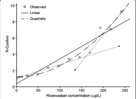
ForIn vitrospiked studies known concentration of rivar-oxaban was used. The correlation between rivarrivar-oxaban concentration and R-C using a simple linear correlation yielded an R-square coefficient of 0.910 (p< 0.0001) (Fig. 1); using a quadratic model, the R-square coefficient was 0.971 (p< 0.0001) (Fig. 1). The corresponding parame-ters and constant are depicted in Table 1. As mentioned above, the “goodness of fit” was significant using the quadratic approach. Overall, these correlation coeffi-cients indicate an excellent correlation between rivarox-aban concentration and R-C test. As seen in Fig. 1, it seems feasible to interpret the curvilinear quadratic graph as being composed of two linear components (dashed lines). This finding warranted further exploration.
Measurements in plasma patients receiving oral therapy with rivaroxaban
The correlation between rivaroxaban concentration mea-sured as aFXa in patients plasma and R-C is shown in Table 2. The correlation between plasma rivaroxaban con-centration and R-C at trough (24 h of drug ingestion) yielded a R coefficient of 0.688 (R2: 0.473,p< 0.001) at the linear model; at peak (after 2 h of drug ingestion) the strength of the association was weaker, with an R linear correlation coefficient of 0.39 (R2: 0.15;p= 0.03).
Rivaroxaban-test coagulometric method
To evaluate the rivaroxaban coagulometric method, where the patient should be at both peak and trough times the
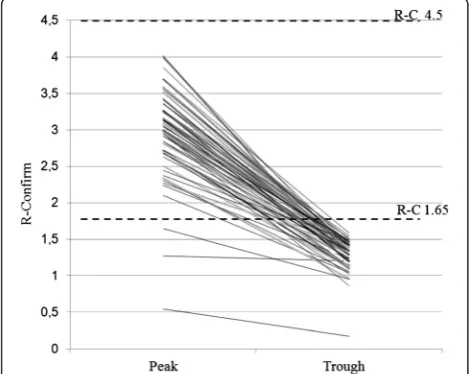
Russel’s viper venom-confirm® method was used in 158 plasma samples from 58 patients. Considering cut-offs of 1.65 and 4.5 as limits of therapeutic levels, 10 determi-nations (6.3 %) (Fig. 2) were above the bleeding risk level (R-C 5.39 ± 1.01, median 5.13) and 66 tests (42 %) (Fig. 3) were below the theoretical prevention cut-off (R-C 1.31 ± 0.18, median 1.35).
Discussion
Although quantitative anti-FXa chromogenic assay has been proposed to measure rivaroxaban plasma levels for patients under rivaroxaban therapy, it is not widely per-formed or available [4–6]. With the absence of widely accepted therapeutic target levels, speculative cut-off levels for safety and prevention were used in the current manuscript.
Taking into account the hemorrhagic level of patients with Factor X deficiency as well as their haemostatic level, this prospective study based on an empirically validated numerical model was designed to investigate cut-off levels in patients receiving rivaroxaban therapy.
Rivaroxaban blocks FX activity, and the unbounded FXa remains available in the clotting system. Even if clinical trials prove the safety of the fixed dose regimen, patients should get frequent follow-up attention by nurses regarding any side effects that do not occur in daily practice.
The cut-off for prevention was based on the plasma activity of vitamin K-dependent clotting factors mea-sured in patients on warfarin, a treatment with a target INR range of 2.5 ± 0.3. In Dargaud et al. study [10] the Factor X level in patients without complications was 19 ± 10 IU dL−1, and the level in patients with throm-bosis was 16 ± 5 IU dL−1. Moreover, bleeding is infre-quent in patients with F X levels above 20 % [11].
Fig. 1In vitrospiking studies. Correlation between rivaroxaban concentration and R-C. ForIn vitrospiked studies known of rivaroxaban concentrations between 0 and 241μg/L were used. Statistically significant R-Squares of 0.910 (p< 0.0001) using a linear correlation and an R-Square of 0.971 using a quadratic equation (p< 0.0001) indicate an excellent correlation between rivaroxaban concentration and R-C test. Apparently the curved line has two lineal components (dashed lines)
Table 1Model summary and parameter estimates
Equation Model summary Parameter estimates
R square F df1 df2 Sig. Constant b1 b2
Linear .901 100.405 1 11 .000 .371 .031
Quadratic .971 169.965 2 10 .000 1.195 .000 .000
Dependent variable: R-C
The independent variable is Rivaroxaban concentration
Table 2Chromogenic substrate test
Correlation at trough (24 h) R-C
aFXa Rivaroxaban 0,6881
P= 0.0001 Correlation at peak (2 h)
aFXa Rivaroxaban 0,3908
P= 0.033 Coefficient of correlations between R-C and rivaroxaban in plasma
Congenital or acquired FX deficit allows us to speculate on the safety of patients undergoing rivarox-aban therapy. Bleeding symptoms tended to correlate with FX levels: mild (>6–10 %), moderate (1–5 %) or severe (<1 %). Severe clinical symptoms, such as intra-cranial hemorrhage, gastrointestinal bleeding and hemarthrosis, are not common in patients with FX levels >2 % [12]. Patients with FX levels <10 % present with mucocutaneous bleeding, whereas those with moderate to severe deficiency may have symptoms
such as hemarthrosis, intracranial hemorrhage, and gastrointestinal bleeding. These patients experience spontaneous bleeding when their plasma Factor X con-centration is below 1 % [13, 14]; patients with higher levels of Factor X can also bleed in a traumatic situ-ation. Patients with amyloidosis [15] developed severe gastrointestinal bleeding as a result of a significant decrease in Factor X levels, but a patient who pre-sented with spontaneous retroperitoneal bleeding had 22 % FX activity [16].
Based on these data and that reported in our previous publication [7], Factor X concentration curves were cre-ated. Based on these curves, 1.56 % FX corresponded with a R-C of 4.5, and 20 % FX corresponded with a R-C of 1.65. These values were designated as cut-offs for safety and thromboembolic prevention in the current study.
An optimal drug level should be obtained to achieve the correct balance between antithrombotic efficacy and bleeding risk. Brummel-Ziedinset al.[17] found a marked interindividual variation of FXa generation in a healthy population (from 49 to 163 %) and in individuals with a known DVT history (from 58 to 174 %). with statistically significant differences between groups. These differences were also related to gender, BMI and oral contraceptives [17]. Patients taking 20 mg rivaroxaban had a daily con-centration peak that varied between 177 and 361 μg/L and a trough of 9.02 to 147μg/L [18, 19]. Similarly, at a dose of 10 mg, the daily concentration peak was 75.1 to 177μg/L and a trough of 1.35 to 37.2μg/L. These inter-individual variations were also observed with other DOACs [20, 21].
In the current study based onin vitro spiking studies, it is apparent that the DOAC-rivaroxaban coagulometric method is suitable for measuring non-rivaroxaban boun-ded plasma-activated Factor X ex vivo. The results ob-tained with spiked plasma may not be similar to those obtained fromex vivopatient plasma due to the absence of drug metabolites. At high concentrations, rivaroxaban or its metabolites may have additional effects on clotting mechanisms that are currently unknown [22].
Thus, the question arises over whether rivaroxaban concentrations measured by an anti-FXa chromogenic assay indicate the extent of clotting inhibition mecha-nisms as well as a coagulometric assay. Given the marked variation between healthy individuals with thrombosis as well as patients with liver, kidney or bowel disease, one must assume that a significant fraction of patients will be exposed to either very low or very high drug levels [23]. Safety in daily practice could be improved with an easy-to-use coagulometric method. In the absence of widely accepted therapeutic target levels, we speculate cut-off points for identifying patients with an increased risk of hemorrhage, R-C ≥4.5, or those exhibiting a low Fig. 2DOAC-test coagulometric method. Ten plasmas among 158
assayed for R-C value were above R-C of 4.5 proposed as cut-off for increased bleeding tendency. Patients were above range at peak time only. The value at peak R-C is 5.39 ± 1.01, with a median of 5.13. Peak vs. trough values:p< 0.001
anticoagulation effect, R-C of ≤1.65, as potential cut-offs of a pro-thrombotic state. Using the proposed therapeutic range for 158 plasma samples, 10 (6.5 %) were above the level of bleeding tendency at the peak (R-C 5.39 ± 1.01, median 5.13) and 66 (42 %) were below the prevention cut-off at the trough (R-C 1.31 ± 0.18, median 1.35), which indicates that treatment with rivaroxaban warrants dose adjustment. The paper by Beyer-Westendorfet al.[24], describe that in real life, rates of rivaroxaban-related major bleeding may be lower and that the outcome may at least not be worse than that of major vitamin K antagonist bleeding. We assume that in patients on rivaroxaban, routine measure of anti-coagulant activity using a coagulometric method as R-C could lower risk of bleeding and/or thromboembolism compared with no routine coagulation monitoring. Our proposal should be considered hypothesis-generating; it requires further clinical studies to show that in real life, this monitoring strategy actually reduces bleeding and risk of ischemic stroke.
Competing interests
The authors declare that they have no competing interests.
Authors’contributions
RA designed the study, performed the experiments, analyzed the data, reviewed the manuscript. CDG designed the study, performed the statistical analysis and reviewed the manuscript. All authors read and approved the final manuscript.
Acknowledgements
Diana Garcia, PhD Laboratorio CEDEAC 25 de Mayo, Mar del Plata, Argentina who performed plasma rivaroxaban chromogenic assays.
Author details
1
Centro de Trombosis de Buenos Aires, Buenos Aires, Argentina. 2Department of Pharmacology, School of Medicine, University of Buenos Aires, Buenos Aires, Argentina.
Received: 2 April 2015 Accepted: 29 May 2015
References
1. Rodriguez RA, Carrier M, Wells PS. Non-adherence to new oral anticoagulants: a reason for concern during long-term anticoagulation? J Thrombos Haemost. 2013;11:390–4.
2. Bosworth HB, Bradi B, Granger BB, Mendys P, Brindis R, Burkholder R, et al. Medication adherence: a call for action. Am Heart J. 2011;162:412–24. 3. Davis NJ, Billett HH, Cohen HW, Arnsten JH. Impact of adherence, knowledge,
and quality of life on anticoagulation control. Ann Pharmacother. 2005;39:632–6. 4. Samama MM, Contant G, Spiro TE, Perzborn E, Le Flem L, Guinet C, et al.
Laboratory assessment of rivaroxaban: a review. Thromb J. 2013;11:11. 5. Baglin T, Hillarp A, Tripodi A, Elalamy I, Buller H, Ageno W. Measuring oral
direct inhibitors of thrombin and factor Xa: a recommendation from the Subcommittee on Control of Anticoagulation of the Scientific and Standardization Committee of the International Society on Thrombosis and Haemostasis. J Thromb Haemost. 2013;11:756–60.
6. Cuker A, Siegal DM, Crowther MA, Garcia DA. Laboratory measurement of the anticoagulant activity of the non-vitamin K oral anticoagulants. J Am Coll Cardiol. 2014;64:1128–39.
7. Altman R, Gonzalez CD. Simple and rapid assay for effect of the new oral anticoagulant (NOAC) rivaroxaban: preliminary results support further tests with all NOACs. Thromb J. 2014;12:7.
8. Exner T, Ellwood L, Rubie J, Barancewicz A. Testing for new oral anticoagulants with LA resistant russells viper venom reagents. an in vitro study. Thromb Haemost. 2013;109:762–5.
9. Samama MM, Contant G, Spiro TE, Perzborn E, Guinet C, Gourmelin Y, et al. Evaluation of the anti-factor Xa chromogenic assay for the measurement of rivaroxaban plasma concentrations using calibrators and controls. Thromb Haemost. 2012;107:379–87.
10. Dargaud Y, Hoffman M, Lefrapper L, Lin F-C, Genty A, Chatard B, et al. Bleeding risk in warfarinized patients with a therapeutic international normalized ratio: the effect of low factor IX levels. J Thromb Haemost. 2013;11:1043–52.
11. Menegatti M, Peyvandi F. Factor X deficiency. Semin Thromb Hemost. 2009;35:407–15.
12. Brown DL, Kouides PA. Diagnosis and treatment of inherited factor X deficiency. Haemophilia. 2008;14:1176–82.
13. Shim YJ, Won DI. Pharmacokinetics and prophylactic use of FEIBA® in a child with severe congenital factor X deficiency and recurrent spontaneous intracranial haemorrhage: a case report. Haemophilia. 2013;19:e364–7. 14. Rauch R, Girisch M, Wiegand G, Schroeder W, Hofbeck M, Welisch E, et al.
Factor X deficiency and intracranial bleeding: who is at risk? Haemophilia. 2011;17:759–63.
15. Furuhata M1, Doki N, Hishima T, Okamoto T, Koyama T, Kaito S. Acquired factor X deficiency associated with atypical AL-amyloidosis. Intern Med. 2014;53:1841–5.
16. Enjeti AK, Walsh M, Seldon M. Spontaneous major bleeding in acquired factor X deficiency secondary to AL-amyloidosis. Haemophilia. 2005;11:535–8. 17. Brummel-Ziedins KE, Orfeo T, Gissel M, Mann KG, Rosendaal FR. Factor Xa
generation by computational modeling: an additional discriminator to thrombin generation evaluation. PLoS One. 2012;7, e29178. 18. Mueck W, Schwers S, Stampfuss J. Rivaroxaban and other novel oral
anticoagulants: pharmacokinetics in healthy subjects, specific patient populations and relevance of coagulation monitoring. Thromb J. 2013;11:10. 19. Buller HR, Lensing AW, Prins MH, Agnelli G, Cohen A, Gallus AS, et al.
Einstein-DVT dose-ranging study investigators. a dose-ranging study evaluating once-daily oral administration of the factor Xa inhibitor rivaroxaban in the treatment of patients with acute symptomatic deep vein thrombosis: the einstein-DVT dose-ranging study. Blood. 2008;112:2242–7.
20. Douros A, Schlemm L, Bolbrinker J, Ebinger M, Kreutz R. Insufficient anticoagulation with dabigatran in a patient with short bowel syndrome. Thromb Haemost. 2014;112:419–20.
21. Lippi G, Favaloro EJ. Recent guidelines and recommendations for laboratory assessment of the direct oral anticoagulants (DOACs): is there consensus? Clin Chem Lab Med. 2014. doi:10.1515/cclm-2014-0767.
22. Weinz C, Schwarz T, Kubitza D, Mueck W, Lang D. Metabolism and excretion of rivaroxaban, an oral, direct factor Xa inhibitor, in rats, dogs, and humans. Drug Metab Dispos. 2009;37:1056–64.
23. ten Cate H. New oral anticoagulants: discussion on monitoring and adherence should start now! Thromb J. 2013;11:8.
24. Beyer-Westendorf J, Gelbricht V, Thieme C, et al. Rates, management, and outcome of rivaroxaban bleeding in daily care: results from the Dresden NOAC registry. Blood. 2014;124:955–62.
Submit your next manuscript to BioMed Central and take full advantage of:
• Convenient online submission
• Thorough peer review
• No space constraints or color figure charges
• Immediate publication on acceptance
• Inclusion in PubMed, CAS, Scopus and Google Scholar
• Research which is freely available for redistribution