University of Pennsylvania
ScholarlyCommons
Publicly Accessible Penn Dissertations
2018
Cargo Specific Regulation Of Cytoplasmic Dynein
By Effector Proteins
Mara Olenick
University of Pennsylvania, [email protected]
Follow this and additional works at:
https://repository.upenn.edu/edissertations
Part of the
Biochemistry Commons
,
Biophysics Commons
, and the
Cell Biology Commons
This paper is posted at ScholarlyCommons.https://repository.upenn.edu/edissertations/3167
For more information, please [email protected].
Recommended Citation
Olenick, Mara, "Cargo Specific Regulation Of Cytoplasmic Dynein By Effector Proteins" (2018).Publicly Accessible Penn Dissertations. 3167.
Cargo Specific Regulation Of Cytoplasmic Dynein By Effector Proteins
Abstract
Axonal transport is vital for the development and survival of neurons. The transport of cargo and organelles from the axon to the cell body is driven almost completely by the molecular motor, cytoplasmic dynein. Yet, it remains unclear how dynein is spatially and temporally regulated given the variety of cargo that must be properly localized to maintain cellular function. Previous work has suggested that adaptor proteins provide a mechanism for cargo-specific regulation of motors. During my thesis work, I have investigated the role of mammalian Hook proteins, Hook1 and Hook3, as potential motor adaptors. Using optogenetic and single molecule assays, I found that Hook proteins interact with both dynein and dynactin, to effectively activate dynein motility, inducing longer run lengths and higher velocities than the previously characterized dynein activator, BICD2. In addition, I found that complex formation requires the N-terminal domain of Hook proteins, which resembles the calponin-homology domain of EB proteins yet cannot bind directly to
microtubules. In collaborative studies, we found the Hook domain directly interacts with a helix of the dynein light intermediate chain and this interaction is important for Hook-induced processive motility of dynein. In my final project, I found that Hook1 mediates the transport of TrkB-BDNF signaling endosomes in primary hippocampal neurons. Using live cell microscopy and microfluidic devices, Hook1 depletion resulted in a significant decrease in the flux and processivity of BDNF-Qdots along the mid-axon, an effect specific for Hook1 but not Hook3. Together, my work suggests that dynein effectors like Hook proteins can differentially regulate dynein to allow for organelle-specific tuning of the motor for precise intracellular trafficking.
Degree Type
Dissertation
Degree Name
Doctor of Philosophy (PhD)
Graduate Group
Biochemistry & Molecular Biophysics
First Advisor
Erika L. Holzbaur
Subject Categories
Biochemistry | Biophysics | Cell Biology
CARGO SPECIFIC REGULATION OF CYTOPLASMIC DYNEIN BY EFFECTOR PROTEINS
Mara Alizabeth Olenick
A DISSERTATION
in
Biochemistry and Molecular Biophysics
Presented to the Faculties of the University of Pennsylvania
in
Partial Fulfillment of the Requirements for the
Degree of Doctor of Philosophy
2018
Supervisor of Dissertation
__________________________
Erika L. F. Holzbaur, PhD, Professor of Physiology
Graduate Group Chairperson
__________________________
Kim A. Sharp, PhD, Associate Professor of Biochemistry and Biophysics
Dissertation Committee
E. Michael Ostap, PhD, Professor of Physiology
Roberto Dominguez, PhD, Professor of Physiology
Michael A. Lampson, PhD, Associate Professor of Biology
CARGO SPECIFIC REGULATION OF CYTOPLASMIC DYNEIN BY EFFECTOR PROTEINS
COPYRIGHT
2018
iii
ACKNOWLEDGMENT
Getting a PhD can be a very long journey, full of ups and downs but having a good
support system can make it an amazing ride. First, I would like to thank my mentor, Erika
Holzbaur for always being positive and encouraging throughout my thesis. Without her
encouragement and support along the way, I wouldn’t have the confidence in myself as a
scientist that I have today. I would also like to thank my committee members who always pushed
me to think more analytically and be more rigorous with my science. Special thanks to Roberto
Dominquez for being a great collaborator throughout my thesis. His scientific advice and
structural expertise was critical to my thesis work.
I want to thank the Biochemistry and Molecular Biophysics Graduate Group facility and
staff for being supportive and helpful, especially during the early years when you are just trying to
get use to the graduate school life style. A big thank you to the Pennsylvania Muscle Institute, a
community with such collaborative spirit and excitement for science. The many years of PMI
journal club have taught me so much about critical thinking and the art of science, which no class
could ever achieve.
I would not be writing this thesis without the tremendous support of the Holzbaur lab
members of past and present. First, the biggest thank you to Mariko Tokito, our lab manager for
being the cloning master and keeping our lab running with the upmost precision. I would also like
to thank Karen Wallace Jahn for being the best lab veterinarian and providing the wit and humor
that keeps all of us grounded in the real world. These two women provide such experience and
expertise that is invaluable to the Holzbaur lab. Over the years, I have had the pleasure of
working with the most talented post-docs and graduate students, who provided a variety of
expertise and experience to learn from. Special thank you to Meredith Wilson, Swathi Ayloo,
Sandra Maday and Amy Ghiretti for being my original lab lunch crew and helping me through the
first half of my graduate school career. Their support and friendship were vital during those early
years. Another big thank you to Pallavi Gopal, Chantell Evans, and Andrea Stavoe for being my
iv
crossword breaks kept me sane when the science was driving me insane. I would like to thank
everyone in the Holzbaur lab, past and present, for making lab a great place to grow as a
scientist.
Of course, I could not have survived the many years of graduate school without the
support from my family and friends. I would like to thank all the wonderful friends that I have
gained in graduate school over the years. A special thanks to my fellow 2013 BMB incoming
class members, also known as BAMB. Not only did we make it through those first two years of
class together, but we gained friendship that will last a lifetime. I would also like to thank my
family members who got me away from work every now and then to keep me grounded. A special
thank you to my parents, Jodie and William Olenick for always believing in me and telling me to
shoot for the stars. Finally, I would like to thank my partner in crime and light of my life, Kevin
Walker for always being supportive and loving even when I’m stressed and cranky.
v ABSTRACT
CARGO SPECIFIC REGULATION OF CYTOPLASMIC DYNEIN BY EFFECTOR PROTEINS
Mara A. Olenick
Dr. Erika L.F. Holzbaur
Axonal transport is vital for the development and survival of neurons. The
transport of cargo and organelles from the axon to the cell body is driven almost
completely by the molecular motor, cytoplasmic dynein. Yet, it remains unclear how
dynein is spatially and temporally regulated given the variety of cargo that must be
properly localized to maintain cellular function. Previous work has suggested that
adaptor proteins provide a mechanism for cargo-specific regulation of motors. During my
thesis work, I have investigated the role of mammalian Hook proteins, Hook1 and
Hook3, as potential motor adaptors. Using optogenetic and single molecule assays, I
found that Hook proteins interact with both dynein and dynactin, to effectively activate
dynein motility, inducing longer run lengths and higher velocities than the previously
characterized dynein activator, BICD2. In addition, I found that complex formation
requires the N-terminal domain of Hook proteins, which resembles the
calponin-homology domain of EB proteins yet cannot bind directly to microtubules. In
collaborative studies, we found the Hook domain directly interacts with a helix of the
dynein light intermediate chain and this interaction is important for Hook-induced
processive motility of dynein. In my final project, I found that Hook1 mediates the
transport of TrkB-BDNF signaling endosomes in primary hippocampal neurons. Using
live cell microscopy and microfluidic devices, Hook1 depletion resulted in a significant
vi
specific for Hook1 but not Hook3. Together, my work suggests that dynein effectors like
Hook proteins can differentially regulate dynein to allow for organelle-specific tuning of
vii
TABLE OF CONTENTS
ACKNOWLEDGMENT ... III ABSTRACT ... V LIST OF FIGURES ... IX
CHAPTER 1: INTRODUCTION ... 1
I. Intracellular Transport ... 1
II. Microtubules ... 3
III. Cytoplasmic Dynein ... 7
IV. Dynein Effector Proteins ... 13
V. Hook Proteins ... 24
CHAPTER 2: HOOK ADAPTORS INDUCE UNIDIRECTIONAL PROCESSIVE MOTILITY BY ENHANCING THE DYNEIN-DYNACTIN INTERACTION ... 28
I. Summary ... 29
II. Introduction ... 30
III. Results ... 33
IV. Discussion ... 51
V. Materials and Methods ... 56
CHAPTER 3: A CONSERVED INTERACTION OF THE DYNEIN LIGHT INTERMEDIATE CHAIN WITH DYNEIN-DYNACTIN EFFECTORS NECESSARY FOR PROCESSIVITY ... 62
I. Summary ... 63
II. Introduction ... 64
III. Results ... 66
IV. Discussion ... 87
V. Materials and Methods ... 93
viii
I. Summary ... 102
II. Introduction ... 103
III. Results ... 107
IV. Discussion ... 126
V. Materials and Methods ... 130
CHAPTER 5: DISCUSSION AND FUTURE DIRECTIONS ... 135
ix
LIST OF FIGURES
Figure 1.1 Microtubule dynamics and organization ... 6
Figure 1.2 Dynein-Dynactin schematic ... 12
Figure 1.3 Model of dynein activation ... 16
Figure 1.4 Domain structure of dynein effectors ... 21
Figure 2.1 Hook proteins redistribute peroxisomes to perinuclear region in an optogenetic assay ... 34
Figure 2.2 Hook proteins differentially redistribute peroxisomes to the MTOC ... 36
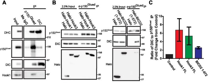
Figure 2.3 Mammalian Hook proteins interact with dynein-dynactin complex ... 38
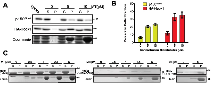
Figure 2.4 Hook proteins bind microtubules indirectly ... 40
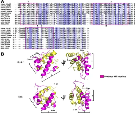
Figure 2.5 Hook proteins lack conserved regions for MT binding ... 42
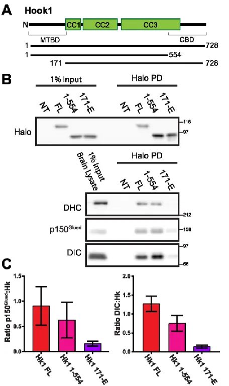
Figure 2.6 Pull-Down (PD) of Halo-Hook1 with dynein-dynactin complex requires N-terminal region ... 44
Figure 2.7 Hook proteins display high velocities and long run lengths ... 46
Figure 2.8 C-terminally truncated Hook proteins display similar motility to full-length ... 49
Figure 3.1 Hook interacts with LIC1 via the N-terminal Hook domain ... 69
Figure 3.S1 Domain architecture of Hook1 ... 71
Figure 3.2 The conserved Helix-1 within the LIC1 effector-binding domain binds the Hook domain... 74
Figure 3.S2 Alignment of the Effector-Binding Domain of LIC sequences ... 76
Figure 3.3 Crystal structure of the Hook domain in complex with LIC1 Helix-1... 79
Figure 3.4 LIC1 Helix-1 mediates the interaction with CC1-Box-containing effectors ... 82
Figure 3.5 The Helix-1-effector interaction is important for processive motility in vitro and in cells ... 85
Figure 3.6 Model for cargo transport by dynein-dynactin-effector complexes ... 91
Figure 3.S3 Table of primers used in this study ... 100
Figure 4.1 Hook1 co-migrates with Rab5- and Rab7-endosomes ... 108
Figure 4.S1 Hook1 Knockdown does not significantly change Rab5 or Rab7 motility ... 111
Figure 4.2 Hook1 KD reduces TrkB-BDNF signaling endosome motility ... 113
x
Figure 4.4 Hook1 localizes to distal axon, while other dynein effectors are enriched in
other compartments ... 117
Figure 4.5 Hook1 KD reduces flux of BDNF from distal axon ... 120
Figure 4.6 Impaired signaling endosome motility reduces BDNF endocytosis ... 122
Figure 4.7 Loss of Hook1 leads to loss of downstream signaling, measured by pCREB levels ... 124
Figure 5.1 Co-IP experiments with other dynein adaptors ... 139
Figure 5.2 Optogenetic recruitment assay with other dynein adaptors ... 140
1
CHAPTER 1: Introduction
I. Intracellular Transport
Spatial and temporal organization of macromolecules and organelles is vital to
cellular functions and is driven by a process called intracellular transport. Transport in
cells was first observed while studying the growth of peripheral nerves. In 1948, Wiess
and Hiscoe described the slow flow, 1mm per day, of axoplasm material from cut or
constricted nerves, which was contradictory to the prevailing view that nerve fibers were
static threads to conduct nerve impulses (Dahlstrom, 2010; Weiss and Hiscoe, 1948).
Subsequent radiolabeling experiments from Samuels et al. in 1951 described the slow
transport of materials in the sciatic nerve over the course of several days (Samuels et
al., 1951). After the discovery of this slow transport, Lubinska and her group described
fast accumulation of acetylcholinesterase in sciatic nerve from rats (Lubinska et al.,
1964). Later, Lasek would repeat radiolabeling experiments from Samuels’ group to
observe the sciatic nerve after a few hours and get rates of transport that were several
hundreds of mm per day, in addition to observing retrograde transport (Lasek, 1967).
Collectively, these studies laid the foundation for what would later be known as axonal
transport.
At the same time as the discovery of axonal transport, intracellular transport was
beginning to be described for the secretory pathway in pancreatic exocrine cells. In the
1960s, pulse chase experiments were used to show the transport of secretory proteins in
pancreatic exocrine cells and tissue slices from guinea pigs (Caro and Palade, 1964;
Jamieson and Palade, 1967). Using this method, they observed transport of synthesized
proteins from the ER to the Golgi via small vesicles. They also found that this transport
2
Antimycin A (Jamieson and Palade, 1968). Following this work, cell-free systems
showed rapid, energy-dependent transport of membranes through the Golgi complex
(Fries and Rothman, 1980).
While these studies provided insights into intracellular transport, the mechanism
behind this transport remained to be revealed. Early work in nerve fibers showed that
colchicine, a drug that disrupts microtubules, induced the blockage of movement of
acetylcholinesterase, implicating the cytoskeleton in intracellular transport (Kreutzberg,
1969), which had already been described in the formation of the mitotic spindle and in
flagella (Ledbetter and Porter, 1964; Phillips, 1966; Roth and Daniels, 1962).
Subsequent work using pharmacological inhibition showed the importance of
microtubules in transport of secretory proteins in liver and thyroid (Le Marchand et al.,
1973; Williams and Wolff, 1972). Studies in the 1980s would then bring insight into the
machines for this microtubule-based transport, the molecular motors, kinesin and dynein
(Allen et al., 1985; Brady, 1985; Paschal and Vallee, 1987; Vale et al., 1985). The next
sections will describe in depth the insights gained over the years into microtubules and
3
II. Microtubules
Microtubules are dynamic polymers of the protein tubulin, which contribute to a
variety of cellular functions including acting as roads for intracellular transport, driving
chromosome separation for cellular division, and maintaining cellular structures including
cilia and flagella. Microtubules are made from heterodimers of α- and β-tubulin (Bryan
and Wilson, 1971; Feit et al., 1971), which organize head-to-tail to give polarity to the
structure, with α-tubulin exposed on the minus-end and β-tubulin exposed at the
plus-end (Amos and Klug, 1974; Bergen and Borisy, 1980; Crepeau et al., 1977). Tubulin
dimers polymerize longitudinally into protofilaments, which associate laterally (usually 13
protofilaments) to form the hollow tubes of microtubules that are about 25nm in diameter
(Tilney et al., 1973).
Microtubule polymerization is powered by the nucleotide guanosine triphosphate
(GTP) (Olmsted and Borisy, 1975; Weisenberg, 1972). Tubulin dimers are added at a
faster rate than GTP hydrolysis, creating what is known as the GTP cap (Mitchison and
Kirschner, 1984). The GTP cap at the end of a growing microtubule allows for continued
polymerization, but hydrolysis to GDP makes the microtubule lattice more prone to
shrinking, also known as catastrophe (Carlier and Pantaloni, 1981; Carlier et al., 1984).
In fact, hydrolysis of GTP on β-tubulin induces conformational changes in α-tubulin
which causes global lattice rearrangements and lattice strain making GDP-microtubules
less stable (Alushin et al., 2014; Mandelkow et al., 1991). Polymerization can occur on
either end of the microtubule but occurs with very different dynamics (Allen and Borisy,
1974; Kristofferson et al., 1986). The microtubule minus end is very stable with slow
growth and very little shrinking, while the microtubule plus end is very dynamic with rapid
4
different proteins including microtubule associated proteins (MAPs) and molecular
motors, which can act to stabilize or increase dynamics.
The C-terminal tails of tubulin are highly charged, acidic, unstructured regions
that project from the outer surface of the microtubule lattice. They are critical for
interactions with proteins on the surface of microtubules. In addition, the tails can be
post-translationally modified (PTM) in a variety of ways including detyrosination,
polyglutamylation, and polyglycylation, which can modulate protein interactions. For
example, proteins with basic microtubule binding domains like CAP-Gly domains, such
as the p150 subunit of dynactin (see next section), interact with the tail tyrosine
specifically (Honnappa et al., 2006; Peris et al., 2006), while the plus-end microtubule
motor kinesin-1 prefers detyrosinated microtubules (Konishi and Setou, 2009; Liao and
Gundersen, 1998). PTMs can also be indicators of microtubule dynamics; for instance,
detyrosination is generally found on long lived stable microtubules, such as those found
in the neuronal axons (Cambray-Deakin and Burgoyne, 1987; Robson and Burgoyne,
1989). The microtubule PTM field has had many advances including the discovery of
tubulin-modifying enzymes and the creation of new tools, which continue to further
insights in this area (reviewed in Janke, 2014).
Microtubule organization is important for cellular maintenance, mitosis, and
creating polarized cells. In general, microtubules are organized with the minus ends
anchored at a microtubule organization center (MTOC) and the plus ends orientated out.
For proliferative cells, the MTOC is at the centrosome near the nucleus and center of the
cell during interphase or when not dividing. Another tubulin isoform, γ-tubulin is
important for microtubule nucleation at the MTOC, where γ-tubulin forms a ring of similar
size to microtubule diameter (reviewed in Kollman et al., 2011). During mitosis,
5
chromosomes and cellular division (Brinkley et al., 1975; Inoué and Sato, 1967; Inoué et
al., 1975). In specialized cells, microtubule organization can be important for creating
cellular polarity. In mammalian neurons, microtubules are organized in a uniform
polarized array in the axon with plus-ends oriented toward the axon terminal (Burton and
Paige, 1981; Heidemann et al., 1981), while the short and highly branched dendrites
have mixed microtubule organization (Baas et al., 1988). In other tissues like intestine
and muscle, differentiated cells have non-centrosomal microtubule arrays to create
polarity needed for function (reviewed in Dyachuk et al., 2016; Sanchez and Feldman,
2017).
The polar nature of microtubules allows for directional transport by the molecular
motors dynein and kinesin. The kinesin family of proteins are mainly plus-end directed
motors with a few exceptions, while cytoplasmic dynein is a minus-end directed motor
(discussed in the next section). There are up to 45 different kinesin genes in mammals,
which can be classified into 15 families based on phylogenetic analyses (reviwed in
Hirokawa and Tanaka, 2015). In contrast, there is only one major form of cytoplasmic
dynein that is responsible for the majority of minus-end directed microtubule transport
and is described further in the next section. The coordination and regulation of these
microtubule motors for efficient transport and proper function is still a major area of
6
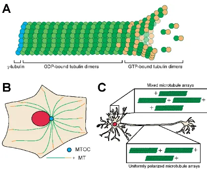
Figure 1.1 Microtubule dynamics and organization
A) Microtubule with dynamic GTP plus-end. B) Organization of microtubules in fibroblast
cells with minus ends at the microtubule organization center (MTOC) and plus-ends
oriented towards the periphery. C) Organization of microtubules in mammalian neurons
7
III. Cytoplasmic Dynein
In the 1960s, the first microtubule motor was discovered in the cilia of
Tetrahymena (Gibbons, 1963; Gibbons and Rowe, 1965). They named this protein
dynein, from the Greek dyne meaning force, due to its ability to slide microtubules in the
cilia using ATP (Gibbons and Rowe, 1965). Since this discovery, sixteen genes have
been found in humans to encode dynein heavy chains, including fourteen that function
within the axoneme and one involved in retrograde intraflagellar transport (Wickstead
and Gull, 2007; Yagi, 2009). The remaining gene encodes cytoplasmic dynein which
drives ATP-dependent minus-end directed microtubule transport, opposite of
conventional kinesins (Lye et al., 1987; Paschal and Vallee, 1987; Paschal et al., 1987).
Cytoplasmic dynein is responsible for the majority of minus-end directed transport in
cells, with a few minus-end directed kinesins contributing to this motility. In animals,
cytoplasmic dynein is essential due primarily to its vital role in cellular division and
development (Gepner et al., 1996; Robinson et al., 1999; Vaisberg et al., 1993).
Cytoplasmic dynein (hence forth referred to as dynein) is a 1.4 MDa motor
complex consisting of dimerized heavy chains (DHC, ~530 kDa), each containing a
N-terminal tail, six AAA+ domains to form a motor ring and a flexible stalk with the
microtubule binding domain. N-terminal tail of dynein allows for homodimerization of the
heavy chains, along with binding sites for other non-catalytic subunits, including two
intermediate chains (DIC, ~74 kDa) and two light intermediate chains (LIC, ~33-59 kDa).
Smaller light chains (LC, ~10-14kDa), including LC7/Roadblock, LC8, and TCTEX1 bind
to the intermediate chains. These non-catalytic subunits provide sites of attachment and
regulation for specific dynein functions. In vertebrates, the intermediate and light chains
are encoded by two genes and expression patterns differ in various cell types, likely due
8
Unlike kinesins, dynein is a member of the AAA+ (ATPases Associated with
various cellular Activities) family of proteins, which have ring-like hexamers of ATPase
domains that can function in a variety of ways to unfold proteins, disassemble
aggregates, and produce mechanical force (Miller and Enemark, 2016). Unlike other
AAA+ proteins, the motor domain of dynein is formed by a single polypeptide to form the
hexamer of AAA+ domains, a linker domain and a microtubule binding domain. The first
four AAA+ domains, AAA1-AAA4 are able to bind ATP, while AAA5 and AAA6 serve as
structural units for the stalk and buttress of the microtubule binding domain. While AAA1
is the primary site of ATP hydrolysis, the nucleotide state of the other AAA domains like
AAA3 is important to coordinate microtubule binding and potentially regulate different
motility properties of the motor (DeWitt et al., 2015; Nicholas et al., 2015a; Takahide Kon
et al., 2004). The microtubule binding domain of dynein is at the end of a 15 nm
antiparallel coiled-coil stalk that projects from the AAA4-AAA5 domain (Burgess et al.,
2003; Gee et al., 1997; Kon et al., 2011).
Dynein motility is coupled to ATP-induced conformational changes in the AAA+
ring, with the bending and straightening of the linker region. ATP binds at AAA1 to
induce a weak microtubule-binding state and release of the microtubule binding domain
(MTBD) from the microtubule. When ATP is present at AAA1, the linker region also
undocks and moves away from the motor ring (Cho and Vale, 2012; Roberts et al., 2012;
Schmidt et al., 2014). The hydrolysis of ATP on the dissociated motor head then causes
a primed conformation to allow for rebinding to the microtubule. The forward step is
coupled to the release of phosphate and ADP to reset the cycle (Holzbaur and Johnson,
1989b, 1989a; Imamula et al., 2007). Unlike the classic power-stroke mechanism, the
9
the stalk and hinging at the microtubule binding domain, instead of tilting the MTBD stalk
as previously thought (Lippert et al., 2017).
Single molecule studies have provided insight into the motility of cytoplasmic
dynein, in addition to finding differences between dynein from different species.
Mammalian dynein without the presence of interacting partners has been found in a
state of autoinhibition, called the phi state, which has low microtubule binding affinity, or
as a flexible dimer with very low processivity (Zhang et al., 2017). This is in contrast to
yeast dynein, which displays slow processive motility and does not require tight coupling
of the two motor domains (DeWitt et al., 2012; Qiu et al., 2012; Reck-Peterson et al.,
2006). Furthermore, mammalian dynein can produce up to 1.1 pN of force, while yeast
dynein has a stall force of 7pN (Gennerich et al., 2007; Mallik et al., 2004; Schroeder et
al., 2010). Dynein also takes many sideways or backwards steps along the microtubule
lattice (Reck-Peterson et al. 2006; Ross et al. 2006) and has a variable stepping pattern,
with predominantly an 8 nm step size with a maximum step size of 32 nm, which is in
contrast to the uniform 8 nm step size of kinesin-1 (DeWitt et al., 2012; Mallik et al.,
2004; Qiu et al., 2012; Reck-Peterson et al., 2006). Due to the flexible nature of dynein,
it has been shown to avoid obstacles better than kinesins (Dixit et al., 2008).
Dynein functions in many different roles in the cell including nuclear positioning,
organelle and virus transport, chromosome dynamics, mitotic spindle orientation,
axonogenesis, and cell migration (reviewed in Reck-Peterson et al., 2018; Roberts et al.,
2013). Loss of dynein is lethal for many organisms including mice and Drosophila
melanogaster (Gepner et al., 1996; Harada et al., 1998). In addition, mutations in dynein
lead to neurological diseases like spinal muscular atrophy with lower extremity
predominance (SMALED), Charcot–Marie–Tooth (CMT) disease, and malformations of
10
(Fiorillo et al., 2014; Lipka et al., 2013; Poirier et al., 2013; Tsurusaki et al., 2012;
Weedon et al., 2011). Since dynein function is vital for proper development, uncovering
the regulatory mechanisms of dynein motility and function has been an imperative area
of research.
Dynactin
Dynein has been found to work in complex with dynactin, a 1 MDa, 23 subunit
complex which is needed for the majority of dynein’s transport functions. Dynactin was
found as an “activating” component that co-purified with 20S dynein and was needed for
in vitro vesicle motility (Gill et al., 1991; Schroer and Sheetz, 1991). With many years of
work and advancements in cryo-EM technology, the components and structure of
dynactin have now been elucidated. Dynactin is comprised of an actin-like Arp1 filament
with a large shoulder of alpha-helices at one end (Schroer, 2004). The Arp1 filament
contains eight Arp1 molecules, one beta-actin and one Arp11 protein. Arp11 interacts
with p25, p27, p62 to form the pointed-end of dynactin. The other end of dynactin is
capped by an actin capping protein, CapZαβ, which forms the barbed-end of the
filament. The shoulder of dynactin sits on the Arp1 filament near the barbed-end. It is
comprised of two copies of p24, four copies of p50 (dynamtin), and two copies of
p150Glued (Chowdhury et al., 2015; Urnavicius et al., 2015).
The p150Glued subunit is the largest subunit of dynactin and projects out of the
shoulder as a coiled-coil with a N-terminal CAP-Gly domain, which binds microtubules
(Waterman-Storer et al., 1995). In early work, p150Glued was found to interact with the
dynein intermediate chain (Karki and Holzbaur, 1995; Vaughan and Vallee, 1995). This
subunit is very flexible and likely can fold back on its self to dock against the dynactin
11
microtubule binding of p150Glued is required to recruit dynein to microtubules and
enhance dynein processivity (Ayloo et al., 2014; Culver–Hanlon et al., 2006; King and
Schroer, 2000). In addition, mutations in p150Glued are associated with
neurodegenerative disorders such as slowly progressing lower MND (motor neuron
disease) and Parkinson-like Perry syndrome (Farrer et al., 2009; Puls et al., 2003;
Stockmann et al., 2013). In the brain, there is also an alternative splice form of p150,
p135 which lacks the CAP-Gly domain (Tokito et al., 1996). Motor complexes with p135
seem to have reduced frequency of motility events in vitro (McKenney et al., 2014), but it
is still unclear in vivo how p135 affects dynein functions compared to p150.
Dynactin is required for most of dynein’s functions. Recent cryo-EM studies have
shown that dynactin reorients the dynein dimer for proper recruitment and positions the
motor heads for proper motility along microtubules (Zhang et al., 2017). Furthermore,
dynactin is vital to retrograde axonal transport of organelles (Haghnia et al., 2007;
Waterman-Storer et al., 1997). The pointed-end of dynactin has been suggested to play
a role in facilitating cargo interaction (Yeh et al., 2012; Zhang et al., 2011), but more
recent work has shown that dynactin interacts with dynein via dynein effectors which
12
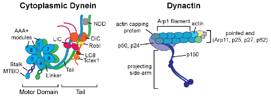
Figure 1.2 Dynein-Dynactin schematic
Schematics of cytoplasmic dynein and dynactin. Abbreviations: NDD=N-terminal
dimerization domain, MTBD=microtubule binding domain, Robl=roadblock, LIC=Light
13
IV. Dynein Effector Proteins
Adaptor and effectors proteins are required to link cargo to the dynein-dynactin
motor complex (Fu and Holzbaur, 2014; Kardon and Vale, 2009). In addition to linking
dynein to cargo, some of these proteins can modulate motility properties of dynein,
either to enhance or inhibit movement, and some can act as motility switches by
interacting with kinesins as well as dynein. This section will highlight the current insights
into dynein effectors proteins.
The Bicaudal D (BICD) proteins were the first family of proteins described to link
dynein motors to cargo. BICD was initially identified in Drosophila where mutations in
BICD cause abnormal development of a double abdomen or bicaudal (meaning
‘two-tailed’) phenotype (Mohler and Wieschaus, 1986). Further analysis of these flies showed
that BICD was vital in mRNA transport and nuclei positioning, suggesting a link to
microtubule-mediated transport (Bullock and Ish-Horowicz, 2001; Mach and Lehmann,
1997; Suter and Steward, 1991; Swan and Suter, 1996; Swan et al., 1999; Wharton and
Struhl, 1989). In mammals, there are two BICD orthologs, BICD1 and BICD2 as well as
two related proteins, BICDR-1 and BICDR-2. The BICD proteins contain long stretches
of coiled-coil domains which interact with dynein and dynactin, with the C-terminal region
bending back on itself to produce an autoinhibited state (Liu et al., 2013; Terawaki et al.,
2015; Urnavicius et al., 2015; Wharton and Struhl, 1989). The C-terminal region of all
BICD proteins, except BICDR-2, can be released from this inhibition via cargo binding to
the small GTPase Rab6 (Hoogenraad et al., 2001; Matanis et al., 2002; Schlager et al.,
2010; Short et al., 2002). The BICD family has been the most well-characterized family
of dynein effectors and has been the starting point for many discoveries into the
14
Activation of Dynein
Recently, a set of coiled coil effector proteins including BICD2, Hook3, Spindly
and NINL have been shown to enhance the dynein-dynactin interaction and induce
superprocessive motility (McKenney et al., 2014; Redwine et al., 2017; Schlager et al.,
2014; Schroeder and Vale, 2016; Splinter et al., 2012). BICD2, the best characterized of
these activators, has been shown to increase the affinity of dynein-dynactin interaction
through coiled-coil contacts along the Arp1 filament that forms the core of dynactin
(Chowdhury et al., 2015; Urnavicius et al., 2015). BICD2 also interacts with the
N-terminal tail of the dynein heavy chain (Chowdhury et al., 2015; Urnavicius et al., 2015)
and the dynein light intermediate chain 1 (LIC1) (Lee et al., 2018; Schroeder et al.,
2014), leading to a stabilization of the dynein-dynactin-effector complex. Some dynein
effectors including BICDR-1 can recruit two dynein dimers to a single dynactin, which
further enhances the force and velocity of the motor complex (Grotjahn et al., 2018;
Schlager et al., 2014b; Urnavicius et al., 2018).
Spindly is another dynein activator which plays a role in mitosis by silencing a
mitotic checkpoint after proper spindle assembly (Barisic and Geley, 2011; Griffis et al.,
2007; McKenney et al., 2014). Spindly recruits dynein to kinetochores which induces the
movement of chromosomes to the poles (Chan et al., 2009; Gassmann et al., 2008;
Griffis et al., 2007). A recent study on Spindly also found new features of dynein
activators, the CC1 box and the Spindly motif (Gama et al., 2017). Using sequence
analysis and biochemistry, the CC1 box was found to be important for interaction with
dynein while the Spindly motif was found to be important for interaction with dynactin. In
addition, sequence alignments with other dynein adaptors showed the presence of these
motifs in other proteins including the BICD family, HAP1, and TRAKs (Gama et al.,
15
alanines were of interest due to previous studies on BICD2, where mutations of these
alanines to valines cause loss of interaction with dynein-dynactin in vitro (Schlager et al.,
2014b) and a loss-of-function phenotype in Drosophila melanogaster (Oh et al., 2000).
Similar alanine to valine mutations in Spindly also resulted in loss of dynein interaction.
The Spindly motif is located after the CC1 box and a stretch of coiled coil and has a
sequence of L(F or A)XE for most of dynein effectors. This region of Spindly was found
to interact with the pointed end of dynactin. The authors chose to mutate the
phenylalanine to an alanine to show a loss of interaction with dynactin but unfortunately,
the phenylalanine is not conserved in other dynein effectors and is actually an alanine in
most cases (Gama et al., 2017). Further work is still needed to determine the importance
of this region in other dynein effectors.
Rab11-FIP3 was also found to activate the motility of dynein. It is part of the
Rab11-FIP family of proteins which are mainly known to regulate the trafficking of
recycling endosomes via a conserved Rab11 GTPase binding domain (reviewed in
Horgan and McCaffrey, 2009; Jing and Prekeris, 2009). Class I FIPs (Rab11-FIP1, 2, 5)
have N-terminal phospholipid-binding C2-domains, while class II FIPs (Rab11-FIP3 and
-FIP4) possess N-terminal EF-hand domains in addition to longer coiled-coil regions
near the C-terminus. Rab11-FIP3 and Rab11-FIP4 seem to have more specialized roles
in endosomal trafficking then class I FIPs (Jing and Prekeris, 2009). Rab11-FIP3 plays
an important role in the cell cycle dependent trafficking of recycling endosomes to the
cleavage furrow but does not affect recycling of proteins in interphase (Horgan et al.,
2010; Inoue et al., 2008; Simon et al., 2008; Wilson et al., 2005). The other class II FIP,
Rab11-FIP4, is highly enriched in the brain but little is known of its function in endosomal
16
interact with dynein, despite the high similarities between FIP3 and FIP4 (Horgan et al.,
2010; McKenney et al., 2014).
Figure 1.3 Model of dynein activation
Dynein is found autoinhibited or in open states of flexible conformations. Following the
addition of dynactin and a dynein activator, the motor domains of dynein reorganizes for
proper microtubule attachment and display processive motility (Zhang et al., 2017).
Adaptors
The following proteins have not been shown to activate dynein motility in vitro like
the activators described above but do share similar coiled coil structure and might be
identified as activators in future work. These adaptors have mostly been identified
through protein interactions, genetic manipulation, and live cell experiments.
One of these identified dynein adaptors is HAP1 (Huntingtin-associated protein
1). HAP1 interacts with huntingtin (htt), which is best known for its causative role in
Huntington’s disease where huntingtin is mutated with an expanded polyglutamine
insertion. Huntingtin is a very large protein that has been shown to interact with a variety
of proteins but notably interacts with the intermediate chain of dynein (Caviston et al.,
2007). HAP1 can interact with p150Glued (Engelender et al., 1997; Li et al., 1998) as well
17
Huntingtin and HAP1 have long been associated with intracellular transport
(Block-Galarza et al., 1997). Huntingtin has been linked to axonal transport of autophagosomes
(Wong and Holzbaur, 2014), synaptic vesicles (Gunawardena et al., 2003; Weiss and
Littleton, 2016), amyloid precursor protein(APP)-positive vesicles, and brain derived
neurotrophic factor (BDNF)-positive vesicles (Colin et al., 2008; Her and Goldstein,
2008). HAP1 has been shown to be required for htt-mediated transport and it has been
suggested that htt/HAP1 together act as a platform for both dynein and kinesin
attachment to vesicles. Due to the large size of htt, it has been difficult to dissect the
mechanism of htt and HAP1-mediated dynein motility in vitro assays.
Milton/TRAK proteins are a family of proteins suggested to act as motor adaptors
for mitochondria. The Milton/TRAK family have a N-terminal coiled-coil region that has
high homology to the HAP1 domain and the C-terminal region interacts with
Mitochondrial Rho GTPase (Miro). In Drosophila, Milton in complex with Miro interacts
with kinesin-1 to deliver mitochondria to neuronal synapses (Glater et al., 2006; Stowers
et al., 2002). The mammalian homologs of Milton, TRAK1 and TRAK2, have been linked
to dynein and kinesin motility and are required for mitochondria distribution in a variety of
cell types including neurons (reviewed in Melkov and Abdu, 2018). TRAK1 binds
dynein-dynactin and kinesin-1, while TRAK2 predominately interacts with dynein-dynein-dynactin (van
Spronsen et al., 2013). Interestingly in neurons, TRAK1 is mainly localized in the axon,
while TRAK2 is localized to the dendrites (Loss and Stephenson, 2015; van Spronsen et
al., 2013), which could reflect the dependence of each compartment for specialized
motors.
Another adaptor, RILP (Rab7-interacting lysosomal protein) has been suggested
to link dynein to Rab7 vesicles, including late endosomes and lysosomes. RILP was
18
(Cantalupo et al., 2001). RILP was then suggested to link Rab7 vesicles with dynein
through co-localization experiments (Jordens et al., 2001). Later, biochemical studies
showed a stepwise process of dynein recruitment by RILP, where RILP and
oxysterol-binding protein–related protein 1L (ORP1L) form a complex with Rab7 and then RILP
can interact with p150Glued of dynactin, which in turn recruits dynein to the vesicle
(Johansson et al., 2007). This stepwise recruitment suggests dynein association with
vesicles can be regulated by cholesterol levels, which are sensed by ORP1L. Consistent
with this mechanism, cholesterol-rich lysosomes are transported to the nucleus, but low
cholesterol vesicles remain at the cell periphery due to conformational changes in
ORPL1, which leads to dissociation of p150Glued (van der Kant et al., 2013; Rocha et al.,
2009). In addition, RILP has been shown to self-interact, likely as a homodimer, similar
to other dynein effectors like BICD2 (Colucci et al., 2005; Wu et al., 2005).
JIP (c-Jun N-terminal kinase (JNK)-interacting proteins) proteins have also been
identified as motor adaptors. There are four mammalian JIPs, JIP1-4, which are highly
expressed in the brain (Dickens et al., 1997; Kelkar et al., 2000, 2005; Yasuda et al.,
1999). While each JIP protein contains a JNK-binding domain near the N-terminus, JIP1
and JIP2 are structurally distinct from JIP3 and JIP4. JIP1/2 each contain a Src
homology-3 domain (SH3) and a phosphotryosine binding domain (PTB) near the
C-terminus (Dickens et al., 1997; Yasuda et al., 1999), while JIP3/4 each contain
N-terminal coiled coil regions, a leucine zipper domain, and a C-N-terminal transmembrane
region (Kelkar et al., 2000, 2005). JIP proteins interact with multiple kinases of the JNK
pathway and p38 MAPK pathway, as positive or negative regulators (Whitmarsh, 2006).
These signaling pathways are involved in a variety of cellular processes including
growth, differentiation, and apoptosis. In addition to signaling factors, JIPs have been
19
with kinesin-1 (Bowman et al., 2000; Montagnac et al., 2009; Verhey et al., 2001). JIP1,
JIP3, and JIP4 have also been implicated in minus-end motility via dynein-dynactin
interaction. JIP1 has been found to coordinate dynein and kinesin motility for
APP-positive vesicles, with phosphorylation used to switch from kinesin to dynein motility (Fu
and Holzbaur, 2013). JIP4 was shown to transport recycling endosomes during
cytokinesis via its interaction with kinesin-1 and dynactin, with ARF6 binding as the
regulatory switch for JIP4 interaction with motors (Montagnac et al., 2009). In
Drosophila, JIP3 (aka Sunday Driver) was found to associate with dynein-dynactin
during the transport of axonal injury signals (Cavalli et al., 2005). Further work
suggested that JIP3 interacts with endosomes for retrograde transport and a small
anterograde vesicle population, potentially for axonal growth (Abe et al., 2009; Watt et
al., 2015). A recent study showed that JIP3 knockout mouse neurons have lysosome
accumulation in axons and impaired maturation, which was previously seen in zebrafish
and C. elegans (Drerup and Nechiporuk, 2013; Edwards et al., 2013; Gowrishankar et
al., 2017).
Regulatory mechanisms
There are several ways that dynein effector proteins can be regulated for proper
spatiotemporal intracellular transport. Some of these adaptor proteins including JIP1 and
huntingtin have phosphorylation sites that regulate interaction with molecular motors
(Colin et al., 2008; Fu and Holzbaur, 2013). Other interacting proteins can also act as
regulators of dynein effector proteins. For example, Miro is a Ca+2 sensor which alters
interaction with Milton and kinesin-1 upon Ca+2 binding, reducing mitochondria motility
(MacAskill et al., 2009; Wang and Schwarz, 2009). Some dynein effectors have also
20
interaction unless properly bound to cargo. For example, the last coiled coil region of
BICD2 folds back on itself but binding its cargo protein Rab6 releases this autoinhibition,
unmasking its dynein-dynactin interface (Terawaki et al., 2015). Differential expression
of these effectors in tissues and during development is another way to control transport
by dynein. For instance, BICD1 has been shown to be highly expressed early in
development and decreases during neurite growth (Schlager et al., 2010). Other
21
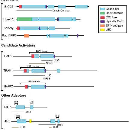
Figure 1.4 Domain structure of dynein effectors
Figure legend at the top right. Gray lines indicate identified dynein-dynactin interaction
22
Other regulators of dynein
Another important regulator of dynein is Lis1. LIS1 was first described as the
mutated gene in type 1 lissencephaly, a neurodevelopmental disease (Reiner et al.,
1993). Deletions or duplications of LIS1 cause serious brain development defects. Lis1
plays a role in neuronal migration and proliferation (reviewed in Reiner and Sapir, 2013).
Lis1 interacts with Nudel and NudE, which also have roles in neurodevelopment
(reviewed in Bradshaw et al., 2013). Lis1 and Nudel/NudE were found to interact with
dynein during screens for nuclear migration defects in filamentous fungi (Efimov and
Morris, 2000; Minke et al., 1999; Xiang et al., 1995), later confirmed to be conserved in
other systems, including Drosophila (Liu et al., 1999, 2000) and mammals (Faulkner et
al., 2000; Sasaki et al., 2000; Smith et al., 2000). In polarized cells like neurons, Lis1 has
been linked to the motility of several different cargos including mitochondria,
endosomes, peroxisomes, and lysosomes, and suggested to be important for motor
loading at microtubule plus-end (Egan et al., 2012; Lenz et al., 2006; Moughamian et al.,
2013; Shao et al., 2013; Yi et al., 2011; Zhang et al., 2010). Lis1 is also involved in
positioning of centrosomes, nuclei (Gambello et al., 2003; Tsai et al., 2007; Xiang et al.,
1995), and spindle poles (Moon et al., 2014; Yingling et al., 2008).
Lis1 has a N-terminal dimerization domain, a coiled-coil region, a disordered
loop, and a β-propeller domain with seven WD repeats(Kim et al., 2004; Tarricone et al.,
2004). The β-propeller domain of Lis1 interacts with the dynein motor domain, at AAA3
and AAA4(Huang et al., 2012; Toropova et al., 2014). Biochemical experiments suggest
that Lis1 increases dynein’s affinity for microtubules and slows down dynein’s velocity in
motility assays in vitro(Huang et al., 2012; McKenney et al., 2010; Toropova et al., 2014;
Yamada et al., 2008). Lis1 does not prevent ATP hydrolysis but has been suggested to
23
challenged the idea that Lis1 plays an inhibitory role in dynein motility. For yeast dynein,
Lis1 is now proposed to induce weak or tight microtubule-binding of dynein, depending
on the nucleotide state of AAA3 and the numberof Lis1 β-propeller domains bound to
the motor domain(DeSantis et al., 2017). In recent studies on mammalian dynein with
dynactin and BICD2, Lis1 was found to actually increase the frequency and velocity of
dyneinmotility, in a concentration-dependent manner(Baumbach et al., 2017; Gutierrez
et al., 2017). Future structural work is needed to better understand the complex of Lis1
with dynein-dynactin-BICD2.
Nudel/NudE are dimeric, coiled-coil proteins that interact with DIC and LC8 via
their N-termini and Lis1 with their C-termini (Stehman et al., 2007; Wang and Zheng,
2011; Zyłkiewicz et al., 2011). Nudel/NudE has been suggested to tether Lis1 to dynein
and be a regulatory module due to their phosphorylation by Cdk5(Hebbar et al., 2008;
Klinman and Holzbaur, 2015; Pandey and Smith, 2011). Lis1 in complex with NudE is
suggested to induce a highload-bearing state in dynein. Optical trapping experiments
showed that Lis1 and NudE allow dynein to have a prolonged force-producing state
(McKenney et al., 2010). Lis1 and NudE/Nudelalso enhanced force production of dynein
24
V. Hook Proteins
With the growing evidence for dynein adaptors, the goal of my thesis work was to
uncover new dynein adaptors and understand their role in the regulation of dynein
motility. My thesis work focuses on the Hook protein family, due to several studies in
other model systems, like Drosophila and filamentous fungi, which suggested roles for
Hook proteins in intracellular transport.
Hook proteins were first investigated in a screen for genes involved in
cell-specific ligand endocytosis in Drosophila (Krämer and Phistry, 1996). Hook mutant flies
have abnormal bristle morphology which makes them look like a “hook”, in addition to
eye degeneration (Krämer and Phistry, 1996; Mohr, 1927). Genetic analysis showed that
mutations in the hook gene inhibit endocytosis, as seen by reduced accumulation of
transmembrane and soluble ligands into multivesicular bodies, and increased the
accumulation of multilamellar late endosomes in the photoreceptor cells of Drosophila
eyes (Krämer and Phistry, 1996, 1999; Sunio et al., 1999). The Drosophila Hook protein
was also localized to vesicular structures, potentially late endosomes marked by Rab7
(Krämer and Phistry, 1996; Szatmári et al., 2014).
In general, Hook proteins are characterized by three conserved regions: a
globular N-terminal putative microtubule binding domain, a central coiled-coil domain,
and a divergent, predicted unstructured C-terminal domain thought to mediate cargo
binding (Walenta et al., 2001). The N-terminal region of Hook proteins was originally
described to interact with microtubules based on homology to the well characterized
microtubule binding domains, calponin-homology domains, and initial microtubule
pelleting assays with cell lysates (Walenta et al., 2001). The central coiled-coil region
was suggested to induce homodimerization by immunoprecipitation and sedimentation
25
region is the most divergent region among species and isoforms but has been
suggested to link Hook proteins to their intended cargo based on the use of truncated
constructs (Bielska et al., 2014; Walenta et al., 2001; Xu et al., 2008; Zhang et al.,
2014).
Hook proteins (HookA or Hok1), were first characterized as potential dynein
adaptors in filamentous fungi (Bielska et al., 2014; Zhang et al., 2014). In Aspergillus
nidulans, HookA was described as an adaptor on early endosomes regulating dynein,
while Hok1 in Ustilago maydis was shown to coordinate dynein and kinesin-3 motors
during early endosome transport (Bielska et al., 2014; Zhang et al., 2014). In both fungal
Hook proteins, the N-terminus is required for interaction with dynein and the C-terminus
allows for attachment to cargo via interaction with Fused Toes (FTS) and FTS and
Hook-Interacting Protein (FHIP) (Bielska et al., 2014; Yao et al., 2014; Zhang et al., 2014).
These interactions were determined by immunoprecipitation experiments from cell
lysates, so direct protein-protein interactions remained to be determined by future
studies.
In mammalian cells, three highly conserved Hook proteins are expressed: Hook1,
Hook2, and Hook3. Each Hook protein has been linked to different cellular functions.
Hook1 has been implicated in spermiogenesis as mutations in Hook1 cause abnormal
spermatozoon head shape (Mendoza-Lujambio et al., 2002). Hook2 has been linked to
centrosomal function and homeostasis (Guthrie et al., 2009; Moynihan et al., 2009;
Szebenyi et al., 2007). Hook3 was originally described as a Golgi-associated protein due
to its localization at the Golgi in cell lines and the disruption of Golgi structure when
Hook3 is overexpressed (Walenta et al., 2001). In addition to the aforementioned work,
Hook1 and Hook3 have been implicated in a variety of endosomal trafficking pathways,
26
al., 2004; Maldonado-Báez et al., 2013; Xu et al., 2008). In HeLa cells, Hook proteins
were found to interact with members of the HOPS complex and to be important for timely
trafficking of EGF through endosomal compartments marked by EEA1, CD63 and
LAMP1, but this study simultaneously knocked down all three Hook isoforms, making it
difficult to determine their individual roles (Xu et al., 2008). A recent study in
hippocampal neurons suggested that Hook1 and Hook3 are involved in Rab5 retrograde
motility in the axon (Guo et al., 2016). Hook1 and Hook3 have been suggested to attach
to cargo through C-terminal interactions with Fused Toes (FTS) and FTS-Hook
Interacting (FHIP) proteins, similar to fungal studies (Guo et al., 2016; Xu et al., 2008;
Yao et al., 2014).
Other studies have looked at individual Hook proteins. Using HeLa cells, Hook1
was found to interact with CD147 to facilitate sorting into Rab22-positive recycling
tubules (Maldonado-Báez et al., 2013). In COS-1 cells, Hook1 was found to interact with
Rab7, Rab9, and Rab11 using immunoprecipitation (Luiro et al., 2004). The variety of
results seen in these studies is likely due to the fluid nature of endosomal pathways and
differential cellular demands on these pathways. Studies on Hook3 have implicated that
it interactions with a variety of proteins including scavenger receptor A, Pericentriolar
Material 1 (PCM1), IIGP, and Salmonella SpiC (Ge et al., 2010; Kaiser et al., 2004; Sano
et al., 2007; Shotland et al., 2003). It remains to be seen if these interactions are
reproducible in other systems and studies.
Throughout my thesis, I studied the role of mammalian Hook proteins as dynein
adaptors using biochemistry, in vitro motility assays, and live cell microscopy. In the first
half of my thesis, I worked to determine how Hook proteins effect dynein motility and
which domains are important for motor interaction. In addition, I collaborated with the
27
proteins and effect processivity. In the second half of my thesis, I studied the role of
28
CHAPTER 2: Hook Adaptors Induce Unidirectional Processive Motility by
Enhancing the Dynein-Dynactin Interaction
This chapter is adapted from:
Olenick, M. A., Tokito, M., Boczkowska, M., Dominguez, R., Holzbaur, E.L.F. (2016)
Hook adaptors induce unidirectional processive motility by enhancing the
dynein-dynactin interaction. Journal of Biological Chemistry 291, 18239-18251.
We gratefully acknowledge Karen Wallace for technical assistance, Michael Woody and
Jeffery Nirschl for assistance with custom image analysis programs, and Chanat
29
I. Summary
Cytoplasmic dynein drives the majority of minus-end directed vesicular and
organelle motility in the cell. Yet, it remains unclear how dynein is spatially and
temporally regulated given the variety of cargo that must be properly localized to
maintain cellular function. Recent work has suggested that adaptor proteins provide a
mechanism for cargo-specific regulation of motors. Of particular interest, studies in
fungal systems have implicated Hook proteins in the regulation of microtubule motors.
Here, we investigate the role of mammalian Hook proteins, Hook1 and Hook3, as
potential motor adaptors. We used optogenetic approaches to specifically recruit Hook
proteins to organelles and observed rapid transport of peroxisomes to the perinuclear
region of the cell. This rapid and efficient translocation of peroxisomes to microtubule
minus ends indicates that mammalian Hook proteins activate dynein rather than kinesin
motors. Biochemical studies indicate that Hook proteins interact with both dynein and
dynactin, stabilizing the formation of a supramolecular complex. Complex formation
requires the N-terminal domain of Hook proteins, which resembles the
calponin-homology domain of EB proteins yet cannot bind directly to microtubules. Single
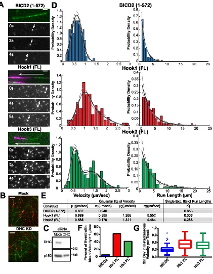
molecule motility assays using TIRF microscopy indicate that both Hook1 and Hook3
effectively activate cytoplasmic dynein, inducing longer run lengths and higher velocities
than the previously characterized dynein activator BICD2. Together, these results
suggest that dynein adaptors can differentially regulate dynein to allow for
30
II. Introduction
Microtubules provide a polarized highway to facilitate transport of organelles and
vesicles throughout the cell. The minus ends of microtubules are usually nucleated near
the cell center with the plus ends oriented outwards, toward the cell periphery. This
polarity ensures that microtubule motors drive motility in a specific direction; kinesin
motors generally drive plus-end motility while minus-end traffic is primarily driven by
cytoplasmic dynein. Regulation of these opposing motors is vital for cell survival,
particularly in specialized cells like neurons that require efficient transport over long
distances (Maday et al., 2014). However, it remains unclear how microtubule motors are
spatially and temporally regulated to control the intracellular trafficking of specific cargo.
As a single major form of cytoplasmic dynein drives the transport of a wide array of
cargos, including endosomes, RNA granules, and mitochondria (van Niekerk et al.,
2007; van Spronsen et al., 2013; Xiang et al., 2015), it is likely that the transport
properties of dynein are modulated by the binding of cargo-specific adaptor molecules.
A number of dynein regulatory and adaptor proteins have been identified to date,
including dynactin, Lis1, BICD2, and, more recently, Hook proteins. The first major
regulator to be identified was dynactin, a large multi-subunit protein complex required for
most functions of dynein within the cell. Dynactin forms a co-complex with dynein
(Chowdhury et al., 2015; Karki and Holzbaur, 1995; Urnavicius et al., 2015; Vaughan
and Vallee, 1995) that enhances the initial recruitment of dynein to the microtubule
(Ayloo et al., 2014; Moughamian and Holzbaur, 2012) and mediates association of
dynein with some intracellular cargos (Holleran et al., 2001; Muresan et al., 2001; Yeh et
al., 2012; Zhang et al., 2011). A second major dynein regulator, Lis1, binds to the dynein
motor domain and blocks the required linker swing in the mechanochemical cycle for
31
microtubule (Toropova et al., 2014). In contrast to the inhibitory effect of Lis1 on dynein
motility, the dynein adaptor BICD2 has been shown to induce superprocessive motility of
dynein, potentially through enhanced stability of the dynein-dynactin complex
(McKenney et al., 2014; Schlager et al., 2014b). Yet another mechanism that adaptors
can use to regulate transport is coordination of different motors on the same cargo. For
instance, JIP1 acts as a switch between dynein- and kinesin-1-mediated transport
depending on its phosphorylation state (Fu and Holzbaur, 2013). Given the wide variety
of cargo that must be properly localized within eukaryotic cells, it is likely that many
additional adaptors and their underlying regulatory mechanisms remain to be identified
and characterized.
Here we focus on another family of potential dynein adaptors, Hook proteins.
Genetic screens in fungal model systems have provided evidence that Hook proteins are
required for early endosome trafficking. In general, Hook proteins are characterized by
three conserved regions: a globular N-terminal putative microtubule binding domain, a
central coiled coil domain, and a variable and predicted unstructured C-terminal domain
thought to mediate cargo binding (Walenta et al., 2001). In Aspergillus nidulans, HookA
was described as an adaptor on early endosomes regulating dynein, while Hok1 in
Ustilago maydis was shown to coordinate dynein and kinesin-3 motors during early
endosome transport (Bielska et al., 2014; Zhang et al., 2014). In both fungal Hook
proteins, the C-terminus attaches to cargo through interaction with the proteins, Fused
Toes (FTS) and FTS and Hook-Interacting Protein (FHIP) (Bielska et al., 2014; Yao et
al., 2014; Zhang et al., 2014).
These studies led us to ask if such functions of Hook proteins were conserved in
mammalian systems. There are three Hook isoforms expressed in humans: Hook1,
32
localizes to the Golgi (Walenta et al., 2001), Hook2 is recruited to centrosomes
(Szebenyi et al., 2007), and Hook1 is implicated in endosomal transport (Luiro et al.,
2004; Maldonado-Báez et al., 2013; Xu et al., 2008). To explore adaptors with roles in
cargo transport, we focused on Hook1 and Hook3 in our studies.
We used complementary optogenetic and single molecule approaches to
establish mammalian Hook proteins as motor adaptors enhancing unidirectional minus
end-directed motility driven by dynein. We show that both Hook1 and Hook3 enhance
the formation of a dynein-dynactin complex. The formation of this complex requires the
N-terminal globular domain of Hook proteins. Contrary to previous suggestions, this
domain does not bind to microtubules directly. In single molecule assays, we find that
both Hook1 and Hook3 induce highly processive dynein motility, resulting in both longer
run lengths and faster velocities than the previously characterized dynein activator,
BICD2. Together these results support a model in which organelle-specific adaptors
33
III. Results
Differential regulation of dynein-mediated cargo transport by Hook proteins
To assess the role of different adaptors on cargo transport within the cell, we
used a light-induced dimerization system to observe changes in cargo motility after
recruitment of different adaptors and regulators. In this system, we use the dimerizer
cTMP-Htag, a small molecule made of a HaloTag (Htag) ligand linked to a photocaged
trimethoprim (TMP). This molecule heterodimerizes HaloTag proteins (Halo) and
Escherichiacoli DHFR (eDHFR)-tagged proteins. In our experiments, dimerization
between a Halo-tagged cargo and a DHFR-tagged adaptor/motor is induced using
405nm light to cleave photocaged cTMP-Htag (Ballister et al., 2015) (Fig. 2.1A). We
used peroxisomes as a model organelle since they are not very motile under
endogenous conditions and are uniformly distributed throughout the cell (Smith and
Aitchison, 2013), making them ideal to observe changes in motility.
In live cell experiments in HeLa cells, recruitment of either Hook1 or Hook3 to
peroxisomes through light-induced dimerization resulted in a pronounced redistribution
of peroxisomes toward the perinuclear region (Fig. 2.1B,C). The organelle redistribution
induced by either of the Hook proteins was similar to that observed upon recruitment of
the known dynein activator BICD2, suggesting Hook proteins also act as dynein
adaptors (Fig. 2.1B,C). In contrast, recruitment of the dynein-binding protein p150Glued
was not sufficient to induce robust global redistribution of peroxisomes in this assay. We
used K560 (a constitutively active construct of kinesin-1), as a control for kinesin motility
and observed robust motility to the periphery of the cells (Fig. 2.1B,C). For additional
34
or in the absence of photoactivation and saw no effects on peroxisome location or
motility (data not shown).
Figure 2.1 Hook proteins redistribute peroxisomes to perinuclear region in an
optogenetic assay
A) Schematic of inducible dimerization assay and corresponding constructs. B) Using a
photoactivatable dimerization system (cTMP-Htag dimerizer) (Ballister et al., 2014),
35
labeled) by 405 nm light and the resulting motility was observed by live cell confocal
microscopy. Scale bars=10 μm. Arrows indicate peroxisome clustering after recruitment.
C) Overlay of pre- and post-dimerization images of peroxisomes.
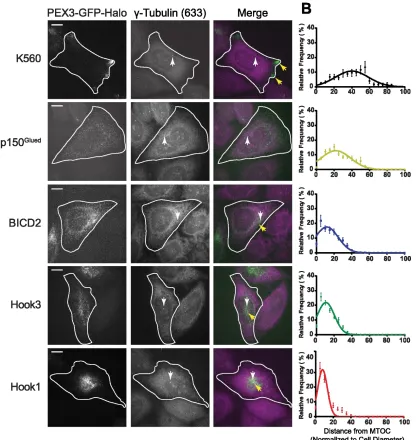
To quantify the redistribution phenotype of each adaptor, we measured the
distance of each peroxisome from the microtubule organization center (MTOC) in cells
that were fixed 45 minutes after addition of an uncaged TMP-Htag dimerizer and then
stained with a gamma-tubulin antibody to visualize the MTOC. Analysis of cells from 3
independent repeats showed that Hook1, Hook3, and BICD2 each induced a significant
concentration of peroxisomes near the MTOC in contrast to either p150Glued or K560 (Fig.
2.2). Direct comparison of the distributions shows that Hook1 induced the tightest
clustering of peroxisomes near the MTOC, while recruitment of either Hook3 or BICD2
induced similar distributions (Fig. 2.2). Of note, both Hook3 and BICD2 have been linked
to Golgi transport (Hoogenraad et al., 2001; Walenta et al., 2001), while Hook1 has been
linked to endosomal transport (Maldonado-Báez et al., 2013). While p150Glued was
efficiently recruited to peroxisomes in this assay (Fig. 2.2), this recruitment was not
sufficient to induce marked peroxisome motility or redistribution, consistent with the idea
that dynactin alone is insufficient to induce superprocessive motility. Together, these
observations suggest that like BICD2, both Hook1 and Hook3 can activate dynein
motility, and that differences among these activators may tune dynein activity to regulate
36
Figure 2.2 Hook proteins differentially redistribute peroxisomes to the MTOC
A) Dimerization assay in fixed cells stained with gamma-tubulin antibody. Images are
maximum projections of confocal z-stacks. Scale bars=10μm. White arrows point to
gamma-tubulin stained MTOC and yellow arrows point to peroxisome clusters. Cell
outlines were determined from corresponding X-mCH-DHFR images (not shown). B)
37
(analyzed using Cell Profiler (Carpenter et al., 2006)). The endosomal-linked adaptor
Hook1 tightly clusters peroxisomes to the MTOC compared to Hook3 and BICD2. (Cells
analyzed per condition: K560: n=36, p150Glued: n=19, BICD2: n=26, Hk1: n=23, Hk3:
n=32) Error bar are S.E.M based on number of cells.
Mammalian Hook proteins interact with dynein-dynactin
To characterize the interactions of mammalian Hook proteins with dynein and
dynactin, we performed immunoprecipitation experiments using endogenous and
expressed Hook proteins. Using mouse brain lysates, we immunoprecipitated
endogenous dynein and dynactin with monoclonal antibodies to the dynein intermediate
chain (DIC) and the p150Glued subunit of dynactin, respectively. Co-immunoprecipitation
of endogenous Hook1 was seen with the anti-p150Glued antibody but not with the anti-DIC
antibody (Fig. 2.3A). This DIC antibody is known to disrupt the interaction of dynein with
dynactin (Karki and Holzbaur, 1995), as confirmed by a decreased amount of p150Glued
in the DIC IP lane (Fig. 2.3A). These results suggest that Hook proteins either interact
with dynactin directly or with the full dynein-dynactin complex, rather than interacting
solely with dynein.
Next, we performed IP experiments using Halo-tagged Hook proteins expressed
in Cos7 cells. Again, using the p150Glued antibody, we observed co-immunoprecipitation
of expressed full length human Hook1 and Hook3 with the dynein-dynactin complex.
With the expression of Hook proteins, we also observed an increase in the
co-precipitation of dynein by the anti-p150Glued antibody, as compared to a control
experiment in which only Halo-tag was expressed (Fig. 2.3B). We quantified the ratio of
DIC:p150Glued in the immunoprecipitates from each condition, and observed that