University of Pennsylvania
ScholarlyCommons
Publicly Accessible Penn Dissertations
1-1-2015
Controlling Thermogenesis: Understanding the
Role of PRDM16 in the Development and
Function of Brown Fat
Matthew James Harms
University of Pennsylvania, [email protected]
Follow this and additional works at:
http://repository.upenn.edu/edissertations
Part of the
Biology Commons
,
Developmental Biology Commons
, and the
Molecular Biology
Commons
This paper is posted at ScholarlyCommons.http://repository.upenn.edu/edissertations/1059 For more information, please [email protected].
Recommended Citation
Harms, Matthew James, "Controlling Thermogenesis: Understanding the Role of PRDM16 in the Development and Function of Brown Fat" (2015).Publicly Accessible Penn Dissertations. 1059.
Controlling Thermogenesis: Understanding the Role of PRDM16 in the
Development and Function of Brown Fat
Abstract
The alarming rise in the incidence of obesity found throughout the world has precipitated a need to look for novel methods to increase energy expenditure to counter weight gain. Recently it was discovered that adult humans possess a substantial mass of brown adipose tissue (BAT), a tissue that consumes stored lipid to produce heat. Although the primary physiologic role for BAT is to protect mammals from the cold, it is currently thought that enhancing BAT mass or activating BAT in humans is a novel way to decrease adiposity. However, before BAT can be effectively utilized for therapeutic purposes a better understanding of the transcriptional regulation underlying BAT function is required. Here, we investigated the role of the transcription factor PRDM16 in BAT. We found that PRDM16 is not required for BAT development, however it is required to maintain BAT identity in adult mice. The loss of PRDM16 in adult mice led to a loss of BAT functionality and an inability to produce heat. We found that PRDM16s ability to drive a thermogenic program is due to its recruitment of Med1/the Mediator Complex to BAT-selective genes. Without PRDM16 in BAT a loss of higher order chromatin structure and a corresponding loss of transcription takes place at genes required for BAT identity and function.
Degree Type
Dissertation
Degree Name
Doctor of Philosophy (PhD)
Graduate Group
Cell & Molecular Biology
First Advisor
Patrick Seale
Keywords
brown fat, Prdm16, thermogenesis, Ucp1
Subject Categories
Biology | Developmental Biology | Molecular Biology
CONTROLLING THERMOGENESIS: UNDERSTANDING THE
ROLE OF PRDM16 IN THE DEVELOPMENT AND FUNCTION OF
BROWN FAT
Matthew J. Harms
A DISSERTATION
in
Cell and Molecular Biology
Presented to the Faculties of the University of Pennsylvania in
Partial Fulfillment of the Requirements for the Degree of Doctor of Philosophy
2015
Supervisor of Dissertation:
________________________
Patrick Seale, Ph.D.
Assistant Professor of Cell and Developmental Biology
Graduate Group Chairperson:
________________________
Daniel S. Kessler, Ph.D.
Associate Professor of Cell and Developmental Biology
Dissertation Committee:
Paul J. Gadue, Ph.D., Assistant Professor of Pathology and Laboratory Medicine
Mitchell A. Lazar, MD, Ph.D., Sylvan H. Eisman Professor of Medicine and Genetics
Michael L. Atchison, Ph.D., Professor of Biochemistry
CONTROLLING THERMOGENESIS: UNDERSTANDING THE ROLE OF PRDM16 IN THE DEVELOPMENT AND FUNCTION OF BROWN FAT
COPYRIGHT 2015
Matthew James Harms
iii
ABSTRACT
CONTROLLING THERMOGENESIS: UNDERSTANDING THE ROLE
OF PRDM16 IN THE DEVELOPMENT AND FUNCTION OF BROWN
FAT
Matthew J. Harms Patrick Seale, P.hD.
iv
TABLE OF CONTENTS
ABSTRACT
... III
LIST OF FIGURES
... VI
CHAPTER 1: BROWN AND BEIGE FAT: DEVELOPMENT, FUNCTION AND
THERAPEUTIC POTENTIAL
... 1
The Development of Brown/Beige Adipocytes ... 7
Developmental regulation of brown and beige adipocytes by Prdm16 ... 12
Role of brown/beige fat in regulating weight and metabolism ... 14
Sympathetic nerve control of brown/beige fat ... 17
Novel BAT/beige fat recruiters/activators ... 24
Outlook and challenges ... 28
CHAPTER 2: PRDM16 IS REQUIRED FOR THE MAINTENANCE OF BROWN
ADIPOCYTE IDENTITY AND FUNCTION IN ADULT MICE
... 30
Abstract ... 31
Introduction... 32
Results ... 34
• Prdm16 is dispensable for embryonic BAT development ... 34
• Prdm16 recruits Ehmt1 to repress the expression of white fat-selective genes ... 36
• Prdm16 maintains iBAT identity during aging ... 39
v
• Reduced BAT function in Myf5-ΔPrdm16 mice ... 46
• Prdm3 compensates for the loss of Prdm16 to preserve brown fat fate in young mice ... 50
Materials and Methods ... 58
CHAPTER 3: PRDM16 BINDS MED1 AND CONTROLS CHROMATIN
ARCHITECTURE TO DETERMINE A BROWN FAT TRANSCRIPTIONAL
PROGRAM
... 64
Abstract ... 65
Introduction... 66
Results and Discussion ... 67
• PRDM16 binding is enriched at BAT-selective genes ... 67
• PRDM16 recruits MED1 to BAT-selective genes ... 70
• PRDM16 controls higher order chromatin structure ... 76
• Prdm16 controls BAT-selective super-enhancers ... 77
Materials and Methods ... 81
DISCUSSION AND FUTURE DIRECTIONS
... 86
vi
LIST OF FIGURES
CHAPTER 1: Brown and beige fat: development, function and therapeutic potential Figure 1 Differences between Brown and Beige Adipocytes 3 Figure 2 Genetic Models Resistant to Weight Gain Through
Enhanced Brown and Beige 5 Figure 3 Transcriptional Regulation of Brown and Beige Adipocyte
development 10
Figure 4 Catecholamine and Natriuretic induction of
Thermogenesis 18
Figure 5 Secreted factors that recruit brown/beige adipocytes 24 CHAPTER 2: Prdm16 is required for the maintenance of brown adipocyte identity and function in adult mice
Figure 1 Prdm16 is dispensable for embryonic BAT development 34 Figure S1 Prdm16 deficiency in BAT does not increase the
expression of skeletal muscle genes. 35 Figure 2 Prdm16 represses the expression of white fat-selective
genes 37
Figure S2 Prdm16 deficient iBAT expressed a white fat-related gene
profile. 38
Figure 3 Prdm16 is required for the maintenance of iBAT fate in
adult animals 41
Figure S3 Prdm16-deficiency causes a loss of interscapular brown
adipose tissue identity in adult mice 42 Figure 4 Prdm16 is required for activation of the brown fat-selective
gene program in cultured brown fat precursors 44 Figure S4 Prdm16 is required cell autonomously 45 Figure 5 Myf5-∆Prdm16 mice have severely deficient BAT function
but are not prone to obesity or metabolic disease 47 Figure S5 Myf5-ΔPrdm16 mice do not have metabolic defects. 49 Figure 6 Prdm3 compensates for the loss of Prdm16 in juvenile
BAT 51
Figure S6 Prdm16 or Prdm3 are required for the postnatal
vii
CHAPTER 3: PRDM16 binds MED1 and controls chromatin architecture to determine a brown fat transcriptional program
Figure 1 PRDM16 binding is enriched at BAT-selective genes 68 Figure S2 PRDM16 is preferentially bound to BAT-selective genes 69 Figure 2 PRDM16-deficiency reduces MED1 levels at
BAT-selective genes 71
Figure S2 Loss of PRDM16 does not affect PPARγ or C/EBPβ
binding levels at BAT-selective genes 72 Figure 3 PRDM16 binds to MED1 and recruits it to BAT-selective
genes 74
1
2
Published: Harms M, Seale P. (2013) Brown and beige fat: development, function and therapeutic potential. Nature Medicine. 2013 Oct; 19(10):1252-63.
Sedentary living and calorie dense food has precipitated a dramatic rise in obesity throughout the developed world. This is particularly alarming due to the vast array of associated diseases, including type 2 diabetes, heart disease, insulin
resistance, hyperglycemia, dyslipidemia, hypertension and many types of cancer1,2. The
result is an expanding population of chronically sick people, staggering health care expenses and a prediction that for the first time, the current generation will have a shorter life span than previous generations3-5. There is thus an urgent need for new
weight loss-treatments. Brown adipose tissue (BAT) is a key site of heat production (thermogenesis) in mammals that has been considered for many decades as an attractive target to promote weight loss. The heat produced by BAT is essential for the survival of small mammals in cold environments and for arousal in hibernators. Brown adipocytes in BAT are packed with mitochondria that contain Uncoupling Protein-1 (Ucp1). Ucp1, when activated, short circuits the electrochemical gradient that drives ATP synthesis and thereby stimulates respiratory chain activity. Heat is generated from the combustion of available substrates6 and is distributed to the rest of the body via the
circulation.
Clusters of Ucp1-expressing adipocytes with thermogenic capacity also develop in white adipose tissue (WAT) in response to various stimuli7. These have been named
3
their multilocular lipid droplet morphology, high mitochondrial content, and expression of a core set of brown vs. white fat-specific genes (e.g. Ucp1, Cidea, Pgc-1α). Despite a common ability to undergo thermogenesis, brown and beige cells have many
distinguishing characteristics and should be considered as distinct cell types (Figure 1).
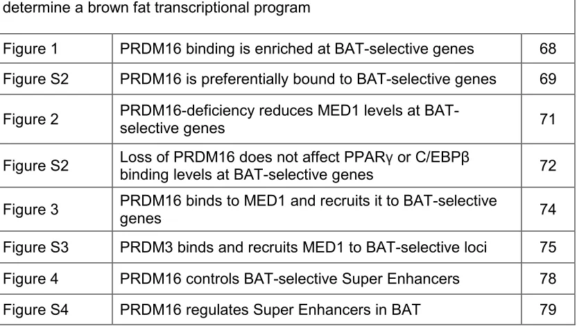
Figure 1. Differences between Brown and Beige Adipocytes
Brown adipocytes express high levels of Ucp1 under basal conditions, whereas clusters of beige adipocytes can only be easily recognized in WAT after cold/β-adrenergic stimulation. Enriched markers of brown versus beige adipocytes have recently been identified, including: brown markers, Zic1, Lhx88,9, Eva110, Epsti111; and beige markers, CD137, TMEM2610,
Tbx18,10, Cited111, Shox212. Among the activators that have been studied in both compartments, Irisin is the only one that
has selective actions in beige but not brown adipocytes.
Firstly, beige cells, at least those in the mouse subcutaneous depot, do not derive from the same embryonic (Myf5-expressing [see later]) precursors that give rise to brown adipocytes13. Secondly, a number of quantitative trait loci are associated with
the induced development of beige but not brown adipocytes14, suggesting that these cell
types are differentially regulated. Thirdly, brown and beige adipocytes express distinct and distinguishing gene signatures10,11. Finally, a striking difference is that brown
adipocytes express high levels of Ucp1 and other thermogenic genes under basal (unstimulated) conditions, whereas beige adipocytes only express these genes in response to activators, like β-adrenergic receptor or Pparγ agonists8,15. Importantly, this
4
genes (e.g. Ucp1) during adipogenesis in culture from preadipocytes without addition of classical activators.
An obvious question is whether brown and beige fat cells have different
functions. The answer to this is still unknown and has not been well studied. However, a recent study suggests that fully stimulated brown and beige adipocytes contain
comparable levels of Ucp1, suggesting that their thermogenic capacities are similar10.
Based on this, the name “beige” might be misleading and is more applicable to describe the tissue that has undergone “browning” rather than the Ucp1+ adipocytes themselves. Aside from thermogenesis, it seems highly likely that beige and brown adipocytes have other cell type-specific actions that have yet to be studied. For example, beige
adipocytes may secrete certain factors that affect WAT function and/or systemic metabolism.
The biomedical interest in brown and beige adipocytes is centered on the
capacity of these cell types to counteract metabolic disease, including obesity and type 2 diabetes. Indeed, increased brown and/or beige adipose activity is linked to obesity resistance in many mouse models (Table 1). In humans, it was assumed for many years that there was too little brown fat in adults to affect body weight. However, in 2009, imaging studies revealed the presence of substantial deposits of Ucp1-expressing adipocytes whose mass and/or activity are lower in obese and older subjects16-20. The
5
Mitochondrial uncoupling has already been tried as a weight loss therapy. The chemical uncoupler, 2, 4-Dinitrophenol (DNP) allows protons to leak across the mitochondrial membrane, mimicking the effect of activated Ucp121. In the 1930s, DNP was widely
used as an effective diet pill to treat obesity, providing proof-of-concept for mitochondrial uncoupling as an approach for weight loss. However, at high doses (variable in different people), unregulated respiratory uncoupling in all cells causes dangerous side-effects including hyperthermia and death. Thus, the goal is to develop strategies that enhance respiratory uncoupling selectively in adipose tissue by exploiting the mechanisms that naturally evolved to do this in brown and/or beige fat cells.
Figure 2. Genetic Models Resistant to Weight Gain Through Enhanced Brown and Beige Fat Development
Gene Induce
Beige
Increase
Brown Gain of function models
Cox222 X
Cox2 over-expressing mice have increased beige fat and are resistant to weight gain, demonstrating the role of prostaglandins in the recruitment of beige fat
FoxC223-25 X X Overexpression of FoxC2 in adipose increases the expression of the R1α
regulatory subunit of PKA, making the cells more sensitive to catecholamines Prdm1626 X Fat-selective Prdm16 transgenic mice have increased beige fat.
Pten27 X X Increases in Pten inhibits PI3K, which drives a thermogenic program
Ucp128,29 X Transgenic expression of Ucp1 increases thermogenesis in WAT and
prevents weight gain
Loss of function models
4E-BP130 X
4E-BP1 KO mice have an increased metabolic rate, an induction of thermogenic genes in WAT depots, and an increases in eIF4F phosphorylation
4E-BP231 X
Treatment with a antisense oligo. caused weight loss and an increase of the β3-adrenergic receptor in both WAT and BAT. BAT showed a PGC-1α independent increase in Ucp1
ActRIIB32,33 X
Neutralizing antibodies show an increase in BAT mass without affecting WAT. Loss of ActRIIB activates Smad3 signaling to increase thermogenic genes
Aldh1a134 X KO results in a buildup of Retinaldehyde. This activated the retinoic acid
receptor – which recruited PGC-1α to the Ucp1 promoter Arrdc335 X X
Arrdc3 interacts directly with β-adrenergic receptors. Loss of Arrdc3 sensitized adipocytes to catecholamines and thus increased thermogenic programs in BAT and WAT
ATG736 X X
BAT from KO mice showed an increase in thermogenic proteins, and WAT had an increase in thermogenic signatures. Studies demonstrate role of autophagy in adipose development
ATF437 X X WAT showed an increase of PGC-1α and Ucp2, while BAT was enriched for
Ucp1 and Ucp3
Bace138 X An increase in Ucp1 in BAT and Ucp2/3 in skeletal muscle
Cidea39 X
KO mice are lean, have increased oxygen consumption, and defend core temperature against a cold challenge. Direct interactions with Ucp1 could explain Cidea’s repressive effect
Cnr140 X Mice are lean. In vitro, Cannaboid receptor type 1m antagonists are able to
6
Crfr241 X An increase in glucose tolerance and a further increase of Ucp1 in BAT
Ffar242 X Mice resist weight gain and have increased core temperature
FoxO143 X Adipose specific dominant negative mice had increased oxygen consumption
and a BAT specific increase in thermogenesis
Fsp2744 X Mice have increased BAT specific genes and mitochondrial in WAT. The
mechanism is thought to involve a loss of pRb and RIP140.
Ghsr45 X Mice are protected from the age-associated decline of thermogenesis
Grk246 X
Knockout mice have an increased core temperature and thermogenic program in BAT and WAT. Interestingly, the phenotype appears to be age related.
Id147 X Increased oxygen consumption and an increase in thermogenic genes in
BAT
Ikbke48 X WAT has increased Ucp1 transcript and protein
Lipe49 X The Increase in Ucp1 is attributed to a decrease in RIP140 and pRb
LPR650 X KO animals gain less weight and a diminished mTORC1 activity in BAT
causes an increase in thermogenic proteins LXRα51 X Recruitment of RIP140 to displace PPARγ/PGC-1α
Mstn52 X Increase of thermogenic program in WAT
Nprc53
X X
Loss of the natriuretic peptide (NP) clearance receptor causes increased circulating NPs which increase thermogenic activity
Oprd154 X Mice are resistant to weight gain and have enhanced thermogenesis in BAT
p10755 X Loss of p107 causes a loss of pRb and increased browning of WAT.
Pctp56 X BAT showed enlarged mitochondrial and an increase thermogenic genes
pRb57 X pRB binds to and represses the PGC-1a promoter.
Pref-158 X BAT has increased PGC-1α and Ucp1. C/EBPδ binds and activates the
Pref-1 promoter Prkar2b59 X
The loss causes a compensatory increase in RIα, which binds cAMP with higher affinity, causing increased basal PKA activity –increasing thermogenesis
Prkcb60
X WAT had increased β1 and β3 adrenergic receptors. This resulted in a p38/MAPK mediated increase of PGC-1α and Ucp1
Prlr61 X
Prolactin receptor KO mice have increased thermogenic genes and altered pRb/Foxc2 levels in WAT. This indicates a novel paracrine or endocrine role of prolactin
Rip14062-64 X RIP140 directly interacts with PGC-1a to inhibit its transcriptional activity ;
Recruits DNMTs and HMTs to silence Ucp1
Scd165 X X Skin-specific KO mice result in increased thermogenesis in BAT and WAT,
indicating cross talk between the different tissues
Sfrp566 X KO mice are resistant to weight gain and isolated KO adipocytes have
increased oxidative respiration
Smad367 X Smad3 represses PGC-1α expression. Loss of Smad3 induces transcripts
that correspond to increased thermogenesis
Them152 X An increase in thermogenic genes in brown fat and a decrease in markers of
inflammation in white adipose
Tif268 X Tif2 competes with the activator Src2 for PGC-1a binding. Tif2 binding
prevents PGC-1α from interacting with PPARγ
Tnrf169 X Knockout of tumor necrosis factor-alpha receptor 1 results in increased Ucp1
in BAT and Ucp3 in muscle resulting in increased O2 consumption Trpv470 X KO mice are resistant to weight gain and have increased thermogenic gene
expression in WAT, mediated by a loss of ERK1/2 effects on Pgc-1α Twist-171 X Twist-1 binds to and inhibits PGC-1α activity at target genes.
Vegfa72 X An induction of the thermogenic program in WAT with associated resistance
to weight gain
Vgf73 X Knockout of secreted protein VGF caused increased Ucp1 expression in
7
The Development of Brown/Beige Adipocytes
Brown adipocytes
BAT forms during embryonic development, before other fat depots, and is assumed to contain a uniform population of adipocytes. The major BAT depots in rodents are in the interscapular region (interscapular, axillary and cervical pads), embedded in and around deep back muscles. An interscapular BAT depot has also been noted in human infants, which regresses and is absent in adults12,74. Most brown fat cells originate from
precursor cells in the embryonic mesoderm that also give rise to skeletal muscle cells and a subpopulation of white adipocytes13,75,76. These precursors transiently express
Myf5 and Pax7, two genes that were previously thought to selectively mark skeletal myogenic cells in the mesoderm (Figure 2A)13,76. Consistent with a developmental
relationship between brown fat and muscle, brown fat precursor cells express a muscle-like gene signature77, and brown fat and muscle have related mitochondrial proteomes78.
However, whether Myf5+ cells are multipotent or whether there are separate pools of
Myf5+ precursors that contribute to muscle, brown fat and white fat remains to be tested.
Beige Adipocytes
The embryonic origin and cell hierarchy of beige adipocytes is less clear. Beige and brown adipocytes likely come from distinct cell lineages, given that beige cells, at least in the subcutaneous depot, do not have a history of Myf5 expression13,75. In
8
Since then, Cinti and others have provided substantial evidence in support of the idea that large unilocular “white” adipocytes transform into beige adipocytes in response to cold/β3-adrenergic agonists7.
A new study from the Scherer lab used a pulse-chase fate-mapping technique in mice to revisit this issue. Wang et al. pulse labeled the mature adipocytes in WAT with LacZ expression80. This labeling is indelible and heritable such that LacZ is
constitutively expressed in the pulsed adipocytes and any of their descendents. After being “pulsed”, the mice were exposed to cold or treated with β3-adrenergic agonists to induce the formation of beige adipocytes. The results were clear- the large majority of newly acquired Ucp1+ adipocytes in the subcutaneous inguinal depot are not marked by LacZ. This proves that most, if not all, beige adipocytes, at least in this subcutaneous depot, arise from a precursor population rather than from pre-existing adipocytes (Figure 2B).
The thermogenic profile of beige adipocytes is reversible. Beige adipocytes acquired in WAT during cold-exposure lose Ucp1 and are retained after mice are moved back to warmer conditions (Figure 2B)80,81. When these animals are re-exposed to cold,
the same (marked) cells again induce Ucp181. Interestingly though, the cells marked by
9
Another important question is whether beige and white adipocytes arise from different types of precursors. Petrovic et al., found that a subset of adipocytes differentiated in vitro from the stromal vascular fraction (SVF, an enriched source of preadipocytes) of WAT activate Ucp1 expression in response to treatment with Pparγ activators8; this suggests that some but not all preadipose cells are thermogenically
competent. Recently, the Spiegelman lab used limited dilution to clone preadipocyte cell lines from the stromal vascular fraction of subcutaneous (inguinal) WAT10. Through
10
Figure 3. Transcriptional Regulation of Brown and Beige Adipocyte development
11
Beige adipocytes are most abundant in the inguinal WAT, a major subcutaneous depot in rodents7. However, Ucp1-expressing adipocytes are evident in most (if not all)
WAT depots in response to cold exposure7,79,82. In peri-gonadal (visceral) fat of male
mice, beige adipocytes develop from a population of precursors that also differentiates into white adipocytes (Figure 2B)83. These bi-potent white/beige precursors express
Platelet-derived growth factor receptor-α (Pdgfrα) and are closely associated with blood vessels. Upon treatment of mice with β3-adrenergic agonists, these precursor cells proliferate, then lose Pdgfrα expression and differentiate into Ucp1+ adipocytes. Conversely, high fat diet stimulates the differentiation of Pdgfrα+ cells into white adipocytes83. This is consistent with the finding that most/all white adipocytes are
descendent from Pdgfrα-expressing cells84. Importantly, cell culture analyses shows that
single Pdgfrα+ cells give rise to both Ucp1- and Ucp1+ (beige) adipocytes.
In the mature adipocyte tracing studies of Wang et al. (discussed above), very little beige fat recruitment but a surprising amount of white adipogenesis was detected in the perigonadal WAT of mice exposed to cold for 1-3 days or treated with β-agonist for 7 days80. Why new white fat cells develop during cold exposure is unclear. It is also
surprising that so few Ucp1+ cells were detected. Perhaps the exposure was too short to elicit a full beige recruitment in the newly developed adipocytes? It would be
interesting to examine the effect of chronic cold in these mice, since this is known to extensively brown the WAT depots.
The prevalence of beige adipocytes within different human WAT depots has not been carefully evaluated. However, it is known that human WAT contains precursor cells that are capable of expressing Ucp1 and other brown/beige characteristics,
12
it was (and still is) unclear whether the deposits of Ucp1-expressing adipocytes identified by Fluorodeoxyglucose - Positron Emission Topography (FDG-PET) in adult humans are analogous to beige or brown fat. Wu et al. and Sharp et al., reported that
supraclavicular tissue, the largest FDG-PET+ depot, expresses selective markers of rodent beige versus brown fat cells 10,11. By contrast, Jesperson et al., found that tissue
and in vitro differentiated adipocytes from this depot expresses both brown- and beige-specific markers9. A different depot in the neck region was shown to possess the
molecular characteristics of murine brown fat86. Typing these depots as “brown” or
“beige” based on the expression levels of a few mouse marker genes that have no known function(s) has not been conclusive thus far. Functional marker genes or assays are needed to better categorize the different human (as well as mouse) depots/cell types. The field must continue to study the biology and therapeutic potential of both the classic/developmental BAT and (inducible) beige fat.
Developmental regulation of brown and beige adipocytes by Prdm16
Prdm16 (PRDI-BF1 and RIZ homology domain containing protein-16) is a large zinc-finger containing transcriptional factor that is expressed at high levels in murine BAT relative to visceral WAT87. Prdm16 expression is also substantially enriched in
human “BAT” relative to adjacent subcutaneous WAT20,88. Ectopic Prdm16 expression
converts myoblasts and white fat precursors into thermogenic, Ucp1-containing
13
characteristics of brown fat cells while also causing an increase in the expression of white fat- and muscle-specific genes. Together, these studies have strongly suggested that Prdm16 is a key driver of brown fat cell fate.
The importance of Prdm16 in brown fat cell differentiation prompted us to examine whether Prdm16 also played a role in the development of beige adipocytes. Upon analyzing various murine WAT depots, we noted that Prdm16 was expressed at higher levels in the depots that are most prone to beiging, especially the inguinal WAT26.
Importantly, reduction of Prdm16 blocks the induction of a thermogenic program in cultured subcutaneous adipocytes and decreases the recruitment of beige adipocytes in WAT in response to β-adrenergic or Pparγ agonists26,91. Conversely, transgenic
expression of Prdm16 in adipose tissues of mice stimulates beige adipocyte development to counteract high fat diet-induced weight gain and improve glucose tolerance26.
Several factors have been shown to regulate brown/beige adipocyte
differentiation by modulating Prdm16 expression/activity. Notable among these is Bone morphogenic protein-7 (Bmp7), an essential signal for brown fat development, which increases Prdm16 mRNA levels in brown and white fat precursor cells92-94. Additionally,
thiazoledinediones (TZDs), which agonize Pparγ, induce thermogenic gene expression in fat cells through effects on Prdm16 (see later). Interestingly, the muscle-enriched microRNA, miR-133 directly targets and reduces Prdm16 levels to block both brown and beige adipose development95-46. Notably, cold-exposure suppresses miR-133
expression in fat cells, which leads to increased levels of Prdm16 and downstream thermogenic target genes95. Mice lacking miR-133 express higher levels of Prdm16 in
14
levels in adult muscle stem cells where it suppresses Prdm16 expression97. Reduction of
miR-133 in regenerating muscle causes the ectopic development of brown adipocytes and an associated increase in energy expenditure.
Role of brown/beige fat in regulating weight and metabolism
BAT has long been viewed as a critical tissue for defending body temperature in response to cold. In 1979, Rothwell and Stock first reported that BAT was also activated in rodents when they overeat as a mechanism to preserve energy balance and limit weight gain- so called diet-induced thermogenesis (DIT)98. Consistent with this, mice
genetically engineered to have less BAT gain more weight than control animals99.
However, for many years it was unclear why Ucp1-deficient mice, which are cold intolerant (and thus have defective BAT), resisted rather than developed obesity100.
An important study by the Cannon and Nedergaard group in 2009 revealed that Ucp1-deficient mice gain more weight than wildtype controls, but only when they are housed under thermoneutral (28-30ºC) conditions101. At room temperature (20-22ºC),
mice are cold and must therefore expend extra energy to defend their body temperature. Ucp1-deficient mice, which can’t use BAT, activate alternative thermogenic
mechanisms102,103. This is thought to conceal the effect of brown fat/Ucp1 on energy
balance. Consistent with this, old Ucp1-deficient animals, that are larger and less cold-sensitive than younger mice, become obese even at ambient temperature104. The
15
thermoneutrality with the aid of clothing, heating, etc., a compelling argument could be made that all/most metabolic studies in mice should be conducted under thermoneutral conditions.
The obesogenic effect of Ucp1-deficiency in warm mice indicates that BAT/beige fat activity can affect energy balance, but the magnitude (and significance) of this effect in free-living mice or humans is uncertain. It should also be noted that previous studies in rats housed at thermoneutrality failed to find any significant contribution of BAT activity to diet-induced thermogenesis105. Moreover, Kozak and colleagues did not observe
changes in adiposity in their studies of Ucp1-knockout animals when housed under varying temperature conditions106. Finally, increases in BAT/Ucp1 activity in response to
high fat feeding are not consistently observed107. These divergent findings may provide
an opportunity to identify modifying factors that affect BAT/Ucp1 activity and energy balance. Are there specific dietary components that are needed to recruit BAT
efficiently? What are the genetic/strain-specific effects? Does the microbiome or other environmental factors in different vivariums play a role?
Regardless of whether BAT plays a major physiological role in body weight regulation in mice or humans, there is no question that expanding BAT/beige fat activity in mice, through genetic manipulation, drugs, or transplantation suppresses metabolic disease (Table 2 and 26,28,108,109-112) This implies that counter-regulatory mechanisms
16
Similarly, transgenic expression of Prdm16 in all fat tissues promotes beiging of WAT and resistance to obesity without increasing BAT mass or Ucp1 mRNA levels26,28.
Finally, transgenic expression of Ucp1 in adipocytes suppresses obesity in spite of a reduction in BAT mass28. These results raise an obvious question - do beige adipocytes
play a more important physiological role in fighting obesity? This seems unlikely, given that high fat diet generally decreases the levels of thermogenic genes in WAT,
coincident with increases in WAT mass107.
Mice with increased brown and/or beige fat activity resist weight gain, but also display improvements in systemic metabolism, including improved glucose tolerance and increased insulin sensitivity. Along these lines, activated brown fat takes up and
metabolizes large quantities of lipid from the bloodstream113, which has beneficial effects
on metabolism. In models where beige fat appears to be selectively increased, such as Prdm16 fat transgenics26 and Irisin-treated animals108, the improvement in glucose
tolerance seems disproportional to the modest effects on body weight. We speculate that the increased proportion of beige to white adipocytes in WAT modulates systemic insulin action through non-thermogenic mechanisms, perhaps via altering the secretome of adipose tissue. Additionally, thermogenic fat cells, not yet classified as brown or beige, that surround blood vessels (perivascular adipose) have been suggested to protect against the development of atherosclerosis114. Thus, the potential therapeutic
17
Sympathetic nerve control of brown/beige fat
Cold is a dominant regulator of many aspects of BAT biology. Mice lacking BAT activity are cold intolerant due to defective non-shivering thermogenesis100. Cold, sensed
by various mechanisms, including through thermoreceptors in the skin, elicits sympathetic outflow to BAT through an intricate neural circuitry (reviewed in 115). In
addition to nerve terminals, alternatively activated macrophages in BAT produce catecholamines in response to cold116. Norepinephrine (NE) agonizes adrenergic
receptors on adipocytes which triggers a signal transduction cascade leading to adaptive increases in the expression of thermogenic genes (Figure 3)117. Prolonged cold
exposure also stimulates the proliferation and differentiation of brown precursor cells to expand BAT mass and increase thermogenic capacity118. Conversely, at warmer
housing temperatures or in surgically denervated BAT, Ucp1 and other thermogenic factors are dramatically reduced119,120.
Sympathetic nerve activity also acutely stimulates heat production by activating Ucp1 function. Classic studies showed that fatty acids, rapidly released from lipid droplets in response to nerve activity, increase proton leak through Ucp1 (reviewed by
121). Fedorenko et al. recently discovered that long chain fatty acids generated in the
inner mitochondrial membrane by a phospholipase A2 (PLA2) bind directly to Ucp1 and are required for proton transport122 (Figure 3). An important, but often overlooked tenet
is that Ucp1 does not increase the respiratory activity of cells under basal
conditions123,124. Therefore, therapeutic approaches which expand brown and/or beige
18
people, an expanded BAT/beige fat compartment may be sufficiently activated by daily “tonic” stimuli (e.g. food, cold, exercise, etc.) to achieve therapeutic effect.
Figure 4. Catecholamine and Natriuretic induction of Thermogenesis
19
Cold is also the classic activator of beige adipocyte development and function. Animals housed in the cold undergo a dramatic remodeling of their WAT, characterized by an accumulation of beige adipocytes - this can be mimicked by treating animals with β3-adrenergic activators like CL 316,2437,79,109,110,125-128. Interestingly, the propensity of
WAT depots to undergo beiging is highly correlated with the density of sympathetic nerve fibers129. However, other adipose cell/tissue autonomous factors must be involved
because systemic β3-agonist administration (thus bypassing the central nervous system) causes certain depots to beige more than others. Many of the effects of chronic cold on adipose tissues are recapitulated in mice that express elevated levels of FoxC2 in adipocytes23 Specifically, FoxC2 increases BAT mass, induces beige fat cell
development, drives mitochondrial biogenesis and promotes angiogenesis in fat
tissue25,130,131. FoxC2 functions in fat cells, to a large extent, by driving the expression of
the R1α regulatory subunit of PKA (Prkar1a)23,24, thus sensitizing adipocytes to the
effects of catecholamines. These results suggest that the adipocytes instigate most of the tissue remodeling that occurs in response to NE.
The discovery of the murine β3-adrenergic receptor (β3-AR), which is expressed mainly in fat and whose agonism activates thermogenesis, generated tremendous excitement for therapeutic possibilities in humans. However, treatment of humans with β3-AR agonists never lived up to the forecasted predictions132. Difficulties appear to be
due to receptor differences between mice and humans - leading to off-target effects in humans, as well as poor pharmacokinetic properties and oral bioavailability133. These
20
BAT/beige fat recruiters. Alternatively, it would be worth considering whether prescribed cold exposure could be used to activate BAT/beige fat after augmentation via other pathways.
Cold exposure, which induces thermogenic features in adipose cells, also affects the developmental programs of other cell types in adipose tissue to coordinate and optimize heat production. For example, and as noted above, cold activates alternatively activated macrophages in BAT to produce catecholamines116. Cold also stimulates
sympathetic nerve branching/recruitment during the browning response of WAT129.
Finally, cold exposure induces the sprouting and growth of blood vessels in adipose to facilitate oxygen delivery and heat exchange118,120,134. This angiogenic effect is regulated
through increased production of Vascular endothelial growth factor (Vegf), through a mechanism that does not involve hypoxia135-137. Interestingly, Vegf secreted by adipose
tissue also enhances the recruitment of brown and beige adipocytes via an unknown mechanism (Figure 4). In cultured brown adipocytes, Vegf enhanced cell survival and proliferation whereas Vegf neutralizing antibodies caused apoptosis138. Strikingly,
overexpression of Vegf in adipose tissues of mice increases BAT mass, stimulates beiging and promotes a healthy metabolic profile139,140. Curiously, Vegf-inhibition has
also been shown to reduce metabolic disease in mice, though this is in the context of already dysfunctional obese WAT72,140. Further studies are needed to elucidate the
21
Pparγ Coactivator-1α (Pgc-1α) controls the thermogenic activation of adipocytes
Pgc-1α was discovered as a cold-induced interacting partner of Pparγ in brown fat141. Based on hundreds of studies, Pgc-1α is now recognized as a master regulator of
mitochondrial biogenesis and oxidative metabolism in many cell types. In adipocytes, Pgc-1α also induces the expression of Ucp1 and other thermogenic components141,142.
Surprisingly however, BAT develops normally without Pgc-1α143, probably due to
compensation by the related family member, Pgc-1β. Although not required for tissue development, Pgc-1α is essential for the cold/β-agonist-induced thermogenic activation of brown adipocytes144,145 and for the expression of thermogenic genes in WAT146
(Figure 2). Thus, Pgc-1α is a central transcriptional effecter of adrenergic activation in thermogenic adipocytes.
Pgc-1α expression levels and activity are directly regulated by the β-adrenergic signaling pathway147, providing a link between the physiological activator of brown fat
thermogenesis and the transcriptional machinery in brown adipocytes (Figure 3). Specifically, Pgc-1α is phosphorylated and thereby activated by p38 MAPK in response to sympathetic stimulation147,148. Activated Pgc-1α1regulates thermogenic gene levels
through its interaction with Pparγ, Pparα, Thyroid Receptor and other factors, though a detailed mechanism to account for its selective effects at brown fat-specific genes is lacking. Pgc-1α transcription also rises in response to β-adrenergic agonists through increases in the function of Activating transcription factor-2 (Atf2)147.
22
adipose55,57. Notably, pRb activity declines during the β-adrenergic-induced beige
conversion of WAT57. The nuclear co-repressor, RIP140 binds to Pgc-1α and blocks its
transcriptional activity at certain target genes149. The nuclear receptor, LXRα also blocks
Ucp1 expression by recruiting RIP140 and displacing Pgc-1α at an LXR binding site51.
Brown/beige-specific functions for the general adipogenic machinery
Pparγ and members of the C/EBP protein family orchestrate the general differentiation program in all adipose lineages150, but are also deployed to activate
specific thermogenic genes in brown/beige adipocytes. For example, C/EBPβ is present at higher levels in BAT relative to WAT and protein levels increase further in response to cold 90. In WAT, β-adrenergic agonists increase C/EBPβ levels through miRNA-mediated
degradation of Hoxc8, a repressor of C/EBPβ transcription (Figure 3)151. Loss of C/EBPβ
is associated with defective thermogenesis, whereas increasing the levels of C/EBPβ in white fat cells triggers a brown fat transcriptional profile90,152-154.
The master adipogenic factor, Pparγ also controls the expression of brown fat-specific genes, including Ucp1, particularly in response to β-adrenergic activators
147,148,155. Genome-wide analyses demonstrate that Pparγ binds and regulates distinct
target genes in brown and white fat cells156,157. We recently discovered that Ebf2, a
helix-loop-helix transcription factor, regulates Pparγ activity to drive the expression of Prdm16 and a brown fat fate156,157 (Figure 2). Ebf2 appears to function, at least in part, by
facilitating the recruitment of Pparγ (and likely other factors) to brown fat-specific genes. Ebf2-deficient mice develop fatty tissue with the molecular and morphological
23
Activation of Pparγ by synthetic Thiazolidinedione (TZD) agonists enhances thermogenic gene expression in both white and brown adipocytes8,158-162. TZDs induce
Ucp1 expression and increase mitochondrial biogenesis in adipocytes from mice and humans8,85,163. This enables TZD-treated adipocytes to undergo Ucp1-mediated
increases in respiration in response to β3-adrenergic activators8,85,91. Mechanistically,
TZDs appear to act, in large part, through Prdm16 to activate a thermogenic program. In particular, TZD treatment stabilizes Prdm16 protein to increase its levels in fat cells91
and also enhances the interaction of Prdm16 with Pparγ (Figure 2). SirT1 plays a role in this TZD-driven process by deacetylating Pparγ to facilitate the docking of Prdm16. In vivo, activation of SirT1 promotes browning of WAT and resistance to obesity112,
suggesting that SirT1 activators might have a use as weight loss agents.
In the clinic, TZDs, though associated with unwanted side effects, are highly effective in the treatment of type 2 diabetes through enhancing insulin action. Given that beige adipocytes improve insulin sensitivity, it is reasonable to speculate that TZDs may act, at least in part, by inducing beige fat development. However, non-TZD Pparγ modulators, like MRL24, promote insulin sensitivity but have little effect on Ucp1 expression91,164. Moreover, TZDs are associated with weight gain and increased
adipocyte development in rodents and humans rather than weight loss. This may be due to a blunting effect that TZDs have on the sympathetic activation of adipocytes165,166
24
Novel BAT/beige fat recruiters/activators
Sympathetic nerve activity was widely believed to be the primary or only physiological signal which activates BAT thermogenesis and induces beige adipocyte development. Though β-AR signaling is undoubtedly a central regulator of these processes, several other hormones and factors have now been shown to regulate energy expenditure in adipose tissue (Figure 3); these have been discussed
comprehensively in recent reviews 15,167,168. Here, we comment on secreted/systemic
factors that affect brown/beige fat and appear particularly promising for therapeutic development.
Figure 5. Secreted factors that recruit brown/beige adipocytes
In rodents, a number of tissues and cell types have been found to secrete factors that regulate brown and beige adipose activity through systemic, autocrine and paracrine mechanisms. Neurons and alternatively activated macrophages secrete norepinephrine (NE); cardiac tissue – natriuretic peptides; liver and BAT – Fgf21; muscle- Irisin, thyroid hormone-T4. BAT also produces Bmp8b, and Vegf which increase thermogenic function in an autocrine manner. Additionally,
25
Irisin
In skeletal muscle, Pgc-1α orchestrates the adaptive response to exercise, including increased mitochondrial biogenesis, fast to slow muscle fiber switching, and angiogenesis169. Unexpectedly however, raising Pgc-1α levels in muscle protects
sedentary animals against obesity 170. In a search for effectors of the enhanced energy
expenditure in these animals, Spiegelman and colleagues discovered that the WAT of Pgc-1α transgenic mice contained more beige adipocytes108. They identified FNDC5
(Fibronectin type III domain containing 5) as a Pgc-1α target gene that was secreted from myocytes in the form of a novel hormone, named Irisin. Irisin stimulates the
browning of WAT through specific actions on the beige preadipocyte population10(Figure
4). Circulating Irisin levels increase in rodents and humans by exercise training.
Remarkably, a modest increase in the serum levels of Irisin in mice stimulates beige fat development leading to enhanced glucose tolerance and suppressed weight gain108.
Irisin is thus a very compelling hormone for clinical development since it has marked beneficial effects when used at near-physiological levels in mice.
Of course, as with any new hormone, there are many outstanding questions. What is the Irisin receptor(s) in beige fat precursors and how does it signal to the transcriptional machinery? Is the cleavage of FNDC5 into Irisin a regulated process? And, what effects does Irisin have on other tissues?
Fibroblast Growth Factor 21 (Fgf21)
26
thermogenesis171,172. Interestingly, there is a dramatic burst of Fgf21 production from the
neonate liver in response to suckling- this is likely to be critical for activating BAT thermogenesis, at a time when animals are especially vulnerable to hypothermia173.
Consistent with this, administration of Fgf21 to fasted neonates augments the
thermogenic gene program in BAT. In WAT, Fgf21 increases Pgc-1α protein levels to drive beige adipocyte recruitment in response to cold174,175.
Fgf21 has many desirable effects on metabolism in fed animals, including
increased glucose uptake into peripheral tissues, improved insulin sensitivity and weight reduction174,176,177. Some of these actions may be mediated, at least in part, by
stimulating fatty acid oxidation and energy dissipation pathways in adipocytes.
Unfortunately however, Fgf21 has also been shown to cause bone loss, which will need to be overcome for clinical applications in obesity178.
Natriuretic Peptides (NPs)
The natriuretic peptides, atrial NP (ANP) and brain-type NP (BNP) are released by the heart in response to heart failure or pressure overload. These factors reduce blood volume, blood pressure and cardiac output by dilating blood vessels and by promoting salt and fluid excretion from the kidneys. ANP was also known to promote lipolysis in adipocytes. Notably, high circulating levels of NPs had also been associated with weight loss in humans179,180.
Bordicchia et al. recently discovered that increased levels of NPs in mice
promotes beige adipocyte development in WAT and increases thermogenic gene levels in BAT53. This effect is due to a direct effect of NPs on adipose cells. Mechanistically,
27
(PKG). PKG works in parallel with the more familiar β-adrenergic/PKA pathway to trigger lipolysis and stimulate thermogenesis (Figure 3).
The effect of NPs on brown and beige adipogenesis suggests that the control of adaptive thermogenesis is more complex than is currently appreciated.
Cardiomyocytes, a cell type thought to have little cross-talk with adipocytes can dramatically alter gene expression and function of adipose through the secretion of potent cardiometabolic hormones. Importantly, cold increases NP levels, suggesting that this browning system may have evolved, perhaps in epidcardial fat, to safeguard cardiac function in animals during cold exposure. Systemic elevation of NPs would likely have a large number of undesirable off-target effects, but pharmacological targeting of this pathway in adipocytes could be considered.
BAT activators with central and peripheral actions
Bmp8b is produced by mature brown fat cells and functions to amplify the thermogenic response of brown adipocytes to adrenergic activators181 (Figure 4).
Interestingly, Bmp8b is also expressed in certain hypothalamic nuclei. Central treatment with Bmp8b increases sympathetic outflow to BAT but not other tissues and leads to weight loss. More studies are needed to assess the effect of Bmp8b on other tissues. But, at this stage, Bmp8b is a very promising target for therapeutics.
Other factors have been shown to augment BAT activity through both central and peripheral actions. For example, thyroid hormone directly induces thermogenic genes in brown adipocytes via the action of Thyroid receptors and also functions centrally to activate BAT182-184. Along similar lines, the neurotransmitter, Orexin augments BAT
28
precursor differentiation (Figure 4)185,186. Targeting molecules like Bmp8b, Orexin and
Thyroid hormone that both recruit and activate brown fat may be particularly effective in promoting energy expenditure and weight loss.
Outlook and challenges
There is persuasive evidence from animal models that enhancement of brown and/or beige adipose function in humans could be very effective for treating type 2 diabetes and/or obesity. Moreover, there is now an extensive variety of factors and pathways that could potentially be targeted for therapeutic effect. In particular, the discoveries of circulating factors like Irisin, Fgf21 and NPs that enhance brown and beige fat function in mice has garnered tremendous interest. However, there are several challenges/issues to consider with regard to brown/beige fat-targeted therapies.
First, many of the thermogenic inducers, like Irisin, Bmp8b, Orexin, NPs, Sirt1, etc. were identified very recently as having effects on brown/beige fat biology. While the early findings are very promising, many more studies are needed to assess the potency of these factors on brown/beige fat under a variety of experimental conditions. On a related point, very few studies have explored the mechanisms of brown/beige adipocyte recruitment in human cells/tissues. Given the depot-specific mechanisms of beige fat recruitment in mice, this trait is likely to be highly variable amongst human fat depots. Defining the cell type(s) within human fat depots that can undergo efficient thermogenic activation and examining which pathways promote this process will be an important avenue of future research.
29
housed below their thermoneutrality, which consequently have increased sympathetic outflow to fat. Thus, brown/beige fat-based therapies will likely need to expand the number of thermogenic fat cells(s) and/or activate them. Molecules like Bmp8b that increase the sensitivity of brown fat cells to adrenergic stimuli could be particularly valuable.
Finally, energy balance is tightly controlled by homeostatic mechanisms. Despite enormous fluctuations in food intake and physical activity, the average person is
relatively weight stable over long periods of time. By virtue of this, most individuals that lose weight tend to gain it back. Even if brown fat thermogenesis can be ramped up to increase calorie consumption, the body may compensate for the calorie “deficit” by increasing hunger or increasing the metabolic efficiency of other tissues, like muscle.
Acknowledgements
30
31
Published: Matthew J. Harms1,2, Jeff Ishibashi1,2, Wenshan Wang1,2, Hee-Woong Lim1,3, Susumu
Goyama4, Tomohiko Sato4, Mineo Kurokawa4, Kyoung-Jae Won1,3, Patrick Seale1,2,* (2014)
Prdm16 is required for the maintenance of brown adipocyte identity and function in adult mice.
Cell Metabolism, 2014 Apr 1;19(4):593-604. 33(19):8534-40.
Abstract
32
Introduction
The global rise in obesity and type-2 diabetes has precipitated the need for novel approaches to reduce adiposity. Obesity is caused by prolonged periods of positive energy balance in which energy taken in from food exceeds energy expenditure. Brown and beige adipose cells expend chemical energy in the form of heat and can thus oppose obesity in mice. Higher levels of brown and/or beige adipose activity are also correlated with reduced adiposity in people 18.
Brown adipocytes reside in discrete brown adipose tissue (BAT) depots whereas beige adipocytes are intermingled with white adipocytes in white adipose tissue (WAT). Both cell types have a multilocular morphology, large numbers of mitochondria, and express a common set of brown fat (versus white fat)-selective genes such as
Uncoupling protein-1 (Ucp1) 187. Upon activation, Ucp1 dissipates the mitochondrial
proton gradient, which results in loss of respiratory control and the production of substantial amounts of heat from the combustion of available substrates 120. The heat
produced by brown and/or beige fat is necessary for the survival of small mammals in the cold and also reduces fat deposition in animals fed a high fat/low protein diet 98,101,120.
The development of therapies aimed at increasing the amount of brown or beige adipocytes will require a detailed mechanistic understanding of how these cell types are formed. PR (PRD1-BF1-RIZ1 homologous)-domain containing 16 (Prdm16) is a
transcriptional co-regulator that has been shown to powerfully regulate the differentiation of brown and beige fat cells 187. Notably, increased expression of Prdm16 in mouse
WAT promotes beige adipocyte development and suppresses metabolic disease 26. By
33
188. Surprisingly, the deletion of Prdm16 in adipocytes, at relatively late stages of their
differentiation, does not affect BAT function 188.
BAT forms during embryonic development before other fat depots, and is an essential source of heat production in neonates. Lineage analyses indicate that brown adipocytes and skeletal myocytes originate from precursor cells in the somite that express Engrailed-1, Pax7 and Myf513,76,189. Previous studies showed that Prdm16
controls a bidirectional cell fate switch between brown fat and skeletal muscle in this somite-derived lineage 13,90,97,190,191. However, the requirement for Prdm16 in regulating
brown adipocyte development and function in vivo had not been thoroughly assessed. In this study, we used the Myf5Cre mouse strain to delete Prdm16 in the
progenitors for brown adipocytes and muscle but not beige adipose cells. BAT
34
Results
Prdm16 is dispensable for embryonic BAT development
To investigate the genetic requirement for Prdm16 in BAT development and function, we deleted Prdm16 in the brown fat lineage by intercrossing Myf5Cre mice 192 with
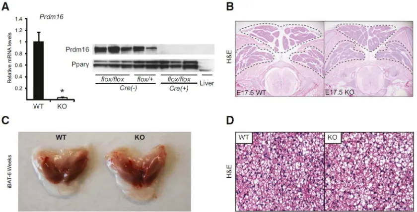
Prdm16flox/floxmice (Figure S1A). Myf5Cre is activated in the somitic precursors that give rise to brown adipocytes. The resulting Myf5Cre/+;Prdm16flox/flox (Myf5-ΔPrdm16) mice were born in normal Mendelian ratios and were grossly indistinguishable from their wildtype (WT) littermates. Prdm16 mRNA and protein expression were almost completely ablated in Myf5-ΔPrdm16 BAT (Figure 1A).
Figure 1: Prdm16 is dispensable for embryonic BAT development
35
Surprisingly, there were no differences in the morphology or size of BAT depots between WT and Myf5-ΔPrdm16 mice at E17.5 of development (Figure 1B). At 6 weeks of age, WT and Prdm16-deficient iBAT depots were also grossly and histologically similar (Figures 1C, D). The other major site of Myf5Cre-mediated DNA recombination
during development is skeletal muscle where Prdm16 mRNA is not normally detected 87.
Consistent with this, Prdm16 mRNA levels were equivalent in WT and Myf5-ΔPrdm16
muscles (Figure S1B). Previous studies indicated that Prdm16 can suppress the expression of certain muscle-specific genes 13; however, we did not observe elevated
expression of muscle-specific genes in the iBAT of Myf5-ΔPrdm16 mice(Figure S1C). Taken together, these results indicate that Prdm16 is dispensable for BAT formation in mice.
Figure S1. Prdm16 deficiency in BAT does not increase the expression of skeletal muscle genes.
(A) Gene targeting strategy for creating Prdm16floxmice. (B) Prdm16 mRNA levels in interscapular brown adipose tissue (iBAT) and several skeletal muscles (quadricep, tibialis anterior [TA], extensor digitorum longus [EDL], gasterocnemius, diaphragm and soleus) of wildtype (WT) and Myf5-ΔPrdm16 mice. (C)
36
Prdm16 recruits Ehmt1 to repress the expression of white fat-selective genes
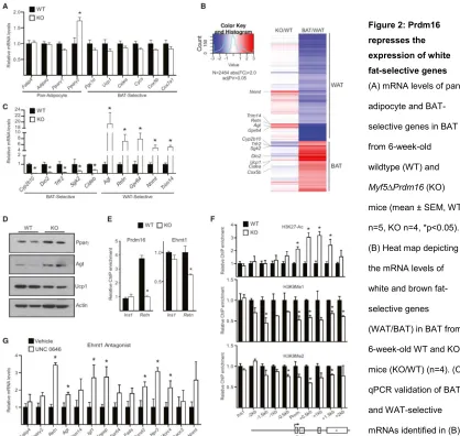
We next analyzed the molecular phenotype of iBAT from 6-week-old WT and
Myf5-ΔPrdm16 mice. Pan-adipocyte genes like Fabp4 and Adipoq were equivalently expressed in WT and Prdm16 knock-out (KO) iBAT (Figure 2A), although KO tissue expressed higher levels of Pparγ2,the adipose-selective isoform of Pparγ. The levels of brown fat-selective(Pgc1α, Ucp1 and Cidea) and mitochondrial (Cycs, Cox5b, Cox7a1) genes were mildly but not significantly decreased in Prdm16 KO tissue (Figure 2A).
To search for genes/pathways that were sensitive to Prdm16 levels in BAT, we compared the global gene expression profiles of iBAT from 6-week-old WT and Myf5-ΔPrdm16 miceusing cDNA microarrays. Gene ontology analysis revealed that genes involved in lipid-metabolism, including “lipid biosynthetic process” and “lipid metabolic process”, were increased in the absence of Prdm16 (Figure S2A). This suggested that loss of Prdm16 shifted adipocyte metabolism to favor a white fat-like energy storage phenotype. We thus specifically analyzed the impact of Prdm16-deficiency on the complete set of BAT- and WAT-selective genes (Figure 2B). Consistent with qPCR analysis, most typical brown-selective genes (e.g. Ucp1, Cidea, Cox5b) were only slightly reduced in Prdm16-deficient BAT. However, the expression of a few BAT-selective genes were dramatically diminished in KOBAT, including Dio2 (deiodinase, iodothyronine, type II), an important regulator of brown adipocyte function 182 (Figure 2B,
37
increase in Agt,and 6- to 8-fold increases in Retn, Gpr64, Nnmt and Trim14 (Figure 2C). These results reveal that Prdm16 is required in BAT to suppress the expression of many white fat-selective genes.
Figure 2:Prdm16
represses the
expression of white
fat-selective genes
(A) mRNA levels of pan-adipocyte and BAT-selective genes in BAT from 6-week-old wildtype (WT) and
38
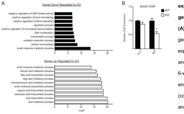
We used Retn as a model locus to investigate the mechanism by which Prdm16 represses white fat-selective genes. Chromatin immunoprecipitation (ChIP) for Prdm16 in WT and KO BAT showed that it was specifically enriched at the Retn promoter relative to non-specific control sites (Figure 2E). The repressive chromatin modifier, Ehmt1 (G9a-like protein), an interacting partner of Prdm16 90,190, was also bound to the Retn
promoter in BAT and its binding there was reduced by ~40% in Prdm16 KO BAT relative to WT BAT (Figure 2E). Importantly, the loss of Prdm16 and Ehmt1 binding at Retn was associated with increased levels of H3K27-Ac, a histone mark correlated with active transcription; and decreased levels of H3K9-Me1 and H3K9-Me2, modifications laid down by Ehmt1 and associated with gene repression (Figure 2F). Ehmt1 binding atthe
Agt promoter was also significantly decreased in Myf5-ΔPrdm16 BAT(Figure S2B). These data suggest that Prdm16 recruits Ehmt1 to certain white fat-selective genes to decrease their transcription. In accordance with this, treatment of cultured brown adipocytes with UNC 0646, an Ehmt1 antagonist, increased the expression of many white fat-selective genes, including Retn and Agt (Figure 2G).
Figure S2. Prdm16 deficient iBAT
expressed a white fat-related
gene profile.
(A) Gene Ontology (GO) analysis of genes thar are differentially expressed betweeb wildtype (WT) and Prsm16-deficient BAT (KO) of 6-week-old mice. (B) Relative ChIP enrichment for Ehmt1 at the Ins1
39
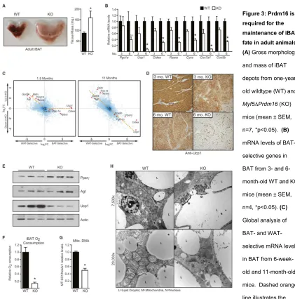
Prdm16 maintains iBAT identity during aging
In contrast to juvenile mice, we noticed that older (>6 months of age) Myf5-ΔPrdm16 animals exhibited a profound morphological “whitening” of their iBAT. This included a ~50% increase in the size of the tissue, a switch from multilocular to unilocular morphology, and increased lipid content (Figures 3A, S3A). To determine when this phenotype emerged, we analyzed gene expression in iBAT from WT and
Myf5-ΔPrdm16 mice at 3 and 6 months of age. In 3-month-old animals, Prdm16-deficiency resulted in a modest decline in the expression of brown fat-selective genes, including ~30-40% reductions in the levels of Ucp1, Pparα and many mitochondrial genes (Figure 3B). The reduction of brown fat-specific gene expression in KO BAT was much more pronounced in 6-month-old mice. At that age, Ucp1 mRNA levels were decreased by >90% and Cidea and Pparα levels were reduced by ~70% (Figure 3B). Hematoxylin and eosin staining showed that lipid droplet size increased dramatically in the KO BAT from 3 to 6 months of age (Figure S3B). Microarray analyses revealed that
40
We next examined the consequences of Prdm16-deficiency on mitochondrial mass and function. Using a Clark electrode, we measured oxygen consumption in isolated iBAT from 9-month-old WT and Myf5-ΔPrdm16 mice. Remarkably, basal
(unstimulated) respiration in the KOiBAT was ~85% lower than in WT tissue(Figure 3F), indicative of a loss of mitochondrial mass. Indeed, PCR analysis revealed that KO BAT had 50% less mitochondrial DNA than WT tissue (Figure 3G). Transmission electron microscopy studies showed that WT adipocytes were packed with mitochondria
41
Figure 3: Prdm16 is
required for the
maintenance of iBAT
fate in adult animals
(A) Gross morphology and mass of iBAT depots from one-year-old wildtype (WT) and
Myf5∆Prdm16 (KO) mice (mean ± SEM, n=7, *p<0.05). (B)
mRNA levels of BAT-selective genes in BAT from 3- and 6-month-old WT and KO mice (mean ± SEM, n=4, *p<0.05). (C)
Global analysis of BAT- and WAT-selective mRNA levels in BAT from 6-week-old and 11-month-6-week-old mice. Dashed orange line illustrates the change in gene expression pattern. Data is presented as a log2FC scatter plot. (n=4). (D) Immunohistochemical staining
42
The interscapular depot is the largest BAT depot in adult mice and was the focus of our study. However, we also investigated the impact of Prdm16-deficiency on the axillary and cervical BAT (aBAT and cBAT). The aBAT and cBAT depots in 6-month-old
Myf5-ΔPrdm16 appeared paler than those in WT mice, but there was no difference in aBAT or cBAT mass between WT and Myf5-ΔPrdm16 mice (Figure S3D). Interestingly, the white fat-selective genes were markedly elevated in Prdm16-deficient aBAT and cBAT but brown fat-specific gene levels were not affected (Figure S3E). These results suggest that interscapular BAT is particularly reliant on Prdm16 for maintaining the expression of brown fat-selective genes during aging.
Figure S3. Prdm16-deficiency causes a
loss of interscapular brown adipose
tissue identity in adult mice
(A) Hematoxylin and eosin (H&E) staining of sections from the interscapular brown adipose tissue (iBAT) of one-year-old WT and Myf5-ΔPrdm16 (Prdm16 KO) mice. (B)
H&E staining of WT and Myf5-ΔPrdm16 (KO) iBAT from 3 and 6 month-old-mice.
(C) Myf5-Cre driven DNA recombination of the Prdm16 locus in iBAT from 9-month-old WT and KO mice. (D) Gross morphology and mass of dissected axillary and cervical BAT depots from WT and KO mice. (E)
43
Prdm16 is required for induction of the brown fat gene program in isolated precursors
The aging-associated decline of iBAT identity in Myf5-ΔPrdm16 mice raised the question of whether Prdm16 was required cell autonomously for proper brown adipocyte differentiation in this depot. To study this, we isolated primary brown adipocyte
precursors from the iBAT of WT and Myf5-ΔPrdm16 mice and examined their
differentiation into adipocytes under defined culture conditions. WT and KO precursor cells from newborn iBAT differentiated with equivalent efficiently into mature lipid droplet-containing adipocytes that expressed similar levels of pan-adipocyte genes, including
Fabp4 and Adipoq (Figure 4A, S4A). KO cultures displayed a >90% reduction in Prdm16 mRNA and protein levels (Figure 4B, D), indicating that most or all of the precursor cells in BAT descend from Myf5Cre-expressing cells. Strikingly,
Prdm16-deficient cultures expressed dramatically lower levels of brown adipocyte-specific genes as compared to WT cultures, including 90-95% reductions in the mRNA levels of Ucp1,
Cidea and Dio2; and 60-80% decreases in Pgc1α, Pparα, Cox7a1, Cycs, and Errγ
(Figure 4B). WT adipocytes also had four times more mitochondrial DNA than KO cells (Figure 4C). Western blot analysis showed that Ucp1 protein, like its mRNA, was dramatically lower in KO adipocytes (Figure 4D). Importantly, retroviral expression of Prdm16 in KO preadipocytes efficiently activated the expression of thermogenic genes like Ucp1 and Cidea (Figure 4E), indicating that the KO cells were competent to induce the brown-selective gene program. Immortalized brown fat cell lines from newborn BAT of Myf5-ΔPrdm16 mice displayed a similarly severe defect in activating the