Abstract
Having in mind the fact that cerebrovascular disease (CVB) takes today in medicine, in spite of diagnostic and therapeutic modernisation, the third place of mortality causes in the world (behind cordial and malignant dis-eases, but in front of depressions), and the second place of invalidity cause (right after trauma) as well as the sec-ond place of dementia cause (after Alzheimer disease), it urges primary prophylaxis.
Developing countries, but before all countries of East and Middle Europe, where is our country, are highly risked areas where CV disease has trend of incidence and total frequency increase. In the neighbouring Croatia today CV disease is at the first place of mortality causes. In the world today 5 million people annually suffer CV disease, in Europe about 700.000, but frequency of suf-fering on Balkan is about 5 prom.
Age of CV disease effecting unfortunately moved towards young age, and today 49% of effected by CV disease are of 46 to 59 years of age. Early detection and treatment of risk factors (before hypertension, smoking, diabetes mellitus, hyperlipidaemia, stress and physical non-activities) are the first aspect of CV disease prophy-laxis.
Together with this aspect of primary prophylaxis is early detection of complications of mentioned risk factors on the walls of blood vessels, before all changing in sense of arteriosclerosis, with consequence of disorder of cerebral haemodynamics.
With that objective – verification of circulator and total haemodynamic disorders, there is obvious disclose of non-invasive diagnostic methods, and one of them is Transcranial Doppler Sonography (TCD).
TCD is method comfortable for patient, reliable and rather precise, dynamic, and can be repeated several times, without side effects and in comparison with others rather cheap.
Key words: cerebrovascular disease (CVB), Transcranial Doppler Sonography (TCD)
Educational article
Transcranial Doppler Sonography is non-invasive diag-nostic method, which ensures insight into the status of circulation of intra cranial part of posterior – VB basin as well as intra cranial, ending part of front – carotid basin and their anastomosis on the brain basis, which is Willis circle. Method is based on application of Doppler effect
or principle, which consists of application or shift of received frequencies between two bodies in move. When those two bodies are drawing closer received frequency increase and when drawing away received frequency decrease. (This knowledge Doppler published in his work named “About coloured light of double stars and some other sky’s bodies”).
As source of the waves serves piezo electric crystal, which oscillate and emit ultrasound wave of known fre-quency, which is directed through probe of apparatus to blood vessel, but before all to red corpuscles, which flow by certain velocity, reflects from them as well as from blood vessel wall.
On the basis of change of frequency emitted and reflect-ed ultrasound wave is calculatreflect-ed velocity of blood flow, which serves as indicator of blood vessel status.
Frequency change (Doppler frequency change) is calcu-lated as per the following formula:
2fe x v x cos α
Df = c
The above mentioned abbreviations show the following values:
fe = frequency of emitted ultrasound wave v = middle velocity of blood flow
a = angle between probe and blood vessel c = velocity of ultrasound through tissue
(1540 m/sec in soft tissues)
Blood velocity flow is proportional with Doppler shift and it is calculated as per the following formula:
Df x c V = 2 fe x cos α
Frequency value is calculated in hertz (Hz), and blood flow velocity in m or cm / sec.
Results are presented in measure units for velocity, as proposed Doppler Committee for Nomenclature of American Echocardiography Society 1986.
Velocity of ultrasound in blood depends of temperature,
Transcranial doppler sonography as diagnostic method
\elilovi}-Vrani} Jasminka
content of proteins and haematocrit value. Flow can be calculated only by application of pulsating wave, which provides precise estimation of the spot at which the observation is concentrated with responding angle between probe and blood vessel.
It is known that through middle part of blood vessel flow, red corpuscles, erythrocytes, and towards sides (i.e. wall of vessel) are moving thrombocytes and other cell’s ele-ments.
This is normal – laminar blood flow in blood vessel, but in case of stenosis of the vessel, since as vessel is more narrow velocity is higher and considering physics princi-ples that through narrow space the same quantity of blood has to pass within unit of time, that flow of blood after stenosis passing over into turbulent flow, which is sign of pathologic state.
Picture 1Schematic review of changes in stenosis – laminar flow passing over at narrow spot into high frequency flow, then comes turbulences and later on flow is established again.
In 1955 Laksell found out, during echo encephalo-graphic X ray, that ultrasound can intrude through scull and brain structure of different density and velocity of conductivity, when one may establish lesion. However, problem was how and which ultra-sound frequency can achieve such.
Norway physiologist and cybernetic Rune Aaslid resolved that problem, which could not be bridged over at that time, by construction of probe of 2MHz. Until that time investigation of intra cranial circulation by ultra-sound was possible with children up to one year of age, whose large fontanel is not closed or during intra neuro-logical intra surgery, because scull bonds were large obstacles and cause weakening of ultrasound waves. When probe of 2MHz is placed on the thinnest part of temporal bond it is achieved direct insight into state of intra cranial circulation.
Kinds of ultrasound waves
Doppler diagnostic today applies two kinds of ultrasound waves: Doppler with continuing Doppler provides ultra-sound wave, which constantly emits from piezo – electric crystal, placed in the same probe where is a receiver of reflected waves (i.e. returned ultrasound waves), but pul-sated ultrasound wave provides crystal with purpose to emit and receive reflected waves, and emission is from time to time. Ultrasound is directed to the certain depth (different from continuing), goes through tissue (average velocity is 1540 m/sec), it returns, suffers weakening (attenuation), which is as rule 10 decibels per centimetre of tissue depth (always double because of two ways of shift). It is considered that ultrasound travels 13 millisec-onds per centimetre of depth.
Brain requires permanent and proper blood supply, because of which circulation resistance is very weak. In case of increase of resistance at first happens decrease of diastolic flow. In arteries responsible for brain supply, quantity of diastolic flow is function of circulation resist-ance of that area and can be presented through formula, which Pourcelot introduced 1971.
A - D
R = A
The above abbreviations show the following values:
R = Resistance index
A = Maximum velocity of flow in systole D = Maximum velocity of flow in diastole
Vertebral arteries are difficult to access because of its anatomic localisation, starting from palpation, ausculta-tion, as well as testing on Doppler principle. Apart from that they are in bone channel subject to often asymmetry and anomaly at the very beginning.
Transcranial Doppler Sonography – TCD, became very important non-invasive method for testing of intra cranial circulation; it is different from angiographies, which is invasive method used only in some cases, mostly in pre-surgery state.
Apart from the above TCD method is very reliable and because of that it displaced angiographies. Following lit-erature TCD method is in correlation with angiographies about 75%, what understands high percent of accuracy of a method itself.
a minute and testing can be repeated numberless times. This method is much cheaper in comparison with others, with special importance in economic sense.
Diagnostic areas of TCD are as follows:
• Detection of intra cranial stenosis and occlu-sions
• Haemodynamic evaluation of tendem’s (extra – intra cranial) lesion;
• Evaluation of collateral possibilities,
• Estimation and monitoring of vasospasm
• Diagnosis of artery – vein malformations (AVM)
• Intra surgery monitoring and monitoring in units of intensive care
• Estimation of brain death
• Classification of cerebrovascular insult
• Evaluation of manifested physiological reactions
• Early detection and prevention of cerebrovas-cular insult
Considering VB basin, TCD method found the applica-tion in case of suspected stenosis, occlusion or asymme-try of vertebral arteries.
Doppler signals can be registered intra cranial only at the places where bound of the scull are very thin and provide passing of UZ waves, called bound windows. “Lighting” of the scull provides disclosing of the windows on tem-poral bound, side part of front bound, on orbit, orbital roof and in the area of occipital bound. Size of the win-dow varies from person to person. The most often, in younger persons, is possible to achieve rather good sig-nal from relatively large area, but in older persons some-times is very difficult to achieve it because of hyper ossi-fication of bounds. That happens more often in females. (5, 6, 11)
Temporal windows are used usually for testing of circu-lation of branches of Willis circle, orbital for search of art. ophthalmia and sub-occipital for vertebral and basilar arteries.
Picture 2Bound windows on scull
Techniques of testing
Every blood vessel, when tested, has certain rules, such as: through which window is the easiest to verify it, at which usual depth, and when defined ones, it is necessary to recognise its spectrum, determine direc-tion of flow, whether towards probe or from the probe and at the end its mean, i.e. average velocity of blood flow.
In the process of identification of blood vessels, in par-ticular in intra cranial part, it is important to determine segment of individual blood vessel.
Having in mind that Doppler shift of frequency is pro-portional with cosine of angle between probe and blood vessel, as angle is smaller the mistakes are less and irrel-evant.
The smallest angle of insonation through temporal win-dow has medial cerebral artery, where is that angle about 0 degrees, and achieved values are close to absolute val-ues of blood flow velocity, but through sub-occipital window, the smallest angle of insonation has basilar artery, which is always below 30 degrees.
Through temporal window are disclosed the easiest arter-ies of Willis circle as follows: MCA, ACA, PCA, as well as SC, AcoA and PcoA, but VA and BA are disclosed through sub-occipital or transforamental window. Testing can be performed in sitting position (most often) or position of lying on the stomach. Method is comfort-able for patient, without side effects, and it is enough if patient co-operates during testing, to be quite and do not talk. In order to improve the contact with blood vessel is used cream, which is applied, in thin layer on the spot.
Picture 3Techniques of testing
Blood flow velocity in large brain arteries can be expressed as systolic, diastolic or average blood flow velocity. Average blood flow velocity has the largest physiological importance, because it depends less from central cardio-vascular factors like heart activity, con-tractions, total peripheral resistance and aortic compli-ance, than systolic and diastolic value.
Average value in respect of time (time – mean velocity) has better correlation with values of perfusion than max-imum and other values. Following literature, reference value for adults are blood flow velocity in cm/sec as per Aaslid. (7, 11)
Table 1 shows: depth of insonation, direction of blood flow and value of SBSK for arteries of Willis circle and VB basin.
In the most cases value of average blood flow velocity has higher value in carotid than in VB basin.
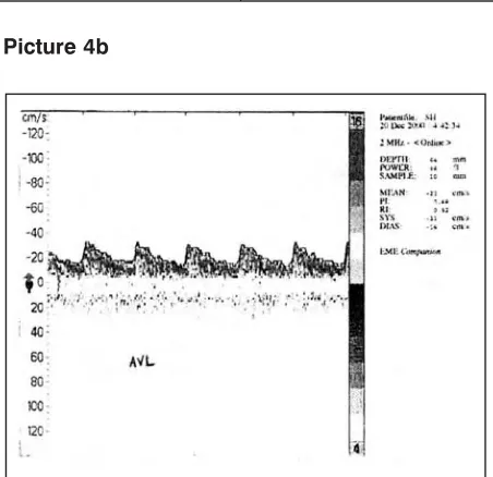
During testing was observed the following: normal (lam-inar) flow – Picture 4a, weakened flow – picture 4b, and flow of high frequency – picture 4c.
Picture 4a
Picture 4b
Weakened flow is present when received ultrasound wave is deafened by red corpuscles, because of some rea-sons. Spectrum is entirely of lower maximum velocity with expressed peripheral resistance, but without neces-sarily disturbed flow. Typical weakened flow appears with calcificated blood vessel (Picture 4-b)
High frequency is noticed when lesion or process, which caused it, reaches critical point and creates clear haemo-dynamic disturbance of circulation. Lesion is critical when reduce inside diameter of blood vessel lumen and create increased pressure down the narrowed part (pic-ture 4-c) (11, 12, 13, 14).
Blood vessel Depth in mm Blood flow direction SBSK values
ACA 65-75 From probe 50+- 12 cm/sec
CS 60-80 54+- 23 cm/sec
MCA 40-60 Towards probe 62+- 12 cm/sec
PCA 55-70 Towards probe 42 +- 10 cm/sec
BA 65-100 From probe 42+- 10 cm/sec
VA 50-60 From probe 36+- 9 cm/sec
Sa`etak
Transkranijalna doppler sonografija kao dijagnosti~ka metoda
Imaju}i u vidu ~injenicu da je cerebrovaskularna bolest (CVB) danas u medicini, i pored dijagnosti~kih i terapeut-skih napredaka, na 3. mjestu uzroka smrtnosti u svijetu (iza kardijalnih i malignih bolesti, a ispred depresija), na 2. mjestu uzroka invaliditeta (odmah iza traume) i tako|e na 2. mjestu uzroka demencija (iza Alchajmerove bolesti) to se nala`e hitnost primarne prevencije. Zemlje u razvoju, prije svega zemlje Isto~ne i Srednje Evrope, a gdje spada i na{a zenlja, su visoko rizi~na podru~ja u kojima CV bolest ima tendenciju rasta incidence i ukupne prevalence. U nama susjednoj Hrvatskoj danas je CV bolest na 1. mjestu uzroka smrtnosti.
U svijetu godi{nje oboli od CV bolesti oko 5 miliona ljudi, u Evropi je taj podatak oko 700.000, dok se prevalenci-ja na ovim na{im balkanskim prostorima kre}e oko 5 promila.
Starosna dob oboljevanja od CV bolesti se na`alost pomjerila ka mla|oj dobi, tako da je danas 49% oboljelih od CV bolesti u `ivotnoj dobi 46-59 godina `ivota. Rano otkrivanje i lije~enje faktora rizika (prije svega hipertenzije, pu{enja, dijabetes melitusa, hiperlipidemije, stresa i fizi~ke neaktivnosti) su prvi vid prevencije CV bolesti. Uz to rano otkrivanje komplikacija navedenih faktora rizika na zidovima krvnih sudova, prije svega promjena u smislu arterioskleroze, {to ima za posljedicu poreme}aj cerebralne hemodinamike, je tako|e vid primarne preven-cije. U tom cilju – u verificiranju cirkulatornih i ukupnih hemodinamskih poreme}aja, danas se daje prednost grupi neinvazivnih dijagnosti~kih metoda, a jedna od tih je Transkranijalna Doppler Sonografija (TCD).
TCD je metoda komforna za pacijenta, pouzdana i prili~no ta~na, dinami~ka, mo`e se ponavljati vi{e puta, bez {tet-nih efekata i uz to, u odnosu na druge relativno jeftina.
Klju~ne rije~i: Cerebrovaskularna bolest (CVB), Transkranijalna dopler sonografija (TCD)
Edukativni ~lanak
Bibliography
1. Aaslid R, Lindegaard KF, Sorteberg W, Vomes H, Cerebral Auto-regulation dynamics in humans, Stroke, 1989, 20:45-52
2. Aaslid R, Lindegaard KF, Cerebral Haemodynamics, U. Waslid R, Transcranial Doppler Sonography, Wien-New York, Springer Verlag 1986.
3. Ackerstaff R G A, Ultrasonic duplex scanning in atherosclerotic diseases of the vertebrobasilar arterial system, Netherlands, University of Utreht, 1985, 195 str. Disertacija
4. Adams RJ, Nicholas FT, Hess DC: Normal Values and Physiological Variables U: Newell DW, Aaslid R, Transcranial Doppler, Raven Press, 1992, 41-48.
5. Berni A, Tromba L, Cavaiola S, Trombesi T, Castellani: Classification of the subclavian steal syndrome with transcranial Doppler, Journal of Cardiovascular Surgery, 1997, 38(2), 141-145.
6. Bluth EI, Merritt CRB, Sullivan MA.A screening test for carotid stenosis: preliminary feasibility study. Radiology 1997:205(S):696-671.
7. Bray JM, Missoum A, Dubas F, Emile J, Lhoste P,: Detection of vertebrobasilar intracranial stenosis: Transcranial Doppler Sonography versus angiography, Journal of Ultrasound in Medicine, 1997, 16(3), 213-218. 8. Breyer B. Fizika ultrazvuka. U: Kurjak A. Ultrazvuk u klini~koj medicini, Medicinska naklada, Zagreb, 1997,
1-13.
9. Breyer B. Medicinski dijagnosti~ki ultrazvuk, Školska knjiga, Zagreb, 1982, 37-103. 10. Caplan LR, Migraine and vertebrobasilar ischemia, Neurology, 1991, 41: 55-61.
11. Demerin V, Transkranijalni Doppler (TCD). U: Demerin V, Štitkovac M, Thaller N, Doppler – sonografija krvnih `ila, Školska knjiga, Zagreb, 1990, 89-126.
12. \elilovi} J., Vrijednost TCD u dijagnostici VB insuficiencije, Sarajevo, Medicinski fakultet 2001. 21-27, Magistarski rad
13. Gavranovi} M: Terapija cerebralnih komplikacija kod hipertenzije, Soc. Med. 1997, 2-91.
14. Illuminati G, Calio FG, Bertagni A, Vietri F, Martinelli V: Transcranial Doppler validation of haemodynamic vertebrobasilar insufficiency diagn., Revista EuropaPer Le Scienze Mediche Farmacologiche, 1996, 18(5-6): 213-216.