1834–1846 Nucleic Acids Research, 2018, Vol. 46, No. 4 Published online 27 December 2017 doi: 10.1093/nar/gkx1291
Sp1 phosphorylation by ATM downregulates BER and
promotes cell elimination in response to persistent
DNA damage
Sally C. Fletcher
1, Claudia P. Grou
1, Arnaud J. Legrand
1, Xin Chen
1,2, Kalle Soderstrom
3,
Mattia Poletto
1,*and Grigory L. Dianov
1,4,5,*1Department of Oncology, CRUK & MRC Oxford Institute for Radiation Oncology, University of Oxford, Old Road
Campus Research Building, Oxford OX3 7DQ, UK,2Department of Marine Technology, College of Ocean, Nantong
University, Nantong, Jiangsu, 226007, China,3Nuffield Department of Orthopaedics, Rheumatology and
Musculoskeletal Sciences, Botnar Research Centre, University of Oxford, Oxford OX3 7LD, UK,4Institute of Cytology
and Genetics, Russian Academy of Sciences, Lavrentyeva 10 Novosibirsk 630090, Russian Federation and
5Novosibirsk State University, Pirogova 2, Novosibirsk 630090, Russian Federation
Received July 11, 2017; Revised December 13, 2017; Editorial Decision December 14, 2017; Accepted December 19, 2017
ABSTRACT
ATM (ataxia-telangiectasia mutated) is a central molecule for DNA quality control. Its activation by DNA damage promotes cell-cycle delay, which facil-itates DNA repair prior to replication. On the other hand, persistent DNA damage has been implicated in ATM-dependent cell death via apoptosis; however, the mechanisms underlying this process remain elu-sive. Here we find that, in response to persistent DNA strand breaks, ATM phosphorylates transcrip-tion factor Sp1 and initiates its degradatranscrip-tion. We show that Sp1 controls expression of the key base exci-sion repair gene XRCC1, essential for DNA strand break repair. Therefore, degradation of Sp1 leads to a vicious cycle that involves suppression of DNA repair and further aggravation of the load of DNA damage. This activates transcription of pro-apoptotic genes and renders cells susceptible to elimination via both apoptosis and natural killer cells. These findings constitute a previously unrecognized ‘gate-keeper’ function of ATM as a detector of cells with persistent DNA damage.
INTRODUCTION
Failure to preserve genome stability underlies the decline of every organism through pathophysiological processes such as ageing, neurodegeneration and cancer. For this reason, the cellular genome is constantly guarded against DNA le-sions generated by both exogenous and endogenous muta-gens. In order to maintain genome stability, cells exploit a
number of DNA repair systems. Amongst these, the DNA base excision repair (BER) pathway constitutes the front-line defense against endogenously-generated DNA damage including DNA base lesions and single strand breaks (SSBs) (1,2). BER is robust pathway that, under normal circum-stances, is sufficient to cope with endogenous DNA damage. However, low BER efficiency can occur as a result of mal-function of BER components or overload of cellular repair capacity by acute DNA damage. This can either lead to cell death, or result in accumulation of mutations and induc-tion of tumorigenesis (3–5). It is still poorly understood how cells decide to trigger apoptosis and eliminate potentially dangerous cells, rather than attempting DNA repair of ex-cessively damaged DNA. The ATM (ataxia-telangiectasia mutated) protein is the primary candidate for this role of decision maker (6). In fact, ATM’s kinase activity has been shown to respond to a wide range of genome-threatening le-sions including DNA single and double strand breaks (7,8) and to mobilize a cascade of phosphorylation events that delay cell cycle, providing additional time for DNA repair prior to replication (9). At the same time, ATM has been implicated in the initiation of programmed cell death in response to DNA damage (10,11). Despite this, although ATM-dependent cell cycle delay is known to be mainly ac-complished through effectors such as p53 and p21 (12), the mechanisms driving an ATM-dependent switch to apopto-sis are unclear.
Among a number of proteins that are phosphorylated by ATM in response to DNA damage, transcription factor Sp1 has been shown to regulate the expression of multiple genes (13–15), including cellular components involved in apopto-sis (16–18). While ATM-dependent phosphorylation of Sp1 at serine 101 has been previously documented (13–15), the
*To whom correspondence should be addressed. Tel: +44 1865617325; Email: [email protected]
Correspondence may also be addressed to Mattia Poletto. Email: [email protected]
C
The Author(s) 2017. Published by Oxford University Press on behalf of Nucleic Acids Research.
biological role of this modification remains unclear. Here we report that, in response to persistent DNA strand breaks, ATM phosphorylates transcription factor Sp1 at serine 101, initiating its proteasomal-dependent degradation. We also find that Sp1 controls the expression of the key BER gene
XRCC1 and that degradation of Sp1 decreases DNA re-pair capacity and aggravates the load of DNA damage. This feeds a vicious cycle that further supports Sp1 degra-dation. Furthermore, we demonstrate that downregulation of Sp1 upon DNA damage primes fibroblasts to apopto-sis and to elimination by natural killer (NK) cells. We sug-gest that this mechanism allows the detection of potentially pre-cancerous cells bearing persistent DNA strand breaks, prompting their removal either through apoptosis or via the innate immune system.
MATERIALS AND METHODS
Cell culture and drug treatments
Normal human TIG-1 fibroblasts were from the Coriell Institute Cell Repository (AG06173). Cells were cultured in Dulbecco’s modified Eagle’s medium low glucose (Life Technologies) supplemented with 15% foetal bovine serum (FBS) at 37◦C in a humidified atmosphere with 5% CO2.
The Nishi NK cell line has been previously described, and is derived from the peripheral blood mononuclear cells of a boy with chronic active Epstein–Barr virus infection complicated with NK leukaemia. The phenotype of this NK leukaemia is: CD94/NKG2A and LIR-1/ILT-2 pos-itive, but CD3,␣ TCR, ␥ ␦TCR, KIR3DL1, KIR2DL1, KIR2DL2, KIR2DS1, KIR2DS2 negative. CD16 expres-sion is low (19). NK cells were grown in IMDM Glu-taMAX™medium (Life Technologies) supplemented with 10% FBS, 2% heat-inactivated human serum (Sigma-Aldrich), 100 IU/ml penicillin, 100 g/ml streptomycin and 10 ng/ml recombinant human IL-15 (PeproTech) at 37◦C in a humidified atmosphere with 7.5% CO2. Cells
were routinely checked for mycoplasma. H2O2,
camp-tothecin, cycloheximide, the Chk1 inhibitor (UCN-01) and midostaurin were from Sigma. Zeocin was from Life Tech-nologies. Mithramycin A and MG132 were from Enzo Life Sciences. The ATM inhibitors (KU55933 and KU60019), the DNA-PK inhibitor (Inhibitor III) and staurosporine were obtained from Millipore, while the Chk2 inhibitor (CCT 241533) was from Tocris. The ataxia telangiectasia and Rad3-related (ATR) inhibitor (VE-821) was a kind gift from Dr Anderson Ryan (University of Oxford). Navitoclax (ABT-263) was purchased from Cayman Chemical.
Cell viability assays
Cell viability was assessed using resazurin (Sigma). For co-culture experiments, TIG-1 cells were treated as described and a suspension of NK cells was aliquoted onto adherent fibroblasts at the indicated NK:TIG-1 ratio. Cell cytotoxic-ity was assessed after co-incubation by washing off NK cells and evaluating the viability of fibroblasts using a resazurin assay.
Comet assays, immunostaining and high-throughput mi-croscopy
Alkaline comet assays were carried out as previously described (7). Immunostaining and high-throughput mi-croscopy were carried out as described in (20).
Protein expression and purification
The plasmid pN3-Sp1FL, containing full-length Sp1 was a gift from Guntram Suske (Addgene plasmid #24543). A pET-28a plasmid expressing His(6)-tagged recombinant Sp1
was generated by sub-cloning the Sp1 cDNA from pN3-Sp1FL. Protein expression was carried out in Rosetta™ Es-cherichia colicells and recombinant Sp1 was purified under denaturing conditions (6 M guanidine hydrochloride) us-ing a HisTrap column (GE Healthcare). Recombinant Sp1 was refolded over 96 h by sequential dialysis against 10 mM Tris–HCl, pH 7.5, 200 mM NaCl, 50M ZnSO4, 0.4 M
L-Arginine, 5% glycerol for 48 h, followed by 10 mM Tris– HCl, pH 7.5, 200 mM NaCl, 5% glycerol for further 48 h.
In vitrophosphorylation assays
Phosphorylation reactions were carried out by combining recombinant Sp1 (500 ng) and active recombinant ATM (100 ng - Millipore) in phosphorylation buffer (50 mM HEPES pH 7.5, 50 mM KCl, 10 mM MgCl2, 10 mM
MnCl2, 1 mM adenosine triphosphate (ATP), 1 mM
dithio-threitol (DTT) and 5% glycerol). Reactions were incubated for 2 h at 30◦C and halted by adding sodium dodecyl sulphate-polyacrylamide gel electrophoresis loading buffer.
In vitroligation assays
Nuclear cell extracts were prepared as described previously (21). Ligation assays were carried out using 1g of nuclear extract essentially as described in (22), with minor modifi-cations. Briefly, reactions were performed in 50 mM Tris– HCl pH 7.5, 10 mM MgCl2, 10 mM DTT, 1 mM ATP at
23◦C for the indicated time; the oligonucleotide substrate (50 nM) has been described (22) and was 5-labeled with IRDye®800 (IDT). Reactions were halted with 96%
for-mamide and 10 mM EDTA and analysed by electrophoresis on a 20% denaturing polyacrylamide gel. The percentage of substrate converted to product was determined by using an Odyssey image analysis system (Li-Cor Biosciences).
Luciferase assays and chromatin immuno-precipitation (ChIP)
1836 Nucleic Acids Research, 2018, Vol. 46, No. 4
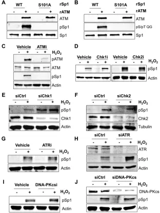
Figure 1. ATM is the main kinase responsible for serine 101 phosphorylation of Sp1. (AandB) Representativein vitrokinase assay using recombinant ATM (rATM) and wild-type (WT) or Ser101-Ala mutant (S101A) recombinant Sp1 proteins (rSp1). ATM can phosphorylate WT Sp1 but is unable to
phosphorylate the S101A mutant. Reactions were probed with either the phosphorylated Ser101-specific antibody (A) or an ATM/ATR substrate-specific
pS/pT QG antibody (B). (C) Representative Western blot analysis on TIG-1 cells showing Sp1 phosphorylation at Ser101 upon H2O2treatment (200M,
1 h). Loss of phosphorylation occurs in the presence of ATM inhibitor (ATMi: KU55933, 10M, 2 h prior to H2O2). (D) Representative Western blot
analysis as in (C), in the presence of Chk1 inhibitor (UCN-01, 10M for 2 h prior to H2O2) or Chk2 inhibitor (CCT 241533, 3 nM for 2 h prior to H2O2).
(EandF) Representative Western blot analysis as in (C), on cells depleted of either Chk1 (E) or Chk2 (F) using siRNA. Phosphorylation of Sp1 is still
detected after treatment with H2O2. (G) Same as in (C), but cells were treated with an ATR inhibitor (VE-821, 1M for 2 h prior to H2O2) (H) Same as
in (E), but cells were treated with an ATR siRNA. (I) Same as in (C), but cells were treated with a DNA-PKcs inhibitor (DNA-PK Inhibitor III, 10M
for 2 h prior to H2O2). (J) Same as in (E), but cells were treated with DNA-PKcs siRNA. Data information: In (A–J) either actin or tubulin was used as
loading control.
Electrophoretic mobility shift assays (EMSA)
Electrophoretic mobility shift assays (EMSAs) were carried out essentially as in (20). AnXRCC1promoter probe (−145 to−128 bp) was obtained by annealing a 5-IRDye®
800-labeled oligonucleotide (5-GTGTGGCGGAGGGAGGC GGGGCTGGAGGAAACG-3 (Integrated DNA Tech-nologies)), with its complementary sequence. An Sp1 consensus probe (5-ATTCGATCGGGGCGGGGCGAG C-3) was used as cold competitor in order to confirm bind-ing specificity.
Flow cytometry
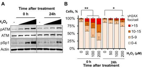
Figure 2. Phosphorylation of Sp1 at Ser101 correlates with the amount
of DNA damage. (A) Representative Western blot analysis on TIG-1 cells
showing dose-dependent induction of ATM phosphorylation at Ser1981
and Sp1 phosphorylation upon treatment with H2O2(50–100–200M, 1
h) and release for either 0 or 24 h. Actin was used as loading control. (B)
Quantification of␥H2AX foci in TIG-1 cells treated as in (A). Foci were
scored using high-throughput microscopy. At least 500 cells/condition
were analyzed. The histogram reports the foci distribution as mean±SD
from three independent experiments and confirms persistency of DNA
double strand breaks at the highest H2O2 dose 24 h after treatment *P
<0.05; **P<0.01.
Statistical analyses
Statistical analyses were performed with the two-tailed Stu-dent’st-test using either Microsoft Excel or SPSS (IBM). Sample size is indicated for each experiment.
RESULTS
Direct phosphorylation of Sp1 at serine 101 by ATM in re-sponse to DNA damage
Although a number of studies have demonstrated that Sp1 phosphorylation at Ser101 depends on ATM (13–15), it is not clear whether Sp1 phosphorylation is accomplished by ATM itself or can also be mediated by other ATM-activated kinases. In order to unequivocally demonstrate that ATM is responsible for direct phosphorylation of Sp1 at Ser101 we employed an in vitrokinase assay using purified proteins. Co-incubation of active recombinant ATM and recombi-nant purified Sp1 showed that Sp1 is robustly phosphory-lated by ATM at Ser101 (Figure1A). Importantly, an Sp1 mutant in which Ser101 was changed into alanine (S101A) could not be phosphorylatedin vitro(Figure1A), confirm-ing the specificity of the antibody used in this study. Fur-thermore, when the phosphorylation reaction was probed with an antibody specific to the phospho-ATM/ATR sub-strate motif (pS/pT QG), the Sp1 S101A mutant did not show any phosphorylation (Figure 1B), suggesting that Ser101 is the major site phosphorylated by ATM under these conditions. Consistent with previous findings (14,15), Sp1 phosphorylation was readily induced by H2O2
treat-ment in normal human fibroblasts (Figure 1C). Modifica-tion of Sp1 was prevented by inhibiModifica-tion of ATM kinase ac-tivity (Figure 1C), confirming that Ser101 could be mod-ified by either ATM or by another kinase downstream to ATM in vivo. Using specific kinase inhibitors we demon-strated that Sp1 phosphorylation at Ser101 was not depen-dent on either Chk1 or Chk2 (Figure 1D) activity. These observations were also substantiated by siRNA-mediated knockdown of either Chk1 (Figure 1E) or Chk2 (Figure
1F), leading us to conclude that these kinases are not in-volved in Sp1 phosphorylation at Ser101 in response to DNA damage. Additionally, we checked if this phospho-rylation was dependent on other members of the phos-phatidylinositol 3-kinase family involved in the DNA dam-age response (DDR), namely ATR and DNA-dependent protein kinase catalytic subunit (DNA-PKcs). Neither ATR inhibition (Figure1G), nor siRNA-mediated knockdown (Figure1H) abrogated Ser101 phosphorylation upon treat-ment with H2O2. Similarly, neither inhibition (Figure1I),
nor siRNA-mediated knockdown of DNA-PKcs (Figure
1J) led to a loss of Ser101 phosphorylation, indicating that Ser101 phosphorylation was not dependent on either ATR or DNA-PKcs. These data demonstrate that, in response to DNA damage, ATM is the major kinase responsible for di-rect Sp1 phosphorylation at Ser101.
Unrepaired DNA strand breaks promote phosphorylation-dependent degradation of Sp1
Human fibroblasts challenged with H2O2are known to
ac-tivate ATM in a dose-dependent manner (14) and this is ac-companied by accumulation of Ser101-phosphorylated Sp1 (Figure2A). We observe here a striking correlation between ATM activation, the Sp1 phosphorylation pattern and the amount of DNA double strand breaks arising after a short H2O2treatment, as measured by scoring␥H2AX foci
(Fig-ure2B). Notably, dose-dependent Sp1 phosphorylation ob-served immediately after H2O2 treatment (Figure2A,
left-hand side) correlated with the amount of DNA double strand breaks generated (Figure2B, left-hand side). Phos-phorylation persisted 24 h after treatment (Figure2A, right-hand side) as a consequence of unrepaired DNA strand breaks produced at the highest H2O2dose tested (Figure2B,
right-hand side). Consistent with this, other DNA strand-break inducing agents such as topoisomerase I inhibitor camptothecin and radiomimetic zeocin were also able to trigger robust phosphorylation of Sp1 after a brief treat-ment (Figure3A and B).
The functional consequences of Sp1 phosphorylation are poorly understood. To address this gap in our knowledge we investigated the fate of phosphorylated Sp1 after per-sistent DNA damage induced by prolonged exposure of cells to H2O2or zeocin. Interestingly, we observed a
time-dependent reduction of both phosphorylated and total Sp1 (Figure3C and D), suggesting that Sp1 phosphorylation at serine 101 may trigger destabilization of the protein. Fur-thermore, cycloheximide treatment demonstrated reduced Sp1 half-life after treatment with zeocin (Figure3E and F). In addition, inhibition of proteasome activity by MG132 prevented zeocin-induced Sp1 depletion suggesting persis-tent DNA damage may negatively affect Sp1 stability (Fig-ure3G and H). In order to demonstrate that ATM kinase activity is ultimately responsible for the modulation of Sp1 protein levels, we incubated cells with zeocin in the presence of an ATM inhibitor. Inhibition of ATM kinase activity prevented Sp1 downregulation induced by zeocin (Figure
1838 Nucleic Acids Research, 2018, Vol. 46, No. 4
Figure 3. Unrepaired DNA strand breaks promote phosphorylation-dependent degradation of Sp1. (AandB) Representative Western blot analysis on TIG-1 cells treated with camptothecin (10M, 1 h) (A), or zeocin (50g/ml, 2 h) (B) and showing ATM and Sp1 phosphorylation. (CandD) Representative
time-course on TIG-1 cells treated with H2O2(200M) (C), or zeocin (50g/ml) (D) showing time-dependent ATM and Sp1 phosphorylation, followed
by degradation of Sp1. (E) Representative cycloheximide time-course on TIG-1 cells (50g/ml) showing reduced Sp1 half-life upon zeocin treatment
(50g/ml). (F) Densitometric quantification of the data presented in panel (E). Data are expressed as mean±SD from three independent experiments
and confirm that Sp1 downregulation upon zeocin is due to increased protein turnover. (G) Representative Western blot analysis on TIG-1 cells showing
that Sp1 downregulation upon zeocin treatment (50g/ml, 24 h) can be prevented by inhibition of proteasomal activity with MG132 (10M, 6 h). (H)
Densitometric quantification of the data presented in panel (F). Data are expressed as mean±SD from three independent experiments and confirm that
Sp1 downregulation upon zeocin is proteasome dependent. (I) Representative Western blot analysis on TIG-1 cells showing decreased amount of Sp1 upon
zeocin treatment (50g/ml, 24 h) and recovery upon co-incubation with an ATM inhibitor (ATMi: KU60019, 10M, 24 h). NS: not significant; *P<
0.05; **P<0.01. Data information: In (A–I) actin was used as loading control.
Depletion of Sp1 impairs repair of endogenous DNA damage via BER
Having established that in response to unrepaired DNA strand breaks Sp1 undergoes ATM-dependent phosphory-lation and degradation, we next sought to understand the consequences of a reduction in Sp1 levels on the ability of cells to repair DNA.
Alkaline comet assays showed that siRNA-mediated de-pletion of Sp1 leads to an increased amount of endoge-nously generated DNA strand breaks, even in the absence
con-Figure 4. Depletion of Sp1 leads to XRCC1 downregulation and impairs repair of endogenous DNA damage via BER. (A) Alkaline comet assay on TIG-1
cells 72 h after Sp1 depletion showing accumulation of DNA damage (N=3). Relative DNA damage represents the percentage of DNA in the comet tail
normalized to the level in siCtrl cells. (B) Representative western blot analysis on cells treated as in (A). Sp1 depletion results in a reduction in XRCC1 and LigIII protein levels. Actin was used as loading control. (C) Densitometric quantification of data presented in panel (B) confirming Sp1 depletion results
in a reduction in XRCC1 and LigIII protein levels (N=4). (D) Representative micrographs on cells treated as in (A) showing downregulation of XRCC1
upon Sp1 knockdown. Scale bars 50m. (E)In vitroassay measuring the nick ligation activity in Sp1- and XRCC1-depleted nuclear cell extracts. Sp1
downregulation impairs cellular nick ligation efficiency (N=3). Data information: In (A, C and E), data are reported as mean±SD from the indicated
number (N) of independent experiments *P<0.05; ***P<0.001.
firmed byin vitroligation assays using nuclear cell extracts and a nick-containing oligonucleotide duplex (Figure4E).
Basal transcription ofXRCC1is modulated by Sp1
As depletion of Sp1 decreased XRCC1 protein levels (Fig-ure 4B and C), we next tested whether Sp1 might be di-rectly involved in the regulation of XRCC1 gene expres-sion. Several lines of evidence were in support of this. First, XRCC1 mRNA levels were reduced in both Sp1-depleted cells (Figure5A) and in cells treated with the Sp1 inhibitor mithramycin A (Figure5B). Secondly, using luciferase as-says we measured the transcriptional activity of the ge-nomic region spanning∼4 kb upstream from theXRCC1
transcription start site. These experiments confirmed that Sp1 modulates cellular activity of theXRCC1promoter, as depletion of Sp1 resulted in a significant reduction of the reporter signal (Figure5C). Additionally, ChIP assays al-lowed us to confirm that Sp1 binds a region between−196 and−17 bp within theXRCC1promoter (Figure5D). Fi-nally, using an oligonucleotide containing the putative Sp1 binding site in theXRCC1promoter in electrophoretic mo-bility shift assays (EMSAs) we detected Sp1 binding activity in nuclear cell extracts (Figure5E). EMSA experiments al-lowed us to narrow the Sp1 binding site to a region between
1840 Nucleic Acids Research, 2018, Vol. 46, No. 4
Figure 5. Basal transcription ofXRCC1is modulated by Sp1. (A) qPCR analysis showing reduction inXRCC1transcription following Sp1 depletion (N= 4). (B) qPCR analysis of XRCC1 transcript levels in TIG-1 cells treated with the Sp1 inhibitor mithramycin A (MTM, 1M) for the indicated time. The level
of XRCC1 transcript decreases in a time-dependent manner (N=3). (C) Histogram showing reduction inXRCC1promoter activity in cells depleted of Sp1,
as measured by luciferase assays (N=4). (D) ChIP analysis on TIG-1 cells assessing ability of Sp1 to bind to theXRCC1proximal promoter. Sp1 is enriched
at theXRCC1promoter under basal conditions. Enrichment is lost upon Sp1 depletion. Results are expressed as fold enrichment relative to the unspecific
IgG (N=3). (E) Representative EMSA measuring Sp1 binding activity to anXRCC1proximal promoter probe.Left:TIG-1 cells were treated with
the indicated siRNA and nuclear extracts were used to assess Sp1 binding activity. The arrowhead indicates formation of a Sp1-containing protein–DNA
complex, which is lost upon Sp1 depletion. Comp: unlabeled Sp1 competitor sequence shows specificity of protein–DNA complex formation.Right: EMSA
using recombinant Sp1 protein and assessing its binding activity to theXRCC1promoter probe. BSA was used as negative control. (F) Representative
EMSA measuring Sp1 binding activity to anXRCC1proximal promoter probe in the presence of Sp1 inhibitor (MTM). Increasing concentrations of Sp1
inhibitor prevent formation of the protein–DNA complex. (GandH) qPCR analysis of XRCC1 transcript levels in TIG-1 cells treated with H2O2(125
M, 72 h) (G) or zeocin (50g/ml, 72 h) (H) in presence of an ATM inhibitor (ATMi: KU60019, 10M 72 h), as indicated. Suppression ofXRCC1
transcription upon DNA damage is ATM dependent (N=3). Data information: In (A, B, C, D, G and H) data are reported as mean±SD from the
the only factor able to bind the promoter probe, as siRNA-mediated depletion of Sp1 resulted in a loss of binding ac-tivity in cell extracts (Figure5E, lane 3). Binding was also affected by supplementing the nuclear extracts with the Sp1 inhibitor mithramycin A (Figure5F). Moreover, Sp1 bind-ing to the XRCC1probe was completely abolished in the presence of an Sp1 consensus competitor oligonucleotide (Figure5E, lane 4), while recombinant purified Sp1 was suf-ficient to achieve efsuf-ficient binding of the probe (Figure5E, lane 6). Our findings strongly suggest that Sp1 modulates basal transcription ofXRCC1in normal human fibroblasts. As we demonstrated that Sp1 degradation occurred in an ATM-dependent manner in response to persistent unre-paired DNA strand breaks (Figure3I), we checked whether this could affect XRCC1 expression. In line with our hy-pothesis, XRCC1 mRNA levels were significantly decreased in cells treated with H2O2 or zeocin, and this could be
prevented by inhibition of ATM (Figure5G and H). This indicates that ATM-dependent Sp1 phosphorylation and degradation modulate XRCC1 expression in response to persistent DNA strand breaks.
We conclude that Sp1 is responsible for modulation of XRCC1 gene expression and that activation of the ATM/Sp1 axis in response to DNA damage has a negative impact onXRCC1transcription and BER efficiency.
The ATM/Sp1 cross-talk primes cells bearing persistent DNA damage to apoptosis and accelerates their elimination through NK cells
Our data demonstrate that persistent DNA lesions activate ATM, which phosphorylates Sp1 and triggers a cellular re-sponse that leads to suppression of DNA repair and fur-ther DNA damage. We hypothesized that this somewhat counterintuitive process could be a mechanism of ampli-fication of a signal that allows cells with persistent DNA strand breaks to be flagged for irreversible elimination. Al-though previous studies indicated that ATM promotes pro-grammed cell death (23,24) and that BER deficiency can lead to apoptosis (4,25) the mechanism involved was not clear.
In order to test our hypothesis we performed a series of experiments. First, we examined cell morphology after either Sp1 depletion, or zeocin treatment. We found that both conditions led to noticeable changes in cell morphol-ogy inlcuding an intensified cytoskeleton network, elon-gated fusiform cellular shape and shrinkage of cellular ex-tremities, as assessed by staining for the cytoskeleton pro-tein ␣-tubulin (Figure 6A). Importantly, this phenotype was dependent on ATM activity, as ATM inhibition com-pletely restored cellular morphology upon zeocin treat-ment (Figure 6A). Altogether, these changes are consis-tent with intense cellular stress, and are reminiscent of the very early morphological profile observed in apoptotic fi-broblasts (26). Following this conclusion, we investigated whether cells expressing low levels of Sp1 could be in a pre-apoptotic state. This idea was supported by qPCR analy-ses that confirmed transcriptional upregulation of canoni-cal pro-apoptotic genes includingBAXand PUMA (BBC3) in Sp1-depleted fibroblasts (Figure6B and Supplementary Figure S2A). Consistent with qPCR data, Sp1-depleted
cells expressed increased protein levels of BAX (Figure
6C). Importantly, ATM inhibition also effectively prevented zeocin-induced upregulation of pro-apoptotic genes (Fig-ure 6D and Supplementary Figure S2B), consistent with a leading role for ATM in the signaling process. More-over, Sp1-depleted cells showed greater sensitivity to apop-tosis induced by the BH3-mimetic navitoclax (Figure6E), or pro-apoptotic agents such as staurosporine (Figure6F) and midostaurin (Figure 6G). In fact, cell treatment with these agents induced full-blown apoptosis specifically in Sp1-depleted cells, as demonstrated by caspase3/7 activa-tion assays (Figure 6H–J), annexin V/propidium iodide staining (Figure7A and B) and cleavage of caspase 3 (Fig-ure7C). Furthermore, apoptosis regulator BAX underwent cleavage specifically in Sp1-knockdown cells upon treat-ment with navitoclax (Figure 7C). As BAX cleavage has been proposed to boost apoptosis (27,28), we concluded that cells expressing low levels of Sp1 are primed to apop-tosis and that apoptotic agents promptly trigger pro-grammed cell death in these fibroblasts.
Our data suggest that, at the cellular level, downregula-tion of Sp1 in response to persistent DNA strand breaks primes cells to elimination. In a fully developed organism, however, the innate immune response is tasked with the elimination of genetically unstable host cells. In particu-lar, NK cells have been shown to respond to oncogenic stress and DNA damage, whereby upregulation of activat-ing ligands in target cells has been proposed to be dependent on ATM/ATR signaling (29,30). We reasoned that fibrob-lasts bearing unrepaired DNA strand breaks and express-ing lower amounts of Sp1 could be susceptible to recogni-tion and eliminarecogni-tion via NK cells. Consistent with our hy-pothesis, co-culture experiments showed that Sp1-depleted fibroblasts were indeed more susceptible to cell death in-duced by NK cells (Figure7D). Fibroblast hypersensitivity to NK-mediated elimination was also observed when DNA damage was induced using zeocin (Figure7E) and crucially, this phenotype was completely dependent on ATM’s kinase activity (Figure7E).
In conclusion, we have discovered a mechanism whereby activation of ATM by persistent DNA strand breaks leads to downregulation of transcription factor Sp1. This pro-motes BER downregulation, further accumulation of DNA damage and primes fibroblasts to apoptosis. These con-ditions promote elimination of cells exposed to persistent DNA damage by the innate immune system.
DISCUSSION
1842 Nucleic Acids Research, 2018, Vol. 46, No. 4
Figure 6. Downregulation of Sp1 primes fibroblasts to apoptosis. (A) Representative micrographs from TIG-1 cells treated with either control siRNA
(siCtrl), Sp1-targeting siRNA (siSp1) or zeocin (50g/ml, 72 h). An ATM inhibitor (ATMi: KU60019, 10M 72 h) was added where indicated. Both Sp1
depletion and zeocin treatments result in cytoskeleton contraction, as assessed by␣-tubulin staining (red). The phenotype is reversed by ATM inhibition.
Hoechst (blue) was used to stain nuclei. Scale bars: 50m. (B) qPCR analysis assessing transcription ofBaxin TIG-1 cells treated with the indicated
siRNA (N=3). (C) Representative Western blot analysis on TIG-1 cells showing increased BAX protein levels upon Sp1 depletion. (D) qPCR analysis
assessing transcription ofBaxin fibroblasts treated with zeocin (50g/ml, 6 h). An ATM inhibitor (ATMi: KU60019, 10M) was added where indicated.
Induction ofBaxis completely prevented by ATM inhibition (N=4). (E–G) Sensitivity of TIG-1 cells depleted of Sp1 to navitoclax (E), staurosporine
(F) or midostaurin (G). Cells were treated with increasing doses of the indicated drug 48 h after siRNA transfection. Cell viability was measured using resazurin 48 h (E and G) or 24 h (F) after treatment (N=3). (H–J) Caspase 3/7 activity assays showing increased caspase activity in cells treated with
navitoclax (2M, 48 h) (H), staurosporine (125 nM, 24 h) (I) or midostaurin (8M, 48 h) upon Sp1-depletion (J). Bars report the fold change in caspase
activity, normalized to drug-treated siCtrl cells (N=4). Data information: In (B–I) data are reported as mean±SD from the indicated number (N) of
independent experiments *P<0.05; **P<0.01; ***P<0.001. In (C) actin was used as loading control and densitometric quantification is reported below the blot.
this reason, control mechanisms must be in place to pro-mote elimination of cells harboring unrepaired DNA strand breaks to ultimately prevent genomic instability and finally malignant transformation.
The main question addressed by this study is what are the molecular mechanisms that detect cells deficient in the repair of endogenous DNA lesions and direct them towards elimination?
We propose a model whereby downregulation of tran-scription factor Sp1 in response to a signal radiating from
Figure 7. Downregulation of Sp1 primes fibroblasts to apoptosis and elimination by NK cells. (AandB) Apoptosis in cells depleted of Sp1 upon treatment
with navitoclax. Cells were transfected with the indicated siRNA and treated with either DMSO (Veh) or navitoclax (Nav: 2M, 48 h) before Annexin V/PI
staining and FACS analysis (N=4). A representative density plot showing increased early- and late-apoptotic cells (bottom rightandtop rightquadrant,
respectively) is shown (B). (C) Representative Western blot analysis on cells treated as in (A) and showing cleaved caspase 3 and BAX in cells depleted
of Sp1 upon treatment with navitoclax (Nav). Quantification of total BAX is reported at the bottom of the gel; actin was used as loading control. (D)
Survival of TIG-1 fibroblasts upon incubation with NK cells. TIG-1 cells were treated with the indicated siRNA for 48 h before co-culture with NK cells at the indicated ratio for a further 24 h. Cell viability was measured using resazurin. Data show dose-response curves representative of three independent biological replicates. (E) Survival of TIG-1 fibroblasts upon incubation with NK cells. Cells were treated as indicated for 48 h, followed by co-incubation with NK cells for further 14 h. Cell viability was measured using resazurin. Data show dose-response curves representative of three independent biological
replicates. Data information: In (A, D and E) data are reported as mean±SD from the indicated number (N) of independent experiments *P<0.05; **P
<0.01; ***P<0.001.
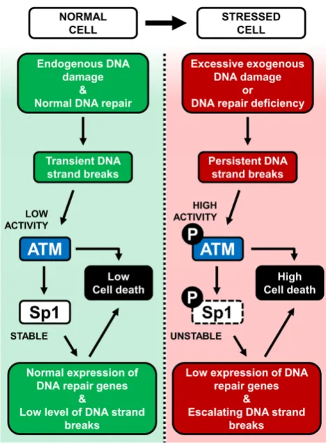
downregulation of XRCC1transcription, decreased levels of DNA ligase III and reduction in BER capacity. This mechanism leads to accumulation of further DNA strand breaks, resulting in a self-accelerating cycle (Figure8, right). This kind of scenario may represent a response to exces-sive acute DNA damage, or to BER failure. In both cases this will result in cell elimination either through apoptosis or by components of the innate immune system. We believe this represents a protective pro-survival mechanism to en-sure the removal of cells with persistent unrepaired DNA strand breaks.
Sp1 Ser101 phosphorylation in response to DNA dam-age has been previously described in a number of studies (13–15) although the specific trigger for Ser101 phosphory-lation was not known. We show that this occurs directly via ATM (Figure1) in response to genome-threatening lesions including both SSBs and DSBs (7,8). These findings led us to hypothesise that DNA strand breaks in general are likely the major trigger for Ser101 phosphorylation. However, the
1844 Nucleic Acids Research, 2018, Vol. 46, No. 4
Figure 8. Graphical summary of the model supported by this study. Left: in a normal cell, endogenous DNA damage is kept under control by DNA repair mechanisms. Low ATM activity ensures stable Sp1 protein levels, promoting efficient expression of DNA repair genes and low levels of cell death. Right: genomically stressed cells can arise under conditions of exces-sive exogenous DNA damage, or DNA repair deficiency. This leads to per-sistent DNA strand breaks and ATM activation, which triggers Sp1 phos-phorylation and degradation. In this situation the cellular load of DNA strand breaks is aggravated by decreased expression of DNA repair genes, leading to further ATM phosphorylation, activation of a pro-apoptotic cascade and death through apoptosis or cell elimination.
and duration of DNA damage are likely important consid-erations for determining the kinetics of the Sp1 response. Given that Sp1 destabilization in TIG-1 normal human fibroblasts did not occur immediately after genotoxic in-sult, but only when DNA damage persisted (Figure 3A– D), this suggests dependency on the cellular load of unre-paired DNA strand breaks. Decreased Sp1 protein levels af-ter H2O2treatment has previously been observed in HeLa
cells (37). However, this occurred using a shorter but higher dose of H2O2,thus supporting the hypothesis that the
cel-lular DNA damage load is crucial for determining Sp1 sta-bility. Moreover, the differential response time between fi-broblasts and HeLa cells suggests that cell type is also an important determinant of Sp1 stability after DNA damage. Therefore, further study to fully elucidate the mechanism behind Sp1 degradation is warranted.
We demonstrated that loss of Sp1 is associated with in-creased sensitivity to cell elimination (Figure 7). Immune surveillance in whole organisms by components of the in-nate immune system including NK cells, constitutes an
important defense against tumorigenesis by eliminating stressed, damaged or tumorigenic cells. Recognition of such cells is proposed to occur through upregulation of cytotoxic NK cell receptor NKG2D (NK group 2, member D) lig-ands which include the MIC and ULPBP protein families (29). Interestingly, expression of some activating NKG2D ligands is proposed to depend on the ATM/ATR signal-ing axis (29,30,38). It is therefore tempting to speculate that NK cells recognize and eliminate fibroblasts harboring persistent unrepaired DNA strand breaks through ATM-dependent upregulation of NKG2D ligands.
The consequences of DNA damage are inexorably linked to the severity of the lesion. It remains unknown, how-ever, how cells transition between survival and pro-death responses as a consequence of unrepairable DNA le-sions. Several lines of evidence indicate that the ATM ki-nase performs a key role in this process (6); multiple stud-ies highlight the involvement of ATM in apoptosis in re-sponse to DNA damage (10,11,39) and emerging evidence has linked ATM with promoting immune surveillance (29). Our study supports a role for ATM in the pro-survival/ pro-death decision-making process through ATM-dependent destabilization of Sp1. Of note, ATM has been suggested to undergo selective pressure for inactivation in cancer (40), while loss of ATM function correlates with the occurrence of malignancies (41–43); this could explain how genetically unstable cells might escape this control mechanism, leading to cancer emergence.
While we demonstrate that this protective mechanism ex-ists in normal untransformed fibroblasts, it is of interest to understand whether it remains active in cancer cells, par-ticularly where ATM function is not impaired. It raises the question whether it is possible to target this mechanism us-ing chemotherapeutic drugs to induce elimination of can-cerous cells. Moreover, given the sensitivity to apoptosis observed in Sp1-depleted cells, it is tempting to speculate whether low-Sp1 expressing tumors might show sensitivity to pro-apoptotic agents. Clearly, the potential exploitation of this system requires further study.
Moreover, the existence of a similar mechanism involv-ing downregulation of DNA repair to enable selective cell killing has recently been proposed (44). In their study, Ponath and Kaina suggested that monocytes expressing lower levels of BER components could be eliminated by their differentiated counterpart, macrophages, when these cells generate a reactive oxygen species burst during the in-flammatory response (44).
In summary, we believe that the molecular mechanism we propose could explain how physiological populations of healthy cells are maintained in an organism, by detecting and eliminating cells with DNA repair defects.
SUPPLEMENTARY DATA
Supplementary Data are available at NAR Online.
FUNDING
to G.L.D.]. Funding for open access charge: Medical Re-search Council grant H3RWGJ00.H302.1 to G.L.D.
Conflict of interest statement.None declared.
REFERENCES
1. Lindahl,T. (1993) Instability and decay of the primary structure of DNA.Nature,362, 709–715.
2. Dianov,G.L. and Hubscher,U. (2013) Mammalian base excision repair: the forgotten archangel.Nucleic Acids Res.,41, 3483–3490. 3. Horton,J.K., Watson,M., Stefanick,D.F., Shaughnessy,D.T.,
Taylor,J.A. and Wilson,S.H. (2008) XRCC1 and DNA polymerase beta in cellular protection against cytotoxic DNA single-strand breaks.Cell Res.,18, 48–63.
4. Ochs,K., Sobol,R.W., Wilson,S.H. and Kaina,B. (1999) Cells deficient in DNA polymerase beta are hypersensitive to alkylating
agent-induced apoptosis and chromosomal breakage.Cancer Res.,
59, 1544–1551.
5. Unnikrishnan,A., Raffoul,J.J., Patel,H.V., Prychitko,T.M., Anyangwe,N., Meira,L.B., Friedberg,E.C., Cabelof,D.C. and Heydari,A.R. (2009) Oxidative stress alters base excision repair
pathway and increases apoptotic response in apurinic/apyrimidinic
endonuclease 1/redox factor-1 haploinsufficient mice.Free Radic. Biol. Med.,46, 1488–1499.
6. Roos,W.P., Thomas,A.D. and Kaina,B. (2016) DNA damage and the
balance between survival and death in cancer biology.Nat. Rev.
Cancer,16, 20–33.
7. Khoronenkova,S.V. and Dianov,G.L. (2015) ATM prevents DSB formation by coordinating SSB repair and cell cycle progression. Proc. Natl. Acad. Sci. U.S.A.,112, 3997–4002.
8. Bakkenist,C.J. and Kastan,M.B. (2003) DNA damage activates ATM through intermolecular autophosphorylation and dimer dissociation. Nature,421, 499–506.
9. Shiloh,Y. (2014) ATM: expanding roles as a chief guardian of genome stability.Exp. Cell Res.,329, 154–161.
10. Karlseder,J., Broccoli,D., Dai,Y., Hardy,S. and de Lange,T. (1999) p53- and ATM-dependent apoptosis induced by telomeres lacking
TRF2.Science,283, 1321–1325.
11. Paulson,Q.X., Pusapati,R.V., Hong,S., Weaks,R.L., Conti,C.J. and Johnson,D.G. (2008) Transgenic expression of E2F3a causes DNA
damage leading to ATM-dependent apoptosis.Oncogene,27,
4954–4961.
12. Shiloh,Y. and Ziv,Y. (2013) The ATM protein kinase: regulating the
cellular response to genotoxic stress, and more.Nat. Rev. Mol. Cell
Biol.,14, 197–210.
13. Iwahori,S., Yasui,Y., Kudoh,A., Sato,Y., Nakayama,S., Murata,T., Isomura,H. and Tsurumi,T. (2008) Identification of phosphorylation sites on transcription factor Sp1 in response to DNA damage and its accumulation at damaged sites.Cell. Signal.,20, 1795–1803. 14. Olofsson,B.A., Kelly,C.M., Kim,J., Hornsby,S.M. and
Azizkhan-Clifford,J. (2007) Phosphorylation of Sp1 in response to
DNA damage by ataxia telangiectasia-mutated kinase.Mol. Cancer
Res.,5, 1319–1330.
15. Hau,P.M., Deng,W., Jia,L., Yang,J., Tsurumi,T., Chiang,A.K., Huen,M.S. and Tsao,S.W. (2015) Role of ATM in the formation of the replication compartment during lytic replication of Epstein-Barr virus in nasopharyngeal epithelial cells.J. Virol.,89, 652–668. 16. Grande,L., Bretones,G., Rosa-Garrido,M., Garrido-Martin,E.M.,
Hernandez,T., Fraile,S., Botella,L., de Alava,E., Vidal,A., Garcia del
Muro,X.et al.(2012) Transcription factors Sp1 and p73 control the
expression of the proapoptotic protein NOXA in the response of testicular embryonal carcinoma cells to cisplatin.J. Biol. Chem.,287, 26495–26505.
17. Li,H., Zhang,Y., Strose,A., Tedesco,D., Gurova,K. and Selivanova,G. (2014) Integrated high-throughput analysis identifies Sp1 as a crucial determinant of p53-mediated apoptosis.Cell Death Differ.,21, 1493–1502.
18. Hirose,T. and Horvitz,H.R. (2013) An Sp1 transcription factor coordinates caspase-dependent and -independent apoptotic
pathways.Nature,500, 354–358.
19. Cerboni,C., Mousavi-Jazi,M., Wakiguchi,H., Carbone,E., Karre,K. and Soderstrom,K. (2001) Synergistic effect of IFN-gamma and human cytomegalovirus protein UL40 in the HLA-E-dependent
protection from NK cell-mediated cytotoxicity.Eur. J. Immunol.,31, 2926–2935.
20. Poletto,M., Legrand,A.J., Fletcher,S.C. and Dianov,G.L. (2016) p53 coordinates base excision repair to prevent genomic instability. Nucleic Acids Res.,44, 3165–3175.
21. Markkanen,E., Fischer,R., Ledentcova,M., Kessler,B.M. and Dianov,G.L. (2015) Cells deficient in base-excision repair reveal cancer hallmarks originating from adjustments to genetic instability. Nucleic Acids Res.,43, 3667–3679.
22. McNeill,D.R., Narayana,A., Wong,H.K. and Wilson,D.M. 3rd. (2004) Inhibition of Ape1 nuclease activity by lead, iron, and
cadmium.Environ. Health Perspect.,112, 799–804.
23. Westphal,C.H., Rowan,S., Schmaltz,C., Elson,A., Fisher,D.E. and Leder,P. (1997) atm and p53 cooperate in apoptosis and suppression of tumorigenesis, but not in resistance to acute radiation toxicity. Nat. Genet.,16, 397–401.
24. Duchaud,E., Ridet,A., Stoppa-Lyonnet,D., Janin,N., Moustacchi,E. and Rosselli,F. (1996) Deregulated apoptosis in ataxia telangiectasia: association with clinical stigmata and radiosensitivity.Cancer Res.,
56, 1400–1404.
25. Roos,W.P. and Kaina,B. (2013) DNA damage-induced cell death: from specific DNA lesions to the DNA damage response and apoptosis.Cancer Lett.,332, 237–248.
26. Moodley,Y.P., Caterina,P., Scaffidi,A.K., Misso,N.L.,
Papadimitriou,J.M., McAnulty,R.J., Laurent,G.J., Thompson,P.J. and Knight,D.A. (2004) Comparison of the morphological and biochemical changes in normal human lung fibroblasts and fibroblasts derived from lungs of patients with idiopathic pulmonary fibrosis during FasL-induced apoptosis.J. Pathol.,202, 486–495. 27. Cao,X., Deng,X. and May,W.S. (2003) Cleavage of Bax to p18 Bax
accelerates stress-induced apoptosis, and a cathepsin-like protease
may rapidly degrade p18 Bax.Blood,102, 2605–2614.
28. Wood,D.E. and Newcomb,E.W. (2000) Cleavage of Bax enhances its cell death function.Exp. Cell Res.,256, 375–382.
29. Cerboni,C., Fionda,C., Soriani,A., Zingoni,A., Doria,M., Cippitelli,M. and Santoni,A. (2014) The DNA damage response: a common pathway in the regulation of NKG2D and DNAM-1 ligand expression in normal, infected, and cancer cells.Front. Immunol.,4, 508.
30. Raulet,D.H. and Guerra,N. (2009) Oncogenic stress sensed by the immune system: role of natural killer cell receptors.Nat. Rev. Immunol.,9, 568–580.
31. Bauer,M., Goldstein,M., Christmann,M., Becker,H., Heylmann,D. and Kaina,B. (2011) Human monocytes are severely impaired in base and DNA double-strand break repair that renders them vulnerable to oxidative stress.Proc. Natl. Acad. Sci. U.S.A.,108, 21105–21110. 32. Narciso,L., Fortini,P., Pajalunga,D., Franchitto,A., Liu,P., Degan,P.,
Frechet,M., Demple,B., Crescenzi,M. and Dogliotti,E. (2007) Terminally differentiated muscle cells are defective in base excision
DNA repair and hypersensitive to oxygen injury.Proc. Natl. Acad.
Sci. U.S.A.,104, 17010–17015.
33. Weissman,L., Jo,D.G., Sorensen,M.M., de Souza-Pinto,N.C., Markesbery,W.R., Mattson,M.P. and Bohr,V.A. (2007) Defective DNA base excision repair in brain from individuals with Alzheimer’s
disease and amnestic mild cognitive impairment.Nucleic Acids Res.,
35, 5545–5555.
34. Higo,T., Naito,A.T., Sumida,T., Shibamoto,M., Okada,K., Nomura,S., Nakagawa,A., Yamaguchi,T., Sakai,T., Hashimoto,A. et al.(2017) DNA single-strand break-induced DNA damage response causes heart failure.Nat. Commun.,8, 15104.
35. Wei,S., Chuang,H.C., Tsai,W.C., Yang,H.C., Ho,S.R., Paterson,A.J., Kulp,S.K. and Chen,C.S. (2009) Thiazolidinediones mimic glucose starvation in facilitating Sp1 degradation through the up-regulation
of beta-transducin repeat-containing protein.Mol. Pharmacol.,76,
47–57.
36. Wang,Y.T., Yang,W.B., Chang,W.C. and Hung,J.J. (2011) Interplay of posttranslational modifications in Sp1 mediates Sp1 stability during cell cycle progression.J. Mol. Biol.,414, 1–14.
37. Chuang,J.Y., Chang,W.C. and Hung,J.J. (2011) Hydrogen peroxide induces Sp1 methylation and thereby suppresses cyclin B1 via
recruitment of Suv39H1 and HDAC1 in cancer cells.Free Radic. Biol.
1846 Nucleic Acids Research, 2018, Vol. 46, No. 4
38. Gasser,S., Orsulic,S., Brown,E.J. and Raulet,D.H. (2005) The DNA damage pathway regulates innate immune system ligands of the
NKG2D receptor.Nature,436, 1186–1190.
39. Lee,Y., Chong,M.J. and McKinnon,P.J. (2001) Ataxia telangiectasia mutated-dependent apoptosis after genotoxic stress in the developing nervous system is determined by cellular differentiation status.J. Neurosci.,21, 6687–6693.
40. Negrini,S., Gorgoulis,V.G. and Halazonetis,T.D. (2010) Genomic instability––an evolving hallmark of cancer.Nat. Rev. Mol. Cell Biol.,
11, 220–228.
41. Paull,T.T. (2015) Mechanisms of ATM Activation.Annu. Rev.
Biochem.,84, 711–738.
42. Russell,R., Perkhofer,L., Liebau,S., Lin,Q., Lechel,A., Feld,F.M.,
Hessmann,E., Gaedcke,J., Guthle,M., Zenke,M.et al.(2015) Loss of
ATM accelerates pancreatic cancer formation and
epithelial-mesenchymal transition.Nat. Commun.,6, 7677.
43. Feng,X., Li,H., Dean,M., Wilson,H.E., Kornaga,E., Enwere,E.K.,
Tang,P., Paterson,A., Lees-Miller,S.P., Magliocco,A.M.et al.(2015)
Low ATM protein expression in malignant tumor as well as cancer-associated stroma are independent prognostic factors in a retrospective study of early-stage hormone-negative breast cancer. Breast Cancer Res.,17, 65.