INTRODUCTION
Alloplastic grafts are commonly used as bone substitutes in orthopedic and oral surgeries to treat osseous defects [1,2], due to their capability to promote bone regeneration within an acceptable time frame [3]. In particular, calcium phosphate based ceramics have similar characteristics as the crystal structure and chemistry of bone mineral phase [2,3]. Among synthetic calcium phosphate ceramics are hydroxyapatites (HAs), tricalcium phosphates (TCP), and calcium phosphate cements [4]. These bone grafts need to be osteoconductive to provide a scaffold structure, as well as, to have bioactive prop-erties, which means being capable of adhering to the bone [5]. After graft placement, approximately 70% is resorbed and replaced by newly formed bone [6]. Different methods have been used to measure the resorption rates [7,8]. Although
histological analysis is necessary to determine the actual nature of the tissue of interest, in most cases, such analysis is not performed due to economical and practical reasons. In this sense, the cone beam computed tomography (CBCT) is a less-invasive method, and highly used by surgeons and den-tists. It provides the three-dimensional (3D) analysis of bone and other tissues [9,10], as well as the means to determine their radiopacity.
The radiopacity is a representation of tissue mineral den-sity [11].The radiopacity of an alloplastic bone graft vary over time due to the fraction of TCP and calcium phosphate that is resorbed and replaced by osteoid tissue [4]. However, because of the low resolution and limitations of conventional radio-graphs, the change in radiopacity during this time is not visible until 50% of the biomaterial is replaced by bone [4]. Current literature lacks studies that use CBCT to quantify the varia-tions in radiopacity that occur in a particular time period after alloplastic bone graft implantation.
The present study aimed to evaluate the radiopacity of three different synthetic bone grafts in rabbit calvaria over 3-month period, using CBCT.
*Corresponding author: Ramón Fuentes, Department of Integral Dentistry, Research Centre in Dental Sciences (CICO), Dental School, Universidad de La Frontera. Av. Francisco Salazar 01145, 4781176, Temuco, Chile.
Phone: 56-45-325775. E-mail: [email protected] Submitted: 12 June 2016/Accepted: 30 July 2016
Radiopacity of alloplastic bone grafts measured with
cone beam computed tomography: An analysis in
rabbit calvaria
Cristina Bucchi, Eduardo Borie, Alain Arias, Fernando José Dias, Ramón Fuentes*
Department of Integral Dentistry, Research Centre in Dental Sciences (CICO), Dental School, Universidad de La Frontera, Temuco, Chile
ABSTRACT
Availability of adequate bone structure for dental implants is still a problem in dentistry. Alloplastic grafts, which promote bone regeneration, are used as bone substitutes in orthopedic and oral surgical procedures. The aim of this study was to evaluate the radiopacity of three different synthetic bone grafts in rabbit calvaria, over 3 months, using cone beam computed tomography (CBCT). Four critical-size defects were made on the calvaria of 11 rabbits. The lesions were classified into three groups according to the alloplastic grafts they received: Osteon 70/30,
Osteon collagen, and Osteon II groups. The fourth group received blood clot, and served as a control. The bone samples were collected and
analyzed with CBCT after the 1st, 2nd, and 3rd month. One month after surgery, the lesions that received Osteon 70/30 and Osteon collagen
grafts showed the highest radiopacity compared to the lesions with Osteon II and blood clot. After the 2nd month, the radiopacity values
between the three groups that received the grafts were more similar compared to the group with blood clot. After the 3rd month, the lesions
with Osteon 70/30 graft showed the highest radiopacity values, followed by Osteon collagen and Osteon II groups. The group that received
blood clot showed the lowest radiopacity values. In conclusion, the grafts used in this study had higher radiopacity values compared to blood clot. Among the grafts used, the Osteon 70/30 graft showed the highest radiopacity values in the 3-month period.
KEY WORDS:Alloplastic bone grafts; cone beam computed tomography; critical bone defects
MATERIALS AND METHODS
Surgical procedure
The following study was approved by the Scientific Ethics Committee from the Universidad de La Frontera (Protocol 015/13) and performed according to the Declaration of Helsinki.
Eleven adult New Zealand rabbits (Oryctolagus cunicu-lus - weights of 3.9 ± 0.2 kg) were selected from the Experimental Surgery Center of the Universidad de La Frontera.
The animals were premedicated according to their weight with ketamine (35 mg/kg) and xylazine (2 mg/kg). In addi-tion, the head was shaved, and a local anesthetic (0.4 ml lido-caine 2%) was applied to this region. A median lineal incision was made from the frontal to occipital region, elevating the periosteum, and exposing both parietal bones. Subsequently, four bicortical defects were created with an 8 mm external diameter trephine bur.
The samples were divided into four groups depending on the material used to fill in the defects:
1. Osteon 70/30 group - 70% HA + 30% β-TCP (Genoss,
Suwon, Korea);
2. Osteon II group - Biphasic calcium phosphate coated
with β-TCP (Genoss, Suwon, Korea);
3. Osteon collagen group - HA + β-TCP + collagen
(Genoss, Suwon, Korea); 4. Blood clot group - control.
The periosteum and skin were separately sutured, with a 5-0 absorbable Vicryl suture (Ethicon, Novartis Animal
Health US, Inc., Greensboro, NC, USA). Following the sur-gery, the animals were medicated for the first 3 days with amoxicillin (50 mg/kg) and metamizole (0.2 ml/5 kg, twice a day; Drag Pharma, Chile) and maintained with ad libitum feeding in closed cages with controlled temperature.
After healing period, the animals were euthanized with an overdose of pentobarbital (150 mg/kg) after the 1st (n = 3), 2nd
(n = 4), and 3rd months (n = 4), and the defects were extracted
immediately with safety margins of 2 mm and perfused with 10% paraformaldehyde.
CBCT evaluation
After the rabbits were euthanized, the cranium was ana-lyzed using CBCT (Pax Zenith, Vatech, Korea) with an expo-sition time of 23 s, 100 kvp, and 5 mA. The window size used in all samples was 12 cm × 9 cm, and voxel size was 0.12 mm. Data were imported in DICOM format and analyzed with specific software (EZ3D2009, E-Woo Technology Co., Korea).
Image analysis
Each defect was analyzed separately by locating the course at the center of defect, and the views were readjusted to
perform a multiplanar reconstruction. Subsequently, tracings were performed parallel to the bone with an interval of 0.5 and 1 mm thickness. From all the images obtained, the main cen-tral images were selected for the analysis, and the other images were discarded.
APFill Ink Coverage Calculator software (V.5.2, Insight, AZ, USA) was used to measure the radiopacity of each defect, using a scale from 0 (black) to 250 (white). The radiopacity of each defect corresponded to the mean of radiopacity values of the three cuts or central planes.
Statistical analysis
Statistical analysis was performed using SigmaPlot v.12.0 software (Systat Software, Inc., CA, USA) and a value of p < 0.05 was considered statistically significant. The Shapiro–Wilk test was used to test the normality of data distribution. After deter-mining the normal distribution of the sample, one-way analysis of variance (ANOVA) was used with the Holm-Sidak post-test, which allows the simultaneous analysis of multiple groups. All data are presented as mean ± standard deviation.
RESULTS
Clinically, no allergic reactions or other local complica-tions were observed in the three groups.
Figure 1A and B show the radiopacity values and tendency, respectively, of the blood clot and alloplastic bone grafts over the three time periods. The radiopacity of all defects filled in with the alloplastic bone grafts was the highest after the 3rd month. The Osteon 70/30 grafts demonstrated the
high-est radiopacity, while the blood clot showed the lowhigh-est radi-opacity during the three time periods (Figure 2). The statistical analysis for each time period is summarized in Figure 3A-C.
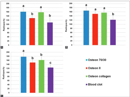
After the 1st month of healing, similar radiopacity values
were detected in Osteon 70/30 (162.17 ± 20.37) and Osteon
collagen groups (160 ± 14.03) (p = 0.745). In addition, similar
radiopacity values were detected in Osteon II (136.17 ± 8.93)
and blood clot groups (124.17 ± 5.56). However, the first two groups showed significant differences in the radiopacity val-ues compared to the second two groups (p = 0.003).
After the 2nd month of healing, similar radiopacity values
were detected in Osteon 70/30 (165.72 ± 16.81), Osteon
col-lagen (155.72 ± 13.2), and Osteon II groups (150.09 ± 11.6). In
addition, the radiopacity values in these groups were signifi-cantly higher than the values in blood clot group (121.76 ± 18.4) (p = 0.001).
After the 3rd month of healing, Osteon 70/30 group
(177.92 ± 20.18) showed significantly higher radiopacity val-ues than the other three groups (p = 0.001). Osteon collagen
similar radiopacity values which were higher than the radi-opacity values in blood clot group (124.71 ± 13.99) (p = 0.012).
DISCUSSION
The calcium phosphate grafts are biodegradable, bioactive, and osteoconductive, with a similar composition as the min-eral phase of bone [2]. However, a highly soluble graft is not recommended because a high release of calcium may inhibit the osteoclast function [5].
Because of the increasing radiopacity values observed over the study period, the three bone grafts used in this study appear to be osteoconductive. In a reconstructive 3D study from 2015, Dadsetan et al. [12] observed that the three coatings (magnesium-substituted β-tricalcium phosphate, carbonated
HA, and biphasic calcium phosphate) loaded in combination with recombinant human bone morphogenetic protein-2 were osteoconductive in critical-size defects, after 6 weeks of implantation.
The Osteon and Osteon collagen grafts showed
sim-ilar radiopacity values at 1st month after the implantation.
Furthermore, 2 months after the implantation, Osteon II
graft showed radiopacity values similar to the values observed for the first two groups, without statistically significant differ-ences between the three grafts. This result might be explained by the fact that Osteon and Osteon collagen have the same
composition, except for the addition of bovine collagen in Osteon collagen. The lower radiopacity values of Osteon II
during the first two months could be due to a slow resorption rate when compared with Osteon and Osteon collagen.
FIGURE 1. Radiopacity values of three bone grafts and blood clot. (A) Radiopacity data grouped by each month. (B) Radiopacity values
over the 3-month period.
A B
FIGURE 2. Cone beam computed tomography image showing the radiopacity (black arrows) of the three biomaterials and blood clot.
(A-D) After the 1st month and (E-H) after 3 months of application.
A
E
B
F
C
G
D
After the 3rd month, the Osteon II and Osteon collagen
grafts showed similar radiopacity values, while Osteon
showed significantly higher radiopacity, indicating that col-lagen or the block presentation of this graft could affect the mineralization of bone graft when compared with the pure Osteon graft. The graft composition and its presentation
directly affect its solubility and the ability to differentiate the osteoclast precursor cells into mature osteoclasts [2,5]. These mature osteoclasts have the ability to release cytokines that influence osteoblasts and bone formation [2]. Furthermore, the bone graft presentation may be influenced by the particle geometry, porosity, and interconnectivity [13].
β-TCP is a resorbable ceramic that bonds to the bone directly. The β-TCP granules are rapidly dissolved and newly formed bone fill in the area of dissolved granules [14]. The graft releases calcium and phosphate ions that stimulate the bone healing [15]. However, if the graft resorption takes place before the new bone is formed, the bone healing rate may be affected [16]. When this ceramic is mixed with HA, which is a low resorption rate graft [2,17], it improves the formation and remodeling of new bone by providing adequate mechanical resistance [18].
The difference in the resorption rate between β-TCP and HA is due to the fact that HA adheres directly to the bone
through a collagen-free layer interface with no preferred ori-entation [14], and thus is more stable than β-TCP. This colla-gen-free layer has an active role absorbing extracellular pro-teins such as growth factors [19]. Thus, bone grafts containing more HA show more radiopacity during the time of heal-ing [18]. The effect of Osteon (70% of HA) graft observed in
our study seems to be related to this process.
The blood clot used as the control exhibited the lowest radiopacity over the study period. It might have acted as a critical size-defect, which is a defect that will not heal sponta-neously during the lifetime of an animal [20]. The critical size defect used in this study concurs with defects having an exter-nal diameter of 8 mm, as suggested by other authors [7,8,16,21]. Despite some controversies regarding the critical-size defect, the results of this study confirmed that the prepared area was sufficient for analysis. The same results for blood clot were observed in a two-dimensional radiographic study by Oporto et al. [8] for a follow-up period of 3 months, where they observed <10% of ossification.
CBCT allows a more accurate analysis of bone com-pared to conventional radiographs [22,23], making it a useful method to quantify bone density [9,24]. Each slice of a CBCT image is composed of pixels that represent the tissue density, thus providing better accuracy [25].
FIGURE 3. Statistical analysis of radiopacity values for each month. (A) 1st month; (B) 2nd month; and (C) 3rd month. a, b, and c – statisti-cally significant difference (p < 0.05).
A
C
A limitation of this study is lack of information on the per-centage and distribution of HA and β-TCP in the Osteon II
graft. This is a limitation because the chemical properties of a graft depend on the HA/β-TCP ratio [5]. Moreover, a histo-logical analysis is necessary to determine the possible causes of differences in radiopacity values between defects filled with different bone grafts [8]; however, this analysis was not the main objective of our study.
As assessed by CBCT, the grafts used in this study had higher radiopacity values compared to blood clot. Among the grafts applied to the bone lesions, the Osteon 70/30
graft showed the highest radiopacity values over the 3-month period.
ACKNOWLEDGMENTS
Funded by Universidad de La Frontera, Project DI15-0009.
DECLARATION OF INTERESTS
The authors declare no conflict of interests.
REFERENCES
[1] Ogose A, Hotta T, Kawashima H, Kondo N, Gu W, Kamura T, et al. Comparison of hydroxyapatite and beta tricalcium phosphate as bone substitutes after excision of bone tumors. J Biomed Mater Res B Appl Biomater 2005;72(1):94-101.
http://dx.doi.org/10.1002/jbm.b.30136.
[2] Shiwaku Y, Neff L, Nagano K, Takeyama K, de Bruijn J, Dard M, et al. The crosstalk between osteoclasts and osteoblasts is depen-dent upon the composition and structure of biphasic calcium phos-phates. PLoS One 2015;10(7):e0132903.
DOI: 10.1371/journal.pone.0132903.
[3] Denry I, Kuhn LT. Design and characterization of calcium phos-phate ceramic scaffolds for bone tissue engineering. Dent Mater 2016;32(1):43-53.
http://dx.doi.org/10.1016/j.dental.2015.09.008.
[4] Larsson S. Calcium phosphates: What is the evidence? J Orthop Trauma 2010;24 Suppl 1:S41-5. DOI: 10.1097/ BOT.0b013e3181cec472.
[5] Yamada S, Heymann D, Bouler JM, Daculsi G. Osteoclastic resorption of calcium phosphate ceramics with different hydroxyapatite/beta-tricalcium phosphate ratios. Biomaterials 1997;18(15):1037-41.
http://dx.doi.org/10.1016/S0142-9612(97)00036-7.
[6] Fellah BH, Gauthier O, Weiss P, Chappard D, Layrolle P. Osteogenicity of biphasic calcium phosphate ceramics and bone autograft in a goat model. Biomaterials 2008;29(9):1177-88. http://dx.doi.org/10.1016/j.biomaterials.2007.11.034.
[7] Borie E, Fuentes R, Del Sol M, Oporto G, Engelke W. The influence of FDBA and autogenous bone particles on regeneration of calvaria defects in the rabbit: A pilot study. Ann Anat 2011;193(5):412-7. http://dx.doi.org/10.1016/j.aanat.2011.06.003.
[8] Oporto VG, Fuentes R, Borie E, Del Sol M, Orsi IA, Engelke W. Radiographical and clinical evaluation of critical size defects in rabbit calvaria filled with allograft and autograft: A pilot study. Int J Clin Exp Med 2014;7(7):1669-75.
[9] Ahmad R, Abu-Hassan MI, Li Q, Swain MV. Three dimensional quantification of mandibular bone remodeling using standard
tessellation language registration based superimposition. Clin Oral Implants Res 2013;24(11):1273-9.
DOI: 10.1111/j.1600-0501.2012.02566.x.
[10] Fernandes TM, Adamczyk J, Poleti ML, Henriques JF, Friedland B, Garib DG. Comparison between 3D volumetric rendering and multiplanar slices on the reliability of linear measurements on CBCT images: An in vitro study. J Appl Oral Sci 2015;23(1):56-63. http://dx.doi.org/10.1590/1678-775720130445.
[11] Marquezan M, Osório A, Sant’Anna E, Souza MM, Maia L. Does bone mineral density influence the primary stability of dental implants? A systematic review. Clin Oral Implants Res 2012;23(7):767-74.
http://dx.doi.org/10.1111/j.1600-0501.2011.02228.x.
[12] Dadsetan M, Guda T, Runge MB, Mijares D, LeGeros RZ, LeGeros JP, et al. Effect of calcium phosphate coating and rhBMP-2 on bone regeneration in rabbit calvaria using poly(propylene fuma-rate) scaffolds. Acta Biomater 2015;18:9-20.
http://dx.doi.org/10.1016/j.actbio.2014.12.024.
[13] Walsh WR, Vizesi F, Michael D, Auld J, Langdown A, Oliver R, et al. Beta-TCP bone graft substitutes in a bilateral rabbit tibial defect model. Biomaterials 2008;29(3):266-71.
http://dx.doi.org/10.1016/j.biomaterials.2007.09.035.
[14] Fujita R, Yokoyama A, Nodasaka Y, Kohgo T, Kawasaki T. Ultrastructure of ceramic-bone interface using hydroxyapatite and beta-tricalcium phosphate ceramics and replacement mechanism of beta-tricalcium phosphate in bone. Tissue Cell 2003;35(6):427-40. http://dx.doi.org/10.1016/S0040-8166(03)00067-3.
[15] LeGeros RZ. Calcium phosphate-based osteoinductive materials. Chem Rev 2008;108(11):4742-53.
http://dx.doi.org/10.1021/cr800427g.
[16] Lee JH, Ryu MY, Baek HR, Lee KM, Seo JH, Lee HK. Fabrication and evaluation of porous beta-tricalcium phosphate/hydroxyapa-tite (60/40) composite as a bone graft extender using rat calvarial bone defect model. Scientific World J 2013;2013:481789.
http://dx.doi.org/10.1155/2013/481789.
[17] Farzadi A, Solati-Hashjin M, Bakhshi F, Aminian A. Synthesis and characterization of hydroxyapatite/β-tricalcium phosphate nano-composites using microwave irradiation. Ceram Int 2011;37(1):65-71. http://dx.doi.org/10.1016/j.ceramint.2010.08.021.
[18] Bansal S, Chauhan V, Sharma S, Maheshwari R, Juyal A, Raghuvanshi S. Evaluation of hydroxyapatite and beta-trical-cium phosphate mixed with bone marrow aspirate as a bone graft substitute for posterolateral spinal fusion. Indian J Orthop 2009;43(3):234-9.
http://dx.doi.org/10.4103/0019-5413.49387.
[19] Hench LL. Bioactive ceramics: Theory and clinical applications. In: Anderson ÖH, Happonen RP, Yli-Urpo A, editors. Bioceramics. Oxford: Butterworth-Heinemann; 1994. p. 3-14.
http://dx.doi.org/10.1016/B978-0-08-042144-5.50005-4.
[20] Schmitz JP, Hollinger JO. The critical size defect as an experimental model for craniomandibulofacial nonunions. Clin Orthop Relat Res 1986;205:299-308.
http://dx.doi.org/10.1097/00003086-198604000-00036. [21] Kitayama S, Wong LO, Ma L, Hao J, Kasugai S, Lang NP, et al.
Regeneration of rabbit calvarial defects using biphasic calcium phosphate and a strontium hydroxyapatite-containing collagen membrane. Clin Oral Implants Res 2015.
http://dx.doi.org/10.1111/clr.12605.
[22] Creanga AG, Geha H, Sankar V, Teixeira FB, McMahan CA, Noujeim M. Accuracy of digital periapical radiography and cone-beam computed tomography in detecting external root resorption. Imaging Sci Dent 2015;45(3):153-8.
http://dx.doi.org/10.5624/isd.2015.45.3.153.
[23] Saidi A, Naaman A, Zogheib C. Accuracy of cone-beam computed tomography and periapical radiography in endodontically treated teeth evaluation: A five-year retrospective study. J Int Oral Health 2015;7(3):15-9.
enhanced-MRI and cone beam-CT: A pilot study. Clin Orthop Relat Res 2008;466(8):1897-904.
http://dx.doi.org/10.1007/s11999-008-0293-5.
[25] Azeredo F, de Menezes LM, Enciso R, Weissheimer A,
de Oliveira RB. Computed gray levels in multislice and cone-beam computed tomography. Am J Orthod Dentofacial Orthop 2013;144(1):147-55.