Research Article
Molecular prevalence of hepatitis C virus
genotypes in district Kohat, Khyber
Pakhtunkhwa, Pakistan
Farman Ullah
1, 2, Nawab Ali
2*, Irum Sabir Ali
3, Shamim Saleha
2, Syed
Tahir Ali Shah
4, Hazir Rahman
5, Shahbaz Ahmad
2, Muhammad Jamil
2and Muhammad Saeed
11. Department of Biosciences, COMSATS Institute of Information Technology, Islamabad-Pakistan. 2. Department of Biotechnology & Genetic Engineering, Kohat University of Science & Technology, Kohat, Khyber Pakhtunkhwa-Pakistan.
3. Department of Surgical C Unit, Post Graduate Medical Institution, Lady Reading Hospital Peshawar, Khyber Pakhtunkhwa-Pakistan.
4. Department of Surgery, Divisional Head Quarter Hospital (DHQ), Kohat, Khyber Pakhtunkhwa-Pakistan. 5. Department of Microbiology, Abdul Wali Khan University Mardan, Khyber Pakhtunkhwa-Pakistan.
*Corresponding author’s email: [email protected] Citation
Farman Ullah, Nawab Ali, Irum Sabir Ali, Shamim Saleha, Syed Tahir Ali Shah, Hazir Rahman, Shahbaz Ahmad, Muhammad Jamil and Muhammad Saeed. Molecular prevalence of hepatitis C virus genotypes in district Kohat, Khyber Pakhtunkhwa, Pakistan. Pure and Applied Biology. Vol. 6, Issue 1, pp237-246.
http://dx.doi.org/10.19045/bspab.2017.60019
Received: 04/12/2016 Revised: 02/02/2017 Accepted: 09/02/2017 Online First: 14/02/2017
Abstract
Hepatitis C Virus is a causative agents of liver cirrhosis, having highly diversified genome. Genotype study has important role in clinical setup and prognosis of hepatitis C virus infection. The aim of this was to investigate the prevalence and risk factors associated with hepatitis C virus genotypes in district Kohat, Khyber Pakhtunkhwa, Pakistan. Hepatitis C virus pre-infected patients (n = 600) were analyzed to assess circulating HCV genotypes, using reverse transcriptase approach. Moreover serum ALT levels were determined followed by correlation with genotypes. In current study cohort, majority of patients (62%) were, having elevated ALT (> 50 U/L). similarly patients with mixed HCV genotypes, 3a/3b have relatively higher ALT levels (~62) compared to 2a/3a (~43) and others. We found significant correlation (p = 0.002) of genotype 2a in connection with ALT (49.48) level. Similarly 3a, 3b, mixed as well as un-typeable genotypes correlations were also found highly significant (p <0.001) with their mean ALT levels (47.14, 31.58, 46.69 and 43.68) respectively. In our studied population, most prevalent genotypes were 3a (25%) followed by 3b (20%) and 2a (15%). Fifteen percent of patient’s infections were untypeable while in 10% patients mixed genotype were observed. Among total 30 (5%) were blood transfusion cases, 90(15%) surgical, 540 (90%) dentistry, 360 (60%) Barber, 492 (82%) Pricks, whereas 180 (30%) cases were those having early type of HCV/HBV infection in their family. In current study, genotypes 3a and 3b were more prevalent, with two potential risk factors; dentistry and barbers. Moreover, genotypes and ALT investigations were found to be more fruitful in prognosis as well as management of HCV infection.
Keywords: HCV; Genotypes; ALT; Risk factors; Kohat
Introduction
Hepatitis C Virus (HCV) is a leading causative agent of liver cirrhosis and
family [1]. It has a single open reading genome of ~9.6 kb, encoding a polypeptide of 3010 amino acids, flanked by un-translated region at both 5ʹ and 3ʹ terminus [2]. This polypeptide is post-translationally modified into various structural (C, E1, E2, and p7) and non-structural (NS2, NS3, NS4A, NS4B, NS5A, and NS5B) proteins by viral and cellular proteins [3]. HCV possessed extreme genetic heterogeneity due to poor RNA-dependent RNA polymerase (NS5B-protein) activity [4]. The route of
transmission of HCV shown very
complexity but it is more intended on futile neutralizing immune responses including unscreened blood transfusions, unsterilized dental and surgical instruments, unhygienic barber conditions and tattooing [5]. Patients
carrying HCV infection shows symptoms
like acute/chronic liver disease, elevated serum ALT level that finally may lead to
hepatocellular carcinoma [6]. There are
~200 million HCV patients which account for ~3.3% of the total population of the
world [7]. Among them, ~10 million people
having HCV infection accounting ~4.7% of the total population of Pakistan [8]. The first report about HCV was reported in 1992 by screening 45 patients from the northern part
of Pakistan [9]. There are six major
genotypes of HCV and more than one hundred subtypes reported throughout the world [10]. Out of these, the genotype-1, genotype-2 and genotype-3 are most popular genotypes in terms of prevalence [10]. The prevalence rate varies from region to region and have variation geographically. The prevalence of genotype 1 is a bit high (7.04%), followed by genotype 2 (3.82%). However, the prevalence rate of genotype 3 is very high 78.96%, while it subtype 3a reported 58.01%, followed by 3b (9.76%). The other genotypes such as 4, 5, 6 are very seldom reported in Pakistan. This revealed that the genotype 3a have high prevalence rate at Khyber Pakhtunkhwa, and Punjab.
The Baluchistan province of Pakistan is reported to have high prevalence of genotype 1a and the least prevalence is for
genotype 3a [11]. There are very high
numbers of patients having HCV infection in Pakistan and this number is increasing on daily basis. Therefore, it is the need of today and tomorrow to investigate the risk factors, occurrence and molecular level of HCV genotypes and search for the possible effective treatment in order to stop the transmission and save the lives of the patients having this chronic disease [12]. District Kohat is a multicultural and highly populated regions of Khyber Pakhtunkhwa having different communities of the local FATA zones in addition to local people. There is still very few report from district Kohat and need updated information in order to plan and eradicate this infection from local population.
Materials and methods
Collection of samples from patients
Total 600 blood samples were collected from HCV positive patients through pre-designed questionnaire including patient details i.e. age, socioeconomic status, risk factors, duration of infection, racial origin, and liver function test. Written consents were signed from patients and the study was initiated with proper approval from ethical committee. Samples were transported to the lab immediately and stored for onward analysis.
Antibody screening and ALT analysis Immuno- chromatographic method was used for the screening of HCV antibodies utilizing the kit (Accurate Diagnostics™ Canada) and the sera of patients were tested by ELISA using commercially available kits (Axen Diagnostic™, Germany). ALT, as
hepatocytes marker was investigated
Genotyping of HCV patients
Approximately 200 µL of the blood was taken and HCV genomic RNA was extracted according the manufacturer’s protocol by using RNA extraction kit of UltrascriptTM (Anagen, USA). The molecular analysis was done with little modification using the Ohno et al., (1997) [13] method.
Then cDNA was made from viral mRNA by using 10µL of RNA with 1µL (200U) of
RevertAidTM M-MuLV reverse transcriptase
enzyme (Fermentas) at 42oC for 30 min
followed by 92oC for 2 min in thermocycler
(Techne, USA). The cDNA was amplified in first round of PCR using forward and reverse primers, one unit of Taq polymerase
and 3mM MgCl2 at 94oC for 3 min followed
by 92oC (45 Sec), 52oC (45 Sec) and 72oC (1
min) with final extension at 72oC (10 min) in PCR. Again, 4µL of the first PCR product was further amplified by genotype specific primers (1a, 1b, 2a, 2b, 3a, 3b, 4, 5a, and 6a) in two mixtures. Mixture-1 contained antisense primers 1b, 1.D-8 (2a), 2b, 2a and 3b while Mixture-2 composition included the antisense primers 1a, 3a, 4, 5a and 6a. The reaction mixture of both mixtures composed of one unit of Taq polymerase and 3mM MgCl2 having PCR conditions as mentioned for the first round of PCR. PCR products were separated by agarose gel electrophoresis on 2% agarose gel along with 100 bp DNA ladder and later on stained with ethidium bromide and checked under UV illuminator (Figure 1).
Figure 1. Gel electrophoresis of the amplified core regions of HCV genome showing different genotypes. Mix. A and Mix. B gel pictures represent mixtures of PCR constituted for HCV primers related to group A & B respectively for based on genotypes identifications
Base pairs Genotypes
Lane 1: 139bp (Mix A) 2a
Lane 2 232bp (Mix B) 3a
Lane 3 Nonspecific Untypeable
Lane 4: 176bp (Mix A) 232bp (Mix B) 3a & 3b (Mixed Genotypes)
Lane 5: 176bp (Mix A) 3b
Lane 6: Nonspecific Untypeable
Statistical analysis
Statistical analysis was performed using Origin Pro 2016 (Origin Lab, Northampton, USA) statistic software. The correlation among HCV genotypes and ALT was analyzed at 95 % confidence intervals (CIs)
using simple t-test, while specific
comparison were made by standard
descriptive analysis. The p-values <0.05 were considered, significant for a particular test.
Results
Clinico-pathological characteristics of patients
In total, 600 patients were studied gender-wise in equal ratio ranging the age groups between 20-80 years. The patients were also reported in terms of marriage, income class and on educational background. The married patients were reported 438 (73%), unmarried
162 (27%), lower income class (≥ 100 US$/month) 462 (77%) and middle income class (500 US$/month) 138 (23%). In addition, 375 (~62%) were illiterate, 225 (~38%) were of middle class. However, no patient among all was found having high income per month (<500 US$/month). Potential risk factors in HCV
transmission
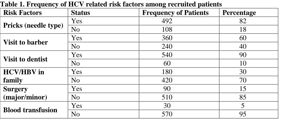
Data reported from patients revealed high prevalence rate of HCV infection among patients experienced dental procedures 540 (90%), followed by unhygienic pricks 492 (82%), barber intervenes 360 (60%), early HBV/HCV running in family 180 (30%), surgical operations 90 (15%). However, the blood transfusion was reported with the least HCV transmission rates 30 (5%) as per information collected through questionnaires from the patients (Table 1).
Table 1. Frequency of HCV related risk factors among recruited patients
Risk Factors Status Frequency of Patients Percentage
Pricks (needle type) Yes 492 82
No 108 18
Visit to barber Yes 360 60
No 240 40
Visit to dentist Yes 540 90
No 60 10
HCV/HBV in family
Yes 180 30
No 420 70
Surgery (major/minor)
Yes 90 15
No 510 85
Blood transfusion Yes 30 5
No 570 95
Frequencies of ALT levels
All the patients under study were followed for measuring their ALT levels up to six months with specific interval. Significant ALT fluctuations were reported. Moreover, 62% of patients had high ALT levels (≥ 50 U/L) and 38% patients were reported with normal ALT level (≤ 50 U/L). In this study, we have found significant increased (61.79 U/L; mean value) in ALT level. Moreover,
Figure 2. Alanine amino transferase (ALT) level (mean) in patients infected with HCV mixed genotypes
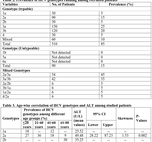
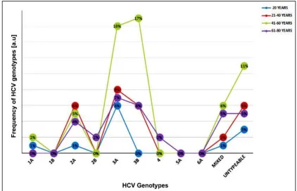
General prevalence of HCV genotype The high prevalence rate among different HCV genotypes was reported for 3a (25%), followed by 3b (20%), 2a (15%), 1a (5%) and 4 (5%). However, the genotypes such as 1b, 6a, and 5a were not reported among the patients under investigation. There was 15% patients which indicated no genotype and 10% of the patients showed mixed infection of HCV genotypes. The mixed genotypes were reported in decreasing order as followed; 2a/3a (45%), 3a/3b (35%) and 5% in cases of 2a/2b, 3b/1a, 1a/2a and 4/2a (Table 2). The genotypes prevalence in terms of age groups is reported as: genotype 1a was with high prevalence rate in age group of ≤20 years followed by 41-60 years; genotype 2a had high prevalence in age groups 21- 40 years, followed by age group of ≤20 years, then 41-60 years with lowest
Table 2. Prevalence of HCV genotypes running among recruited patients
Variables No, of Patients Prevalence (%) Genotype (typable)
1a 30 5
2a 90 15
2b 30 5
3a 150 25
3b 120 20
4 30 5
Mixed 60 10
Total 510 85
Genotype (Untypeable)
1b Not detected 0
5a Not detected 0
6a Not detected 0
Total 90 15
Mixed Genotypes
2a/3a 54 45
3a/3b 42 35
2a/2b 6 5
3b/1a 6 5
1a/2a 6 5
4/2a 6 5
Table 3. Age-wise correlation of HCV genotypes and ALT among studied patients
Genotypes
Prevalence of HCV
genotypes among different age groups (%)
ALT (U/L) (mean values)
95% CI
Skewness P-Values ≤20
years
21-40 years
41-60 years
61-80
years Lower Upper
1a 18 -- 12 -- 25.52 -- -- -- --
2a 27 36 18 9 49.48 28.22 87.23 1.53 0.002
2b -- -- -- 30 35.25 -- -- -- --
3a 30 45 45 30 51.33 42.76 57.86 0.39 <0.001
3b -- 30 60 30 47.14 38.39 51.67 0.51 <0.001
4 -- -- -- 30 31.58 -- -- -- --
Mixed 6 6 30 30 46.69 33.34 60.04 0.42 <0.001
Figure 3. Age-wise distribution of HCV genotypes among recruited HCV patients
HCV genotypes and alanine amino transferase correlation
Alanine amino transferase (ALT) is the most potent hepatocytes marker to detect the degree of liver cirrhosis as a result of ongoing HCV infection. We analyzed serum ALT level for a period of six months and
correlate with the respective HCV
genotypes. A significant correlation (p = 0.002) of genotype 2a with ALT (49.48) level was reported. Similarly 3a, 3b, mixed as well as untypeable genotypes were found highly significant (p <0.001) with their mean ALT levels (51.33, 47.14, 46.69 and 43.68), respectively. However genotypes 1a, 2b and 4 were observed with lower prevalence to meet statistics criteria (Table 3).
Discussion
Hepatitis C virus become asymptomatic at the beginning of infection, called as silent killer. HCV has no clear clinical feature and varies depending on the age, sex, genotype
and viral load of the one individual to another individual [14]. The main objectives of the study were to investigate the risk factors and prediction for early diagnosis in different age, sex and genotypes based on the serum marker evaluation. Most of the patients were reported as illiterate (~62%) and of lower income class (77%). This revealed that mostly poor and uneducated people have high exposure to HCV virus due to their unawareness of the risk factors and transmission routes of HCV infection. In similar fashion, different risk factors studied has shown high prevalence 90%, 82%, 60%, 30%, 15% and 5% in case of dental procedure, unhygienic pricks, barbers intervenes, family history, surgical cases and
blood transfusion, respectively. The
infection. This study is supported by previous literature where blood transfusion, un-sterilized dental & surgical instrument,
multiple sex, contaminated syringes;
needles, unsafe intravenous drugs users, and community barber shops were reported the
major sources of HCV transmission [15].
Similarly blood transfusion as well as sedentary life style has been found to be associated positively with HCV infection
among Pakistani population [16]. Other
aspect of this study was to investigate the role of ALT in the early diagnosis of HCV patients. ALT has been reported as a hepatocyte marker to evaluate the treatment efficiency as well as physiological condition in liver cirrhosis[17].
The ALT levels of patients under study within repeated interval for six months duration revealed significant fluctuation in the sera of each pateint. Moreover, the ALT values were also correlated with HCV genotypes of the patients with different age groups and the severity of infection was evaluted statistically. The results showed significant correlation, and concluded that ALT might be directly related to age of patient as well as with the degree of liver cirrhosis. These results are in parallel with the previously reported work [16].
Moreover, the prevalence of different HCV genotypes were also investigated. The genotypes 3a, 3b, and 2a were found more prevalent in the patients of the Kohat district of Khyber Pakhtunkhwa. These genotypes are also reported in the previous literature from the patients of other areas of Pakistan as the most common prevailing HCV genotypes [18]. In 10% of the patients under study, mixed genotypes were identified and among mixed genotypes the 2a/3a were in higher percentage (45%) followed by 3a/3b genotypes. These combined infections are also reported in previous literature and the
reason could be the inter-individual
mutations and transfusions of mixed viral
strains [19]. These mixed genotypes
exposure might be very dangerous in future in comparison to single genotypes and may results certain highly pathogenic variants and causing severe HCV infection for which treatment may not be effective.
The HCV infection was also reported to be more prevalent among the lower class and uneducated people in comparison to middle/upper class and educated people. This could be due to frequent exposure and unawareness of poor and uneducated people to the unhygienic local barbar, syringes, blood transfusions and contaminated dental instruments [20-22]. This study suggested the importance of using sterilized syringes,
surgical and dental instruments, and
transfusion of HCV and HIV screened bloods. Therefore, long term attention of the competent authorities/health department is required in order to control the transmission of the hepatitis C virus.
Conclusion
The utmost conclusion of this work is that the most prevalent genotypes of HCV in the area of Kohat were 2a, 3a and 3b, which might be fruitful in order to prepare the community-based vaccines for controlling HCV infections in certain areas.
Authors’ contributions
Conceived and designed and performed the experiments: FUllah, SAhmad & M Jamil, Analyzed the data: S Saleha, N Ali, M Jamil & M Saeed, Contributed the reagents: IS Ali, STA Shah & H Rahman, Wrote and corrected the manuscript F Ullah & N Ali. References
1. Shetty S, Kim S, Shimakami T, Lemon SM & Mihailescu MR (2010). Hepatitis C virus genomic RNA dimerization is
mediated via a kissing complex
intermediate. RNA. 16(5): 913–925. 2.Takamizawa A1, Mori C, Fuke I, Manabe
C virus genome isolated from human carriers. J Virol 65(3): 1105-1113.
3. Kato N (2001). Molecular virology of
hepatitis C virus. Acta Med Okayama
55(3): 133-59.
4. Ashfaq UA, Javed T, Rehman S, Nawaz Z & Riazuddin S (2011). An overview of HCV molecular biology, replication and immune responses. Virol J (8): 161.
5. Raja NS & Janjua KA (2008).
Epidemiology of hepatitis C virus
infection in Pakistan. J Microbiol
Immunol Infect 41(1): 4-8.
6. Ahmad W, Ijaz B, Javed FT, Kausar H, Sarwar MT & Gull S et al. (2011). HCV genotype-specific correlation with serum
markers: higher predictability for
genotype 4a. Virol J (8): 293.
7. Aziz H, Raza A, Murtaza S, Waheed Y, Khalid A & Irfan J et al. (2013). Molecular epidemiology of hepatitis C virus genotypes in different geographical regions of Punjab Province in Pakistan and a phylogenetic analysis. Int J Infect Dis 17(4): 247-53.
8. Umar M, Hamama tul Bushra, Ahmad M, Khurram M, Usman S & Arif M et al. (2010). Hepatitis C in Pakistan: A
Review of Available Data. Hepat Mon
10(3): 205–214.
9. Malik IA, Ahmad N, Luqman M, Legters LJ, Khalil U & Zaheeruddin et al. (1992). Hepatitis C as a cause of chronic liver disease in northern Pakistan. J Pak Med Assoc 42(3): 67-8.
10. Cavalheiro Nde P, Filgueiras TC, Melo CE, Morimitsu SR, de Araujo ES & Tengan FM et al. (2007). Detection of HCV by PCR in serum and PBMC of patients with hepatitis C after treatment. Braz J Infect Dis 11(5): 471-4.
11. Attaullah S, Khan S & Ali I (2011). Hepatitis C virus genotypes in Pakistan: a systemic review. Virol J 8: 433.
12. Idrees M & Riazuddin S (2008).
Frequency distribution of hepatitis C
virus genotypes in different
geographical regions of Pakistan and their possible routes of transmission. BMC Infect Dis 8: 69.
13. Ali A, Nisar M, Ahmad H, Saif N,
Idrees M & Bajwa MA (2011). Determination of HCV genotypes and viral loads in chronic HCV infected patients of Hazara Pakistan. Virol J 8: 466.
14. Ohno O, Mizokami M, Wu RR, Saleh
MG, Ohba K & Orito E et al. (1997).
New hepatitis C virus (HCV)
genotyping system that allows for identification of HCV genotypes 1a, 1b, 2a, 2b, 3a, 3b, 4, 5a, and 6a. J Clin Microbiol 35(1): 201-7.
15. Cannon NA, Donlin MJ, Fan X, Aurora
R, Tavis JE & Virahep CSG (2008).
Hepatitis C virus diversity and
evolution in the full open-reading frame during antiviral therapy. PLoS One 3(5): 2123.
16. Channa NA & Khan H (2010) Risk
factors for hepatitis C disease in Tando Allahyar, Pakistan: A case-control study. Bangladesh J of Med Sci 10(3): 163-169.
17. Qureshi H, Arif A, Riaz K, Alam SE, Ahmed W & Mujeeb SA (2009). Determination of risk factors for hepatitis B and C in male patients suffering from chronic hepatitis. BMC Res Notes 2: 212.
18. Couzigou P, Richard L, Dumas F,
Schouler L & Fleury H (1993). Detection of HCV-RNA in saliva of patients with chronic hepatitis C. Gut 34(2): 59-60.
19. Dor-Mohammadi T, Daryani NE,
Bashashati M, Hashtrudi AA,
C-infected patients. Indian J Gastroenterol 24(2): 49-51.
20. Waqar M, Khan AU, Rehman HU,
Idrees M, Wasim M & Ali A et al. (2014). Determination of hepatitis C virus genotypes circulating in different districts of Punjab (Pakistan). Eur J Gastroenterol Hepatol 26(1): 59-64.
21. Roque-Afonso AM, Ducoulombier D,
Di Liberto G, Kara R, Gigou M &
Dussaix E et al. (2005).
Compartmentalization of hepatitis C
virus genotypes between plasma and peripheral blood mononuclear cells. J Virol 79(10): 6349-57.
22. Hu YW, Balaskas E, Furione M, Yen
PH, Kessler G & Scalia V et al. (2000) Comparison and application of a novel
genotyping method, semiautomated
primer-specific and mispair extension analysis, and four other genotyping assays for detection of hepatitis C virus
mixed-genotype infections. J Clin