Diabetic retinopathy screening update
Seema Garg, MD, PhD, and Richard M. Davis, MDD
iabetic retinopathy is the leading cause of blindness among adults aged 20–74 years in the United States, which notably includes the working-age population. With early detection, diabetic retinopathy can be treated with modalities that have been proven to decrease the risk of severe vision loss by > 90%.Given the proven benefits of early detection, guidelines for screening for diabetic retinopathy have been established by national profes-sional organizations such as the American Diabetes Association (ADA) and the American Academy of Ophthalmology (AAO). Unfor-tunately, on average, < 50% of diabetic patients in the United States meet these recommendations.1–3 In
fact, 60% of patients who require vision-preserving laser surgery do not receive treatment.4 The barriers
to recommended eye examinations are numerous and include insuf-ficient referrals, socioeconomic factors, and poor geographical access to care.
Recent advances in digital imag-ing have opened new avenues for assessing retinopathy, which may provide better access to diagnosis and management for this treatable, but often blinding, condition. This article will provide an overview of the epidemiology of diabetic retin-opathy, its pathophysiology and classification, and the multicenter prospective clinical trials that have provided rigorous evidence for
its screening and treatment. The technological advances relevant to screening will be discussed, and finally, the important role of primary care providers in retinal screening for patients with diabetes will be examined.
Epidemiology
Approximately 8% of the U.S. popu-lation has diabetes, which equates to > 23.6 million children and adults.5
The prevalence of diabetes varies among ethnicities: 5.2% for European Americans, 9% for Native Americans, 11% for African Americans, and 10.4% for Mexican Americans.6 The
World Health Organization estimates that > 180 million people worldwide have diabetes. This number is likely to more than double by the year 2030. At least 10% will likely develop visual impairment secondary to diabetic retinopathy.7
The prevalence of diabetic retinopathy is high; 20 years after diagnosis, > 90% of patients with type 1 diabetes and > 60% of those
with type 2 diabetes will have some degree of retinopathy.8,9 The major
risk factors for developing dia-betic retinopathy are duration of diabetes8,9 and severity of
hypergly-cemia.10–14 Other important factors
include hypertension15,16 and elevated
serum lipid levels.17,18
Pathophysiology and Classification
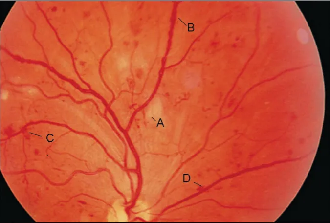
The natural history of diabetic retin-opathy typically follows an orderly and predictable pattern. Long-term hyperglycemia causes vascular endothelial dysfunction resulting in loss of endothelial cells and pericytes. The retina then develops micro-aneurysms, intraretinal hemorrhages, and focal areas of retinal ischemia (cotton-wool spots). At this point, the retinopathy is classified as nonprolif-erative diabetic retinopathy (NPDR). As the retinopathy progresses, the vessels become further damaged, resulting in retinal nonperfusion and more widespread ischemia. Clinically, the retina can have signs of vascular damage including venous beading, intraretinal microvascular abnormalities, and more severe hem-orrhages (Figure 1). At this point, the retinopathy is classified as severe NPDR. Even at this stage, most patients are asymptomatic.
With further ischemic injury, compensatory chemical mediators, most notably vascular endothelial growth factor, induce the growth of fragile new blood vessels at the inner surface of the retina.19 This stage,
called proliferative diabetic
retin-I n B r retin-I e f
opathy (PDR), is characterized by neovascularization of the optic disc and neovascularization elsewhere. When these fragile vessels bleed, the vitreous hemorrhage causes symp-toms of “floaters” or, if severe, loss of vision. Over time, the new vessels fibrose and can contract, resulting in tractional retinal detachments, which can cause significant vision loss.
Macular edema, the leading cause of vision loss among patients with diabetes, can occur at any stage of diabetic retinopathy. Damaged retinal vessels result in increased vascular permeability, causing an accumulation of intraretinal fluid and/or lipid, which is clinically apparent with direct ophthalmos-copy. The retina appears thickened and may contain yellow hard exu-dates (lipid). Macular edema may cause symptoms of blurry vision, or it may cause no symptoms at all.
Landmark Studies
Several multicenter randomized controlled clinical trials have dem-onstrated that diabetic retinopathy can be prevented or that its natural
course can be altered. The landmark Diabetes Control and Complications Trial (DCCT) involved 1,441 subjects with type 1 diabetes, ages 13–39 years, at 29 medical centers in the United States and Canada. Study participants had either no disease or early diabetic retinopathy and were randomized to either intensive blood glucose control (mean A1C 7.2%) or conventional blood glucose control (mean A1C 9.1%). The study demon-strated that intensive blood glucose control reduced the risk of progres-sion of diabetic retinopathy by 54%, reduced the development of severe NPDR or PDR by 47%, reduced the need for laser surgery by 56%, and reduced the risk of diabetic macular edema by 23%.10,11
The U.K. Prospective Diabetes Study (UKPDS) confirmed the protective effect of intensive blood glucose control in patients with type 2 diabetes and also evaluated the effect of hypertension. A total of 1,148 patients with type 2 diabetes and hypertension were enrolled and treated with either an
angio-tensin-converting enzyme inhibitor (captopril) or a β-blocker (atenolol). This study demonstrated that patients with tight blood pressure control (< 150/85 mmHg) compared to patients with blood pressure less tightly controlled (< 180/95 mmHg) were found to have a 37% risk reduc-tion in microvascular changes, 34% risk reduction in the need for laser treatment, and 47% risk reduction in decreased vision.12,13,15
Several randomized trials have also demonstrated the value of surgical treatments to minimize the complications of diabetic retinopa-thy. In the Diabetes Retinopathy Study (DRS) of more than 1,700 patients at 15 medical centers, pan-retinal photocoagulation (PRP; laser treatment to the peripheral retina) reduced the risk of severe (defined as 5/200 or worse) vision loss from PDR from 15.9% in untreated eyes to 6.4% in treated eyes.20 Once a patient
reached the PDR stage (fragile new blood vessels), it was observed that argon laser treatment of the retina resulted in regression of the neovascularization.
The Early Treatment of Diabetic Retinopathy Study (ETDRS) enrolled 3,711 patients and provided valuable information regarding management of diabetic retinopa-thy. First, it demonstrated that PRP can reduce the risk of severe vision loss to < 2% if administered at the appropriate stage (severe NPDR or PDR). Second, focal laser treatment (treatment to the macular area with an argon laser) was found to reduce moderate visual loss (doubling of the visual angle) by 50%. Finally, it was found that aspirin did not alter rates of progression of diabetic retinopa-thy and did not increase the risk of vitreous hemorrhage.21–23
The Diabetic Retinopathy Vitrectomy Study (DRVS) showed that there was a benefit to early vitrectomy (surgical removal of
reous) in very severe PDR in patients with type 1 diabetes. Two years after surgery, 36% of the early vitrectomy group and 12% of the late vitrectomy group had visual acuity of 20/40 or better.24,25
Current Screening Guidelines
These landmark studies have demonstrated that the blinding com-plications from diabetes can be largely prevented medically, by glycemic and blood pressure control, as well as by early detection and timely treatment of diabetic retinopathy with pho-tocoagulation/surgical techniques. Therefore, screening guidelines have been developed by national profes-sional organizations such as the ADA26 and AAO.27
Adults and children > 10 years of age with type 1 diabetes should have an initial dilated examination by an ophthalmologist within 5 years of the onset of diabetes. Because people may already have type 2 diabetes before they are aware of symptoms and up to 20% of patients with type 2 diabetes have retinopathy at the time of diagnosis, they should have an initial dilated examination by an ophthalmologist at the time of their diabetes diagnosis.9 Subsequent
examinations should be yearly or more frequently if retinopathy is progressing. Pregnant women with preexisting diabetes should have a dilated eye examination early in the first trimester of pregnancy because pregnancy can potentiate rapid progression of retinopathy.
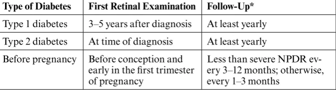
Close follow-up should continue throughout pregnancy and 1 year postpartum. Current recommended screening guidelines are summarized in Table 1.
The retinopathy screening para-digm is based on clinical trials that have demonstrated the benefits of screening. However, current care falls far below these recommenda-tions. Insufficient screening may be partially related to lack of access to eye care specialists. The advent of retinal imaging and digital tech-nology may provide an avenue for greater compliance with screening recommendations.
In 2004, the American Telemedicine Association estab-lished consensus recommendations that provided guidelines for clini-cal, techniclini-cal, and operational performance standards for diabetic retinopathy screening. The develop-ment of retinal imaging and, more recently, digital retinal photography may help address the barriers to access for retinopathy screening. Telehealth (telecommunication to promote health) or telemedicine (telecommunication for diagnos-tic and therapeudiagnos-tic intervention) programs based on retinal imaging with or without remote interpreta-tion may facilitate early diagnosis of diabetic retinopathy and timely treatment, hence preserving vision.28
Methods of screening for diabetic retinopathy include direct and indi-rect ophthalmoscopy, stereoscopic
color film fundus photography, mydriatic or nonmydriatic digital color (Figure 2), and monochromatic photography. Traditionally, ophthal-mologists have screened for diabetic retinopathy by dilating the pupil and performing indirect ophthalmo-scopy, in which the entire retina is examined. This method of screening is successful where access to eye care is sufficient. However, the increasing rate of patients with diabetes will soon outpace the supply of eye care providers, both in the United States and worldwide. At present, some communities have poor or even no access to ophthalmologic care. In these settings, remote interpretation of film-based or digital photographs of the retina may be employed.
The gold standard for the detec-tion of diabetic retinopathy consists of 30-degree stereoscopic photog-raphy of seven standard fields on color film, as developed for the ETDRS—Classification of Diabetic Retinopathy.29 This has a sensitivity
and specificity for the detection of diabetic retinopathy that is superior to direct30 and indirect31
ophthal-moscopy by ophthalmologists. The efficacy of trained readers has been demonstrated in a systematic review, in which the interpretation of mydriatic (dilated) retinal pho-tography provided the most sensitive screening and monitoring test for sight-threatening retinopathy, with sensitivities > 80%.32 However, this
technique is labor intensive, has a long turnaround time, and requires expensive equipment and trained retinal photographers and read-ers. From a patient’s perspective, it can be time-consuming, and the required pupillary dilation may be uncomfortable. Thus, seven-field ste-reoscopic fundus photography is an ideal gold standard but is not ideal for widespread implementation.
The development of digital retinal photography has facilitated rapid
Table 1. Recommended Eye Examination Schedule for Patients with Diabetes27
Type of Diabetes first Retinal Examination follow-Up*
Type 1 diabetes 3–5 years after diagnosis At least yearly Type 2 diabetes At time of diagnosis At least yearly Before pregnancy Before conception and
early in the first trimester of pregnancy
Less than severe NPDR ev-ery 3–12 months; otherwise, every 1–3 months
acquisition and interpretation of fundus images, quantitative analy-sis of data for documentation and progression of retinopathy, and the rapid deployment of retinal imag-ing worldwide. Currently, there are no universally accepted criteria for the detection of diabetic retinopa-thy using digital imaging. However, several systems are being studied and validated. Retinal imaging can be performed using digital retinal photographs with (mydriatic) or without (nonmydriatic) dilating the pupil. The digital photographs may be interpreted by trained readers or forwarded to a reading center for interpretation and grading (“store and forward”).
Several studies have examined the sensitivity and specificity of digital imaging. Two-field mydriatic33 and
two-field nonmydriatic34 digital
photography performed favorably compared to ophthalmoscopy and seven-field stereophotography.
Because of their ease of use and associated patient comfort, non-mydriatic cameras have facilitated retinal imaging for patients with
diabetes in primary care settings, including family practice, inter-nal medicine, and endocrinology offices. The cameras do not require operation by a trained retinal pho-tographer and their use has been validated.35,36 Furthermore, an AAO
meta-analysis determined that there was sufficient evidence from ran-domized clinical trials (Level 1) that single-field digital fundus photog-raphy can serve as a screening tool for diabetic retinopathy to identify patients with retinopathy for refer-ral for ophthalmologic evaluation and management.37 Another study
evaluated single-field 45-degree nonmydriatic monochromatic images and found them to be highly correlated (κ = 0.97, P = 0.0001) to the gold standard of the stereoscopic seven-field mydriatic images.38
As an alternative to a single field, some cameras can photograph three 45-degree fields. A recent report evaluated single-field versus three-field nonmydriatic images compared to the seven-field gold standard and concluded that three color 45-degree nonmydriatic images had a
sensitiv-ity and specificsensitiv-ity of 82 and 92%, respectively, and may also be an effective tool in a screening setting to determine levels of diabetic retin-opathy for specialist referral.39
Recognizing the importance of diabetic retinal imaging, several countries have implemented national screening programs such as the National Plan for Screening in the United Kingdom and the OPHDIAT program in France. The OPHDIAT telemedicine system comprises 11 screening centers equipped with nonmydriatic cameras. Fundus photographs are acquired by techni-cians, with remote interpretation by ophthalmologists who grade the images. In 28 months, 15,307 diabetic retinopathy screening examinations were performed, and diabetic retinopathy was detected in 3,350 patients (23.4%).40 It was found
that the rates of diabetic retinopathy screening improved from 50% before to more than 70% after the imple-mentation of OPHDIAT.41
Acquisition of digitized reti-nal images allow for novel image analysis methods and Web-based connectivity to create models of remote, computer-assisted, or even automated diagnosis and man-agement of diabetic retinopathy. Several systems are in development and are currently being clinically validated.42,43
Although retinal imaging pro-grams are important in improving access to care and identifying patients who need further evalu-ation, they do not replace comprehensive eye exams by oph-thalmologists. A full evaluation is required when a screening retinal photograph is unreadable and for follow-up of abnormalities detected by the screening system. In addi-tion, non–diabetes-related ocular conditions such as cataract, hyper-tensive retinopathy, and glaucoma
are optimally evaluated during a comprehensive eye exam.
Role of Primary Care Providers
The importance of systemic factors such as glycemic and blood pressure control in preventing and slowing the progression of diabetic retinopathy was conclusively demonstrated in the DCCT and UKPDS clinical trials. Primary care physicians play a significant role in optimizing glyce-mic control and managing other risk factors such as hypertension and hyperlipidemia, which can potentially affect eye health.
Appropriate referral by primary care providers at recommended intervals for diabetic retinopathy eye examinations is crucial, because timely treatment with panretinal and focal laser photocoagulation surgery has been proven in the ETDRS, DRS, and DRVS trials to decrease vision loss from diabetes. Primary care physicians can educate their patients with diabetes about the importance of retinal examina-tions, as diabetic retinopathy is often asymptomatic. Encouragement by primary care providers may increase the likelihood that patients will keep their ophthalmology appointments.
In addition, primary care pro-viders can communicate with ophthalmologists to convey the rea-son for referrals and supply patient information such as A1C results and presence of any other comorbid con-ditions. (A sample communication form is provided by Sinclair et al.44)
After patients get eye examinations, primary care providers can expect to receive assessments from the eye care providers and then can reinforce to patients any recommendations for ophthalmological follow-up care.
Finally, as telehealth and tele-medicine programs are implemented, the role of primary care providers may become even more encompass-ing, as screening retinal photographs
may be obtained directly in the pri-mary care office, and, perhaps in the future, primary care providers may even be trained to evaluate retinal photographs.45
REfEREnCES
1Brechner RJ, Cowie CC, Howie
LJ, Herman WH, Will JC, Harris MI: Ophthalmic examination among adults with diagnosed diabetes mellitus. JAMA 270:1714–1718, 1993
2Lee PP, Feldman ZW, Ostermann J,
Brown DS, Sloan FA: Longitudinal rates of annual eye examinations of persons with diabetes and chronic eye diseases.
Ophthalmology 110:1952–1959, 2003
3Paz SH, Varma R, Klein R, Wu J, Azen
SP; Los Angeles Latino Eye Study Group: Noncompliance with vision care guidelines in Latinos with type 2 diabetes mellitus: the Los Angeles Latino Eye Study. Ophthalmology 113:1372–1377, 2006
4Ferris FL 3rd, Davis MD, Aiello LM:
Treatment of diabetic retinopathy. N Engl J
Med 341:667–678, 1999
5American Diabetes Association: All
about diabetes [article online]. Available from http://www.diabetes.org/about-diabetes.jsp. Accessed 22 May 2009
6Cowie CC, Rust KF, Byrd-Holt DD,
Eberhardt MS, Flegal KM, Engelgau MM, Saydah SH, Williams DE, Geiss LS, Gregg EW: Prevalence of diabetes and impaired fasting glucose in adults in the U.S. popu-lation: National Health and Nutrition Examination Survey 1999–2002. Diabetes
Care 29:1263–1268, 2006
7World Health Organization: What is
dia-betes? [article online] Available from http:// www.who.int/mediacentre/factsheets/fs312/ en/index.html. Accessed 22 May 2009
8Klein R, Klein BE, Moss SE, Davis MD,
DeMets DL: The Wisconsin Epidemiologic Study of Diabetic Retinopathy. IX. Four-year incidence and progression of diabetic retin-opathy when age at diagnosis is less than 30 years. Arch Ophthalmol 107:237– 243, 1989
9Klein R, Klein BE, Moss SE, Davis MD,
DeMets DL: The Wisconsin Epidemiologic Study of Diabetic Retinopathy. III. Prevalence and risk of diabetic retinopathy when age at diagnosis is 30 or more years.
Arch Ophthalmol 102:527–532, 1984
10DCCT Research Group: Progression of
retinopathy with intensive versus conven-tional treatment in the Diabetes Control and Complications Trial. Ophthalmology 102:647–661, 1995
11DCCT Research Group: The
relation-ship of glycemic exposure (HbA1c) to the risk of development and progression of retinopathy in the Diabetes Control and Complications Trial. Diabetes 44:968–983, 1995
12UK Prospective Diabetes Study
Group: Intensive blood-glucose control with sulphonylureas or insulin compared with
conventional treatment and risk of com-plications in patients with type 2 diabetes (UKPDS 33). Lancet 352:837–853, 1998
13Kohner EM, Stratton IM, Aldington
SJ, Holman RR, Matthews DR; UK Prospective Diabetes Study (UKPDS) Group: Relationship between the severity of retinopathy and progression to photoco-agulation in patients with type 2 diabetes mellitus in the UKPDS (UKPDS 52). Diabet
Med 18:178–184, 2001
14Wong TY, Liew G, Tapp RJ, Schmidt
MI, Wang JJ, Mitchell P, Klein R, Klein BE, Zimmet P, Shaw J: Relation between fasting glucose and retinopathy for diagnosis of dia-betes: three population-based cross-sectional studies. Lancet 371:700–702, 2008
15UK Propspective Diabetes Study
Group: Tight blood pressure control and risk of macrovascular and microvascular compli-cations in type 2 diabetes: UKPDS 38. BMJ 317:703–713, 1998
16Snow V, Weiss KB, Mottur-Pilson C;
Clinical Efficacy Assessment Subcommittee of the American College of Physicians: The evidence base for tight blood pressure control in the management of type 2 diabetes mel-litus. Ann Intern Med 138:587–592, 2003
17van Leiden HA, Dekker JM, Moll AC,
Nijpels G, Heine RJ, Bouter LM, Stehouwer CD, Polak BC: Blood pressure, lipids, and obesity are associated with retinopathy: the Hoorn study. Diabetes Care 25:1320–1325, 2002
18Klein R, Sharrett AR, Klein BE, Moss
SE, Folsom AR, Wong TY, Brancati FL, Hubbard LD, Couper D; ARIC Group: The association of atherosclerosis, vascular risk factors, and retinopathy in adults with diabe-tes: the Atherosclerosis Risk in Communities study. Ophthalmology 109:1225–1234, 2002
19Boulton M, Foreman D, Williams G,
McLeod D: VEGF localisation in diabetic retinopathy. Br J Ophthalmol 82:561–568, 1998
20Diabetic Retinopathy Study Research
Group: Photocoagulation treatment of proliferative diabetic retinopathy: clinical application of Diabetic Retinopathy Study (DRS) findings, DRS Report Number 8.
Ophthalmology 88:583–600, 1981
21ETDRS Research Group: Early
photo-coagulation for diabetic retinopathy (ETDRS report number 9). Ophthalmology 98 (5 Suppl.):766–785, 1991
22ETDRS Study Research Group:
Photocoagulation for diabetic macular edema (ETDRS report number 1). Arch
Ophthalmol 103:1796–1806, 1985
23ETDRS Study Group: Effects of aspirin
treatment on diabetic retinopathy (ETDRS report number 8). Ophthalmology. 98 (5 Suppl.):757–765, 1991
24DRVS Research Group: Early
vit-rectomy for severe proliferative diabetic retinopathy in eyes with useful vision: results of a randomized trial (DRVS report number 3). Ophthalmology 95:1307–1320, 1988
25DRVS Research Group: Early
diabetic retinopathy: four year results of a randomized trial (DRVS report number 5).
Arch Ophthalmol 108:958–964, 1990
26American Diabetes Association:
Standards of medical care in diabetes—2009.
Diabetes Care 32 (Suppl. 1):S13–S61, 2009
27American Academy of Ophthalmology
Retina Panel: Preferred practice pat-tern guidelines: diabetic retinopathy. San Francisco, American Academy of Ophthalmology, 2008. Available online from http://one.aao.org/CE/PracticeGuidelines/ PPP_Content.aspx?cid=d0c853d3-219f-487b-a524-326ab3cecd9a
28Davis RM, Fowler S, Bellis K, Pockl
J, Al Pakalnis V, Woldorf A: Telemedicine improves eye examination rates in individuals with diabetes: a model for eye-care delivery in underserved communities. Diabetes Care 26:2476, 2003
29ETDRS Research Group: Grading
diabetic retinopathy from stereoscopic color fundus photographs: an extension of the modified Airlie House classification (ETDRS report number 10). Ophthalmology 98 (5 Suppl.):786–806, 1991
30Harding SP, Broadbent DM, Neoh C,
White MC, Vora J: Sensitivity and specificity of photography and direct ophthalmoscopy in screening for sight threatening eye disease: the Liverpool Diabetic Eye Study. BMJ 311:1131–1135, 1995
31Kalm H, Egertsen R, Blohmé G:
Non-stereo fundus photography as a screening procedure for diabetic retinopathy among patients with type II diabetes: compared with 60D enhanced slit-lamp examination. Acta
Ophthalmol (Copenh) 67:546–553, 1989
32Hutchinson A, McIntosh A, Peters J,
O’Keeffe C, Khunti K, Baker R, Booth A: Effectiveness of screening and monitoring tests for diabetic retinopathy: a systematic review. Diabet Med 17:495–506, 2000
33Scanlon PH, Malhotra R, Greenwood
RH, Aldington SJ, Foy C, Flatman M, Downes S: Comparison of two reference standards in validating two field mydriatic
digital photography as a method of screen-ing for diabetic retinopathy. Br J Ophthalmol 87:1258–1263, 2003
34Boucher MC, Gresset JA, Angioi
K, Olivier S: Effectiveness and safety of screening for diabetic retinopathy with two nonmydriatic digital images compared with the seven standard stereoscopic photographic fields. Can J Ophthalmol 38:557–568, 2003
35Bursell SE, Cavallerano JD,
Cavallerano AA, Clermont AC, Birkmire-Peters D, Aiello LP, Aiello LM; Joslin Vision Network Research Team: Stereo nonmy-driatic digital-video color retinal imaging compared with Early Treatment Diabetic Retinopathy Study seven standard field 35-mm stereo color photos for determining level of diabetic retinopathy. Ophthalmology 108:572–585, 2001
36Cavallerano AA, Cavallerano JD,
Katalinic P, Tolson AM, Aiello LP, Aiello LM; Joslin Vision Network Clinical Team: Use of Joslin Vision Network digital-video nonmydriatic retinal imaging to assess diabetic retinopathy in a clinical program.
Retina 23:215–223, 2003
37Williams GA, Scott IU, Haller JA,
Maguire AM, Marcus D, McDonald HR: Single-field fundus photography for diabetic retinopathy screening: a report by the American Academy of Ophthalmology.
Ophthalmology 111:1055–1062, 2004
38Lin DY, Blumenkranz MS, Brothers
RJ, Grosvenor DM: The sensitivity and specificity of single-field nonmydriatic monochromatic digital fundus photography with remote image interpretation for diabetic retinopathy screening: a comparison with ophthalmoscopy and standardized mydri-atic color photography. Am J Ophthalmol 134:204–213, 2002
39Vujosevic S, Benetti E, Massignan F,
Pilotto E, Varano M, Cavarzeran F, Avogaro A, Midena E: Screening for diabetic retinopa-thy: 1 and 3 nonmydriatic 45-degree digital fundus photographs vs 7 standard Early
Treatment Diabetic Retinopathy Study fields.
Am J Ophthalmol 148:111–118, 2009
40Massin P, Chabouis A, Erginay A,
Viens-Bitker C, Lecleire-Collet A, Meas T, Guillausseau PJ, Choupot G, André B, Denormandie P: OPHDIAT: a telemedical network screening system for diabetic retin-opathy in the Ile-de-France. Diabetes Metab 34:227–234, 2008
41Chabouis A, Berdugo M, Meas T,
Erginay A, Laloi-Michelin M, Jouis V, Guillausseau PJ, M’bemba J, Chaine G, Slama G, Cohen R, Reach G, Marre M, Chanson P, Vicaut E, Massin P: Benefits of OPHDIAT, a telemedical network to screen for diabetic retinopathy: a retrospective study in five reference hospital centres. Diabetes
Metab 35:228–232, 2009
42Rudnisky CJ, Tennant MT, Weis E, Ting
A, Hinz BJ, Greve MD: Web-based grading of compressed stereoscopic digital photogra-phy versus standard slide film photographotogra-phy for the diagnosis of diabetic retinopathy.
Ophthalmology 114:1748–1745, 2007
43Chaum E, Karnowski TP, Govindasamy
VP, Abdelrahman M, Tobin KW: Automated diagnosis of retinopathy by content-based image retrieval. Retina 28:1463–1477, 2008
44Sinclair SH, Delvecchio C: The
inter-nist’s role in managing diabetic retinopathy: screening for early detection. Cleve Clin J
Med 71:151–159, 2004
45Farley TF, Mandava N, Prall FR,
Carsky C: Accuracy of primary care clini-cians in screening for diabetic retinopathy using single-image retinal photography. Ann
Fam Med 6:428–434, 2008