Open Access
Review
Action potential evoked transmitter release in central synapses:
insights from the developing calyx of Held
Lu-Yang Wang*, Michael J Fedchyshyn and Yi-Mei Yang
Address: Program for Neurosciences and Mental Health, The Hospital for Sick Children and Department of Physiology, University of Toronto, 555 University Avenue, Toronto, Ontario, M5G, 1X8, Canada
Email: Lu-Yang Wang* - [email protected]; Michael J Fedchyshyn - [email protected]; Yi-Mei Yang - [email protected]
* Corresponding author
Abstract
Chemical synapses are the fundamental units that mediate communication between neurons in the mammalian brain. In contrast to the enormous progress made in mapping out postsynaptic contributions of receptors, scaffolding structures and receptor trafficking to synaptic transmission and plasticity, the small size of nerve terminals has largely precluded direct analyses of presynaptic modulation of excitability and transmitter release in central synapses. Recent studies performed in accessible synapses such as the calyx of Held, a giant axosomatic synapse in the sound localization circuit of the auditory brainstem, have provided tremendous insights into how central synapses regulate the dynamic gain range of synaptic transmission. This review will highlight experimental evidence that resolves several long-standing issues with respect to intricate interplays between the waveform of action potentials, Ca2+ currents and transmitter release and further conceptualize
their relationships in a physiological context with theoretical models of the spatial organization of voltage-gated Ca2+ channels and synaptic vesicles at release sites.
Background
Information transfer between nerve cells is largely medi-ated by fast chemical neurotransmission across their junc-tions or synapses. Electric pulses or action potentials (APs or spikes) are initiated near the soma of presynaptic neu-rons and propagated along the axon to the nerve terminal where voltage-gated Ca2+ channels (VGCCs) open and
transiently raise intracellular Ca2+ concentration. Binding
of Ca2+ to the release sensor (i.e. synaptotagmin) triggers
membrane fusion between synaptic vesicles (SVs) and presynaptic active zones (AZs) via engagement of SNAREs. Upon opening of the fusion pore, neurotrans-mitters are unloaded into the synaptic cleft and activate postsynaptic receptors to generate excitatory or inhibitory postsynaptic potentials (EPSPs or IPSPs), ultimately
affecting postsynaptic spike initiation and firing patterns that encode information.
Although it is well accepted that neurotransmission across chemical synapses is a highly conserved process in all spe-cies, our understanding of how the waveform of APs mod-ulates Ca2+ influx and quantal output is primarily based
on classical work in invertebrate synapses from squid, crayfish and Aplysia [1-6]. This is mainly because typical mammalian central nerve terminals are too small to be readily accessible to electrophysiological analyses, limit-ing our ability to probe the intricate interplay between spike waveform, Ca2+ currents, and transmitter release.
Recently, optical recording techniques with fast Ca2+- and
voltage-sensitive dyes, and new preparations that are
con-Published: 25 November 2009
Molecular Brain 2009, 2:36 doi:10.1186/1756-6606-2-36
Received: 20 August 2009 Accepted: 25 November 2009
This article is available from: http://www.molecularbrain.com/content/2/1/36 © 2009 Wang et al; licensee BioMed Central Ltd.
ducive to direct electrophysiological recordings from nerve terminals have been developed, to study these vari-ables and their crosstalk in several central synapses includ-ing the cerebellar granule cell-Purkinje cell synapses [7], the mossy fiber-CA3 synapses in hippocampus [8] and the calyx of Held synapses in the auditory brainstem [9-11]. These studies have provided the first quantitative descrip-tion of the biophysical behavior of VGCCs in response to an AP, and the downstream coupling of Ca2+ influx to
vesicular release in mammalian central synapses. Since APs and synaptic transmission have been elegantly reviewed [12,13], this article will instead focus on a number of controversial issues with respect to the effects of AP on Ca2+ channels and transmitter release.
First, in contrast to studies performed in invertebrate syn-apses where only a small fraction of VGCCs (~10%) are activated by an AP [2,3], work in mammalian central syn-apses demonstrates that an AP can effectively open a majority of presynaptic VGCCs (>70%) [8,14,15,15-17]. However, the mechanisms underlying this difference are not well understood. Second, the waveform of an AP is known to be altered by neuromodulators or to undergo activity- and development-dependent changes [6,18-20], but opposing views remain with respect to how AP width affects presynaptic VGCCs. In squid giant synapse, AP broadening appears to increase the number of activated VGCCs, but it alters mainly the gating kinetics of VGCCs in central terminals [21,22]. Third, in non-mammalian synapses, release of single vesicles can be triggered by the opening of as few as one channel [3,23-25] and AP broad-ening leads to linear increase in quantal output [3]. In contrast, at mammalian nerve terminals, the relationship between Ca2+ influx and quantal output in response to AP
broadening usually follows a highly nonlinear power function [8,14,15], implying that the cooperative action of multiple channels lead to the fusion of single synaptic vesicles [21,22,26,27]. These studies raise the possibility that different synapses may exhibit distinct coupling modalities between VGCCs and SVs, underpinning the heterogeneity of release probability (Pr). Finally, the peak Ca2+ concentration experienced by Ca2+ sensors on SVs in
the vicinity of AZs was estimated to be more than ~100 μM in non-mammalian synapses but as low as ~10 μM in mammalian synapses during an AP [28-33]. Such a dis-crepancy in spatiotemporal profile of Ca2+ transients is
not fully understood.
The giant calyx of Held synapse-a powerful
experimental model
Recent studies using the calyx of Held synapse as an exper-imental platform have made tremendous progress towards addressing the aforementioned issues. This syn-apse at maturity faithfully relays inputs from the contral-ateral cochlear nucleus (CN) to the ipsilcontral-ateral medial and
lateral superior olives, where neurons encode the relative interaural timing/intensity differences of sound stimuli received at each cochlea as cues for sound localization [34]. From synapse formation at postnatal day 1-3 (P1-3) to the onset of hearing at P10-12 and final maturation around P16-18, this axosomatic synapse achieves the capacity of high-fidelity transmission at extraordinarily rates (up to 600 Hz) [20,34]. Its clearly defined "critical period of development" and rapid functional maturation make this synapse an ideal model for studying synaptic transmission and developmental plasticity. Meanwhile, as the waveform of presynaptic APs shortens dramatically within this developmental window, questions related to interplay between APs, Ca2+ currents (I
Ca) and transmitter
release become relevant to physiological functionality of this synapse. The large dimension and compact structure of this synapse obviously present technical advantages for direct biophysical analysis of synaptic properties [9-11,26,35]. For example, since each postsynaptic neuron receives a single calyx input at the soma, where adequate voltage-clamp of pre- and postsynaptic elements can be simultaneously attained, the shape and size of the recorded currents can be reliably used for deconvolution analysis of the amount of transmitter release and the kinetics and density of postsynaptic receptors with mini-mal concern for voltage-clamp errors. The accessibility of both pre- and postsynaptic elements to patch electrodes offers the option of using either cell-attached non-inva-sive recording mode with excellent signal-to-noise ratio (seals at ~GΩ level) [36] or invasive whole-cell recordings. The measurements of current, voltage, and capacitance in designated intracellular ionic and buffer conditions facili-tate direct studies of pre- and postsynaptic mechanisms [37]. In particular, simultaneous Ca2+ imaging and
pho-tolysis of caged Ca2+ makes quantitative cross-correlation
analyses between Ca2+ and quantal output possible,
inde-pendent of Ca2+ entry from the extracellular space [31,32].
Whole-cell capacitance recordings from the calyx provide means to directly study exocytosis and endocytosis inde-pendent of postsynaptic receptors while cell-attached capacitance recordings from the release face of the calyx make it possible to examine kinetics and conductance of fusion pore and VGCCs at release sites [38-40]. More importantly, presynaptic APs at the developing calyx of Held synapse undergo dramatic shortening in half-width, the quantal output (Q) is paradoxically maintained (in rats) or even markedly increases (in mice) [18,19]. Know-ing that the input-output relationship at any given syn-apse can be described by a power function Q [Ca2+]n,
where n denotes Ca2+ cooperativity (3~5) [41], one would
predict that spike narrowing should yield a much smaller Ca2+ influx and therefore a nonlinear decline in Q.
compen-sate for the reduction in Ca2+ and boost quantal output, in
order to establish the robust high-fidelity character observed at this synapse [19]. Hence, investigation of mechanisms underlying such a compensation may not only unravel important answers for the physiological par-adox at the developing calyx of Held synapse, but also insightful inferences for resolving long-standing discrep-ancies on neurotransmission in non-mammalian and mammalian synapses. The following sections will recapit-ulate the main findings of a series of studies on sequential steps of synaptic transmission, and present new theoreti-cal framework and conceptual models to account for the experimental data.
Relationships between the waveform of action
potentials and Ca
2+influx
Taschenberger and von Gersdorff (2000) first reported that presynaptic APs at the developing calyx of Held syn-apse become progressively narrower in half-width but remain unchanged in amplitude during the first two post-natal weeks [19]. Spike narrowing, in this case, arises from speeding of both depolarization and repolarization phases due to up-regulated Na+ and K+ conductances
[19,42-45]. To better characterize the impact of spike nar-rowing on transmitter release, Yang and Wang (2006) took a reductionist approach and employed a series of pseudo-APs to systemically study the distinctive effects of AP depolarization, plateau and repolarization phases on ICa [46]. Slowing depolarization or plateau phase selec-tively increases ICa by recruiting an increasing number of VGCCs without changing their gating kinetics. Similar changes can also been induced by prolonging the repo-larization phase. However, once the ICa amplitude satu-rates, further broadening serves to slow ICa gating kinetics (both activation and deactivation), which significantly expands the total Ca2+ integral beyond the maximal I
Ca
integral generated by changing AP depolarization or pla-teau phase. Using paired voltage-clamp recordings of ICa and excitatory postsynaptic currents (EPSCs or IEPSC)
evoked by pseudo- and real-APs, Yang & Wang (2006) fur-ther demonstrated that immature and mature APs (with the half width: 0.4 and 0.27 ms) activate 50% and 35% of the total VGCCs in the terminal respectively, and that the ICa-IESPC relationships are well described by the integral,
but not the amplitude, of ICa in the classical Hill function. These observations may help reconcile the opposing views on the effect of spike broadening on presynaptic ICa
among different synapses.
At the squid giant synapse, classical work has shown that a single AP opens only ~10% of presynaptic VGCCs [2,47]. In these synapses, AP broadening enhances cal-cium entry, primarily by increasing the number of acti-vated VGCCs, which manifests as increases in the
amplitude of ICa [3]. In contrast, both theoretical simula-tions and experimental data from central synapses, including the calyx of Held synapse, suggest that an AP effectively opens more than 70% of available VGCCs and that AP broadening increases calcium influx mainly by extending the duration of calcium currents with minimal effect on the amplitude of ICa [8,14-17]. However, these
results were obtained largely from immature mammalian central synapses, in which propagating APs usually have wide waveforms (0.5~1 ms) [8,14-17] that typically acti-vate near maximal number of available VGCCs. As a result, AP broadening in these synapses will only reveal changes in the gating kinetics of ICa but not its amplitude. When narrower APs with a half-width of 0.4 ms and 0.27 ms respectively were used as voltage-command templates [46], the fraction of activated VGCCs (50% vs. 35%) was clearly much lower than previously estimated (>70%).
Based on the above, the initial AP width, defined by the depolarization and repolarization time course, likely dic-tates whether AP broadening induces changes in the amplitude or the kinetics of ICa. It should be noted that experiments in invertebrate synapses are usually con-ducted at lower temperatures (e.g., <18°C for squid giant synapse) than those in central synapses, which may signif-icantly reduce the fraction of VGCCs recruited by an AP (i.e. 10%) and slow the gating kinetics of these VGCCs. In these aforementioned studies, effects of AP amplitude have not been extensively examined, although Wu et al. suggested that small changes in AP amplitude can pro-foundly impact quantal output [48]. In the developing calyx of Held synapse where AP amplitude remains stable [46], it appears that speeding of AP depolarization and repolarization rates mainly shortens the AP half-width, reduces the number of VGCCs activated during an AP and accelerates activation and deactivation kinetics of ICa, ulti-mately ensuring the brevity of Ca2+ transients for
synchro-nized release of transmitters.
Coupling of Ca
2+influx to vesicular release
Paired recordings from the calyx of Held synapse in both mice and rats reveals a significant leftward shift of the input-output relationship (ICa-IEPSC), indicating a devel-opmental upregulation in the release efficiency [46,49]. It has been well documented that the ICa density, or total number of VGCCs on the presynaptic terminal, remains relatively unchanged throughout the development of the calyx of Held synapse [36,50]. This raises the question: how smaller ICa, evoked by a narrow AP at mature syn-apses, can yield higher quantal output than that evoked by a wide AP at immature synapses? Intuitively, downstream coupling of Ca2+ entry to vesicular release must enhanceThere are two possible mechanisms underlying such an enhancement: (1) the spatial coupling between VGCCs and SVs in AZs tightens so that the Ca2+ sensors on SVs are
exposed to higher local Ca2+ concentrations near the
mouth of VGCCs opened during an AP, and/or (2) the Ca2+ sensors (e.g. synaptotagmin) acquire higher Ca2+
sen-sitivity during development and can readily detect smaller Ca2+ transients from fewer activated VGCCs.
To test the first possibility, Fedchyshyn and Wang (2005) showed that injection of a Ca2+ buffer with slow binding
kinetics (EGTA; 10 mM), which can not intercept Ca2+
influx from VGCCs in the immediate vicinity of SVs but is capable of capturing Ca2+ in transit from distant VGCCs,
potently attenuated transmitter release in immature ter-minals (P8-12 mice) as previously demonstrated in rats [26]. Surprisingly, the same manipulation had little effect on mature synapses (P16-18), suggesting that SVs in young synapses are loosely coupled to VGCCs, but transi-tion to a more tightly coupled spatial arrangement during development [36]. Similar conclusion was reached by applying capacitance measurements of exocytosis under different buffer conditions [51]. When a voltage para-digm, which specifically recruited an increasing number of VGCCs without changing kinetics of ICa or the driving force for Ca2+, was used to generate a series of graded I
Ca
and IEPSC, Fedchyshyn and Wang (2005) estimated Ca2+
channel/domain cooperativity (m, the exponent of the ICa-IEPSC power relationship), from the slope of linearized Log(ICa)-Log(IEPSC) plots (i.e. IEPSC ∝ [ICa]m) for immature
and mature synapses [36]. Note that m derived from this protocol has a distinct meaning from the classical Ca2+
cooperativity (n), which is usually interpreted as the number of Ca2+ ions (3~5) which bind cooperatively to
the release sensor and trigger vesicular fusion (i.e. molec-ular cooperativity). The term m refers to the mean number of engaged Ca2+ channels/domains (m) that cooperatively
increase Ca2+ concentration and satisfy the Ca2+ binding
requirements of the Ca2+ sensor (n = 3~5). n is remarkably
consistent across experimental systems [49,52] while m is more variable [25]. Indeed, it has been shown that m val-ues are significantly higher in immature synapses than that in mature synapses, as would be expected from a tighter spatial coupling between VGCCs and SVs with development. This finding implies that the number of VGCCs/Ca2+ domains required for triggering release of
single SVs also decreases [36,49,52]. Pharmacological studies with subtype-specific toxins against different VGCCs further demonstrated that transmitter release requires both N- and P/Q-type VGCCs in P8-12 synapses, but becomes entirely dependent on P/Q-type VGCCs in older synapses where these channels are associated with release sites that specifically displayed low m values [27,53,54].
Collectively, these observations demonstrate that, at the calyx of Held synapse, there is a developmental transfor-mation from "microdomain," the coupling modality involving cooperative action of many loosely associated N- and P/Q-type VGCCs, to "nanodomain", the coupling modality in which opening of fewer tightly coupled P/Q-type VGCCs effectively gates a fusion event. Incidentally, a recent study by Bucurenciu et al. (2008) showed nano-domain coupling modality also operates at the hippocam-pal basket cell-granule cell inhibitory synapses [55]. Unlike previous studies debating the coupling nature of VGCCs and SVs between different synapses, these experi-ments from both excitatory and inhibitory synapses were done in similar conditions ([Ca2+]
o, 1~2 mM), and
pro-vided compelling evidence that microdomain and nano-domain coupling modalities are likely distinct physical entities, and may operate in the same or different central synapses depending on their functionalities or develop-mental stages.
To test the second possibility, two independent studies applied simultaneous Ca2+ imaging and flash photolysis
of caged Ca2+ indicators (i.e. DM-nitrophen) with paired
patch-clamp recordings to directly measure the sensitivity of the Ca2+ sensors in the developing calyx of Held
syn-apse (P9-11 vs. P16-19 mice, [52]; P8-9 vs. P12-15 rats, [49]). Flash photolysis of caged Ca2+ can produce step-like
increases in intracellular Ca2+ concentrations ([Ca2+] i) in a
spatially homogeneous manner in the terminal. Such increases bypass the dynamic coupling process of Ca2+
entry through VGCCs to the release machinery during an AP or a voltage-step, and directly trigger transmitter release correlated with a known [Ca2+]
i. With appropriate
experimental paradigms [31,32], the size of readily releas-able pool (RRP) and release rates per vesicle at any given [Ca2+]
i in individual synapses are estimated. When release
rates are plotted against [Ca2+]
i, their relationship can be
fitted with Ca2+ binding-release models similar to the
five-site kinetic model proposed by Schneggenburger and Neher (2000). Using this approach, the sensitivity of the release machinery, or the Ca2+ sensor(s), to Ca2+ can be
estimated. Surprisingly, experimental data showed that Ca2+ sensitivity of the release sensor in fact decreases with
synaptic maturation while molecular cooperativity (n) remains unchanged. KD values increase from ~81 (P9-P11) to ~123 μM (P16-P19) [52]. Similarly, developmen-tal increases in KD values were also reported in the rat
Mechanisms underpinning developmental
plasticity in AP-evoked transmitter release
When all aforementioned experimental results are taken into consideration, we can readily resolve the issue of how decreasing ICa, due to AP narrowing in the developing calyx of Held synapse, becomes more effective in trigger-ing transmitter release. The answer resides in the two com-pensatory developmental processes in which AP narrowing attenuates the total Ca2+ influx during an AP,
while increasingly intimate spatial coupling of VGCCs to SVs in AZs enhances fusion efficiency [36,46,49,50,52]. These opposing effects are, at the very least, independent of changes in the sensitivity of the Ca2+ sensors on SVs but
most likely also serve to compensate for decreasing sensi-tivity with development [49,52]. From a physiological perspective, both developmental AP narrowing and trans-formation of the release modality are important adapta-tions allowing the calyx of Held synapse to function as a temporally precise relay in the auditory brainstem. Nar-row APs enable high-frequency firing by shortening the refractory period of APs while brief Ca2+ transients during
APs lead to the rapid rise and collapse of local [Ca2+]
nan-odomains, promoting highly synchronized release with minimal synaptic delay, temporal jitter, and asynchro-nous release during transmission at mature calyx syn-apses. Short Ca2+ transients will also reduce the likelihood
of presynaptic Ca2+ overload that can occur during
pro-longed high-frequency transmission, lowering the energy required for Ca2+ clearance via ion pumps and exchangers
[56]. Therefore, the combination of AP narrowing and concurrent tightening of spatial coupling between VGCCs and SVs are critical developmental adaptations enabling the calyx of Held synapse to control the quantal output per AP and the efficient use of the RRP of SVs during high-frequency neurotransmission.
Conceptual models for topographic
organization of SVs and VGCCs at the release
site
With accumulating evidence from the developing calyx of Held synapse, it is important to further conceptualize the spatial reorganization of VGCCs and SVs in AZs to inter-pret experimental data. However, lack of precise knowl-edge of the subsynaptic distribution patterns of VGCCs, their single-channel conductance at physiological concen-trations of [Ca2+]
o, and the number of VGCCs per AZ,
pre-vents establishment of a realistic model for release site
topography. Nevertheless, measurements of the Ca2+
channel/domain cooperativity (m) and the EGTA induced attenuation of transmitter release provide sufficient con-straints for establishing theoretical models to recapitulate the nature of Ca2+-secretion coupling at the developing
calyx of Held synapse.
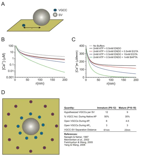
The linearized buffered Ca2+ diffusion model of Naraghi
and Neher (1997) allows for the calculation of the [Ca2+]
at any distance from the mouth of a single VGCC (Figure 1, also see Appendix 1 for details) provided that the com-position of Ca2+ buffers contained within a presynaptic
recording pipette is known [57]. A particularly useful aspect of a linearized system is that it allows for the use of the superposition principle in considering greater num-bers of VGCCs. Thus, the peak [Ca2+] can be determined
at an arbitrary distance (r) from any desired arrangement of VGCCs simply by summating their individual contribu-tions. This freedom allows us to assume any distribution of VGCCs around a SV while being able to determine the [Ca2+] at that point. This provides a constraint governing
the peak [Ca2+] experienced by the release sensor for a
par-ticular set of buffer conditions.
To determine the [Ca2+] at the release sensor, we selected
a simple ring of VGCCs, at equal r from the SV, as a sim-plified topography. It is well established that, at least in the immature synapse, VGCCs are most likely at variable distances from their SV, located in clusters or some other heterogeneous spacing arrangement [58-60]. However, assuming a more simplistic arrangement of a ring of VGCCs simplifies the determination of [Ca2+] arising
from multiple VGCCs and sufficiently address whether VGCC-SV coupling differences can account for develop-mental changes in SV release at this synapse.
Previous modeling studies, performed with data acquired from the immature synapse, have hypothesized an aver-age VGCC-SV separation of ~80 nm [58]. In addition, morphological data, at least from the immature synapse, can provide some constraints when choosing and testing potential VGCC-SV separation distances. AZs at the immature calyx of Held are approximately 0.01 μm2 in
area [61]. If we assume that the AZ is approximately circu-lar in shape, then the diameter of the AZ is ~120 nm and allows for two docked SVs on average [50,60]. This approximate AZ geometry implies that VGCCs can be sep-arated by no more than ~100 nm from any given SV, assuming VGCCs are in the AZ, thus providing an upper limit on the testing ranges for VGCC-SV separation. Simi-larly, the minimum separation distance between SVs and VGCCs is limited by the presence of the release machin-ery, and its many associated proteins, at the base of the SV. The SNARE complex (ring) is estimated to have a diameter of ~20 nm or larger depending on the size of the SV [61], thus we considered 20 nm to be a lower limit for VGCC-SV separation in our modeled VGCC-SV arrangements.
Given these limits, we simulated a number of possible VGCC-SV separation distances with different numbers of VGCCs contributing to the [Ca2+] transient in each case
(see Appendix 1 and Table 1 for parameters). Upon inva-sion of an immature AP into the calyx of Held,
approxi-mately 50% of VGCCs open [46] and the Ca2+-domain
Linearized Buffered Diffusion & Release Site Topography Model
Figure 1
Linearized Buffered Diffusion & Release Site Topography Model. (A) Schematic representation of the single VGCC for which the buffered diffusion model determines [Ca2+] as a function of r. (B) [Ca2+] as a function of r for the various buffer
Therefore, we summated the [Ca2+] contribution from 6
of the 12 VGCCs and set this as the peak [Ca2+] for the
input into a kinetic release model [31,32,57,62]. For the immature synapse, we found that a ring of 12 VGCCs at a distance of r = 61 nm reproduce experimental data well, in line with the average spacing determined by Mein-renken et al. (2002) [58]. Given that VGCC-SV coupling tightens in the mature synapse and fewer VGCCs are required to release a SV, we hypothesized a different topography of VGCCs from that of the immature synapse. In the mature synapse where the narrower APs activate
approximately 35% of VGCCs [46] and Ca2+ channel/
domain cooperativity values for this developmental stage is around 3 [36], we found that the summated contribu-tion of 3 channels per AP from a ring of 9 VGCCs at a mean distance of r = 23 nm to the peak [Ca2+] reproduces
our experimental findings well. These models represent a simplified interpretation of release site topography which allows for simplification of the calculations required to reproduce main experimental observations including developmental increases in quantal output and the extent of EGTA-induced attenuation of EPSCs in immature and mature synapses.
Spatiotemporal profile of Ca
2+transients seen by
Ca
2+sensors on SVs during an AP
The rise and collapse of local Ca2+ domains near the
mouth of VGCCs during APs are difficult to directly resolve (but see [62]). However, these can be
quantita-tively extrapolated from the spatiotemporal profiles of local Ca2+ by using parameters obtained from kinetic
models describing the [Ca2+]
i-release rate curves derived
from flash photolysis experiments [32]. Ca2+ transients are
reproduced by first deconvolving the release profiles of AP-evoked EPSCs, using the respective average mEPSC waveforms, followed by simulating the local [Ca2+]
i
tran-sients at the sites of vesicle fusion required to match the release profiles of individual synapses. This can be achieved by using inverted waveforms of the simulated calyceal ICa, beginning with the Hodgkin-Huxley (HH) model [16] as the initial approximation, and then gradu-ally adjusting the amplitude and decay time courses of [Ca2+]
i transients until the experimental release profiles of
native synapses are accurately reproduced. Simulations performed from data recorded in the mouse calyx of Held, as summarized in Figure 2, demonstrated that with devel-opmental tightening of the spatial coupling between VGCCs and SVs, the local [Ca2+]
i "seen" by the Ca2+
sen-sors increases from 35 to 56 μM and the SV release rate increases from ~600 SVs/ms to 1000 SVs/ms despite smaller and briefer ICa [52]. Similar results, regarding
developmental increased in peak [Ca2+]
i, were also
derived from the developing rat calyx of Held synapse [49].
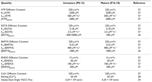
These results reinforce the idea that tightening of the spa-tial coupling between VGCCs and SVs plays a predomi-nant role over other developmental adaptations in Table 1: Parameters for Buffered Ca2+ Diffusion Models
Quantity Immature (P8-12) Mature (P16-18) Reference
ATP Diffusion Constant 220 μm2/s 220 μm2/s 57
Kd (ATP) 2300 μM 2300 μM 57
kon (ATP) 500 μM-1•s-1 500 μM-1•s-1 57
[ATP]pipette 2000 μM 2000 μM 57
EGTA Diffusion Constant 220 μm2/s 220 μm2/s 57
Kd (EGTA) 0.18 μM 0.18 μM 57
kon (EGTA) 2.5 μM-1•s-1 2.5 μM-1•s-1 57
[EGTA]pipette 500/10000 μM 500 μM 36
BAPTA Diffusion Constant 220 μm2/s 220 μm2/s 57
Kd (BAPTA) 0.22 μM 0.22 μM 57
kon (BAPTA) 400 μM-1•s-1 400 μM-1•s-1 57
[BAPTA]pipette 1000 μM 1000 μM 36
ENDO Diffusion Constant 15 μm2/s 15 μm2/s 57
Kd (ENDO) 50 μM 50 μM 57
kon (ENDO) 100 μM-1•s-1 100 μM-1•s-1 57
[ENDO]pipette 500 μM 500 μM 57
Ca2+ Diffusion Constant 220 μm2/s 220 μm2/s 57
Resting [Ca2+]
i 50 nM 50 nM 58
enhancing quantal output at the calyx of Held synapse. This observation may also explain the significant discrep-ancies in estimated AP-driven peak [Ca2+]
i values (10~300
μM) described in previous work [28-33]. That is, this het-erogeneity in the distance between VGCCs and SVs in
dif-ferent experimental systems may contribute to observed variations in the measurement of Pr values in this and other central synapses.
Concluding remarks & prospects
The calyx of Held synapse has emerged as one of the most prominent and important preparations in the field of syn-aptic transmission and developmental plasticity. Studies performed in this model have addressed many important and previously unresolved issues related to the fraction of VGCCs evoked by APs, changes in the amplitude and kinetics of ICa during spike broadening, the downstream
coupling modalities between VGCCs and SVs, as well as the efficacy of Ca2+-dependent transmitter release. In
par-ticular, experimental data and theoretical simulations that confirm the developmental transformation from micro-domain to nanomicro-domain release modalities is of central importance for reconciling opposing views on the nature of coupling between VGCCs and SVs and for presenting the proof-of-principle for idealistic topographic arrange-ments of VGCCs and SVs in AZs.
However, these studies have also raised new questions. Since a majority of functional and morphological adapta-tions at the developing calyx of Held synapse appear to occur after the onset of hearing (at P11/12), the question remains, what are the roles of sensory activity, if any, in driving developmental plasticity and what signaling cas-cade(s) are involved in this transformation[63]. And, is functional plasticity reciprocally coupled to morphologi-cal remodeling at gross and subsynaptic levels during development? Furthermore, neither the molecular sub-strates differentiating microdomain and nanodomain coupling modalities, nor the exact topographic arrange-ments for these distinct release modalities, are known. Many synaptic proteins including syntaxin, Rim, Munc, CAST, synaptotagmin and etc have been implicated in interactions with VGCCs and "positional priming" (i.e. a process for SVs to become tightly associated with Ca2+
influx) in the vicinity of release sites before fusion [64], but critical elements underpinning the proximity of VGCCs and SVs remain mysterious. With the increasing availability of transgenic mice, in which key synaptic pro-teins are deleted or mutated, as well as potential applica-tions of novel viral infection and sRNAi technologies in
vivo [65], future work at the calyx of Held synapse will
undoubtedly lead to further ground breaking discoveries to resolve these critical questions. As answers come to the forefront from this unique synapse, it will undoubtedly continue to make indispensable contributions to our understanding of this and other central synapses.
Competing interests
The authors declare that they have no competing interests. Summary of developmental changes in the temporal profiles
of the local [Ca2+]
i transient seen by the Ca2+ sensor and
glutamate release during an AP
Figure 2
Summary of developmental changes in the temporal profiles of the local [Ca2+]
i transient seen by the Ca2+ sensor and glutamate release during an AP. A, Sche-matic diagram illustrating developmental tightening in the coupling between synaptic vesicles and VGCCs. Immature calyces (A1) have fewer docked vesicles that are loosely coupled to VGCCs, whereas mature calyces (A2) possess approximately a twofold larger number of release-competent vesicles that more closely colocalize with Ca2+ channels. B,
Timing (right) of the local [Ca2+]
i (red) and release (blue)
transients relative to the presynaptic APs (black) for P9--P11 (B1) and P16--P19 (B2) calyces. Traces in B1 and B2 were aligned relative to the onset of the presynaptic APs (dotted line). The average time course of the AP-evoked local [Ca2+] i
transients was obtained from 16 of P9--P11 and 18 of P16--P19 synapses. The peak of the local [Ca2+]
i transient
Authors' contributions
LYW, MJF and YMY wrote this article jointly. LYW designed the conceptual model and MJF performed com-puter simulations. All authors read and approved the final manuscript.
Appendix 1
If the standing [Ca2+] gradients are assumed to be at
steady-state around an open VGCC and buffer saturation is small, the differential equations describing buffered Ca2+ diffusion can be linearized, considerably simplifying
the mathematical description of the system [57,66]. The assumption of low buffer saturation has been proven valid, even for Ca2+ buffers with high binding ratios and
short length constants like BAPTA and conservatively assuming single VGCC flux of around 0.3 pA or smaller [57]. In our simulations, this approximation should also hold as we have chosen single VGCC flux to be well below this threshold at 0.2 pA.
At steady state, the [Ca2+] carried by three Ca2+ buffers can
be described by the following;
Where Φ is the single VGCC flux and;
DN is the diffusion coefficient of the Nth buffer species
and;
In these equations, BN is the concentration of the Nth
buffer species, KN is the dissociation constant of the Nth
buffer species, kNoff is the Ca2+ unbinding rate of the Nth
buffer species, and kNon is the Ca2+ binding rate of the Nth
buffer species. The choices for the above parameters are shown in Table 1 and are taken directly from Naraghi & Neher (1997) with the exception of Φ which was taken as
an average from Stanley (1993) and Shahrezaei & Delaney (2004). Given the necessary parameters, equation 2 can be solved to determine the [Ca2+] at any distance r from a
single VGCC for N described buffer species.
Using the above equations, we have shown the [Ca2+] as a
function of r for various buffer compositions (Figure 1B). In addition, Figure 1C shows [Ca2+]•r as a function of r,
which eliminates the 1/r dependence of [Ca2+] diffusion,
and shows more clearly the characteristic length constant of the given buffer compositions (as in Naraghi & Neher, 1997). Note that the hypothesized endogenous mobile buffer properties, recently uncovered at the immature calyx of Held, have not been included here [67]. This assumption is due to the fact that any endogenous mobile buffers contained within the calyx of Held are likely dia-lyzed rapidly with those contained in the much larger vol-ume of the presynaptic patch pipette.
Acknowledgements
We would like to thank other members of Wang Laboratory for assistance and discussions. This work was supported by individual operating grants (MOP-143867 and MOP-14692) from Canadian Institutes of Health Research (CIHR) and Canada Research Chair (to LYW).
References
1. Llinas R, Steinberg IZ, Walton K: Relationship between presyn-aptic calcium current and postsynpresyn-aptic potential in squid giant synapse. Biophys J 1981, 33:323-352.
2. Llinas R, Sugimori M, Simon SM: Transmission by presynaptic spike-like depolarization in the squid giant synapse. Proc Natl Acad Sci USA 1982, 79:2415-2419.
3. Augustine G: Regulation of transmitter release at the squid giant synapse by presynaptic delayed rectifier potassium cur-rent. J Physiol 1990, 431:343-364.
4. Augustine GJ, Adler EM, Charlton MP: The calcium signal for transmitter secretion from presynaptic nerve terminals. Ann N Y Acad Sci 1991, 635:365-381.
5. Delaney K, Tank DW, Zucker RS: Presynaptic calcium- and sero-tonin-mediated enhancement of transmitter release at cray-fish neuromuscular junction. J Neurosci 1991, 11:2631-2643. 6. Klein M, Kandel ER: Mechanism of calcium current modualtion
underlying presynaptic facilitation and behavioral sensitiza-tion in Aplysia . Proc Natl Acad Sci USA 1980, 77:6912-6916. 7. Sabatini BL, Regehr WG: Timing of neurotransmission at fast
synapses in the mammalian brain. Nature 1996, 384:170-172. 8. Geiger JR, Jonas P: Dynamic control of presynaptic Ca(2+)
inflow by fast-inactivating K(+) channels in hippocampal mossy fiber boutons. Neuron 2000, 28:927-939.
9. Forsythe ID: Direct patch recording from identified presynap-tic terminals mediating glutamatergic EPSCs in the rat CNS, in vitro. J Physiol 1994, 479:381-387.
10. Borst JG, Helmchen F, Sakmann B: Pre- and postsynaptic whole-cell recordings in the medial nucleus of the trapezoid body of the rat. J Physiol 1995, 489:825-840.
11. Takahashi T, Forsythe ID, Tsujimoto T, Barnes-Davies M, Onodera K:
Presynaptic calcium current modulation by a metabotropic glutamate receptor. Science 1996, 274:594-597.
12. Bean BP: The action potential in mammalian central neurons.
Nat Rev Neurosci 2007, 8:451-465.
13. Lisman JE, Raghavachari S, Tsien RW: The sequence of events that underlie quantal transmission at central glutamatergic syn-apses. Nat Rev Neurosci 2007, 8:597-609.
14. Sabatini BL, Regehr WG: Control of neurotransmitter release by presynaptic waveform at the granule cell to Purkinje cell synapse. J Neurosci 1997, 17:3425-3435.
d d d f f B B B
e r C C r C r 1 21 3 1 1 ⎛ ⎝ ⎜ ⎜ ⎜ ⎞ ⎠ ⎟ ⎟ ⎟= − ⋅ −
− − (1)
d
p d
[Ca ]
DCa
D j
DCa Bj
j 2 1 3 4 + =
= Φ −
∑
(2)C D DCa D D DCa D D DCa D
D DCa D
= + + 1 1 1 1 1 1 2 1 1 1 3 1 1 2 1 2 2 1 2 2 2 2 t k t k t k t k t t k t DDCa
D D DCa D
D DCa
D
D DCa D DCa
k t k t k t t k t 2 3 2 2 3 1 3 3 3 2 3 3 1 3 3 3 3 + ⎛ ⎝ ⎜ ⎜ ⎜ ⎜ ⎜ ⎜ ⎜⎜⎜ ⎞ ⎠ ⎟ ⎟ ⎟ ⎟ ⎟ ⎟ ⎟⎟ = ⋅ − − − ⎛ ⎝ ⎜ ⎜ ⎜ ⎜ ⎜ ⎜ ⎜⎜ ⎞ ⎠ ⎟ ⎟ ⎟ f p k t k t k t Φ 4 1 1 1 2 2 2 3 3 3 DCA D D D ⎟⎟ ⎟ ⎟ ⎟⎟
kN BN K N tN
Ca K N kNoff kNon Ca
= ⋅ + +
(
)
⎛ ⎝ ⎜ ⎜ ⎜ ⎜ ⎞ ⎠ ⎟ ⎟ ⎟ ⎟ = + ⋅ +(
)
[ ][ 2 ] 2 [ ]
15. Bischofberger J, Geiger JR, Jonas P: Timing and efficacy of Ca2+ channel activation in hippocampal mossy fiber boutons. J Neurosci 2002, 22:10593-10602.
16. Borst JG, Sakmann B: Calcium current during a single action potential in a large presynaptic terminal of the rat brain-stem. J Physiol 1998, 506:143-157.
17. Borst JG, Sakmann B: Effect of changes in action potential shape on calcium currents and transmitter release in a calyx-type synapse of the rat auditory brainstem. Philos Trans R Soc Lond B Biol Sci 1999, 354:347-355.
18. Joshi I, Wang LY: Developmental profiles of glutamate recep-tors and synaptic transmission at a single synapse in the mouse auditory brainstem. J Physiol 2002, 540:861-873. 19. Taschenberger H, von Gersdorff H: Fine-tuning an auditory
syn-apse for speed and fidelity: developmental changes in presy-naptic waveform, EPSC kinetics, and sypresy-naptic plasticity. J Neurosci 2000, 20:9162-9173.
20. von Gersdorff H, Borst JG: Short-term plasticity at the calyx of Held. Nat Rev Neurosci 2002, 3:53-64.
21. Wu LG, Saggau P: Pharmacological identification of two types of presynaptic voltage-dependent calcium channels at CA3-CA1 synapses of the hippocampus. J Neurosci 1994,
14:5613-5622.
22. Mintz IM, Sabatini BL, Regehr WG: Calcium control of transmit-ter release at a cerebellar synapse. Neuron 1995, 15:675-688. 23. Yoshikami D, Bagabaldo Z, Olivera BM: The inhibitory effects of
omega-conotoxins on Ca2+ channels and synapses. Ann N Y
Acad Sci 1989, 560:230-248.
24. Stanley EF: Single calcium channels and acetylcholine release at a presynaptic nerve terminal. Neuron 1993, 11:1007-1011. 25. Gentile L, Stanley EF: A unified model of presynaptic release
site gating by calcium channel domains. Eur J Neurosci 2005,
21:278-282.
26. Borst JGG, Sakmann B: Calcium influx and transmitter release in a fast CNS synapse. Nature 1996, 383:431-434.
27. Wu LG, Westenbroek RE, Borst JGG, Catterall WA, Sakmann B:
Calcium channel types with distinct presynaptic localization couple differentially to transmitter release in single calyx-type synapses. J Neurosci 1999, 19:726-736.
28. Adler EM, Augustine GJ, Duffy SN, Charlton MP: Alien intracellular calcium chelators attenuate neurotransmitter release at the squid giant synapse. J Neurosci 1991, 11:1496-1507.
29. Yamada WM, Zucker RS: Time Course of Transmitter Release Calculated from Simulations of A Calcium Diffusion-Model.
Biophys J 1992, 61:671-682.
30. Llinas R, Sugimori M, Silver RB: Microdomains of High Calcium-Concentration in A Presynaptic Terminal. Science 1992,
256:677-679.
31. Bollmann JH, Sakmann B, Gerard J, Borst G: Calcium sensitivity of glutamate release in a calyx-type terminal. Science 2000,
289:953-957.
32. Schneggenburger R, Neher E: Intracellular calcium dependence of transmitter release rates at a fast central synapse. Nature 2000, 406:889-893.
33. Roberts WM: Localization of calcium signals by a mobile cal-cium buffer in frog saccular hair cells. J Neurosci 1994,
14:3246-3262.
34. Trussell LO: Synaptic mechanisms for coding timing in audi-tory neurons. Ann Rev Physiol 1999, 61:477-496.
35. Wang LY, Kaczmarek LK: High-frequency firing helps replenish the readily releasable pool of synaptic vesicles. Nature 1998,
394:384-388.
36. Fedchyshyn MJ, Wang LY: Developmental transformation of the release modality at the calyx of held synapse. J Neurosci 2005,
25:4131-4140.
37. Schneggenburger R, Forsythe ID: The calyx of held. Cell and Tissue Res 2006, 326:311-337.
38. Sun JY, Wu LG: Fast kinetics of exocytosis revealed by simul-taneous measurements of presynaptic capacitance and post-synaptic currents at a central synapse. Neuron 2001,
30:171-182.
39. He LM, Wu XS, Mohan R, Wu LG: Two modes of fusion pore opening revealed by cell-attached recordings at a synapse.
Nature 2006, 444:102-105.
40. Renden R, von Gersdorff H: Synaptic vesicle endocytosis at a CNS nerve terminal: Faster kinetics at physiological
temper-atures and increased endocytotic capacity during matura-tion. J Neurophysiol 2007, 98:3349-3359.
41. Dodge FA Jr, Rahamimoff R: Co-operative action a calcium ions in transmitter release at the neuromuscular junction. J Physiol 1967, 193:419-432.
42. Leao RM, Kushmerick C, Pinaud R, Renden R, Li GL, Taschenberger H, Spirou G, Levinson SR, von Gersdorff H: Presynaptic Na+ chan-nels: Locus, development, and recovery from inactivation at a high-fidelity synapse. J Neurosci 2005, 25:3724-3738.
43. Dodson PD, Billups B, Rusznak Z, Szucs G, Barker MC, Forsythe ID:
Presynaptic rat Kv1.2 channels suppress synaptic terminal hyperexcitability following action potential invasion. J Physiol 2003, 550:27-33.
44. Elezgarai I, Diez J, Puente N, Azkue JJ, Benitez R, Bilbao A, Knopfel T, Donate-Oliver F, Grandes P: Subcellular localization of the volt-age-dependent potassium channel Kv3.1b in postnatal and adult rat medial nucleus of the trapezoid body. Neurosci 2003,
118:889-898.
45. Ishikawa T, Nakamura Y, Saitoh N, Li WB, Iwasaki S, Takahashi T:
Distinct roles of Kv1 and Kv3 potassium channels at the calyx of Held presynaptic terminal. J Neurosci 2003, 23:10445-10453. 46. Yang YM, Wang LY: Amplitude and kinetics of action potential-evoked Ca2+ current and its efficacy in triggering transmit-ter release at the developing calyx of held synapse. J Neurosci 2006, 26:5698-5708.
47. Pumplin DW, Reese TS, Llinas R: Are the Pre-Synaptic Mem-brane-Particles the Calcium Channels. Proc Natl Acad Sci USA 1981, 78:7210-7213.
48. Wu XS, Sun JY, Evers AS, Crowder M, Wu LG: Isoflurane inhibits transmitter release and the presynaptic action potential.
Anesthesiol 2004, 100:663-670.
49. Kochubey O, Han YY, Schneggenburger R: Developmental regu-lation of the intracellular Ca2+ sensitivity of vesicle fusion and Ca2+-secretion coupling at the rat calyx of Held. J Physiol 2009, 587:3009-3023.
50. Taschenberger H, Leao RM, Rowland KC, Spirou GA, von Gersdorff H: Optimizing synaptic architecture and efficiency for high-frequency transmission. Neuron 2002, 36:1127-1143.
51. Leao RM, von Gersdorff H: Synaptic vesicle pool size, release probability and synaptic depression are sensitive to Ca2+ buffering capacity in the developing rat calyx of Held. Braz J Med Biol Res 2009, 42:94-104.
52. Wang LY, Neher E, Taschenberger H: Synaptic Vesicles in Mature Calyx of Held Synapses Sense Higher Nanodomain Calcium Concentrations during Action Potential-Evoked Glutamate Release. J Neurosci 2008, 28:14450-14458.
53. Wu LG, Borst JGG, Sakmann B: R-type Ca2+ currents evoke transmitter release at a rat central synapse. Proc Natl Acad Sci USA 1998, 95:4720-4725.
54. Iwasaki S, Momiyama A, Uchitel OD, Takahashi T: Developmental changes in calcium channel types mediating central synaptic transmission. J Neurosci 2000, 20:59-65.
55. Bucurenciu I, Kulik A, Schwaller B, Frotscher M, Jonas P: Nanodo-main coupling between Ca2+ channels and Ca2+ sensors promotes fast and efficient transmitter release at a cortical GABAergic synapse. Neuron 2008, 57:536-545.
56. Kim JH, Sizov I, Dobretsov M, von Gersdorff H: Presynaptic Ca2+ buffers control the strength of a fast post-tetanic hyperpo-larization mediated by the alpha 3 Na+/K+-ATPase. Nature Neurosci 2007, 10:196-205.
57. Naraghi M, Neher E: Linearized buffered Ca2+ diffusion in microdomains and its implications for calculation of [Ca2+] at the month of a calcium channel. J Neurosci 1997,
17:6961-6973.
58. Meinrenken CJ, Borst JGG, Sakmann B: Calcium secretion cou-pling at calyx of held governed by nonuniform channel-vesi-cle topography. J Neurosci 2002, 22:1648-1667.
59. Meinrenken CJ, Borst JGG, Sakmann B: Local routes revisited: the space and time dependence of the Ca2+ signal for phasic transmitter release at the rat calyx of Held. J Physiol 2003,
547:665-689.
Publish with BioMed Central and every scientist can read your work free of charge
"BioMed Central will be the most significant development for disseminating the results of biomedical researc h in our lifetime."
Sir Paul Nurse, Cancer Research UK
Your research papers will be:
available free of charge to the entire biomedical community
peer reviewed and published immediately upon acceptance
cited in PubMed and archived on PubMed Central
yours — you keep the copyright
Submit your manuscript here:
http://www.biomedcentral.com/info/publishing_adv.asp
BioMedcentral 61. Cho WJ, Jeremic A, Jena BP: Size of supramolecular SNARE
complex: Membrane-directed self-assembly. J Amer Chem Soc 2005, 127:10156-10157.
62. Bollmann JH, Sakmann B: Control of synaptic strength and tim-ing by the release-site Ca2+ signal. Nat Neurosci 2005,
8:426-434.
63. Erazo-Fischer E, Striessnig J, Taschenberger H: The role of physio-logical afferent nerve activity during in vivo maturation of the calyx of held synapse. J Neurosci 2007, 27:1725-1737. 64. Neher E, Sakaba T: Multiple roles of calcium ions in the
regula-tion of neurotransmitter release. Neuron 2008, 59:861-872. 65. Young SM, Neher E: Synaptotagmin Has an Essential Function
in Synaptic Vesicle Positioning for Synchronous Release in Addition to Its Role as a Calcium Sensor. Neuron 2009,
63:482-496.
66. Neher E: Vesicle pools and Ca2+ microdomains: New tools for understanding their roles in neurotransmitter release.
Neuron 1998, 20:389-399.