Abstract
Whitehurst, Christopher Benton. Structure and Assembly of the Sindbis Virus E1 and E2 Transmembrane Proteins. (Under the direction of Dr. Dennis T. Brown.)
Sindbis virus is composed of two nested T=4 icosahedral protein shells containing 240 copies each of three structural proteins: E1, E2, and Capsid in a 1:1:1 stoichiometric ratio. E2 is a 423 amino acid glycoprotein with a membrane spanning domain 26 amino acids in length. A previous study had determined that deletions in the transmembrane domain could affect virus assembly and infectivity (Hernandez et al, 2003 J.Virol 77(23), 12710-9). Unexpectedly, a single deletion mutant (from 26 to 25 amino acids) resulted in a 1000-fold decrease in infectious virus production while another deletion of eight amino acids had no affect on virus production. To further investigate the importance of these mutants, other single deletion mutants and another eight amino acid deletion mutant were constructed. We found that deletions located closer to the cytoplasmic (inner) leaflet of the membrane bilayer had a more detrimental effect on virus assembly and infectivity than those located closer to the luminal (outer) leaflet of the membrane bilayer. We also found that selective pressure can restore single amino acid deletions in the transmembrane domain but not necessarily to the wild type sequence. These results suggest the position of the deletion and the length of the C terminal region of the E2 transmembrane domain are vital for normal virus production. Deletion
A complex network of disulfide bonds in the E1 and E2 glycoproteins is developed through a series of intermediates as virus maturation occurs. E1 and E2 cysteine residues were labeled with iodoacetamide in the native virus particle and analyzed by mass spectrometry. This analysis identified cysteines of E1 and E2 that were found to be free in the native virus particle as well as those that were either solvent inaccessible or blocked by their involvement in disulfide bonds. Native virus labeled with iodoacetamide yielded a four log decease in viral infectivity. This suggests either the bound iodoacetamide alone may be involved in the loss of infectivity by destabilizing the virus particle or a rearrangement of disulfide bonds, which is required for infectivity, is blocked by the presence of iodoacetamide. In addition, cysteines that were determined to be free in the E1 glycoprotein were mutated to serine. Mutation of these cysteines greatly decreased the amount of infectious virus production suggesting a disulfide bond rearrangement may occur as virus maturation proceeds.
Structure and Assembly of the Sindbis Virus E1 and E2 Transmembrane Proteins
by
Christopher Benton Whitehurst
A dissertation submitted to the Graduate Faculty of North Carolina State University
In partial fulfillment of the Requirements for the degree of
Doctor of Philosophy Biochemistry
Raleigh, NC 2006 Approved by:
_________________________
__________________________ _________________________
__________________________ _________________________
Dedication
To my wife, Jane, my two boys, Watson and Henry, and to any future offspring I may spawn. To my parents, Howard and Madeline, my bother, Vincent, his wife, Ann, their son, Wren, and their soon to be #2, and my sister, Annette.
To my father and mother-in-law, Charlie and Jane Sutherland, and to their other children, Taylor, James, and Morgan.
Biography
I was born Christopher Benton Whitehurst in Laurinburg, NC on November 29,1973 to Howard Hugh and Madeline Condino Whitehurst. I have one older brother, Vincent, and a younger sister, Annette. In 1981, we moved to Abbeville, South Carolina. I attended Abbeville High School and graduated in 1992. I received my BS in Biochemistry from Clemson University. I met my wife, Jane, there. We broke up. Got back together. Broke up. I moved to Raleigh. She moved to Milwaukee. I worked at Surgicot and Bayer. Work sucked. I decided to go to graduate school. I moved to Milwaukee, WI and moved in with Jane (but only because I got a good deal on the rent) and attended the University of Wisconsin - Milwaukee. Got married to Jane in 2000 (got a better deal on the rent). I received an MS in Chemistry in 2001. I came to NC State University to pursue a PhD in Biochemistry. Watson was born in 2002, and Henry was born in 2004. My wife was the mother of both. I will finish this summer, 2006, with a PhD in Biochemistry. Next I will go to the University of North Carolina for a post doc.
Christopher B. Whitehurst
Department of Molecular and Structural Biochemistry North Carolina State University, Raleigh, NC 27695
PhD Candidate (graduating summer 2006)
Home Address: 1211 Ashburton Rd. Raleigh, NC 27606 Phone: (919) 858-9806 (h) (919) 515-5786 (school)
(919) 971-5134 (c) email: [email protected]
Education:
Clemson University, Clemson, SC. B.S. Biochemistry 1992-1996
University of Wisconsin – Milwaukee, Milwaukee, WI M.S. Chemistry 1998-2001
Thesis: Studies of Vaccinia Virus, VP11, and the MAPK Kinase Pathway North Carolina State University, Raleigh, NC.
Ph.D. Biochemistry 2001- 2006 (pending)
Research: Structural Properties of the Sindbis Virus E1 and E2 Glycoproteins Experience:
Clemson University -Undergraduate and summer research (1995-1996) Surgicot – Lab Technician (1997)
Bayer – Lab Technician (1997-1998) Professional Memberships:
American Society of Virology (Since 2000) Awards and Offices Held:
American Society for Virology Travel Grant (2005)
University of Wisconsin – Milwaukee Travel Grant (2001)
North Carolina State Biochemistry Graduate Student Association Secretary and Social Chair (2004)
Presentations:
Characterization of the Sindbis Virus E2 Transmembrane Domain. Whitehurst, C., Willis, J., Sinodis, C., Hernandez, R. and Brown, D. American Society for Virology Conference. 2005. Pennsylvania State University, University Park, PA
Characterization of the Sindbis Virus E2 Transmembrane Domain. Whitehurst, C., Willis, J., Sinodis, C., Evans, D., Hernandez, R. and Brown, D. American Society for Virology Conference. 2004. McGill University, Montreal, Cananda
Proline-directed kinase motifs are essential for proper functioning of
the major phosphoprotein of Vaccinia: p11 (F18R). Evans, D., Afshan, G. Du, Y., Ohigashi, Y., Whitehurst, C. and Reddy, M. International Poxvirus Meeting. 2002. Lake Placid, NY
A Connection Between MAP/ERK and the Poxviral p11 Phosphoprotein? Ohigashi, Y., Whitehurst, C. and Reddy, M. American Society for Virology Conference. 2001. University of Wisconsin, Madison, WI
Poxviral VP11: A Big Role for a Small Protein. Reddy, M., Afshan, G., Ohigashi, Y., Du, Y., and Whitehurst, C. American Society for Virology Conference. 2000.
Colorado State University, Fort Collins, CO
Publications:
Location of free and disulfide bound cysteines in the Sindbis virus E1 and E2
glycoproteins. Christopher B. Whitehurst, Erik J. Soderblom, Michael B. Goshe, and Dennis T. Brown. In preparation.
Acknowledgments
Teachers: I have had a few good ones, Simone Simmons, Crystal Dunlap, and Conrad Leo.
College Professors: Dr. Jessup Shively (Clemson advisor – always believed in me more than I believed in myself and made me want to try harder), Dr. Robinson (Clemson – for calling on the guy with the hat to teach class for a week straight – MADE me study.), Dr. AP Wheeler (Clemson- allowed me to do senior research in his lab), Dr. Mike Reddy (my boss at UWM – maybe the best teacher I have ever met.), Dr. Graham Moran (UWM – for general advice), Dr. Michael Goshe (NC State - for all the mass spec help), and Drs. Dennis Brown and Raquel Hernandez (NC State – for putting me in position to actually do something with my life).
Committee members: Douglas Lyles (Wake Forest), John Mackenzie (NC State), John Cavanagh (NC State), Robert Rose (NC State), and Dennis Brown (NC State). Thanks for all the guidance.
Lab members: Steevenson Nelson, John West, Usha Mudiganti, Shelly Horton, Christine Sinodis, and Gongbo Wang.
Childhood enemy: Donnie Sutton.
College friends: Geoff Chambers, Brian Morris, Peter Kahle, Aaron Kimmerling, Kirsten G. Pierson, Jason Hemphill, Patrick Hughes, and Nancy Neal.
College enemy: Elvino Menoldovo (I hope I screwed up the spelling of your name).
After college friends: Josh Deverse, Loren Lenzen, Joel Hartman, and Doug Viney.
Family: all the Sutherlands (you guys are actually decent in-laws), my brother, his wife and child, and my sister (thanks for all the confidence and encouragement), Mom and Dad (thanks for the money, support, time, teachings, advice, punishment, and love), my children, Watson and Henry (for your smiles when you greet me at the door and for convincing your mom and I that we actually got something right), and my wife (you’re a good one – I love you).
Table of Contents
List of Figures ... ix
List of Tables ... xi
Chapter 1: Introduction to Sindbis Virus... 1
Chapter 2: Single and multiple deletions in the transmembrane domain of the Sindbis virus E2 glycoprotein identify a region critical for normal virus Growth ... 11
Abstract ... 12
Introduction ... 13
Results ... 18
Discussion... 31
Materials and Methods ... 38
Chapter 3: Identification of free and disulfide bound cysteines in the Sindbis virus E1 and E2 glycoproteins... 42
Abstract ... 43
Introduction ... ... 44
Results ... 48
Discussion... 63
Materials and Methods ... 72
Chapter 4: Conclusions and Future Directions ... 78
List of Figures
Figure 1.1: Genome organization of Sindbis virus ... 3 Figure 1.2: Cross-section of Sindbis virus from a cryo-electron microscopy
reconstruction... 5 Figure 1.3: A schematic representation of the organization of the Sindbis virus
structural proteins in the membrane of the Endoplasmic Reticulum... 7 Figure 1.4: Viral release from the cell ... 9 Figure 2.1: A schematic representation of the organization of the Sindbis virus
structural proteins in the membrane of the Endoplasmic Reticulum... 15 Figure 2.2: Representations of the E2 transmembrane ... 19 Figure 2.3: Infectious virus production and relative infectivity of the TM25,
TM18, FF, and FF25 mutants... 20 Figure 2.4: Production of infectious virus by the TM25 and TM18 mutants
before and after serial passage... 27 Figure 2.5: Compensatory mutations (CM) found in the E2 transmembrane
Domain... 28 Figure 2.6: Comparison of the TM18C, CM18C, and TM22 growth and
Sequence ... 30 Figure 3.1: Representation of labeled cysteine residues in the amino acid
sequence of the Sindbis virus E1 protein as determined by mass
spectrometry ... 49 Figure 3.2: Representation of labeled cysteine residues in the amino acid
sequence of the Sindbis virus E2 protein as determined by mass
spectrometry ... 50 Figure 3.3: Representation of labeled cysteine residues in the amino acid
sequence of RNase B as determined by mass spectrometry... 54 Figure 3.4: Representation of labeled cysteine residues in the amino acid
sequence of the Sindbis virus E1 protein as determined by mass
Figure 3.5: Representation of labeled cysteine residues in the amino acid sequence of the Sindbis virus E2 protein as determined by mass
List of Tables
Table 2.1: Relative density of viral proteins produced from selected mutants ... 25 Table 3.1: Free and bound cysteines of the Sindbis virus E1 and E2 proteins ... 51 Table 3.2: Infectivity of purified virus alkylated with iodoacetamide as
Chapter 1
Sindbis virus was first isolated in August 1952 from a pool of mosquitoes in the Sindbis health district, which is located about 30 km from Cairo, Egypt (Hurlbut, 1953; Taylor and Hurlbut, 1953). It is a member of the alphavirus genus within the togavirus family. The Alphaviruses currently have 26 members (Calisher et al., 1988) and of the genomes sequenced they share about 45% sequence identity in the structural proteins and about 60% identity in the nonstructural proteins (Strauss and Strauss, 1994). Alphaviruses are arthropod borne with mosquitoes being the usual vector. They have a very wide geographical distribution and have been isolated from every continent except Antarctica (Strauss and Strauss, 1994). Alphaviruses are a potential threat to humans as Eastern Equine Encephalitis Virus and Western Equine Encephalitis virus both cause fatal encephalitis in both North and South America (Griffin, 1986; Peters, 1990). Sindbis virus, the prototype alphavirus, is usually considered to be avirulent in humans.
as the template for a new shorter 26S positive strand molecule, which is equivalent to the 3’ end of the genome. The 26S positive strand RNA is then capped and tailed and is translated into a polyprotein. A series of cleavages of the translated
polyprotein results in the formation of the structural proteins: Capsid, E3, E2, 6K and E1.
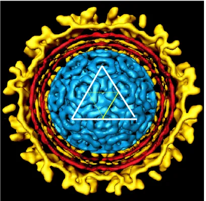
The mature virus particle contains three of the structural proteins (E1, E2, and capsid) in a 1:1:1 stoichiometric ratio. The Sindbis virus particle is 70 nm in
diameter, contains a cell membrane derived bilayer, and serves as an excellent model for structural studies. Sindbis virus is organized into two geometrically identical T=4 icosahedral shells containing 80 equivalent units composed up of 240 copies of each structural protein (E1, E2, and capsid)(Paredes et al., 1993)(Figure 1.2). The outer protein shell is composed solely of the E1: E2 dimers which are then organized into trimers (Anthony and Brown, 1991; Carleton et al., 1997; Pletnev et al., 2001). Each one of these trimers represents an equivalent unit on the surface of the viral particle. The inner protein shell, which is surrounded by the outer protein shell, is composed entirely of the capsid protein. The 11.7 kB genomic RNA is packaged within the inner shell. A host derived membrane bilayer is positioned between the inner and outer shell of the Sindbis virus particle and is penetrated by the transmembrane domain anchors of the E1 and E2 proteins (Lilijestrom and Garoff, 1991; Rice et al., 1982; Strauss, Lenches, and Strauss, 2002). The
Figure 1.2: Cross-section of Sindbis virus from a cryo-electron microscopy
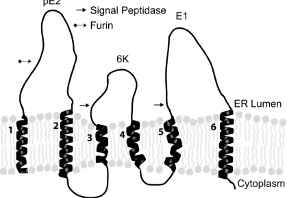
The 26S mRNA produced by Sindbis virus encodes a polyprotein that is post-translationally cleaved into Capsid, E1, E2, E3, and 6K. Auto-protease activity removes the capsid protein after its translation on the cytoplasmic ribosome and exposes a transmembrane signal sequence that is necessary for the insertion of the polyprotein into the membrane bilayer. The transmembrane polyprotein contains six membrane spanning domains (Figure 1.3) (Liljestrom and Garoff, 1991) and is cleaved into its structural components by signalase (PE2, 6K, and E1) (Liljestrom and Garoff, 1991). 6K, which is only found at low levels in the mature virus particle (Gaedigk-Nitschko and Schlesinger, 1990), is necessary for correct integration of E1
into the membrane and plays a role in the efficient budding of viral particles (Liljestrom and Garoff, 1991). E1, which contains a functional domain involved in virus entry, is then folded into a stable high energy configuration as it is assembled into heterodimers with PE2 (Carleton et al., 1997; Mulvey and Brown, 1994; Mulvey and Brown, 1995; Mulvey and Brown, 1996) . E1/PE2 is then transported from the ER into the trans-golgi where PE2 is processed to E2 and E3 by furin (Moehring et al., 1993). E3 is released and is not found in the mature virus particle. The E1/E2 heterotrimer is then recruited to the cell surface. As transport of the E1/E2
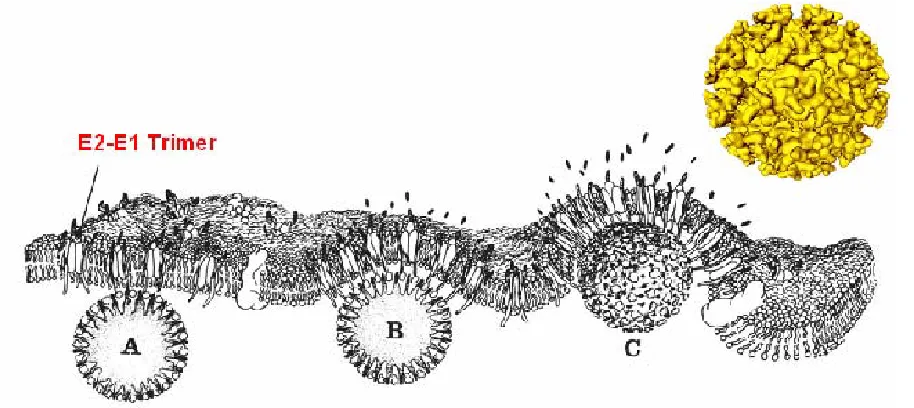
heterotrimer to the plasma membrane occurs, a second membrane-spanning
(Lee and Brown, 1994; Lee et al., 1996; Oates et al., 1992; Owen and Kuhn, 1996; Owen and Kuhn, 1997). The association of the E2 endodomain with the capsid protein gives stability to the structure of the virus (Lee, Ricker, and Brown, 1994) and plays a critical role in the formation of the outer protein shell around the preformed inner protein shell as the process of envelopment takes place (Ferreira et al., 2003)(Figure 1.4). The mature virus particle is then released.
This thesis focuses on two aspects of the E1 and E2 glycoprotein structure. The first deals with investigating the role of the Sindbis virus E2 transmembrane domain. Previously, sequential deletions were made in the 26 amino acid E2 transmembrane domain to investigate the interaction of the membrane-spanning domain with the membrane bilayer (Hernandez et al., 2003). Deletion of a single amino acid in the transmembrane domain resulted in a four-log loss of infectious virus production (Hernandez et al., 2003). This was a surprising find since 25 amino acids is long enough to span the membrane bilayer. Previous results also showed that deleting eight amino acids from the N terminal half of the transmembrane region had no effect on normal virus growth. To further investigate these results a series of single amino acid deletions were made within the E2 transmembrane domain along with another mutant where eight amino acids were deleted from the C terminal half of the transmembrane domain. The results revealed that deletions located closer to the cytoplasmic face of the transmembrane domain resulted in a greater loss of infectivity than deletion located closer to the lumenal side of the membrane
normal virus growth. This may be explained by allowing for more degrees of freedom so that the E2 endodomain may interact properly with the capsid protein (Whitehurst et al., 2006). There is also evidence to suggest that the E1 and E2 transmembrane domains may interact with each other (Strauss, Lenches, and Strauss, 2002; Whitehurst et al., 2006).
The second part of the thesis focuses on identifying free and disulfide bound cysteines in the E1 and E2 proteins by using mass spectrometry as a tool. Little is known about the E2 disulfide network. Gidwitz et al. (Gidwitz et al., 1988) reported that all cysteines located in the E2 protein were either involved in disulfide bonds or were inaccessible when treated with 14C iodoacetamide. Much more is known for the E1 protein. The Semliki Forest virus E1 crystal structure was solved and the disulfide bonds present were indicated (Roussel et al., 2006).
CHAPTER 2
Single and multiple deletions in the
transmembrane domain of the Sindbis virus E2
glycoprotein identify a region critical for normal
virus growth
Christopher B. Whitehurst, John H. Willis, Christine N. Sinodis,
Raquel Hernandez, and Dennis T. Brown
Published in:
Abstract
Sindbis virus is composed of two nested T = 4 icosahedral protein shells containing 240 copies each of three structural proteins: E1, E2, and Capsid in a 1:1:1 stoichiometric ratio. E2 is a 423 amino acid glycoprotein with a membrane spanning domain 26 amino acids in length and a 33 amino acid cytoplasmic endodomain. The interaction of the endodomain with the nucleocapsid is an
essential step in virus maturation and directs the formation of the outer protein shell as envelopment occurs. A previous study had determined that deletions in the
transmembrane domain could affect virus assembly and infectivity (Hernandez et al., 2003. J. Virol. 77 (23), 12710–12719). Unexpectedly, a single deletion mutant (from 26 to 25 amino acids) resulted in a 1000-fold decrease in infectious virus production while another deletion of eight amino acids had no affect on infectious virus
and the length of the C terminal region of the E2 transmembrane domain is vital for normal virus production. Deletion mutants resulting in decreased infectivity produce particles that appear to be processed and transported correctly suggesting a role involved in virus entry.
Introduction
Sindbis virus (SV) is an Alphavirus, which is propagated in nature via a complicated life cycle involving insect vectors and mammalian hosts. Mature SV contains 240 copies each of three structural proteins E1, E2, and Capsid (C) in a 1:1:1 stoichiometric ratio. These proteins are organized into two geometrically identical T=4 icosahedral shells (Paredes et al., 1993). The outer protein shell is composed of the E1 and E2 glycoproteins organized in trimers of heterodimers (Anthony and Brown, 1991; Carleton et al., 1997; Pletnev et al., 2001). This outer shell surrounds the inner shell, composed of protein C, which is assembled around the viral RNA. A host-derived membrane bilayer is positioned between the two shells and is penetrated by the transmembrane (TM) domain anchors of E1 and E2 (Lilijestrom and Garoff, 1991; Rice et al., 1982; Strauss, Lenches, and Strauss, 2002).
around the preformed inner protein shell as the process of envelopment takes place (Ferreira et al., 2003). At the plasma membrane the E1-E2 heterotrimers are
recruited into the outer protein shell by association of the E2 endodomain with the icosahedral nucleocapsid (Owen and Kuhn, 1997).
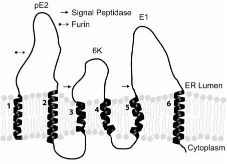
In mature SV only transmembrane regions two (E2) and six (E1) remain membrane associated (see Figure 2.1). The amino acid sequences of the
transmembrane regions are not conserved among the alphaviruses, however the hydrophobic nature and approximate length (26-28 amino acids) are conserved. Previously, sequential deletions were made in the E2 transmembrane domain to investigate the interaction of the 26 amino acid membrane-spanning domain with the membrane bilayer. Deletion of a single methionine at position 379 (TM25) resulted in a 3 to 4 log loss in production of infectious virus (Hernandez et al., 2003). Many different factors could contribute to the observed phenotype of the TM25 mutant. i) The observed phenotype could be a result of a single specific amino acid deletion at position 379. ii) The angular location of the deletion on the face of the
transmembrane helix may account for the loss of infectivity (ie. if the deletion is made 90 degrees, 180 degrees or another angle from a set point of reference). iii) The location of the deletion vertically (ie closer to the luminal side or closer to the cytoplasmic side of the membrane) may contribute to the observed phenotype. iv) A specific interaction between the E1 and E2 transmembrane regions may be
and E2 transmembrane domains using a chimeric virus composed of Ross River E1 and Sindbis virus E2 (Strauss, Lenches, and Strauss, 2002).
To further characterize the E2 membrane-spanning domain and investigate the importance of the factors presented above, additional single deletion mutants and an eight amino acid deletion mutant were constructed in the E2 transmembrane domain. Mutant sites were chosen to encompass all faces of the helix and to
traverse the helix from N terminal to C terminal within the E2 transmembrane domain. It was found that deletions located closer to the cytoplasmic face of the membrane region resulted in a greater loss of infectivity than deletions located closer to the lumenal side of the membrane. Also deletions located on one face of the helix and close to the cytoplasmic interface resulted in reduced infectious virus production. The results presented herein suggest i) the observed phenotype of the TM25 mutants is not due to a single specific amino acid deletion, ii) the location of the deletion vertically is important for normal virus growth, iii) the angular location of the deletion on the face of the helix does not affect normal virus growth and iv) an interaction may be occurring between the E1 and E2 transmembrane domains in the cytoplasmic half of the transmembrane domain. In addition, other factors may
Results
Virus Production by Sindbis virus E2 single amino acid (TM25) mutants
Transmembrane domain two (see Figure 2.1) was chosen for mutagenesis because of E2’s interaction with both the inner and outer icosahedral protein shells (Hernandez et al., 2003). Previously, a series of sequential deletions in the E2 transmembrane domain were produced (Hernandez et al., 2003). The goal of these experiments was to elucidate the requirements placed on the length of the
transmembrane domain for integration into cell membranes and for assembly of infectious virus. In that study, a single deletion of a methionine at position 379 yielded a decrease in infectious virus production of four orders of magnitude (Hernandez et al., 2003). The relative infectivity of this mutant measured as the particle/pfu ratio was approximately 105 suggesting that virus assembly occurred,
but that the resulting virus population was largely noninfectious. To further
determine the effect of single deletion mutations in the transmembrane domain on virus assembly and infectivity, a series of single amino acid deletions were produced in the E2 transmembrane domain (Figure 2.2). Deletions were placed at positions ranging from the luminal to the cytoplasmic side of the membrane bilayers. In addition, the deletions also covered all faces of the transmembrane helix (Figure 2.2).
V371 S373 C 26 A 25 C 24 L 23 V 22 A 21 V 20 T 19 V 18 G 17 I 16 M 15 M 14 A 13 V 12 T 11 A 10 S 9 A 8 V 7 A 6 L 5 I 4 T 3 Y 2 V 1 M379 V376 A385 V386
NH3+
COO
-A370 A372
wt VYTILAVASATVAMMIGVTVAVLCAC 26AA
ΔA370 VYTIL-VASATVAMMIGVTVAVLCAC 25AA
ΔV371 VYTILA-ASATVAMMIGVTVAVLCAC 25AA
ΔA372 VYTILAV-SATVAMMIGVTVAVLCAC 25AA
ΔS373 VYTILAVA-ATVAMMIGVTVAVLCAC 25AA
ΔV376 VYTILAVASAT-AMMIGVTVAVLCAC 25AA
ΔM379 VYTILAVASATVAM-IGVTVAVLCAC 25AA
ΔA385 VYTILAVASATVAMMIGVTV-VLCAC 25AA
ΔV386 VYTILAVASATVAMMIGVTVA-LCAC 25AA TM18N VYTILAV---IGVTVAVLCAC 18AA TM18C VYTILAVASATVAM---LCAC 18AA FF VYTILAVASATVAMMIGVTVAVLCACF 27AA FF25 VYTILAVASATVAM-IGVTVAVLCACF 26AA
Figure 2.2: Representations of the E2 transmembrane. (A) A helical wheel
representation of the E2 transmembrane domain from the N-terminal region to the C-terminal region. Locations of single amino acid deletion mutations are noted with arrows. Mutations cover all faces of the helix and span the entire membrane
domain. Boxed labels indicate mutants, which produced106 pfu/ml, and others
represent virus, which produced 108 pfu/ml. (B) Sequence of the deletions in the E2 transmembrane domain (transmembrane domain #2 from Fig. 1). (-) denotes
location of the deletion. The resulting number of amino acids is listed to the right.
A
1.00E+04 1.00E+05 1.00E+06 1.00E+07 1.00E+08 1.00E+09 1.00E+10 A 370 V 371 A 372 S373 V 376 M 379 A 385 V 386 TM 18 N TM 18 C FF2 5 FF SVH R
P
fu
/m
l
Mutant
Δ Δ Δ Δ Δ Δ Δ ΔN C
1.00E+00 1.00E+01 1.00E+02 1.00E+03 1.00E+04 1.00E+05 1.00E+06 1.00E+07 1.00E+08 1.00E+09 1.00E+10 A 370 V 371 A 372 S373 V 376 M 379 A 385 V 386 TM 18N TM 18C FF2 5 FF SVH R
P
a
rt
ic
le
/p
fu
Mutant
Δ Δ Δ Δ Δ Δ Δ ΔFigure 2.3: Infectious virus production and relative infectivity of the TM25, TM18, FF, and FF25 mutants. (A) Production of infectious virus by the TM25, TM18, FF, and FF25 mutants from transfection. The arrow at the bottom highlights the relative location of the TM25 deletions from N terminal to C terminal. SVHR represents wt Sindbis Virus Heat Resistant strain. (B) Relative infectivity of the TM25, TM18, FF and FF25 mutants as determined by particle-to-pfu assay. Virus utilized in this experiment was released from the cell into the media and purified (see Methods). Mutant virus particles banded to wt density. Those mutants producing higher titers had lower particle-to-pfu ratios indicating more of the particles produced from these mutants were infectious, with the exception of the FF mutant. Except for FF, the mutants produce similar amounts of particles.
A
ΔV376 produced relatively higher titers of approximately 108 pfu/ml but still an order of magnitude below wild type. Single deletion mutants located in the N terminal region of the E2 transmembrane domain produced two logs more infectious virus than those located in the C terminal region of the transmembrane domain. The difference in infectious virus production from mutants in the N terminal and the C terminal TM region did not represent a gradual decline but a two-log decrease that occurred between position 376 and 379. This suggests that single deletion
mutations located closer to the N terminal portion of the transmembrane domain have a less detrimental effect than those located closer to the C terminal domain. These domains are separated by as few as three amino acids.
Affect of transmembrane domain length on virus infectivity
We have described a mutant, FF391/392 (FF), which inserts an extra amino acid into the E2 glycoprotein at the interface of the cytoplasmic side of the
a titer of approximately 106 pfu/ml, whereas the FF25 mutant produced a titer of
approximately 108 pfu/ml (Figure 2.3A). When these two mutations (FF and
ΔM379) are combined the wt number of amino acids is restored and the level of infectious virus production is rescued. A critical number of amino acids may be required in the C terminal region of E2 for interaction with E1 or with the capsid protein.
Virus production by Sindbis virus containing large E2 TM deletions
We have previously shown that a deletion of eight amino acids (TM18) in the N terminal region of the E2 transmembrane domain results in normal virus
production (Hernandez et al., 2003). This very puzzling observation raised a number of questions regarding the role of the TM domain in virus assembly and infectivity. For example; why does Sindbis virus have 26 amino acids in the TM domain if only 18 will suffice for normal virus production? The data presented above show that single deletions in the E2 TM domain have a significantly less deleterious effect if they are located in the region of the TM domain where the original TM 18 deletion was produced (amino acids 372-379). To further investigate the finding that mutations located closer to the C terminal end of the transmembrane had a greater effect on virus production than those located closer to the N terminal end two TM18 mutants were studied (Figure 2.2), the original eight amino acid N terminal deletion of residues 372-379 (TM18N) and a C terminal deletion of residues 379-386
(TM18C). Three of the single deletion mutants yielding a titer of 108 pfu/ml (ΔA372,
deletion and the single deletion mutants yielding a titer of 106 pfu/ml (ΔM379, ΔA385, and ΔV386) were all located in the C terminal sequence included in TM18C deletion. TM18N produced similar levels of infectious virus as wild type (109 pfu/ml) (Figure 2.3A). Whereas, TM18C produced greatly reduced levels of infectious virus (104 pfu/ml). Similar findings were found when TM18N and TM18C were transfected into an insect cell line (Aedes albopictus clone U4.4). In insect cells TM18C and TM18N yielded a titer of 7.3 × 10³ and 7.0 × 10⁹ pfu/ml respectively. Taken together these data demonstrates the N terminal region of the E2 transmembrane domain is not as essential for normal virus growth in BHK-21 or U4.4 cells as is the C terminal region.
Relative infectivity of the E2 TM deletion mutants
Equal volumes of virus released from the cell by the various TM mutants were purified by equilibrium density gradient centrifugation on linear potassium tartrate gradients as described in Materials and Methods. All of the TM mutants were found to produce virus that banded at a mean density of 1.20 gm/cm3 indicating that they
are similar in composition to wild type virus in that they contain a normal complement of protein, lipid, and RNA.
The relative infectivity (particle-to-PFU ratio) of the TM deletion mutants was determined as described in Materials and Methods. Particle-to-pfu it is a measure of total viral particles to infectious particles which were released into the cell
inverse of the virus production profile shown in Figure 2.3A. All of the TM25 mutants produce similar amounts of virus particles (within 1 log), but the relative infectivity of these viruses differ greatly. Deletion mutants ΔA370, ΔV371, ΔA372, ΔS373, and
ΔV376 yield lower particle/pfu ratios and thus produce virus with a relatively high infectivity. Deletion mutants ΔM379, ΔA385, and ΔV386 all give higher particle/pfu ratios and thus have a relatively lower infectivity.
The FF and FF25 mutants produced particle/pfu ratios of 1.2 X 10³ and 2.8 X 10⁴ respectively. In comparison to ΔM379, ΔA385, and ΔV386 vastly more of the particles produced by FF and FF25 are infectious. This result suggests the loss of infectious virus production for FF may be due to the inability of this mutant to correctly assemble virus particles (Hernandez et al., 2005).
Deletion mutations TM18C and TM18N have particle/pfu ratios of 9.6 X 10⁸ to 1 and 4.5 X 101 to 1 respectively (Figure 2.3B). This demonstrates a large number of particles are produced for TM18C however only a very small amount of the particles assembled are infectious in comparison to TM18N.
SDS-PAGE analysis of purified virus particles produced the same protein banding pattern and ratio of incorporated proteins as wt (Table 2.1). These data indicate that the high particle to PFU ratios obtained for the ΔM379, ΔA385, and
ΔV386 mutants are not the result of a failure to incorporate normal amounts of protein into the virus particles. These data could not be obtained for the TM18C mutant due to the very low yield of virus produced. Electron microscopy of
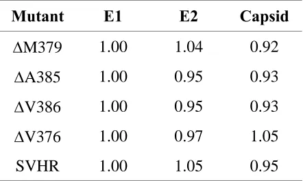
Table 2.1: Relative density of viral proteins produced from selected mutants. Purified virus released from cells was analyzed by SDS-PAGE and visualized by autoradiography. The density of the E1 band for each lane was set to an arbitrary unit of one. The E2 and capsid band densities were reported as a fraction of the E1 value.
Mutant
E1
E2 Capsid
Δ
M379
1.00
1.04
0.92
Δ
A385
1.00
0.95
0.93
Δ
V386
1.00
0.95
0.93
Δ
V376
1.00
0.97
1.05
particle/pfu data supports these conclusions in that the density of the mutant virus particles is similar to wt density suggesting a normal complement of protein, lipid, and RNA. If the altered particle to PFU ratio was due to failure to incorporate structural proteins in normal ratios the density of the virus produced would be expected to be different and the particle to PFU ratio artificially low.
Compensatory mutations for TM mutants
Deletion mutants yielding low amounts of infectious virus (ΔM379, ΔA385,
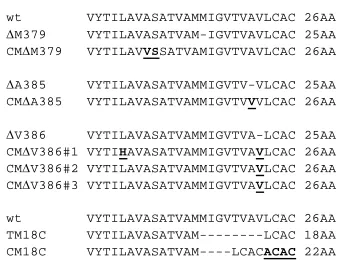
ΔV386, and TM18C) were subjected to serial passage in an attempt to produce mutations, which may rescue virus infectivity (compensatory mutations). Other mutants yielding higher infectious virus production were not subjected to serial passage as their titers were near wild type levels and the lack of selective pressure might result in the production of amino acid changes not related to the original defect. After three passages for ΔM379, ΔA385, and ΔV386 and five passages for TM18C an increase in infectious virus production was observed (Figure 2.4). After passage the single deletion mutants produced infectious virus at the same level as wt and the TM18C mutant infectious virus production was increased by
1.00E+04
1.00E+05
1.00E+06
1.00E+07
1.00E+08
1.00E+09
1.00E+10
1.00E+11
M3
79
A3
85
V3
86
18C
P
fu
/m
l
M utant
Δ Δ Δ
wt VYTILAVASATVAMMIGVTVAVLCAC 26AA
ΔM379 VYTILAVASATVAM-IGVTVAVLCAC 25AA CMΔM379 VYTILAVVSSATVAMIGVTVAVLCAC 26AA
ΔA385 VYTILAVASATVAMMIGVTV-VLCAC 25AA CMΔA385 VYTILAVASATVAMMIGVTVVVLCAC 26AA
ΔV386 VYTILAVASATVAMMIGVTVA-LCAC 25AA CMΔV386#1 VYTIHAVASATVAMMIGVTVAVLCAC 26AA CMΔV386#2 VYTILAVASATVAMMIGVTVAVLCAC 26AA CMΔV386#3 VYTILAVASATVAMMIGVTVAVLCAC 26AA wt VYTILAVASATVAMMIGVTVAVLCAC 26AA TM18C VYTILAVASATVAM---LCAC 18AA CM18C VYTILAVASATVAM----LCACACAC 22AA
Figure 2.5: Compensatory mutations (CM) found in the E2 transmembrane domain. After serial passage and an increase in infectious virus production, virus was
isolated, purified, and RT/PCR followed by sequencing was performed. Mutants compensating for the increase in virus growth are underlined and highlighted in bold lettering. All mutants were examined in triplicate and all three trials resulted in
was inserted adjacent to it. This compensatory mutation restored the wild type length (26 amino acids) to the transmembrane domain but did not restore M at 379. The ΔA385 compensatory mutation inserted a valine at the position of the alanine deletion, which also resulted in a wt length transmembrane domain. The ΔV386 compensatory mutation reverted back to the original wt sequence and length, with the exception of a leucine being converted to a histidine for ΔV386#1 (Figure 2.5). These revertants suggest that wt length is important for normal virus growth in the C terminal half of the transmembrane domain.
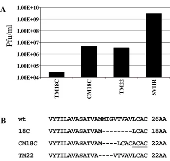
After five passages the TM18C mutant demonstrated a 100-fold increase in infectious virus production. The increase in virus production was accompanied by a four amino acid insertion, ACAC in the C terminal domain of the E2 transmembrane. The ACAC that was inserted contains the same nucleotide sequence as a repeat of the wt AC sequence preceding it (Figure 2.5). The TM18C compensatory mutation results in an increase from 18 to 22 amino acids spanning the transmembrane domain. When this compensatory mutation is compared to a previously described TM22 mutation (Hernandez et al., 2003) it was found that it produces approximately the same amount of infectious virus (106 pfu/ml) and the location of the remaining four amino acid deletion is in approximately the same location (Figure 2.6). This stresses the importance of the size and location of the deletion in the E2
Pfu
/m
l
wt VYTILAVASATVAMMIGVTVAVLCAC 26AA
18C VYTILAVASATVAM---LCAC 18AA
CM18C VYTILAVASATVAM----LCACACAC 22AA
TM22 VYTILAVASATVA----VTVAVLCAC 22AA
1.00E+04 1.00E+05 1.00E+06 1.00E+07 1.00E+08 1.00E+09 1.00E+10
TM
18C
CM
18
C
TM
22
SVH
R
A
B
deletion rather than altering a protein domain with which the mutated sequence is presumed to interact.
Discussion
The data presented herein demonstrate that a single deletion in the E2 transmembrane domain results in a significant loss of ability to produce infectious virus. It also shows that the effect of this deletion varies with its position in the TM sequence. The production of infectious virus is low for deletion mutations ΔM379,
ΔA385, and ΔV386, located near the carboxyl terminus of the TM domain, while
ΔA370, ΔV371, ΔA372, ΔS373, and ΔV376, located near the amino terminal end produce significantly more infectious virus. We also found that the inhibitory effect of a single insertion (FF) at the membrane interface is rescued when a compensating single amino acid deletion is placed in the middle of the transmembrane domain restoring the wild type number of amino acids in the E2 C terminal region (FF25). The relative importance of the two halves of the transmembrane domain was illustrated by the observation that the N terminal region of the E2 transmembrane domain could be deleted (TM18N) and the resulting mutant virus maintained wt growth whereas, deletion of the COOH terminal region (TM18C) resulted in a 10,000 fold decrease in infectious virus production.
deletions in the transmembrane domain is not affected until the deletion is larger than 12 amino acids (Hernandez et al., 2003). The deletion mutants produced all band at wild type virus density indicating that they are similar in composition to wild type virus in that they contain a normal complement of protein, lipid and RNA. SDS-PAGE of proteins in purified virus produced by the mutants indicated that they contained the same relative amounts of E1, E2 and capsid (Table 2.1). The process of assembly of these viruses is dependent upon a strict interaction of the three structural proteins in a 1:1:1 stoicheometric relationship. These data rule out the possibility that the reduced infectivity of the mutant viruses is the result of failure to assemble the normal amounts (240 copies of each) of virus proteins. Such an event would have, also, altered the buoyant density of the virus produced. Therefore, for all of the mutations produced, we have proposed a set of possible explanations to address the hypotheses stated in the introduction. These hypotheses were initially posed to account for the observed loss of infectious virus production produced by the original TM25 mutant (Hernandez et al., 2003):
Hypothesis i) The observed phenotype of the original TM25 deletion
Hypothesis ii) The angular location of the deletion on the face of the helix may account for the loss of infectivity (ie. if the deletion is made 90 degrees, 180 degrees or another angle from a set point of reference). Figure 2.2 reveals that the single amino acid deletions at positions ΔM379, ΔA385 and ΔV386 are located on the same side of the hydrophobic helix. These three deletions resulted in reduced virus growth, however ΔV371 is located on the same face of the hydrophobic helix and produces 100 times more infectious virus. This result suggests that the angular location on the face of the helix does not account for the loss of infectivity.
Hypothesis iii) The location of the deletion vertically (ie closer to the lumenal side or closer to the cytoplasmic side of the membrane) may contribute to the observed phenotype. We found that single deletion mutants located closer to the lumenal face of the transmembrane had a less detrimental effect on virus infectivity than those located closer to the cytoplasmic face (Figure 2.2 and 2.3). This
relationship was also true for the eight amino acid deletion (TM18) mutants where the TM18C mutant grew to a titer of 104 and the TM18N mutant grew to a titer of 109
growth. This phenomenon may be explained by positioning the mutant E2 endodomain in an orientation unfavorable for correct interaction with the capsid protein. Deletions closer to the N terminus may allow for more degrees of freedom for E2 to interact properly with the capsid protein. Mutations located closer to the luminal face may have more flexibility to restore the wild type exit point of the protein from the cytoplasmic face of the transmembrane region.
The single deletion in the FF insertion mutant (FF25) mutant restores normal growth to the FF mutant. The FF25 mutant also restores the wild type number of amino acids in the C terminal region of E2. This result suggests the titer was restored because the length of the C terminal domain is critical for normal virus growth whereas the length in the N terminal domain is not critical. This is supported by the TM18N and TM18C comparison.
transmembrane domain of E2 (I380). In addition structural evidence from Mancini et al (Mancini et al., 2000) shows E1 and E2 protein densities in the transmembrane domain have the greatest degree of separation at the N terminal region and a minimal amount of separation halfway through the transmembrane domain. Zhang et al also suggest that the E1 and E2 proteins are separated at the N terminal region of the transmembrane domain but come together in a coil-coil structure toward the C terminal region of the transmembrane domain (Zhang et al., 2002). These results suggest the E1 and E2 transmembrane domains interact at a point halfway across the membrane bilayer and the association continues through the C terminal region of the transmembrane domain. This data may also explain why eight amino acids may be removed from the N terminal region of the E2 transmembrane domain with no loss of infectivity while deletion of eight amino acids in the C terminal domain results in a five log decrease of infectious virus production. These E1-E2 associations described in the electron cryomicrographs of Mancini et al (Mancini et al., 2000) may be necessary for virus stability and infectivity.
Only the FF insertion mutant seems to have an affect on virus particle assembly. The other mutants described in this study result in the production of approximately the same number of particles although the percentage of virus that is infectious vary greatly. The deletion mutants described in this study are not inhibited in their ability to transport membrane proteins to the cell surface as significant
1997). However, the E2 transmembrane domain deletions described here do not prevent this function. Mutations located closest to the cytoplasmic interface of the E2 transmembrane interface produce the lowest amount of infectious virus but have the highest particle/pfu ratio. This along with SDS-PAGE analysis and negative stains of purified virus particles indicates proteins are being processed correctly, assembled into particles, and released, but the infectivity of these mutants is reduced by some other mechanism – likely by inhibiting cell entry.
After several passages the production of infectious virus by ΔM379, ΔA385, and ΔV386 was restored to wt levels and a 100-fold increase in infectious virus production was observed for the eight amino acid deletion (TM18C) mutant. Compensating mutations were found in the E2 transmembrane domain for all of these mutants (Figure 2.5). For all of the single deletion mutants a restoration to wt length in the TM domain accompanied restoration of wild type phenotype. Wt length in the TM domain may be necessary in the C terminal region of the transmembrane domain to produce a functional interaction with E1 or the capsid protein. The presence of a specific number of amino acids in this region may be necessary for correct protein-protein associations.
18 amino acids in the transmembrane domain suggesting the sequence may be important for normal infectious virus production.
Compensatory mutations in TM18C resulted in a two-log increase in infectious virus production and the insertion of the amino acid sequence ACAC in the C terminal region of the transmembrane domain. The resulting 22 amino acid transmembrane domain deletion matched that of the original TM22 deletion mutant, produced by deletion cloning, (Figure 2.6) and produced the same amount of
infectious virus. This demonstrates that the location of the deletion is important for normal virus production. The insertion of the sequence ACAC in the C terminal region may indicate that it is necessary for normal virus growth. However, when the C at position 388 was converted to a serine from a wt parent no loss in infectious virus production was observed (data not shown). Schlesinger and others mutated both of these cysteines to alanines and found that there was no significant loss of virus infectivity (Ryan, Ivanova, and Schlesinger, 1998) suggesting the cysteines are not vital for normal virus growth. Collectively these results argue that
transmembrane domains may serve functions other than to simply anchor the protein.
Materials and methods
Cell culture and virus titration
Growth of baby hamster kidney cells (BHK-21) were described previously (Renz and Brown, 1976). BHK cells were grown and maintained in minimal essential medium containing Earl’s salts (Invitrogen, Carlsbad Calif.) supplemented with 10% fetal bovine serum (FBS) (Invitrogen), 5% tryptose phosphate broth, and 2 mM glutamine as described previously (Renz and Brown, 1976). The virus strain chosen for mutagenesis was Toto1101 (Rice et al., 1987) or Y420 which contains a
substitution in E2 at position 420 changing a serine to a tyrosine. Y420, and has been described previously (Liu and Brown, 1993). Titration of virus produced was performed using BHK-21 cell monolayers (Renz and Brown, 1976). Virus was titered in triplicate.
TM mutants and reverse transcription (RT)-PCR analysis of mutant viruses The TM mutants were all made by QuikChange site-directed mutagenesis
unique sites and linearized with Xho1. Infectious RNA was transcribed in vitro using SP6 RNA polymerase (New England Biolabs). To confirm that the desired deletions remained in the virus grown in cell culture, the RNA was extracted from the virus, reverse transcribed, and amplified by PCR (RT-PCR) (Hernandez et al., 2003).
In vitro transcription and RNA transfection
The mutant and wild-type cDNA constructs were prepared for transcription and transcribed in vitro as described previously (Hernandez et al., 2000) and
(Hernandez et al., 2003). The RNA transcripts were electroporated into BHK or U4.4 cells. The electroporation was performed as described by Lilijestrom and Garoff (Lilijestrom and Garoff, 1991). Virus was harvested 24 h posttransfection.
Virus growth, purification, quantitation and particle/PFU determination
Virus growth in U4.4 and BHK cells and subsequent purification and quantitation were described previously (Hernandez et al., 2003). The highest
multiplicity of infection possible for each of the mutant viruses was used for infection. An infection of the Toto1101 parent virus using a corresponding multiplicity of
10.8% poly-acrylamide gel to verify normal protein processing and to analyze the stoichiometric ratio of structural proteins produced. The resulting gels were exposed to X-ray film to produce exposures which did not, in any part of the gel, exceed the recording capacity (saturation density) of the film. The resulting protein profiles were scanned with an optical densitometer to obtain optical densities. For each mutant the optical density of the protein E1 was arbitrarily set as 1 and other protein optical densities were presented relative to E1 in Table 2.1.
The number of particles in a preparation of wild-type virus (SVHR) was determined under the electron microscope by the agar filtration protocol described by Kellenberger and Bitterli (Kellenberger and Bitterli, 1976), and the particle count was correlated to the amount of protein as obtained by BCA. The total particle count was divided by the amount of infectious virus particles produced to determine the total number of particles per infectious particle (particle/pfu).
Serial passage of transmembrane mutants
Plaques were then used for a final round of infection. After 24h virus was harvested, extracted, and RT/PCR was performed as described earlier. Resulting PCR
Chapter 3
Abstract
Sindbis virus is a single stranded positive sense RNA virus. It is composed of 240 copies of three structural proteins, E1, E2, and capsid. These proteins are arranged in a 1:1:1 molar ratio and form a mature virus particle composed of two nested T=4 icosahedral shells. A complex network of disulfide bonds in the E1 and E2 glycoproteins is developed through a series of structural intermediates as virus maturation occurs. E1 and E2 cysteine residues were labeled with iodoacetamide in the native virus particle and analyzed by mass spectrometry. This analysis identified cysteines of E1 and E2 which were found to be free in the native virus particle, as well as those that were either solvent inaccessible or blocked by their involvement in disulfide bonds. Native virus particles alkylated with iodoacetamide yielded a four-log decrease in viral infectivity. This suggests that either the bound iodoacetamide alone may be involved in the loss of infectivity by destabilizing the virus particle or that a rearrangement of disulfide bonds, which is required for infectivity, is blocked by the presence of iodoacetamide. To examine if a structural effect due to the alkylation of cysteine, or if a possible disulfide rearrangement was the cause of the loss in virus infectivity, cysteines that were determined to be free in the E1
Introduction
Sindbis virus (SV) is positive sense, single stranded RNA virus of the Alphavirus genus in the Togaviridae family and is propagated in nature by both
insect and mammalian hosts. The mature SV particle contains three structural proteins E1, E2, and capsid (C) in a 1:1:1 stoichiometric ratio. The SV particle is approximately 70 nm in diameter and serves as an excellent model for structural studies. Each virus particle is composed of 240 copies of each of these proteins producing 80 equivalent units which are organized into two geometrically identical T=4 icosahedral shells (Paredes et al., 1993). An outer protein shell, composed of the E1 and E2 glycoproteins, is organized into trimers of E1/E2 heterodimers (Anthony and Brown, 1991; Carleton et al., 1997; Pletnev et al., 2001). The outer protein shell surrounds an inner shell, which is composed entirely of the capsid protein. The 11.7 kb genomic RNA is packed inside the inner capsid shell.
Positioned between the two protein shells is a host-derived membrane bilayer that is penetrated by the transmembrane (TM) domain anchors of E1 and E2 (Lilijestrom and Garoff, 1991; Rice et al., 1982; Strauss, Lenches, and Strauss, 2002). The endodomain of E2 interacts with the capsid protein linking the two shells together.
structural components: PE2, 6K, and E1 (Lilijestrom and Garoff, 1991). The 6K protein plays a role in the correct integration of E1 into the membrane and is
necessary for efficient particle budding (Lilijestrom and Garoff, 1991). 6K is found only at very low levels in mature virus (Gaedigk-Nitschko and Schlesinger, 1990). The E1 glycoprotein, containing a functional domain necessary for viral entry, is folded into a stable compact high-energy conformation as it is assembled into heterotrimers with the precursor to E2, PE2 (Carleton et al., 1997; Mulvey and Brown, 1994; Mulvey and Brown, 1995; Mulvey and Brown, 1996). PE2 is
transported to the cell surface through the trans Golgi network where it is processed to E2 and E3 by furin (Moehring et al., 1993). Evidence suggests that E2 is
responsible for receptor recognition. E3 is not present in the mature virus. During transport of the trimeric complexes to the plasma membrane, a second membrane-spanning region of E2 is retracted from the membrane and a 33 amino acid
endodomain is exposed to the cytoplasm. At the plasma membrane the E1-E2 heteotrimers assemble into the outer protein shell by association of the E2
endodomain with a hydrophobic cleft in the capsid protein (Lee and Brown, 1994; Lee et al., 1996; Oates et al., 1992; Owen and Kuhn, 1996; Owen and Kuhn, 1997) This association of the E2 endodomain with the capsid protein gives stability to the structure of the virus (Lee, Ricker, and Brown, 1994) and plays a critical role in the formation of the viral outer protein shell around the preformed inner protein shell as the process of envelopment takes place (Ferreira et al., 2003).
endodomain consisting of the first 362 residues. Each of these membrane proteins contains 17 cysteine residues (capsid does not contain any cysteines), which are completely conserved across the Alphavirus genus with the exception of E1 C430 and E2 C388 and C390. These non-conserved cysteines are located in the transmembrane domains. Residues C396, C416 and C417 of the E2 protein are located in the 33 amino acid endodomain. Of the remaining cysteine residues, the possibility of eight disulfide bonds in the E1 protein and six in the E2 protein exists.
Little is known about the E2 disulfide network. Gidwitz et al suggested that all cysteines located in the E2 protein were either involved in disulfide bonds or were not accessible when treated with 14[C] iodoacetamide at a 1.1 molar excess of iodoacetamide to total cysteines (Gidwitz et al., 1988). The Semliki Forest Virus, also a member of the Alphavirus genus, E1 crystal structure has been solved at three angstrom resolution (Roussel et al., 2006). Herein the authors report disulfide bonds located at residues 49-144, 62-94, 63-96, 259-271, 376, 328-370, 301-376, and 306-380.
was concluded from these studies that the E1 protein acquires a stable conformation through the sequential formation of disulfide intermediates. It is imperative to
emphasize that attempts to extract the E1 protein from mature virus particles with detergent resulted in the generation of a ladder that may represent all the disulfide intermediates (Mulvey and Brown, 1994). The propensity of E1 to adopt alternate conformations makes isolation away from the mature virus in its native form very difficult. The crystal structure presented by Roussel et al is of the E1 protein which was extracted by detergent based methods (Roussel et al., 2006) and may not represent the native form.
In this report, we identify the location of free and disulfide bound cysteines in the Sindbis virus E1 and E2 proteins by incubation of native virus particles in the presence of iodoacetamide. Iodoacetamide alkylates free sulfhydryl groups and, in this case, revealed the location of free cysteines in the E1 and E2 proteins of the native virus particle. While it was found that virus particles alkylated with
present in the infectious virus particle and that disulfide bond rearrangements may occur as virus maturation proceeds.
Results
Location of the free cysteines in the Sindbis Virus E1 and E2 proteins
Mature virus particles were collected from the media of infected BHK cells and purified over potassium tartrate gradients. Purified virus was then dialyzed against PBS-D and alkylated by the addition of 500 fold molar excess iodoacetamide to total cysteine. After SDS PAGE analysis to separate the viral proteins and
remove excess iodoacetamide, virus particles were reduced with DTT and treated with 20 mM iodomethane (see Materials and Methods). Using this labeling
technique all cysteines containing a free sulfhydryl group (not involved in a disulfide bond) and accessible to solvent would be labeled with iodoacetamide. All other cysteines would be labeled with iodomethane. After labeling samples were examined by LC/MS/MS (ESI). Figure 3.1, Figure 3.2, and Table 3.1 represent a compilation of the data collected for the Sindbis virus E1 protein. This approach labeled14 of the 17 cysteines of E1. E1 cysteines 49, 114, 259, 271, and 430 were found to be labeled by both iodoacetamide and iodomethane. This suggests that these cysteines contained free sulfyhydryl groups but were only partially accessible to iodoacetamide. The remainder of the population would be labeled by
E1 structural protein - (14 of 17 identified)
YEHATTVPNVPQIPYKALVERAGYAPLNLEITVMSSEV
LPSTNQEYIT
C
C
KFTTVVPSPKIK
C
C
C
C
GSLE
C
C
QPAAHADY
T
C
C
KVFGGVYPFMWGGAQCFCDSENSQMSEAYVELSA
D
C
C
ASDHAQAIKVHTAAMKVGLRIVYGNTTSFLDVYVN
GVTPGTSKDLKVIAGPISASFTPFDHKVVIHRGLVYNY
DFPEYGAMKPGAFGDIQATSLTSKDLIASTDIRLLKPSA
KNVHVPYTQASSGFEMWKNNSGRPLQETAPFG
C
C
KIAV
NPLRAVD
C
C
SYGNIPISIDIPNAAFIRTSDAPLVSTVK
C
C
EV
SE
C
C
TYSADFGGMATLQYVSDREGQ
C
C
PVHSHSSTATLQ
ESTVHVLEKGAVTVHFSTASPQANFIVSL
C
C
GKKTT
C
C
NA
E
C
C
KPPADHIVSTPHKNDQEFQAAISKTSWSWLFALFGG
ASSLLIIGLMIFA
C
C
SMMLTSTRR
Blue = iodomethane
Red = iodoacetamide
Green = Both
E2 structural protein - (5 of 17 identified)
SVIDDFTLTSPYLGTCSYCHHTVPCFSPVKIEQVWDEA
DDNTIRIQTSAQFGYDQSGAASANKYRYMSLKQDHTV
KEGTMDDIKISTSGPCRRLSYKGYFLLAK
C
C
PPGDSVTV
SIVSSNSATSCTLARKIKPKFVGREKYDLPPVHGKKIP
C
C
TVYDRLKETTAGYITMHRPRPHAYTSYLEESSGKVYA
KPPSGKNITYE
C
C
KCGDYKTGTVSTRTEITG
C
C
TAIKQCV
AYKSDQTKWVFNSPDLIRHDDHTAQGKLHLPFKLIPST
CMVPVAHAPNVIHGFKHISLQLDTDHLTLLTTRRLGAN
PEPTTEWIVGKTVRNFTVDRDGLEYIWGNHEPVRVYA
QESAPGDPHGWPHEIVQHYYHRHPVYTILAVASATVA
MMIGVTVAVLCACKARRE
C
C
LTPYALAPNAVIPTSLALL
CCVRSANA
Blue = iodomethane
Red = iodoacetamide
Green = Both
105 = 100%
152 = 0.5%
220 = 1.5%
105, 152, 220
201, 411
E2
114 = 90%
259 = 17%
271 = 73%
430 = 100%
49, 114, 259,
271, 430
62, 63, 68, 78,
301, 306, 328,
370, 376, 380
E1
% Free
Free
Inaccessible or
Bound
Table 3.1: Free and bound cysteines of the Sindbis virus E1 and E2 proteins. Residues listed in the “Inaccessible or Bound” column represent cysteines that were only alkylated by iodomethane. Residues listed in the “Free” column were detected alkylated with iodoacetamide. “% Free” is the calculated amount of cysteine labeled with iodoacetamide when both labels were detected.
blocked due to the presence of disulfide bonds. E1 C430 was only detected in the form labeled by iodoacetamide suggesting it was accessible to solvent and was not involved in a disulfide bond. In Table 3.1 a column titled “% free” represents a measure of the percent of the sample alkylated by iodoacetamide (the population that was accessible and had free sulfhydryl groups) when both labels were detected. Precursor ions and extracted ion chromatograms were utilized to obtain this data and offer only an approximation of the true value. The percentage of cysteines alkylated by iodoacetamide for E1 cysteines 114, 259, 271, and 430 was 90%, 17%, 73%, and 100% respectively. The remaining percentage was found alkylated by iodomethane. These values indicate that some cysteines in the native virus particle are more accessible to iodoacetamide while some are less accessible and are buried deeper in the virus particle.
Five of the 17 cysteines were labeled in the E2 protein. C105, C152, and C220 were all labeled, at least partially, with the iodoacetamide reagent
demonstrating that a population of these cysteines have free sulfhydryl groups and are accessible to solvent and therefore cannot be involved in disulfide bonds in the native particle. E2 C201 and C411 were only detected alkylated by iodomethane and may be involved in disulfide bonds. Analysis of precursor ions and extracted ion chromatograms revealed that only a small population of E2 152 and 220 were found to have free sulfhydryl groups (0.5% and 1.5% respectively). E2 C105 was only detected in the “free” state.
are known to be involved in disulfide bonds. Five of the eight cysteines were labeled and detected by mass spectrometry (Figure 3.3). All cysteines detected were only alkylated by iodomethane indicating that they were not accessible to solvent or that they were blocked by the presence of disulfide bonds. This validates the labeling strategy since no cysteines of RNase B were found to have free sulfhydryl groups. This is in agreement with the published disulfide network present in RNase B (Shin et al., 2003).
Effect of iodoacetamide labeling on virus infectivity
Purified virus was incubated at 100 or 500 fold excess iodoacetamide to cysteine at 37 degrees C and pH 8.0 for 2 hours and was titered on BHK cells (Table 3.2). Purified virus treated with 100 fold excess of iodoacetamide resulted in a two log decrease in virus infectivity while treatment of virus with 500 fold excess
iodoacetamide resulted in a three and a half log decrease of infectious virus when compared to wild type purified virus. This data reveals that iodoacetamide is
RNase B - (5 of 8 identified)
KETAAAKFERQHMDSSTSAASSSNYCNQMMKSRNLT
KDR
C
C
KPVNTFVHESLADVQAV
C
C
SQKNVACKNGQTN
C
C
YQSYSTMSITD
C
C
RETGSSKYPN
C
C
AYKTTQANKHIIVAC
EGNPYVPVHFDASV
Blue = iodomethane
Red = iodoacetamide
Green = Both
9.0 x 10
6
pfu/ml
Purified virus
500x
1.5 x 10
8
pfu/ml
Purified Virus
100x
2.0 x 10
10
pfu/ml
Purified virus
Titer
Virus
of cysteine is most efficient at pH 8.0 and since no differences in infectious viral titer were observed at pH 8.0, this pH was adopted for experimentation.
Conformational changes induced with low pH do not alter the location of free cysteines
The location of the free cysteines after the pH shift described above is illustrated in Figure 3.4, Figure 3.5, and Table 3.3. Six of the 17 cysteines of E1 were identified. The reduction in the amount of coverage for the pH shift
experiments compared to the non-shifted sample were probably due to the sample concentration. When the pH was shifted to 5.3 the virus began to precipitate above certain concentrations. Therefore the concentration was adjusted to an amount where no precipitation occurred (see Methods). Both C259 and C271 of E1 were found to be alkylated by iodoacetamide suggesting that they are not involved in disulfide bonds and are accessible to solvent. Cysteines at positions 62, 301, 306, and 328 were only alkylated with iodomethane reagent suggesting that these residues are not available to reagent and that they may be involved in disulfide bonds. The % free (% labeled with iodoacetamide reagent) for C259 was 29% and 100% for C271. This shows a general increase of accessibility to reagent for the pH-shifted sample compared to the sample maintained at pH 8.0. However, no cysteines that were found to have free sulfyhydryl groups at pH 8.0 were found to be inaccessible or blocked after the pH shift or vice versa. No detectable
rearrangement of disulfide bonds was observed after low pH exposure.
E1 structural protein - (6 of 17 identified)
YEHATTVPNVPQIPYKALVERAGYAPLNLEITVMSSEV
LPSTNQEYITCKFTTVVPSPKIK
C
C
CGSLECQPAAHADYT
CKVFGGVYPFMWGGAQCFCDSENSQMSEAYVELSAD
CASDHAQAIKVHTAAMKVGLRIVYGNTTSFLDVYVNG
VTPGTSKDLKVIAGPISASFTPFDHKVVIHRGLVYNYDF
PEYGAMKPGAFGDIQATSLTSKDLIASTDIRLLKPSAKN
VHVPYTQASSGFEMWKNNSGRPLQETAPFG
C
C
KIAVNP
LRAVD
C
C
SYGNIPISIDIPNAAFIRTSDAPLVSTVK
C
C
EVSE
C
C
TYSADFGGMATLQYVSDREGQ
C
C
PVHSHSSTATLQES
TVHVLEKGAVTVHFSTASPQANFIVSLCGKKTTCNAEC
KPPADHIVSTPHKNDQEFQAAISKTSWSWLFALFGGAS
SLLIIGLMIFACSMMLTSTRR