RoseMarie M. Rine, PhD
Michael C. Schubert,
PhD
Susan L. Whitney, PhD
Dale Roberts, MS
Mark S. Redfern, PhD
Mark C. Musolino, PhD
Jennica L. Roche, MS
Daniel P. Steed, MS
Bree Corbin, MS
Chia-Cheng Lin, MS
Greg F. Marchetti, PhD
Jennifer Beaumont, MS
John P. Carey, MD
Neil P. Shepard, PhD
Gary P. Jacobson, PhD
Diane M. Wrisley, PhD
Howard J. Hoffman, MA
Gabriel Furman
Jerry Slotkin, PhD
Correspondence to Dr. Rine:
Vestibular function assessment using the
NIH Toolbox
ABSTRACT
Objective:
Development of an easy to administer, low-cost test of vestibular function.
Methods:
Members of the NIH Toolbox Sensory Domain Vestibular, Vision, and Motor subdomain
teams collaborated to identify 2 tests: 1) Dynamic Visual Acuity (DVA), and 2) the Balance
Accel-erometry Measure (BAM). Extensive work was completed to identify and develop appropriate
software and hardware. More than 300 subjects between the ages of 3 and 85 years, with
and without vestibular dysfunction, were recruited and tested. Currently accepted gold standard
measures of static visual acuity, vestibular function, dynamic visual acuity, and balance were
per-formed to determine validity. Repeat testing was perper-formed to examine reliability.
Results:
The DVA and BAM tests are affordable and appropriate for use for individuals 3 through
85 years of age. The DVA had fair to good reliability (0.41
–
0.94) and sensitivity and specificity
(50%
–
73%), depending on age and optotype chosen. The BAM test was moderately correlated
with center of pressure (
r
5
0.42
–
0.48) and dynamic posturography (
r
5 20.48), depending on
age and test condition. Both tests differentiated those with and without vestibular impairment
and the young from the old. Each test was reliable.
Conclusion:
The newly created DVA test provides a valid measure of visual acuity with the head
still and moving quickly. The novel BAM is a valid measure of balance. Both tests are sensitive to
age-related changes and are able to screen for impairment of the vestibular system.
Neurologyâ2013;80 (Suppl 3):S25–S31
GLOSSARY
BAM5Balance Accelerometry Measure;COP5center of pressure;DS5double-limb feet together stance;DVA5dynamic visual acuity;ETDRS5Early Treatment Diabetic Retinopathy Study;ICC5intraclass correlation coefficient;logMAR5 logarithm of the minimum angle of resolution;NPL5normalized path length;SOT5sensory organization testing;SVA5 static visual acuity;TS5tandem stance;VH5vestibular hypofunction;VOR5vestibulo-ocular reflex;VSP5 vestibulo-spinal outputs.
The vestibular system is an integral component of our sensory experience and sensory-motor
function. Healthy peripheral and central vestibular anatomy is essential for functionally relevant
gaze stability during head motion and postural control.
1However, gaze stabilization and balance
are enabled by multiple sensory inputs (e.g., visual, somatosensory, and vestibular), integration
and interpretation of these redundant sources of orientation, and motor output, each of which
may be affected by age or damage.
1–4Patients with vestibular pathology often report oscillopsia
(due to gaze instability), imbalance, and/or vertigo. The redundancy of the sensory information
confounds distinction of the causal mechanisms of oscillopsia and/or imbalance. However, the
unique contributions of the vestibulo-ocular reflex (VOR) and vestibulospinal outputs (VSP) to
gaze stability and balance, respectively, can direct the identification of tests to screen for
pathol-ogy of the vestibular system.
From the Specialty Therapy Source LLC (R.M.R., B.C.), Jacksonville, FL; Departments of Otolaryngology Head and Neck Surgery (M.C.S., J.P.C.) and Neurology (D.R.), John Hopkins School of Medicine, Baltimore, MD; Departments of Physical Therapy (S.L.W., C.-C.L.) and Bioengineering (M.S.R., J.L.R., D.P.S.) and College of Arts and Sciences (G.F.), University of Pittsburgh, Pittsburgh, PA; Crossroads Consulting, LLC (M.C.M.), Johnstown, PA; Department of Physical Therapy (G.F.M.), Duquesne University, Pittsburgh, PA; Department of Medical Social Sciences (J.B., J.S.), Northwestern University, Chicago, IL; Department of Otorhinolaryngology (N.P.S.), Vestibular and Balance Laboratory, Mayo Clinic, Rochester MN; Department of Hearing and Speech Sciences (G.P.J.), Division of Audiology, Vanderbilt University Medical Center, Nashville, TN; Department of Physical Therapy (D.M.W.), Lynchburg College, Lynchburg, VA; Epidemiology and Statistics Program (H.J.H.), National Institute on Deafness and Other Communication Disorders, NIH, Bethesda, MD; and Rehabilitation Research Chair (S.L.W.), King Saud University, Riyadh, Saudi Arabia.
The semicircular canals and otolith end
or-gans are the primary contributors to the VOR
and VSP, respectively, but not exclusively.
Dam-age to either of these organs will lead to unique
impairments. Investigators have reported that
pa-tients with canal dysfunction have impaired
dynamic visual acuity (DVA),
5–12and damage
to the vestibulospinal system causes postural
con-trol impairment.
13–16Furthermore, adults and
children with unilateral or bilateral vestibular
dysfunction fail balance tests that require
resolu-tion of sensory conflicts regarding posture and
that rely primarily on vestibular afference.
3,17–20Based on a comprehensive review of available
tests and the literature, the NIH Toolbox
Ves-tibular Team (R.M.R., M.C.S., S.L.W., J.P.C.,
N.P.S., G.P.J., D.M.W.) decided to include
one test that attempts to isolate the vestibular
system
’
s contribution to gaze stability (indirect
measure of the VOR), and one that attempts
to isolate the vestibular system
’
s contribution
to postural control (indirect measure of VSP).
Most tests were excluded because of 1) reliance
on self-report of symptoms, 2) high cost, 3)
requirement of expertise to administer and
interpret, and/or 4) lack of sensitivity, validity,
or reliability.
The team selected 2 tests: the Dynamic Visual
Acuity test and a Balance Accelerometry
Mea-sure (BAM) (a modification of the Clinical
Test of Sensory Interaction for Balance.
20Fur-thermore, it was agreed that the static
compo-nent (static visual acuity [SVA]) of the DVA and
BAM would be used as part of vision testing and
as the NIH Toolbox Motor Domain balance
measure, respectively. Although both the DVA
and balance measures have a well-established
his-tory of use clinically and for research, clinical
versions require expertise and have limited
sen-sitivity and specificity. Well-established, existing
computerized versions that quantify the DVA
and postural control are expensive. Thus, new
versions of these tests were developed, modified,
and validated for inclusion in the NIH Toolbox.
METHODSDVA test.Equipment.A low-cost computerized test that minimizes motor, language, and cultural effects was developed (R.M.R., M.C.S., D.R., B.C., J.B., J.S.).21Custom
software was written in Python and C11. Hardware included a 2-GHz Intel dual central processing unit laptop with 2 GB of RAM (IBM Thinkpad; IBM, Armonk, NY). The laptop was connected to a 14403900 resolution monitor that displayed the optotypes. The operator used the built-in laptop display. For the head motion subtest, a single-axis rate sensor (O-Navi, Vista, CA) for detecting horizontal head rotation was attached to a soft bicycle light strap and secured to the head (figure 1). The rate sensor triggered the software to flash an optotype only when head velocity met or exceeded 180° per second.
Technique.The test required an individual to identify an optotype (letter or symbol) presented one at a time at progres-sively smaller sizes. Subjects sat 12.5 ft away from the viewing screen at their eye level. An initial“quick”screening of SVA was completed starting at size 20/50 and presenting a single ran-dom letter per acuity size (steps of 0.1 logarithm of the minimum angle of resolution [logMAR]), either going smaller until one was identified incorrectly or larger until one was identified correctly. Next we collected the subjects’static and dynamic visual acu-ity scores. For SVA, the head was kept stationary. For DVA, the subject actively moved his or her head to the left and right, but we were able to uniquely flash an optotype for rotations to one side only, enabling us to determine a DVA score for left and right rotations. Five optotypes per size were presented. Static testing trials began at 20/80 and continued through to identify the small-est size for which all 5 optotypes were correctly identified (min-imal size to accurately identify all optotypes at a size) and the smallest size for which at least 3 of 5 optotypes were correctly identified (the minimal size to accurately identify the majority of optotypes at a size). The trial stopped automatically when these criteria were met. Dynamic testing was performed similarly, except the trial started at 3 sizes above the static acuity level. Additionally, all children completed training trials as described previously21to assure proper head movement; testing proceeded
when 80% success was achieved in training. During dynamic testing, the optotypes appeared only if the head moved.180° per second (monitored by a rate sensor on a headband; figure 1). All subjects were encouraged to guess even if they were not con-fident (i.e., forced-guess paradigm). Retesting was completed on
the same day within 1 hour by 53 adults and within 5 to 10 days by 246 children.
Scores were determined from each subject performing a single SVA test and 2 DVA tests (right and left).21The difference in
log-MAR scores achieved on the static and dynamic conditions is the DVA score (calculated separately for leftward and rightward rota-tions), which represents the vestibular contribution to gaze stability. We determined the age at which letters vs symbols were most effective, valid, and reliable for testing visual acuity. To identify the optimal optotypes for use with children, subjects younger than 13 years were tested using different optotype sets (Lea, HOTV, and Early Treatment Diabetic Retinopathy Study [ETDRS] test22,23;
table 1) in a single session without rest. Sequence of optotype set presentation was assigned in a random block design to control for the effects of fatigue and/or boredom.
Subjects without (n5301; 51% female, 49% male) and with (n517; 59% female, 41% male) vestibular hypofunction (VH) (bilateral or unilateral, confirmed by rotary and/or caloric tests) participated. Ocular motor examination and medical history review was completed to ensure that exclusion criteria were met: central or peripheral nervous system pathology (except ves-tibular), oculomotor weakness, cognitive deficit, and pregnancy.
SVA was also tested using the gold standard ETDRS testing using backlit (lightbox) charts to enable examination of validity.23
Statistical analysis. Descriptive statistics were calculated to summarize subject characteristics and completion rates. SVA scores were correlated with scores obtained using the lightbox technique to assess validity of the static test. Success rates on each optotype set and correlations among optotypes were examined to determine the optimal optotype for use with children. To examine validity of the DVA, scores from subjects with and without VH were compared using attest, adult DVA scores obtained here were compared with scores on a previously validated and reliable computerized DVA test using attest,5and sensitivity and specificity were calculated. Test-retest
reliability was assessed using intraclass correlation coefficients (ICCs).
Balance Accelerometry Measure.Equipment.Accelerometers have been used to assess postural control24in older adults and to
identify early symptoms of Parkinson disease.25A low-cost tool that
quantifies postural sway using an accelerometer was developed by team members at the University of Pittsburgh.26A dual-axis
accel-erometer (ADXL213AE,61.2 g; Analog Devices, Inc., Norwood, MA) was chosen for its low-frequency characteristics and because it was designed to optimally record mediolateral and anteroposterior acceleration of the pelvis/trunk during standing. The accelerations are transmitted wirelessly using 16-bit bluetooth transmission at 100 Hz. Full description of hardware and algorithms used are avail-able in a previous report.26
Technique.Initially, 26 standing test positions/conditions were explored; the most difficult was a single leg stance on a dense foam pad with eyes closed while moving the head in the yaw plane at approximately 2 Hz. The reliability and feasibility of measuring accelerations of the pelvis for the various conditions was evaluated. Six conditions were chosen to measure vestibulo-spinal function during standing, which the team believed had the potential to differentiate persons by age, and yet be completed by most participants across the age span. These include double-limb feet together stance (DS) or tandem stance (TS) under var-ied vision and surface conditions: 1) eyes open on a solid floor DS, 2) eyes closed on a solid floor DS, 3) eyes open on dense foam DS, 4) eyes closed on dense foam DS, 5) eyes open on solid floor TS, and 6) eyes closed on solid floor TS. After testing several types of foam, the Airex balance pad (Advanced Medical Tech-nology Inc., Watertown, MA) was chosen for the foam test con-dition. This pad created the greatest sway on a force plate compared with 2 other types of frequently used foam pads.
The BAM protocol was tested by the balance team (S.L.W., R.M.R., M.S.R., M.C.M., B.C., G.F.M., J.L.R., C.C.L., G.F., D.P.S., H.J.H., J.S.) using subjects of varying ages with and with-out vestibular pathology. Subjects between the ages of 3 and 85 years without (n5203) and with (n525) VH were recruited, grouped by age (table 2), and tested. A medical history review, and somatosensory, vision, vestibular, and lower extremity strength screening were completed on all subjects to ensure compliance with exclusion criteria as noted above.
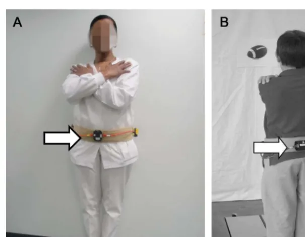
All subjects were asked to stand quietly for 70 seconds, and to look at a symbol or picture placed centrally at eye height, in eyes-open conditions. The first 5 seconds of data were discarded to ensure sta-bility of measures, yielding 65 seconds of data for analysis. The nor-malized path length (NPL) (mG/s; higher values indicate more sway) of the acceleration time series calculated was used for scoring and analyses. Concurrent measures of center of pressure (COP) were obtained from a force plate. For the adults, the BAM was placed ante-riorly around the waist using a gait belt. To prevent children younger than 13 years from disrupting the apparatus, the accelerometer was placed posteriorly (figure 2). All subjects were given 3 attempts to complete 1 trial of each condition. Repeat testing was performed by
Table 1 Reliability of static and dynamic acuity scores by age
Lea HOTV ETDRS
Age, y No.
Static ICCb Dynamic ICC Static ICC Dynamic ICC Static ICC Dynamic ICC
3–4 25 0.71a 0.05 0.75a 0.49a — —
5–6 27 0.80a 0.43a 0.66a 0.52a — —
7–12 136 0.92a 0.26a 0.94a 0.46a 0.85a 0.41a
13–17 24 — — — — 0.75a 0.73a
18–75 24 — — — — 0.84a 0.79a
Abbreviations: ETDRS 5Early Treatment Diabetic Retinopathy Study; ICC5intraclass correlation coefficient.
aSignificance level
p#0.05.
bICC is an assessment of the consistency of measurements made by an instrument.
Table 2 Reliability of BAM across age groupsa
ICC
Age, y No. BAM-1 BAM-2 BAM-3 BAM-4 BAM-5 BAM-6
3–8.5 45 Unable because of lack of variance
8.6–17 62 0.82 0.88 0.97 0.87 0.97 0.73
18–35 31 0.72 0.56 0.33 0.79 0.34 0.48
36–65 41 0.89 0.74 0.73 0.82 0.83 0.27
66–85 29 0.87 0.86 0.88 0.87 0.87 0.06
3–17 107 0.82 0.87 0.97 0.87 0.95 0.70
18–85 101 0.86 0.83 0.74 0.77 0.82 0.36
Abbreviations: BAM 5 Balance Accelerometry Measure; ICC 5 intraclass correlation coefficient.
aBAM conditions: 1
most subjects (n5206) on the same day after a short rest (minimum of 25 minutes). Subjects also completed posturography sensory orga-nization testing (SOT) (higher values indicate less sway),27with
con-current BAM to examine validity.
To examine the impact of accelerometer placement for children, whose locus of balance control is different than adults,3,283.0- to
8.5-year-old children completed testing with the device at the waist and again with the device placed at the upper thoracic level.
Statistical analysis.Reliability (ICCs) was calculated between the test and retest trials at 40, 50, and 60 seconds. BAM was com-pared with COP measures using a Pearson correlation coefficient. Concurrent validity of BAM scores with those obtained from the SOT was evaluated with a Spearman nonparametric correlation coefficient. SOT and BAM scores of subjects who could not com-plete the task because of a loss of balance were assigned a score equal to 3 SDs below the mean (age-group matched). Results obtained from those with and without VH (confirmed as above) were com-pared to determine validity using attest for independent samples. Mean age group differences on BAM scores were tested using a 1-way analysis of variance separately within pediatric and adult age ranges.
RESULTS Dynamic visual acuity.
Examination of
suc-cessful completion rates revealed that 100% of
individ-uals 13 years and older completed ETDRS testing, and
99% of children 5 through 12 years completed HOTV
and Lea testing. Furthermore, among the 3- and
4-year-olds, 91% and 85% completed static testing using
Lea and HOTV optotypes, respectively. However, in
the dynamic condition, only 74% and 69% were able
to complete testing using the Lea and HOTV
opto-types, respectively. There was excellent correlation
between optotypes for HOTV and ETDRS (
r
$
0.93,
p
5
0.001) and for Lea and ETDRS (
r
5
0.90,
p
5
0.001). Furthermore, there was good correlation
(
r
5
0.78,
p
5
0.001) of the NIH Toolbox static
ETDRS scores with those from the ETDRS chart
test-ing with a lightbox. Eighty-two percent of subjects
with pathology were aged 9 years or older, resulting
in minimal variance of scores obtained from the
younger groups (Lea and HOTV). The significantly
larger scores obtained by those with pathology
re-sulted in a greater variance in the age groups including
these subjects. Therefore, the lack of subjects with
pathology in the younger groups would contribute
to minimal variance and the low ICC values, and
an underestimation of reliability for the dynamic test
in the younger groups. However, scores achieved by
children older than 10 years and adults were reliable
(table 1).
DVA scores differed between those with and
with-out VH (
p
#
0.001 for adults;
p
#
0.1 for ETDRS
optotype in children). Effect sizes (mean difference/
SD) ranged from 0.80 to 1.84. Furthermore, dynamic
testing with HOTV, Lea, and ETDRS had fair to good
sensitivity (50%, 67%, and 73%, respectively) and
spec-ificity (73%, 63%, and 69%, respectively) and excellent
negative predictive value (
$
96% for all).
Balance Accelerometry Measure.
Review of the rates of
successful completion on each condition by age
re-vealed that most healthy individuals older than 8 years
could complete conditions 1 through 6. The majority
of children 3 through 5.9 years completed conditions
1 through 4, and those 6 through 8 years completed
conditions 1 through 5. Results of the ICCs support
that analysis of only 40 seconds of BAM data was
needed to yield reliable data for children and adults
(table 2). It must be noted that because of minimal
variance (low number of subjects with pathology),
re-sults are likely an underestimation of reliability.
The correlations of NPL and COP recordings ranged
from poor to excellent. For children,
r
values ranged
from 0.42 to 0.85, and were best with placement of
the accelerometer at the waist. For adults,
r
values ranged
from 0.42 to 0.70 across age groups and trial.
Correlation of scores from BAM condition 4
(stand-ing on foam with eyes closed) and comparable
posturog-raphy condition 5 (standing on sway-referenced support
surface with eyes closed) was moderate for children and
adults (
r
5 2
0.48,
p
5
0.04; and
r
5 2
0.42,
p
5
0.01, respectively). These are the conditions believed to
be most dependent on vestibular function for balance.
The small number of subjects with pathology (6
children and 18 adults) suggests low power. Despite
this, for adults, the NPL measure differed between
those with and without pathology on conditions 2
(
p
5
0.02), 5 (
p
5
0.04), and 6 (
p
5
0.05), with a
trend for significance on 4 (
p
5
0.10). For children,
scores differed only on condition 6 (
p
5
0.001), with
a trend for significance on condition 4 (
p
5
0.09).
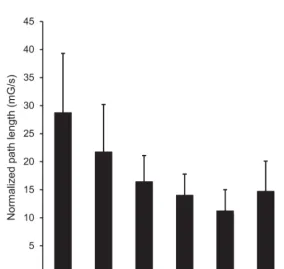
Figure 2 Accelerometer attached to gait belt and placed anteriorly on adultsThe NPL measure also reflected age-related effects
(figure 3).
DISCUSSION Test choice for NIH Toolbox.
Because of
the slightly higher correlation of ETDRS and HOTV
scores, they are the default sets in the NIH Toolbox
for individuals aged 8 through 85 years, and 3 through
7 years, respectively. Lea optotype set will be made
avail-able for use. Completion rates on the SVA test were
lim-ited by cooperation of the youngest groups. Dynamic
test completion rates of the younger children were
affected by the dual task requirement (e.g., moving
the head and identifying symbol). Consequently, rates
were better with the use of Lea symbols. We
acknowl-edge that although passive head movement is more
spe-cific to vestibular function, active movement was chosen
to ensure that the test was independent of the experience
of the tester, and the device assured proper velocity of
head movement.
The moderate correlation of BAM and SOT
scores may be attributable to the shortened duration
(20 seconds) of posturography testing, resulting in
less stable NPL measures. Interestingly, the lowest
correlations were in the older child group (13
–
17
years). Two issues may explain this: 1) this is a critical
period in balance development, particularly for use
of vestibular information
3,28; and 2) during this age
period, children experience significant growth and
body morphology change. Despite this, measures
obtained from the BAM are reliable and valid, based
on results presented here. Final tool
recommenda-tions are that: 1) all condirecommenda-tions be retained for
chil-dren 8 years and older and adults, 2) conditions 1 to 4
be retained for children younger than 8 years, 3) the
device be at the waist for all age levels, and 4) each
subject complete 1 trial (second trial offered only as
needed).
CONCLUSION
Because of the small sample of subjects
with VH, and because most children with VH had
uni-lateral chronic conditions and thus had likely
compen-sated (learned to use remaining vestibular function and
substitute strategies to balance), interpretation of these
results for potential clinical use of the DVA and BAM
is limited. However, the data support that the DVA
and BAM tests meet the requirements of the NIH
Toolbox: they are of low cost, can be completed in less
than 5 minutes, and are appropriate for use with
indi-viduals 3 through 85 years of age. The NIH Toolbox
DVA test is easy to administer and provides valid
and reliable testing of visual acuity with head still or
moving quickly. The NIH Toolbox BAM is reliable
and valid for measuring balance and assists in screening
for vestibular impairment. Both tests are sensitive to
age-related changes. Further study with a larger
num-ber of children and adults with confirmed vestibular
pathology is needed to firmly establish test sensitivity
and specificity. Normative data collection and analyses
on a large number of subjects across the lifespan have
been completed.
AUTHOR CONTRIBUTIONS
Dr. Rine: study concept and design, study supervision, acquisition of data, manuscript writing, data interpretation. Dr. Schubert and Dr. Whitney: study concept and design, acquisition of data, data interpreta-tion, critical revision of the manuscript for important intellectual content. Mr. Roberts: study concept and design, acquisition of data. Dr. Redfern: study concept and design, acquisition of data, data interpretation. Dr. Musolino: study concept and design. Ms. Roche: acquisition of data, data reduction, data interpretation. Mr. Steed: study concept and design, acquisition of data, data interpretation. Ms. Corbin: acquisition of data, reviewing the manuscript for accuracy. Mr. Lin: acquisition of data. Dr. Marchetti and Ms. Beaumont: analysis and interpretation. Dr. Carey, Dr. Shepard, Dr. Jacobson, Dr. Wrisley, Mr. Hoffman: critical revision of the manuscript for important intellectual content. Mr. Furman: acquisition of data, reviewing the manuscript for accuracy. Dr. Slotkin: study con-cept and design, study supervision, data interpretation, critical revision of the manuscript for important intellectual content.
ACKNOWLEDGMENT
The authors extend appreciation to Rohit Varma, MD, and Roberta McKean-Cowdin, PhD, of the University of Southern California for their expert contribution regarding vision acuity testing, and to Susan Magasi, OTR, PhD, of Northwestern University for contributions and collabora-tion on development of the BAM protocol.
STUDY FUNDING
This study was funded in whole or in part with Federal funds from the Blueprint for Neuroscience Research, NIH, under contract no. HHS-N-260-2006-00007-C.
DISCLOSURE
R.M. Rine has received travel reimbursement and honoraria from the British Audiological Society, the Royal Society of Medicine, the Hong Kong Physical Therapy Association, Emory University, Mayo Clinic, and the American Physical Therapy Association, served on the Scientific Review Committee of the Foundation for Physical Therapy Research, and was funded by NIH grant 5R44DC8022-3. M. Schubert was funded by NIH grant K23 007926, has worked as a consultant for a legal proceeding, and holds provisional patent C10585-P10585-02 (02240-283664). S. Whitney is a consultant for Visual Health Information, receives royalties from Oxford Press, and has received honoraria from the American Physical Therapy Association. D. Roberts is funded by NIH grants R21EY019713, R01EY019347, R01EY001849 DOC H, and R21NS059830. M. Redfern is funded by NIH grants R01OH008986, P30 AG024827, and R01 AG03111803, and by NSF grants CNS-0931595, CNS-0931999, and NSF-260116A. He holds US patent nos. 10/840,791 and 61/412,690 (which are not related to the current manuscript). M. Musolino reports no disclosures. J. Roche received funding from NSF grant CNS-0964581. D. Steed received partial funding for a project from Robert Bosch, LLC. B. Corbin and C.-C. Lin report no disclosures. G. Marchetti is a consultant for Eli Lilly Pharmaceuticals and received funding for a trip to Indianapolis, IN as part of consulting services. G. Marchetti has in the past received research support from the Pittsburgh Foundation, the Arthritis Foundation, and the Saunders Corporation. J. Beaumont served as a con-sultant for NorthShore University Health Systems, FACIT.org, and Geor-gia Gastroenterology Group PC, and has received funding for travel as an invited speaker at the North American Neuroendocrine Tumor Sympo-sium. J. Carey has been funded by NIH grants R01 DC005040, R01 DC002390, R01 DC009255, and R01 DC006296. In addition, he is a paid consultant for Otonomy and has provided expert witness testimony for approximately 3 medicolegal cases. N. Shepard has received travel reim-bursement, book royalty and honoraria from the University of Nebraska, Mayo Clinic, University of Southampton, Emory University, American Academy of Otolaryngology, NeuroCom International Inc., MicroMedical Corp., Vanderbilt University, University of Cape Town South Africa, and Plural publishing. G. Jacobson is a consultant for the company Interacous-tics. He receives royalties from Thomson Delmar Learning (Handbook of Balance Function Testing) and Plural Publishing (Balance Function Assessment and Management). He receives compensation for his role as Editor-in-Chief of theJournal of the American Academy of Audiology(American Academy of Audiology). D. Wrisley was funded by the Foundation for Physical Therapy and the New York Physical Therapy Association and served as an educator for NeuroCom International. H. Hoffman,. G. Furman, and J. Slotkin report no disclosures. Go to Neurology.org for full disclosures.
Received June 6, 2012. Accepted in final form August 30, 2012.
REFERENCES
1. Wei D, Hain TC, Proctor LR. Head-shaking nystagmus: associations with canal paresis and hearing loss. Acta Oto-laryngol 1989;108:362–367.
2. Hirabayashi S, Iwasaki Y. Developmental perspective of sen-sory organization on postural control. Brain Dev 1995;17: 111–113.
3. Rine RM, Rubish K, Feeney C. Measurement of sensory system effectiveness and maturational changes in postural control in young children. Pediatr Phys Ther 1998;10:16–22. 4. Gazzola JM, Gananca FF, Aratani MC, Perracini MR, Gananca MM. Clinical evaluation of elderly people with chronic vestibular disorder. Braz J Otorhinolaryngol 2006; 72:515–522.
5. Schubert MC, Migliaccio AA, Clendaniel RA, Allak A, Carey JP. Mechanism of dynamic visual acuity recovery with vestibular rehabilitation. Arch Phys Med Rehabil 2008;89:500–507.
6. Herdman S, Tusa RJ, Blatt P, Suzuki A, Venuto PJ, Roberts D. Computerized dynamic visual acuity test in the assessment of vestibular deficits. Am J Otol 1998;19:790–796.
7. Schubert MC, Herdman SJ, Tusa RJ. Functional measure of gaze stability in patients with vestibular hypofunction. Ann NY Acad Sci 2001;942:490–491.
8. Grossman GE, Leigh RJ. Instability of gaze during loco-motion in patients with deficient vestibular function. Ann Neurol 1990;27:528–532.
9. Grossman GE, Leigh RJ, Bruse EN, Huebner WP, Lanska DJ. Performance of the human vestibuloocular reflex during locomotion. J Neurophysiol 1989;62:264–272. 10. Hillman EJ, Bloomberg JJ, McDonald PV, Cohen HS.
Dynamic visual acuity while walking in normals and labyrinthine-deficient patients. J Vestib Res 1999;9:49–57. 11. Brookouser PE, Cyr DG, Peters JE, Schulte LE. Correlates of vestibular evaluation results during the first year of life. Laryngoscope 1991;101:687–694.
12. Braswell J, Rine RM. Evidence that vestibular hypofunc-tion affects reading acuity in children. Int J Pediatr Oto-rhinolaryngol 2006;70:1957–1965.
13. Mirka M, Black FO. Clinical application of dynamic pos-turography for evaluating sensory integration and vestibu-lar dysfunction. Neurol Clin 1990;8:351–359.
14. Dichigans J, Deiner HC. The use of short- and long-latency reflex testing in leg muscles of neurological patients. In: Struppler A, Weindl A, editors. Clinical Aspects of Sensory Motor Integration. Berlin: Springer-Verlag; 1987:165–175. 15. Allum JHJ, Pfaltz CR. Influence of bilateral and acute unilateral peripheral vestibular deficits on early sway stabi-lizing responses in human tibialis anterior muscles. Acta Otolaryngol Suppl 1984;406:115–119.
16. Allum JH, Bloem BR, Carpenter MG, Honegger F. Dif-ferential diagnosis of proprioceptive and vestibular deficits using dynamic support-surface posturography. Gait Pos-ture 2001;14:217–226.
17. Allum JHJ, Honegger F, Acuna H. Differential control of leg and trunk muscle activity by vestibulo-spinal and pro-prioceptive signals during human balance corrections. Acta Otolaryngol 1995;115:124–129.
18. Herdman SJ, Sandusky AL, Hain TC, Zee DS, Tusa RJ. Characteristics of postural stability in patients with ami-noglycoside toxicity. J Vestib Res 1994;4:71–80. 19. Rine RM, Spielholz NI, Buchman C. Postural control in
children with sensorineural hearing loss and vestibular hypo-function: deficits in sensory system effectiveness and vestib-ulospinal function. In: Duysens J, Smits-Engelsman BCM, Kingma H, editors. Control of Posture and Gait. Amster-dam: Springer-Verlag; 2001:40–45.
20. Shumway-Cook A, Horak FB. Assessing the influence of sensory interaction on balance. Phys Ther 1986;66: 1548–1550.
21. Rine RM, Roberts D, Corbin B, et al. New portable tool to screen vestibular and visual function: National Institutes of Health Toolbox Initiative. J Rehabil Res Dev 2012;49: 209–220.
22. Cotter SA, Tarczy-Hornock K, Wang Y, et al. Visual acu-ity testabilacu-ity in African-American and Hispanic children: the multi-ethnic pediatric eye disease study. Am J Oph-thalmol 2007;144:663–667.
23. Ferris FL, Kassoff A, Bresnick GH, Bailey I. New visual acuity charts for clinical research. Am J Ophthalmol 1982; 94:91–96.
25. Maetzler W, Mancini M, Liepelt-Scarfone I, et al. Impaired trunk stability in individuals at high risk for Parkinson’s disease. PLoS One 2012;7:e32240.
26. Whitney SL, Roche JL, Marchetti GF, et al. A comparison of accelerometry and center of pressure measures during computerized dynamic posturography: a measure of bal-ance. Gait Posture 2011;33:594–599.
27. Monsell EM, Furman JM, Herdman SJ, Konrad HR, Shepard NT. Computerized dynamic platform postur-ography. Otolaryngol Head Neck Surg 1997;117: 394–398.