University of South Carolina
Scholar Commons
Theses and Dissertations
2017
Best Practice for Screening Adult Patients with
Psoriasis for Polyautoimmunity: Celiac Disease,
Rheumatoid Arthritis and Crohn’s Disease
Susan Danielle Ashbaugh University of South Carolina
Follow this and additional works at:https://scholarcommons.sc.edu/etd Part of theNursing Commons
This Open Access Dissertation is brought to you by Scholar Commons. It has been accepted for inclusion in Theses and Dissertations by an authorized administrator of Scholar Commons. For more information, please [email protected].
Recommended Citation
Best Practice for Screening Adult Patients with Psoriasis for Polyautoimmunity: Celiac Disease, Rheumatoid Arthritis and Crohn’s Disease
By
Susan Danielle Ashbaugh
Bachelor of Arts
Mary Washington College, 1995
Bachelor of Science
University of South Carolina, 2011
Submitted in Partial Fulfillment of the Requirements
For the Degree of Doctor of Nursing Practice in
Nursing Practice
College of Nursing
University of South Carolina
2017
Accepted by:
Stephanie Burgess, Major Professor
Abbas S. Tavakoli, Committee Member
Abstract
The purpose of this quality improvement project was to determine if screening
primary care patients with psoriasis will improve early detection of celiac disease (CD),
rheumatoid arthritis, and Crohn’s disease (CrD). The aim of this project is to assess the utility of early screening in patients with psoriasis in order to facilitate earlier diagnosis
of CD, RA and CrD, which would consequently initiate earlier treatment and improve
long-term patient outcomes. Genetic and population-based studies suggest that
individuals with psoriasis have a greater risk of also having CrD, CD or RA, than do
individuals without psoriasis. The literature also suggests that health care providers
would be prudent to evaluate psoriatic patients in a prospective manner for these AI
disorders in order to improve the patient’s long-term health outcomes.
Based on the literature, the DNP project investigator developed a
non-psychometric patient questionnaire to capture data including the signs and symptoms of
CD, CrD and RA and three referral algorithms (one each per CD, CrD and RA). Over
two weeks at a Northern Virginia dermatology clinic, the patient questionnaire was
delivered to 261 adult patients, of which 34 were identified as psoriatic or newly
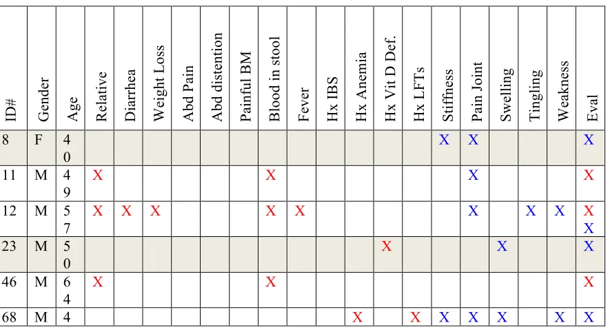
Findings indicated 100% provider compliance documentation for all 34 patients
noting that the patient a) had been screened, and b) if referral was or was not indicated.
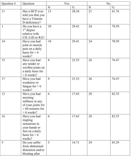
Frequency data indicated that the most reported symptom was a history of vitamin D
deficiency (38.24%). Thirty percent of psoriatic patients reported having a first-degree
relative with celiac disease, Crohn’s disease or rheumatoid arthritis. The most frequently
reported symptoms were for rheumatoid arthritis: daily joint or muscle pain > 6 weeks
(29.41%), daily tender or swollen joints > 6 weeks (23.53%), and weakness or fatigue > 6
weeks (23.53%). The most reported GI symptom was abdominal distention and/or
bloating after eating (14.71%). The least reported symptoms, at 2.94% each, were
abdominal pain after eating, painful bowel movements, and running a fever in the past 4
weeks. This quality improvement project highlights the need to evaluate adult patients
with psoriasis for polyautoimmunity and familial autoimmunity and is consistent with the
Table of Contents
Abstract ... iii
List of Tables ... viii
Chapter 1: Background and Significance ... 1
1.1 Introduction ... 1
1.2 Scope of Problem ... 4
1.3 Analysis of Current Practice ... 12
1.4 Statement of Problem/Purpose ... 23
1.5 Project Questions ... 24
1.6 Definitions ... 25
1.7 Chapter Summary ... 26
Chapter 2: Literature Review ... 28
2.1 Introduction ... 28
2.2 Search Methodology ... 28
2.3 Analysis of the Evidence ... 30
2.4 Genetic Studies. ... 30
2.5 Population Based Studies ... 43
2.6 Synthesis of Findings ... 64
3.1 Methods ... 67
3.2 Design ... 68
3.3 Instrument ... 68
3.4 Unit of Analysis ... 71
3.5 The Setting ... 72
3.6 Sample ... 72
3.7 Description of Intervention ... 73
3.8 Data Management and Analysis Methods ... 76
3.9 Human Subjects ... 78
3.10 Framework/Model of Research ... 80
3.11 Strategies to Reduce Barriers and Increase Supports ... 81
3.12 Summary ... 81
Chapter 4: Results ... 83
4.1 Introduction ... 83
4.2 Description of Sample ... 84
4.3 Analysis of Research Questions ... 84
4.4 Conclusion ... 94
Chapter 5: Discussion ... 97
5.1 Introduction ... 97
5.2 Recommendations for Practice ... 97
5.3 Recommendations for Education ... 98
5.4 Recommendations for Research ... 99
5.6 Limitations ... 102
5.7 Conclusion ... 103
References ... 105
Appendix A: Scottish Intercollegiate Guidelines Network (SIGN) Grading System 1999 – 2012: Levels of Evidence ... 118
Appendix B: Evidence Table ... 119
Appendix C: Scottish Intercollegiate Guidelines Network (SIGN) Grading System 1999 – 2012: Grades of Recommendations ... 141
Appendix D: Patient Questionnaire ... 142
Appendix E: Referral Algorithm for Celiac Disease ... 147
Appendix F: Referral Algorithm for Crohn’s Disease ... 148
Appendix G: Referral Algorithm for Rheumatoid Arthritis ... 149
Appendix H: Coded Identifier List ... 150
Appendix I: Chart Review – Data Collection Form ... 151
Appendix J: Screening Questionnaire – Data Collection Form ... 152
Appendix K: Key For Questions 13b, 14a and 15a ... 154
List of Tables
Table 1.1 The 2010 American College of Rheumatology/European League Against
Rheumatism Classification Criteria for Rheumatoid Arthritis ... 19
Table 1.2 PICO Definitions ... 24
Table 2.1 SNP Disease Overlap ... 36
Table 2.2 Screening Parameters for IBD ... 63
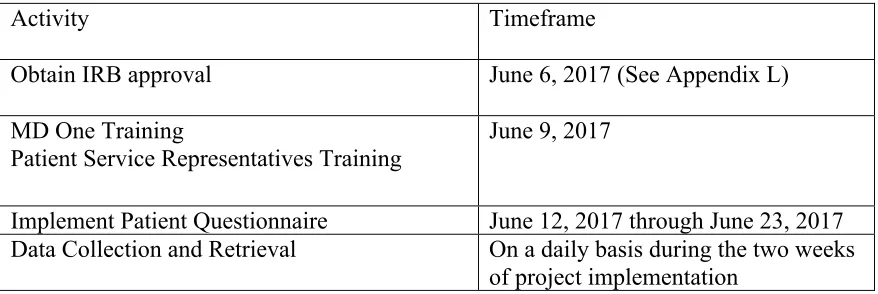
Table 3.1 Time Intervals for Quality Improvement Project ... 76
Table 4.1 Patient Questionnaire Frequency Distributions / Primary Questions ... 85
Table 4.2 Questionnaire Frequency Distributions / First Degree Relative ... 88
Table 4.3 Questionnaire Frequency Distributions / Conditional GI symptoms ... 88
Table 4.4 Questionnaire Frequency Distributions / Conditional Arthritis Symptoms ... 89
Table 4.5 Chart Review ... 92
Chapter 1
Background and Significance
1.1Introduction
Autoimmune (AI) diseases refer to a collection of diseases where the immune
system mistakenly directs the body to attack its own healthy organs, tissues, and blood,
rather than the foreign organisms the immune response was designed to combat. The
result is impaired function of the targeted organ or system. The National Institute of
Health ([NIH], 2005) has identified more than 80 clinically distinct autoimmune
disorders, while the American Autoimmune Related Disease Association ([AARDA],
2011) recognizes greater than 100 known autoimmune diseases. Psoriasis, a chronic,
inflammatory skin disease, is known to be the most prevalent AI disease in humans
(Raychaudhuri, 2014), affecting approximately 2-5% of the world population and as
many as 7.5 million Americans (NIH, 2005).
Psoriasis belongs to a subset of AI diseases classified as Immune-Mediated
Inflammatory Diseases (IMIDs) (Rahman, Inman, El-Gabalawy & Krause, 2010). The
concept of IMIDs describes a group of seemingly unrelated clinical disorders that share
common inflammatory pathways. While all IMIDs are AI diseases, the reverse is not
also true; all AI diseases are not IMIDs. Central to the disease process in IMIDs is the
Hazleton & Astor, 2007; Williams & Meyers, 2002). Cytokines play a pivotal
role in normal immune function. When these molecules are inappropriately expressed,
chronic inflammatory conditions arise. Celiac Disease (CD), Rheumatoid Arthritis (RA)
and Crohn’s Disease (CrD) also belong to this subset.
Another relatively new concept in the autoimmune field is polyautoimmunity,
which is defined as the coexistence of more than one AI disease, or IMID, in a single
individual (Rojas-Villarraga, Amaya-Amaya, Rodriguez-Rodriguez, Mantilla, & Anaya,
2012; Anaya, 2014). Research suggests that individuals with one autoimmune disease
typically will develop an additional one or more separate, and distinct, autoimmune
diseases over the course of their lifetime (Cooper, Bynum & Somers, 2009). Recent
population-based studies, as well as studies that have identified common genes associated
with multiple AI disorders, lend credibility to this idea that polyautoimmunity is not
random, and that there is a true association between different AI disorders. Specifically,
studies suggest that individuals with psoriasis have a greater risk of also having another
IMID, such as CrD, CD or RA, than do individuals without psoriasis (Augustin, Reich,
Glaeske, Schaeffer & Radtke, 2010; Bhatia, Millsop, Debbanch, Koo, Linos & Liao,
2014; Birkenfeld, Dreiher, Weitzman & Cohen, 2009; Cohen, Dreiher & Birkenfeld,
2009; Damasiewicz-Bodzek & Wielkoszynski, 2008; Einarsdottir et al., 2009; Li, Han,
Chan & Qureshi, 2013; Makredes, Robinson, Bala & Kimball, 2009; Qui, Z., Zhang, X.
Qui, Zhou & Li, 2013; Radtke et al., 2015; Tsai et al., 2011; Tsoi et al., 2013; Wolf et al.,
2008; Wu, Nugyen, Poon & Herrington, 2012).
Although these advances in research have fostered a greater understanding of
clinically difficult to recognize categories of disease. According to the AARDA (2011),
it can take patients an average of five years and four health care professionals to obtain a
correct diagnosis. Diagnostically, IMIDs remain a challenge for both patients and health
care providers. The initial presentation of symptoms may be vague and confused with
other disease processes (AARDA, 2011). Additionally, health care professional
education provides minimal training about AI diseases, contributing to a poor
understanding of autoimmunity among primary health care providers. As a result, despite
the proven genetic component in AI disease, health care practitioners do not typically
inquire whether patients have a personal or family history of autoimmune diseases, or
screen for additional autoimmune diseases in the autoimmune patient. Lastly, there are
very few standardized tests for many of the 80-100 AI diseases (AARDA, 2011). These
gaps of knowledge represent lost opportunity for the patient whose disease process
remains unchecked and whose associated AI co-morbidities also go unaddressed.
Despite the differences in heterogenic expression and clinical characteristics
among the IMIDs’ psoriasis, CrD, CD and RA, the shared genetic and pathophysiologic
mechanisms suggest a common origin, and as health care providers, it is imperative to
capitalize upon this information. The potential links between these diseases a) must not
be ignored and b) must be further studied. The purpose of this project is to conduct a
substantive review of the literature and conduct screening of primary care patients with
psoriasis to determine early detection of celiac disease, rheumatoid arthritis, and Crohn’s
1.2Scope of problem
Autoimmune diseases. Autoimmune diseases, to include the immune-mediated
inflammatory diseases, can affect virtually every site in the body, including the nervous,
gastrointestinal, and endocrine systems, as well as skin and other connective tissue, eyes,
blood and blood vessels. These diseases are chronic conditions, for which there currently
is no cure. Autoimmune and immune-mediated inflammatory diseases follow a
progressive path, with more end-organ destruction over the passage of time. Symptoms
tend to increase in severity as the disease progresses and as more tissue destruction
occurs (San Jose Functional Medicine, 2012). While many of these diseases have low
prevalence as a single occurring health disease, collectively autoimmune diseases are the
third most common category of disease in the United States after cancer and heart disease
(NIH, 2005) affecting approximately 5-8% of the population or approximately 23.5
million Americans. For reasons unknown, their prevalence is rising, while paradoxically,
they continue to remain under detected and under diagnosed (NIH, 2005; AARDA,
2011).
Early diagnosis and treatment is key to staving off disease progression and
improving patient outcomes. However, it is also recognized that there is often a delay in
diagnosis due to the fact that symptoms are often vague, misdiagnosed, and treated
symptomatically. The delay in diagnosis and treatment can unfortunately, lead to poorer
clinical outcomes associated with accrued joint and organ damage.
Researchers have been uncertain exactly what triggers an autoimmune response,
but certain modifiable and non-modifiable factors that play a role in autoimmunity have
genetic predisposition toward autoimmunity, while non-modifiable factors could be
anything from viruses, bacteria, medications, pollutants, or hormones. Autoimmune
disorders present disproportionately, and predominately, in the female population, are the
second highest cause of chronic disease (AARDA, 2011) and have been among the top
ten leading causes of death for women in every age group up to 64 years of age (NIH,
2005).
Incidence and Prevalence. In the United States alone, these AI diseases affect
approximately 5-8% of the population, or 14 to 23.5 million individuals (NIH, 2005).
However, the current data about the prevalence of these diseases in the United States is
misleading, since most autoimmune disorders are asymptomatic for years before a
clinical pattern emerges. Due to the silent nature of AI disease onset, one can logically
extrapolate that the true numbers of individuals with autoimmune disorders is actually
much higher than the statistics suggest. Many patients with AI disorders remain
undiagnosed and therefore, have simply not been included in the current numbers.
Furthermore, according to the AARDA (2011), the prevalence of autoimmune disorders
as reported by the National Institute of Health, 14 to 23.5 million individuals was quite
deflated, since the statistic only accounted for 24 of the 80 recognized autoimmune
diseases. The AARDA (2011) estimates that the actual number of individuals that have
autoimmune diseases is closer to 50 million.
There is no doubt that the actual burden of these autoimmune diseases to society,
and to individuals, is enormous. Individually, these chronic and progressively
lost productivity; decreased quality of life; co-morbid mental illnesses, particularly
depression and anxiety; and the disruption of social and family structures (NIH, 2002).
Costs. Quantifying the societal cost burden of AI disease has proven to be a
problematic task, as well. AARDA (2011) and NIH (2002) both agree that the lack of
epidemiological data on autoimmune disorders has made it difficult to calculate the full
direct and indirect cost to the overall health care system due to autoimmune disease. In
2001, the National Institutes of Allergy and Infectious Diseases (NIAID) Director, Dr.
Anthony Fauci estimated that annual autoimmune disease treatment costs were greater
than $100 billion. Again, this number most likely underrepresents the true cost burden of
disease, as the annual costs of only seven autoimmune diseases (Crohn's disease,
ulcerative colitis, lupus, multiple sclerosis, rheumatoid arthritis, psoriasis and
scleroderma), have been estimated to total from $51.8-$70.6 billion annually (AARDA,
2011).
Psoriasis. In 2010, the Centers for Disease Control and Prevention (CDC)
initiated a public health agenda for psoriasis in order to better characterize the burden of
psoriasis on the United States population, and to expand the existing knowledge base.
Examining data from the 2003-2006 and 2009-2010 National Health and Nutrition
Examination Surveys, key indicators such as prevalence, severity, disparities,
health-related quality of life and selected comorbidities were analyzed. The initiative found that
the overall prevalence of psoriasis in the United States is approximately 3.1%, affecting
6.7 million adults aged 20 and greater (Hemlock, Lee-Han, Hirsch, Baird, & Bartlett,
2014). Those with psoriasis tend to have a higher mean age, are more often of
and obesity (Hemlock et al., 2014). The National Foundation of Psoriasis (2008) further
reports that psoriasis is clearly linked with systemic comorbidities, such as cardiovascular
disease and events, hypertension, diabetes, as well as other immune-related diseases.
Psoriasis has also been associated with an increased risk for lymphoma, the strongest risk
was for Hodgkin’s lymphoma, particularly with increased severity of psoriasis (Gelfand
et al., 2006).
A systematic review of the economic burden of psoriasis was published in 2015
(Brezinski, Dhillon & Armstrong). The review concludes that patients with psoriasis
incur annual health care costs that are significantly greater than those of the general
population and may amount to $135 billion annually. In the United States, the economic
burden of psoriasis is substantial because this disease results in considerable negative
physical, psychiatric, and social consequences. The direct psoriasis costs ranged from
$51.7 billion to $63.2 billion, the indirect costs ranged from $23.9 billion to $35.4 billion,
and medical comorbidities were estimated to contribute $36.4 billion annually in 2013.
Patients with psoriasis would pay a lifetime cost of $11,498 for relief of physical
symptoms and emotional health (Brezinski et al., 2015).
Celiac Disease. Celiac disease is one of the recognized immune-mediated
inflammatory diseases, which is caused by a permanent intolerance to the ingestion of
gluten-containing cereals, wheat, rye, and barley, in genetically pre-disposed individuals
(Catassi et al., 2007). Population based studies indicate that the prevalence of CD is
approximately 0.5-1% in Western European and American populations (Tonutti &
Bizzaro, 2014), and the frequency of CD is substantially increased in patients who have a
2013). In patients with CD, the chronic intestinal damage over time carries risk for
adverse health consequences and increased mortality, including an increased risk for
malignancies such as small-bowel adenocarcinoma, cancer of esophagus, B-cell and
T-cell non-Hodgkin lymphomas, and in particular intestinal T-T-cell lymphomas
(Rubio-Tapia et al., 2013). The evidence also suggests that a consequence of untreated CD and
chronic malabsorption of nutrients is an increased prevalence of low bone mineral
density, risk for fractures, and micronutrient deficiencies including iron, folic acid,
vitamins B12 and B6, copper, zinc, and carnitine (Ludvigsson et al., 2014; Rubio-Tapia
et al, 2013). Women with CD have an increased risk of infertility, spontaneous abortions,
preterm deliveries, and delivery of low birth weight infants (Rubio-Tapia et al., 2013). It
is estimated that 83% of Americans who have celiac disease are undiagnosed or
misdiagnosed with other conditions (Fasano et al., 2003). The time from onset of
symptoms to celiac disease diagnosis averages 10 years in the US (Green & Jabri, 2003).
While prevalence is of CD is on the rise, the economic implications of CD are
only just emerging. A 2010 population-based study using administrative data for a cohort
of celiac disease cases and matched controls from Olmsted County, Minnesota were used
to compare direct medical costs one year pre- and post-coeliac disease diagnosis. The
study found that average total costs for patients were reduced by $1,764 in the year
following diagnosis, with a pre-diagnosis cost of $5023 versus a post-diagnosis cost of
$3259 (Long et al., 2010). While additional economic studies are necessary, these results
highlight the importance of early diagnosis and treatment, which may prevent
complications and reduce the economic burden of the disease.
tissue, which if left untreated, progresses to permanent structural damage and long term
disability (Emery et al., 2002). Epidemiological studies of RA indicate a population
prevalence of 0.5-1.0% in Northern European and North American countries
(El-Gabalawy, Guenther, & Bernstein, 2010). According to the CDC (2016), an estimated
1.5 million (0.6%) of US adults aged greater than 18 years had RA in the year 2005.
Data from the past decade indicate that the incidence of RA in women appears to be
rising after four decades of decline (CDC, 2016; El-Gabalawy et al., 2010).
Unchecked disease progression in the RA patient increases the incidence of death
due to infection, renal failure, non-Hodgkin’s lymphoma (Emery et al., 2002) (Johns
Hopkins Arthritis Center, 2016) and cardiovascular disease (John’s Hopkins Arthritis
Center, 2016). Cardiovascular disease, including ischemic heart disease and stroke,
accounts for approximately one-third to one-half of RA-related deaths, and infection is
responsible for approximately one-fourth of RA associated deaths (CDC, 2016). Those
with RA who remain untreated are twice as likely to die as compared to those without RA
of the same age (CDC, 2016; Johns Hopkins Arthritis Center, 2016). Additional risks
associated with RA are anemia, osteoporosis and depression (Johns Hopkins Arthritis
Center, 2016) and the possibility of partial or total joint replacement surgeries (Kumar,
Karthik, Gayathri, & Sivasudha, 2016). Approximately 90% of patients with RA have
some form of disability within two decades of onset (Emery et al., 2002).
In 2012, there were 9,100 hospitalizations with RA listed as the principal
diagnosis (Birnbaum et al., 2010). Women and people aged 45 years and older accounted
for the majority of these stays. A study utilizing administrative claims databases
year 2005 were used to estimate the comprehensive cost of RA patients to society and
individual stakeholders (Birnbaum et al., 2010). According to the study, total hospital
charges amounted to $374 million, with a mean charge of $41,000 per person. Direct
out-of-pocket medical costs for patients were estimated to be $8.4 billion, while indirect
costs were estimated to be $10.9 billion, including earning losses, disability payments
and decreased productivity. Finally, the cost of quality of life deterioration and
premature mortality were calculated to be approximately $39.2 billion (Birnbaum et al.,
2010). Early detection and treatment of RA improve the long term patient outcomes and
economic burden of this disease (Emery et al., 2002).
Crohn’s Disease. Crohn’s disease is a chronic, relapsing inflammatory bowel
disease (IBD) that may potentially affect any portion of the gastrointestinal tract from the
mouth to the anus. This condition is characterized by progressive bowel damage
associated with impaired functioning (Peyrin-Biroulet, Loftus, Colombel, & Sandborn,
2010). The highest prevalence for CrD has been found for Europe, 322 per 100,000
people and in North America, 319 per 100,000 people (Laass, Roggenbuck, & Conrad,
2014). According to the Crohn’s & Colitis Foundation of America ([CCFA], 2016),
Crohn’s disease may affect as many as 700,000 Americans. CrD is not gender-biased;
men and women are equally likely to be affected. Further, the disease may appear at any
age, although CrD is more prevalent among adolescents and young adults between the
ages of 15 and 35 (CCFA, 2016).
Patient’s with CrD may suffer from cardiovascular, hepatic, biliary, pancreatic,
and digestive co-morbidities, as well as metabolic issues and psychiatric problems (San
Blockage of the intestine due to swelling and scar tissue is the most common
complication of Crohn’s (CDC, 2014). Patients with CrD are at risk for early small
bowel and colorectal cancer (Baumgart & Sandborn, 2012). Studies also link
extraintestinal inflammatory symptoms with CrD in the eyes, skin or joints (Lichtenstein,
Hanauer, & Sandborn, 2009), as well as ankylosing spondylitis, non-drug induced
osteoporosis, and other inflammatory-mediated immune diseases (Baumgart & Sandborn,
2012). With initiation of corticosteroid therapy, up to 38% of CrD patients will require
surgery within one year (Lichtenstein et al., 2009). Mortality risk has been calculated as
over 50% greater than the general population (Canavan, Abrams, & Mayberry, 2007).
In addition to associated co-morbidities and quality of life issues, the economic
burden on the individual and the United States is substantial. A systematic literature
review of the costs of CrD in Western industrialized countries was conducted for the year
2006 (Yu, Cabanilla, Wu, Mulani, & Chao, 2008). Findings indicated that direct
medical costs per year were $18,022-$18,932 per patient in the United States.
Hospitalizations accounted for 53-66% of these direct medical costs, with a
per-hospitalization rate of $37,459. The total economic burden of CrD was estimated to be
between $10.9 and 15.5 billion in the United States (Yu et al., 2008).
Summary. By the numbers alone, it is clear that autoimmune disorders, on both
a collective and individual level, should constitute a national, if not global, health crisis
and that additional scrutiny to this category of diseases needs to be made. It is critical
that new methods for facilitating earlier diagnosis of these diseases be developed. Earlier
disease, with the hopes to stave off as much organ destruction as possible, reducing the
long-term consequences, and costs, of the diseases.
1.3Analysis of Current Practice
General Discussion. Obtaining a diagnosis for a particular autoimmune disease
is typically a long and stressful process for most patients. Many of the initial signs and
symptoms, such as fatigue, joint and muscle pain, fever or weight change (NIH, 2005) are
vague and suggestive of many diagnoses. Practitioners end up treating the symptoms,
without further regard for the etiology of these symptoms, while the disease continues to
progress unchecked. Patients are often required to see multiple practitioners and
specialists before they have been able to get answers and a definitive diagnosis.
According to the AARDA (2011), patients, on average, spent five years seeking a
diagnosis; 46% of patients report being told that they were “constant complainers” or
“too concerned with their health” (p. 9).
Diagnosis of an autoimmune disorder begins with a meticulous health history,
including a careful family history, which might point to a familial tendency toward
autoimmunity. The general concept of shared autoimmunity within families, or the
“kaleidoscope of autoimmunity (Somers, Thomas, Smeeth & Hall, 2009, p. 749)” has
gained acceptance among researchers, and should be a cornerstone of autoimmunity
identification. A careful social history should also be documented, which may help to
identify the patient’s environmental or occupational exposures. Additionally, a complete
physical evaluation has assisted the practitioner in more fully understanding the patient’s
issues (U.S. Department of Health and Human Services, Office on Women’s Health,
Laboratory testing remains fundamental to the diagnosis of AI disease in today’s
healthcare setting (Castro & Gourley, 2010). Unfortunately, no one specific test exists to
diagnose autoimmunity. Multiple tests may be run to help support a diagnosis of
autoimmunity, such as the complete blood count (CBC), comprehensive metabolic panel
(CMP), inflammatory markers, autoantibodies, flow cytometry, cytokine analysis, and
HLA typing (Castro & Gourley, 2010). Additionally, non-specific tests, such as the
erythrocyte sedimentation rate (ESR), serum complement markers, ferritin, fibrinogen,
albumin and C-reactive protein (CRP), help to indicate a state of inflammation and allows
the practitioner to evaluate disease activity (Castro & Gourley, 2010). However,
possessing the knowledge about which tests are available for the 80-100+ AI diseases,
and how to interpret them, continues to be a challenge for practitioners today.
In today’s health care arena, AI diseases are managed as individual entities, via
disease-specific profiles, as opposed to a general immune-related profile (Rahman et al.,
2010). Guidelines have been developed to help practitioners with the diagnosis and
treatment of psoriasis, CD, RA and CrD. The standards of care and current diagnostic
approaches for these four IMIDs will be discussed below.
Psoriasis. Psoriasis commonly presents in the primary care setting (Krueger &
Bowcock, 2005) and is a chronic, inflammatory, papulo-squamous skin disease.
Hyperplasia of skin epithelial cells lead to well-circumscribed, raised, red lesions, with
loosely adherent silvery white scales. Common locations are the knees, elbows and
scalp. Approximately 10-30% of patients also develop psoriatic arthritis (PsA), a painful
While a plethora of guidelines exist to direct the management and treatment of
psoriasis, no published diagnostic criteria have been developed. Diagnosis is dependent
primarily on the practitioner’s recognition of characteristic skin and lesion patterns, using
a subjective, qualitative assessment of the patient’s skin (Menter et al., 2008;
Raychaudhuri, Maverakis, & Raychaudhuri, 2014). A timely and proper diagnosis,
therefore, is based wholly upon the practitioner’s general knowledge of psoriatic
morphology and phenotype.
Johnson and Armstrong (2012) published a set of clinical and histologic
diagnostic guidelines for psoriasis, specifically for non-dermatologist practitioners. The
authors acknowledge that while no established criteria exist for skin-limited psoriasis,
trained health care providers should be able to diagnose psoriasis based on clinical
history and skin examination. The guidelines (Johnson & Armstrong, 2012) do not offer
absolute criteria for diagnosing psoriasis, however, they provide a set of
recommendations that providers should take into consideration during the assessment and
diagnosis of psoriasis.
Among these recommendations is to obtain a complete clinical, family and social
history (Armstrong & Johnson, 2012). Clinical history should include onset of lesions,
triggering factors, and associated symptoms (itch, pain, sensitivity, irritation). Family
history should be discussed due to genetics and heritability of this IMID. Social factors
should also be discussed, due to the association of psoriatic exacerbations with stress,
smoking and alcohol. Finally, the provider must conduct a full skin examination to
include the nails, scalp and intertriginous areas. Health care providers should take special
(Armstrong & Johnson, 2012). Although not included in the Armstrong and Johnson
guidelines, psoriasis classification can include the following morphologies, which may
present differently in patients and often, present with overlapping clinical findings:
plaque, inverse, erythrodermic, pustular, guttate, nail disease and psoriatic arthritis
(Menter et al., 2008).
Histopathology and skin biopsy, while an option for the diagnosis of psoriasis, is
not routinely practiced or required. However, if a question remains on the diagnosis,
histopathology can be beneficial in distinguishing psoriasis from other inflammatory skin
diseases (Armstrong & Johnson, 2012).
Celiac Disease. Celiac disease is a chronic, immune-mediated inflammatory
disease that manifests with a range of clinical symptoms in individuals who are
genetically susceptible. The consumption of gluten-containing foods triggers an immune
reaction, and a subsequent, inflammatory state of the duodenal mucosa (Tonutti &
Bizzaro, 2014). The immune response is directed against both the exogenous gluten
antigen and the autoantigen, tissue transglutaminase (tTG), which is a gluten byproduct
created in the small intestine (Kagnoff, 2006).
Clinical manifestations of CD can vary widely, and there is no concrete consensus
regarding which symptoms, laboratory abnormalities or associated diseases require
further evaluation for CD. Generally speaking, diagnosis begins with a health care
provider’s strong suspicion of CD based on the clinical exam and initial laboratory
results.
According to the guidelines developed by the American College of
malabsorption, such as chronic diarrhea, weight loss, iron deficiency anemia (IDA) or
elevated liver enzymes, and/or steatorrhea, postprandial abdominal pain, and bloating,
should be tested for CD (Rubio-Tapia et al., 2013). Guidelines from the British Society
of Gastroenterology (Ludvigsson et al., 2014) state that CD can be suspected in patients
with mild gastrointestinal symptoms, associated conditions, or those at genetic risk, to
include symptomatic first-degree relatives of patients with CD, as well as symptomatic
individuals with Down’s Syndrome and Turner’s Syndrome (Ludvigsson et al., 2014).
However, since the clinical picture of CD varies, and since many patients only have
minor symptoms, it can be challenging for health care providers to make that first
connection, from symptoms to suspicion.
Further confusing the diagnostic process is the fact that CD has been classified
into multiple phenotypes: classic, atypical, silent, latent and refractory (Kagnoff, 2006).
“Classic” CD is the most commonly described form, and patients present due to
gastrointestinal symptoms. Whereas “classic” CD is the most commonly described form,
“atypical” CD is actually the most prevalent form. Patients have little to no GI issues, but
they become identified for other reasons, such as IDA, osteoporosis or infertility.
“Silent” CD describes the cohort of patients who are asymptomatic, but who diagnose
positive for CD as a result of serology or biopsy done for another reason. “Latent” CD
refers to patients who previously have been diagnosed with CD, who responded to a
gluten-free diet (GFD) and who retain normal mucosal histology. “Refractory” CD
represents patients with true CD, who no longer respond to a GFD. (Kagnoff, 2006;
Regardless of how a patient comes to the attention of the practitioner as
potentially having CD, the next steps in diagnosis are well agreed upon. The first step in
diagnosis is serology, followed by a biopsy, which is considered to be the gold standard
for CD diagnosis (Green & Jabri, 2003; Kagnoff, 2006; Ludvigsson et al., 2014;
Rubio-Tapia et al., 2013; Tonutti & Bizzaro, 2014). The Immunoglobulin A (IgA) anti-tissue
transglutaminase (tTG) antibody is the preferred serological test for individuals over the
age of 2 years. Both the sensitivity and specificity of the IgA-tTG for untreated CD is
approximately 95% (Kagnoff, 2006; Rubio-Tapia et al., 2013). If IgA deficiency is a
concern, occurring in 1.7%-2.6% of patients with celiac disease (Green & Jabri, 2003),
total serum IgA should be included in the panel and both IgA and IgG-based testing may
be initiated to include the IgG-deamidated gliadin peptides (DGPs) (Green & Jabri, 2003;
Rubio-Tapia et al., 2013). With known IgA deficiency, both IgG-DGPs and IgG-tTG
serology may be tested. Finally, if serology is negative, but suspicion for CD is high,
intestinal biopsy should be pursued (Green & Jabri, 2003; Kagnoff, 2006; Ludvigsson et
al., 2014; Rubio-Tapia et al., 2013; Tonutti & Bizzaro, 2014).
The intestinal biopsy remains the gold standard in diagnosis because results
reflect the varying degrees by which mucosal villi have been affected over the course of
the disease. The Marsh-Oberhuber classification of architectural changes in the intestine
outline three categories of lesion: “Type 1” describes an infiltrative lesion, “type 2” an
infiltrative-hyperplastic lesion, and “type 3” reports mild, moderate and total levels of
villous atrophy (Tonutti & Bizzaro, 2014). Multiple samples should be taken from the
second or third portion of the duodenum and at least one sample from the duodenal bulb
In the setting where results from the aforementioned tests are not clear, or for
patients already on a GFD, individuals can be genotyped for the gene pairs that encode
HLA class II heterodimer HLA-DQ2 or HLA-DQ8. Almost all patients with CD have
either DQ2 (~95% of CD patients) or DQ8 (the remaining ~5% of CD pts) and the
absence of both of these DQ alleles provide a negative predictive value for the disease of
close to 100% (Kagnoff, 2006; Green, 2003; Ludvigsson et al., 2014).
Rheumatoid Arthritis. The three major pathways for this immune-mediated
inflammatory disease include bone degradation, cartilage and synovial destruction, which
lead to severe disability and premature mortality (Aletaha et al., 2010). While the precise
etiology and pathophysiology of RA is not completely understood, at least 16 different
cytokines, autoantibodies and other mediators are implicated in the disease process
(Kumar et al., 2016). The initial immune response, the pre-articular phase, begins with
the generation of autoantibodies against own tissue components. During the transition
phase, the introduction of autoantibodies and autoantigens in the articular joints becomes
evident, causing joint destruction to occur symmetrically to joints all over the body,
although the distal interphalangeal and cervical spine is typically spared (Kumar et al.,
2016). Permanent structural damage occurs early in the disease course of RA and early
intervention with disease modifying anti-rheumatics drugs (DMARDs) treatment is
critical toward slowing the progression of joint damage, improving quality of life and
long term outcomes for patients (Emery et al., 2002).
The American College of Rheumatology (ACR) and the European League
Against Rheumatism (EULAR) collaborated to update the classification criteria for RA
help researchers classify newly presenting patients and to help determine which patients
would benefit from early treatment (Aletaha et al., 2010). The expert panel identified the
following mandatory criteria: pattern and extent of joint involvement, serology
(rheumatoid factor (RF) and anti-cyclic citrullinated protein (ACPA)), acute-phase
response (C-reactive protein (CRP) and erythrocyte sedimentation rate (ESR)) and
duration of symptoms (Aletaha et al., 2010; Neogi et al., 2010). Two criteria were
deemed essential. First, the patient must present clinically with joint swelling in at least
one joint, indicating synovitis, and second, there must also be an absence of another
condition that could explain the patient’s symptoms. Differential diagnoses include
multiple disorders such as psoriatic arthritis, systemic lupus erythematous (SLE),
osteoarthritis and gout. The authors of the classification criteria suggest that if it is
unclear to the provider which relevant differential diagnoses to consider, a rheumatologist
should be consulted (Aleteha et al., 2010).
The remaining four criteria each contribute differently to the probability of
developing RA and were weighted accordingly during the criteria development (Aleteha
et al., 2010). The table below shows the criteria and scoring, with a required score of 6 or
greater to be classified as having definite RA.
Table 1.1 The 2010 American College of Rheumatology/European League Against Rheumatism Classification Criteria for Rheumatoid Arthritis
The 2010 American College of Rheumatology/European League Against Rheumatism classification criteria for rheumatoid arthritis
Target population (who should be tested?) Patients who:
1) Have at least 1 joint with definite clinical synovitis (swelling), 2) With the synovitis not better explained by another disease.
1 large joint 0
2-10 large joints 1
1-3 small joints (with or without large joint involvment 2
4-10 small joints 3
> 10 joints (at least 1 small joint) 5 B. Serology
Negative RF and negative ACPA 0
Low-positive RF or low-positive ACPA 2
High-positive RF or high-positive ACPA 3
C. Acute-phase reactants (at least 1 test result required for classification)
Normal CRP and normal ESR 0
Abnormal CRP or abnormal ESR 1
D. Duration of symptoms
< 6 weeks 0
≥ 6 weeks 1
(Reproduced from Aletaha et al., 2010)
Although not included in the new classification criteria, radiograph imaging is
used to assess the structural damage associated with RA, and continues to be the best
method for collecting data on joint erosions and bone density. Other markers
traditionally used by providers to diagnose RA, such as the assessment of morning
stiffness and the metacarpal “squeeze test” (Emery et al., 2002), have also been excluded
from the classification criteria.
One point must be elucidated about the classification criteria. These criteria were
deliberately labeled “classification,” as opposed to “diagnostic” criteria, in order to
provide a standardized approach for determining which patients presenting with
undifferentiated synovitis, would have the highest probability of persistent or erosive RA
(Aletaha et al., 2010). As such, the authors acknowledge that the criteria may in fact be
used as a tool for diagnosis, but that easier-to-use tools are in development through
another joint effort by ACR/EULAR for primary care providers.
to the anus. Ulcerative colitis (UC) is the second disease included in the IBD
classification. By comparison to CrD, which is intermittent inflammation and can attack
all bowel wall layers, UC is a continuous inflammation that it is limited to only the
innermost layer of the intestinal linings in the colon and rectum (CCFA, 2016). Both
IBDs are characterized by periods of disease activity interspersed with periods of
remission. Symptoms of both CrD and UC may include fever, bloating, cramping,
nausea, vomiting, severe diarrhea, bloody stool, abdominal pain, weight loss and fatigue.
CrD patients may also have mucous in their stool (Laass et al., 2014). Patients can have
symptoms for many years prior to diagnosis (Burgmann et al., 2006; Pimentel et al.,
2000).
The rationale for defining early Crohn’s disease is to modify the clinical course of
the disease and intervene prior to the onset of bowel damage in the form of stricture,
fistula, or abscess. However, approximately one-fifth of adult patients already have
evidence of structuring or penetrating intestinal complications at diagnosis
(Peyrin-Biroulet et al., 2010). CrD is a seronegative IMID, meaning there is no direct serological
test for detecting disease activity. And presently, there is no gold standard for CrD
diagnosis. Diagnosis integrates patient information and physical exam with objective
data from a combination of laboratory, radiologic, endoscopic and histologic findings
(Laass et al., 2014; Peyrin-Biroulet et al., 2010; Van Assche et al., 2009; (Baumgart &
Sandborn, 2012). Genetic testing is not currently recommended (Lichentstein, 2009;
Peyrin-Biroulet, 2010; Van Assche et al., 2009).
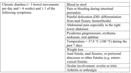
The most common presenting CrD symptom is chronic diarrhea, defined as a
pain is seen in about 70% of patients before diagnosis, and approximately 60% of
patients experience weight loss (Van Assche et al., 2009). In approximately 10% of
patients, the presenting complaint is a perianal fistula (Van Assche et al., 2009).
Providers must take a complete medical history itemizing symptoms and inquire about
family history, as first-degree relatives of patients with IBD have a 10-15 fold risk for
also having IBD (Laass et al., 2014). A full history should also include information about
recent travel, food intolerances and medications. Attention should be paid to proven risk
factors including smoking and recent infectious gastroenteritis (Van Assche et al., 2009).
Physical examination includes general well-being, vital signs, body weight, BMI,
abdominal tenderness or distention, palpable masses, perineal and oral inspection, rectal
digital examination (Van Assche et al., 2009; Laass et al., 2014) plus signs of
extraintestinal disease. Extraintestinal manifestations might present as joint pain,
swelling, redness or stiffness, erythema nodosu, or redness of the eye (Laass et al., 2014).
Clinical laboratory testing continues the inflammatory status assessment, although
of and by themselves, labs are not enough to differentiate CrD from UC or enteric
infection. The initial lab investigations support GI inflammation and are used as an
adjunct to diagnosis (Van Assche et al., 2009). Patients should be assessed for anemia,
fluid depletion and signs of malnutrition or malabsorption via the complete blood count
(CBC). Anemia and thrombocytopenia represent the most common changes in CBC
evaluation of patients with CrD (Van Assche et al., 2009). CRP and ESR are
non-specific acute phase inflammatory markers that should be evaluated. Fecal calprotectin
or lactoferrin provides an estimation of fecal inflammation by measuring for the presence
value of 85-90% in distinguishing IBD from irritable bowel syndrome (IBS). Stool
cultures are beneficial for ruling out infectious colitis caused by viral, bacterial or
parasitic sources (Laass et al., 2014; Van Assche et al., 2009; Lichentenstein et al., 2009).
Providers must also consider lactose intolerance and celiac disease in their list of
differential diagnoses.
Upper or lower GI endoscopy is used to confirm the diagnosis of CrD, assess
disease location, and obtain tissue for pathological examination (Laass et al., 2014; Van
Assche et al., 2009). However, the initial symptoms frequently determine the order of
subsequent testing. For example, colonoscopy, intubation of the terminal ileum, is the
most appropriate initial test for patients presenting with predominant diarrhea, and is used
to establish the diagnosis of ileocolonic CrD. On the other hand, imaging studies may be
more appropriate for those presenting with abdominal pain. Magnetic resonance
enterography is the initial test used to evaluate the small intestine. Wireless video
endoscopy, or video capsule endoscopy (VCE), may also be useful for detecting small
bowel involvement (Laass et al., 2014; Van Assche et al., 2009).
1.4Statement of Problem/Purpose
In adult patients aged 18 years and greater with psoriasis, does screening for
celiac disease, rheumatoid arthritis, and crohn’s disease improve early detection for these
autoimmune disorders? The Population (P) in this question is adult patients aged 18
years and greater in primary care and outpatient settings. The Intervention (I) is the
development and implementation of a simple screening tool in adults patients with
psoriasis for celiac disease, rheumatoid arthritis and crohn’s disease. There is no
improved early detection of celiac disease, rheumatoid arthritis and crohn’s disease in
adult patients with psoriasis. Table 1.1 contains the definitions of population, setting,
intervention, and outcome as defined by Melynk & Fineout-Overholt (2011). The
purpose of this project is to conduct a substantive review of the literature to determine if
screening primary care patients with psoriasis will improve early detection of celiac
disease, rheumatoid arthritis, and Crohn’s disease.
Table 1.2 PICO Definitions
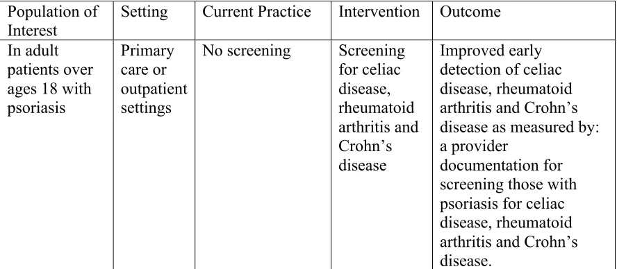
Population of
Interest Setting Current Practice Intervention Outcome In adult
patients over ages 18 with psoriasis
Primary care or outpatient settings
No screening Screening for celiac disease, rheumatoid arthritis and Crohn’s disease
Improved early detection of celiac disease, rheumatoid arthritis and Crohn’s disease as measured by: a provider
documentation for screening those with psoriasis for celiac disease, rheumatoid arthritis and Crohn’s disease.
1.5Project Questions
The project was guided by the following clinical questions:
What is autoimmunity? What is polyautoimmunity? What are IMIDs?
Why is it so difficult for patients with autoimmune diseases to become diagnosed?
Is there evidence that suggests that individuals with one autoimmune disease are
more at risk for developing another autoimmune disease?
Is there evidence that suggests that individuals with psoriasis have a greater risk
What are the current, accepted approaches for diagnosing psoriasis, celiac disease,
rheumatoid arthritis and Crohn’s disease?
Would screening patients with psoriasis for celiac disease, rheumatoid arthritis
and Crohn’s disease improve early detection for these diseases?
1.6Definitions
Adult Patients – Adult patients refer to men and women over the age of 18 years
seeking health care in a primary care setting.
Celiac Disease - Celiac disease is an autoimmune disease that causes an
inflammatory reaction to ingested gluten, a protein found in wheat, rye, and barley. When a person has celiac disease, gluten causes the immune system to react in a way that can cause intestinal inflammation—irritation or swelling—and long-lasting damage (NIH, 2015).
Crohn’s Disease - Crohn’s disease is an autoimmune disease characterized by
chronic, relapsing inflammation to any portion of the gastrointestinal tract from the
mouth to the anus. Also known as one of the inflammatory bowel diseases (IBD), this
condition is caused by an abnormal response to the body's immune system which results
in progressive bowel damage associated with impaired functioning (Peyrin-Biroulet et al.,
2010; CDC, 2014).
Early Detection - Early detection refers to a screening program that detects
disease in asymptomatic persons or in symptomatic persons not yet recognized to have
disease. Relative to background conditions, screening identifies the affected individual at
questionnaire, physical observation or measurement, laboratory test, radiological
procedure, etc.) that are used to help identify persons with unrecognized disease or
unrecognized risk factors for disease (Weissfeld, 2001).
Rheumatoid Arthritis – Rheumatoid arthritis is an autoimmune disease
characterized by chronic, systemic inflammation of the synovial tissue. If left untreated,
RA progresses to permanent structural damage and long-term disability (CDC, 2016;
Emery et al., 2002).
Primary Care Providers - A primary care provider (PCP) is a health care
practitioner who is responsible for monitoring an individual's overall health care needs. The PCP's role is to provide preventative care and teach healthy lifestyle choices, identify and treat common medical conditions, assess the urgency of medical problems and direct the patient to the best place for their care, and make referrals to medical specialists when necessary (She, 2012).
1.7Chapter Summary
Research suggests (Cooper, Bynum & Somers, 2009) that individuals with one
autoimmune disease typically will develop an additional one or more separate, and
distinct, autoimmune diseases over the course of their lifetime. Further, links have been
drawn between psoriasis and celiac disease, rheumatoid arthritis and crohn’s disease
(Augustin, Reich, Glaeske, Schaeffer & Radtke, 2010; Ali & Warren, 2013; Wu, Nugyen,
Poon & Herrington, 2012; Guerin, Zhang, Gauthier, Day & Khan, 2012; Hsu &
Armstrong, 2012; Makredes, Robinson, Bala & Kimball, 2009; Birkenfeld, Dreiher,
Weitzman & Cohen, 2009; Tsai, Wang, Hung, Tsai, Schenkel, Zhang & Tang, 2011).
diseases, prolonging the diagnostic process (Somers et al., 2009). The purpose of this
project is to conduct a substantive review of the literature to determine if screening
primary care patients with psoriasis will improve early detection of celiac disease,
rheumatoid arthritis, and Crohn’s disease. The aim of this project is to assess the utility of early screening in patients with psoriasis in order to facilitate earlier diagnosis of CD,
RA and CrD, which would consequently initiate earlier treatment and improve long-term
patient outcomes.
The next step in the evidenced-based practice process is the literature search and
analysis. The evidence will be organized using an evidence table. This process is
Chapter 2
Literature Review
2.1 Introduction
The goal of this chapter is to appraise and synthesize the evidence for conducting
the DNP project for changes in practice. The discussion that follows describes the
literature search process, and an objective summary and analysis of fourteen research
articles. The purpose of this content is to convey the current state of knowledge, and
significance of, the relationship between psoriasis and the three IMIDs of interest (celiac
disease, rheumatoid arthritis, and Crohn’s disease) to healthcare providers and decision
makers.
2.2 Search Methodology
Evidence-based practice mandates that clinical decisions be driven by the most
current research studies, the clinical experience of the practitioner, and patient
preferences (Melnyk & Fineout-Overholt, 2011). Evidence-based research begins with a
question of interest, followed by the systematic collection, appraisal and synthesis of
evidence. The following information presents the search strategy employed for the
question: in adult patients aged 18 years and greater with psoriasis, does screening for
celiac disease, rheumatoid arthritis, and Crohn’s disease improve early detection for these
This is a background-type PICO question and according to Melnyk and
Fineout-Overholt (2011), the following types of studies are appropriate for review, in descending
order of level of evidence: synthesis of cohort study or case control studies, single cohort
studies or case-control studies, meta synthesis of qualitative or descriptive studies, single
qualitative or descriptive studies, and expert opinion. A search of databases was
performed, accessed through the University of South Carolina’s online library. The
Cumulative Index for Nursing and Allied Health Literature (CINAHL),
PubMed-Medline, Medline OVID, Cochrane Library, Web of Science, Essential Evidence Plus,
Nursing Resource Center, Health Source: Nursing/Academic Edition, Dissertations and
Thesis, Annual Reviews, as well as Google Scholar were included in the search.
Reference lists of acceptable papers were also manually examined for additional
resources.
The main search terms were “psoriasis” and “autoimmunity.” Limitations were
set for the years 2006-2016, in order to review the most up-to-date research and evidence
on the topic, and for English-only papers, to eliminate language barriers. Additional
cross-searching terms utilized were “co-autoimmunity,” “co-existence,” “association,”
“pan autoimmunity,” “immune mediated inflammatory diseases,” or “IMIDs,” and
“screening.” Finally, “psoriasis” was searched specifically against the three AI disorders
of interest for this project: “celiac disease,” “Crohns disease,” and “rheumatoid arthritis.”
A total of fourteen papers were selected. Levels of evidence were appraised
using the Scottish Intercollegiate Guidelines Network (SIGN) rating system (Appendix
A). The rating scale ranges from 1++, the highest level of evidence, reserved for high
to 4, reserved for expert opinion, the lowest level of evidence. Four retrospective cohort
studies with control groups were found (level of evidence 2+). Four case-control studies
(level of evidence 2+), one cross-sectional study (level of evidence 3), two meta-analysis
of genetic studies (level of evidence 1+), one meta-analysis of population based studies
(level of evidence 1+), one prospective cohort study (level of evidence 2+), and one
expert review paper (level of evidence 4) were included.
2.3 Analysis of the Evidence
Evidence was organized in table format with the following headings: brief
reference, type of study/quality ratings, methods, threats to validity/reliability, findings
and conclusions (Appendix B). The table was developed to consolidate methodological
and outcome summaries from the selected articles and used for the purpose of synthesis
and analysis. Each evidence-based, peer reviewed article was systematically appraised
on the individual level, followed by an overall summary of findings. The four genetic
studies are presented first, followed by the population-based studies. For the purposes of
this literature review, when studies include multiple AI disorders, only the four AI
diseases directly related to this study, psoriasis, celiac disease, rheumatoid arthritis, and
Crohn’s disease, will be discussed.
2.4 Genetic Studies.
Four of the fourteen research articles included in this review can be classified as
genetic studies. What follows is a brief discussion to foster a basic understanding of
single nucleotide polymorphisms (SNPs), the main concept in each of the studies.
double helix of deoxyribonucleic acids, or DNA. The DNA double helix is composed of
a long sequence of nucleotide base pairings, adenine (A), thymine (T), guanine (G) and
cytosine (C). These nucleotide bases link in a very specific way: A always pairs with T,
and C always pairs with G. Distinct sequences of these nucleotides organize the
chromosomes into sub-units, which are genes. Genes provide the cell with the
instructions that dictate cell function (National Human Genome Research Institute, 2015).
A SNP is defined as a single nucleotide base change in a DNA sequence that
occurs in a significant proportion (more than 1 percent) of a large population (University
of Utah Health Sciences, 2016). To make an analogy, 99% of the population has a
sequence for “Marie,” while approximately 1% has a sequence for “Maria.” Today’s
challenge for researchers is to identify SNPs, or the “Maria’s,” that correlate with a
particular effect in patients. Genetic association studies, such as the ones to follow,
compare the frequency of genotypes at genetic marker loci, usually single-nucleotide
polymorphisms (SNPs), in individuals with and without a given disease trait from a given
population. The objective of these association studies is to determine whether a
significant statistical association exists between the disease trait and the genetic marker
(Clarke, Anderson, Peterson, Cardon, Morris & Zondervan, 2011). Reliable SNPs could
serve as predictive gene markers that inform decisions about numerous aspects of
medical care, including specific disease diagnosis, predisposition to disease, and the
effectiveness of various drugs and adverse reactions to specific drugs (University of Utah
Health Sciences, 2016).
Association of SNP Gly307Ser (rs763661) with Psoriasis, CD & RA. Qiu,
relationship between the non-synonymous single nucleotide polymorphism (SNP)
Gly307Ser (rs763361) in the CD226 gene that has been reported to be associated with
several AI diseases, including psoriasis, celiac disease, and rheumatoid arthritis. Whether
the presence of this t allele rs763361 confers a risk for multiple AI diseases remains
under investigation. This study was rated a 1+.
The authors conducted a comprehensive search of the U.S. National Library of
Medicine’s PubMed and Embase databases for studies that fulfilled the following
inclusion criteria: (1) were based on case-control design, (2) evaluated the association of
the Gly307Ser (rs763361) polymorphism with multiple autoimmune disorders, (3)
disease diagnosis followed the diagnosis criteria of the World Health Organization
(WHO), (4) genotype frequencies were provided, (5) authors provided sufficient data for
estimating an odds ratio and their 95% confidence interval (CI), and (6) papers were
published with full text articles (Qui, Z., Zhang, X. Qui, Zhou & Li, 2013). Seven
published studies met the inclusion criteria, covering 7,876 cases and 8,558 controls. The
sample sizes varied from 90 to 2,838 and included two European, two Asian, one South
American and three Estonian studies. The studies utilized three different genotyping
methods, polymerase chain reaction-restriction fragment length polymorphism
(PCR-RFLP), TaqMan genotyping and SNPlexTM (Qui, et al., 2013).
Qui et al. (2013) used STATA 12.0 software for statistical analysis and estimated
the association between CD226 Gly307Ser (rs763361) polymorphism and multiple AI
diseases using crude odds ratio (OR) with 95% CIs. As is appropriate for meta-analyses,
the authors assessed the degree of inconsistency in the studies' results, or between-studies
model calculated pooled estimates in case of significant heterogeneity. In cases without
obvious heterogeneity, the Mantel-Haenszel fixed-effect model estimated a summary OR.
The evaluation of the association of CD226 Gly307Ser (rs763361) polymorphism
with multiple AI diseases demonstrated an overall OR 1.19 (95% CI: 1.12-1.27,
Pheterogeneity=0.136), indicating that a significantly increased multiple AI disease risk was
found to be associated with the t allele rs763361 (Qui et al., 2013). The authors also
conducted a subgroup analysis by ethnicity, where increased risks were found for South
Americans (OR=1.31, 95% CI=1.17-1.48, Pheterogeneity = 0.644), Asians (OR=1.23, 95%
CI=1.11-1.38, Pheterogeneity = 0.690), and Europeans (OR=1.13, 95% CI=1.04-1.24,
Pheterogeneity = 0.085) (Qui et al, 2013).
Although heterogeneity was detected in both the overall comparison and in the
subgroup analyses, none were notable (Pheterogeneity = 0.136, I2=29.3% in overall
comparsion; Pheterogeneity = 0.644, I2=0.0% in South Americans; Pheterogeneity = 0.690,
I2=0.0%; Pheterogeneity = 0.085, I2=46% in Europeans). Meta-regression analysis was
performed to explore sources of heterogeneity across studies when statistical
heterogeneity was detected. Publication year was closely related to the heterogeneity in
allele comparison (I2=11.9%, P=0.036), while racial descent, study sample size,
genotyping methods, and controls’ source did not indicate any modifying effect of the
factor (P>0.05) (Qui et al, 2013).
Further, Begg’s test suggested no significant publication bias and the
Hardy-Weinberg equilibrium was demonstrated by using the Fisher’s exact test (p < 0.10).
Finally, the authors conducted a sensitivity analysis to assess the stability of results. The
OR, and that results were statistically robust. The authors, however, acknowledged
limitations to this study. First, this study lacked the original information for the
individuals in the included studies; as such, data could not be stratified by other variables,
such as gender, and mean age at onset (Qui et al, 2013). The lack of original data also
means that the authors would not have been able to validate each case for the
meta-analysis. Therefore, the possibility of diagnoses misclassification in the original studies
cannot be completely excluded. Second, races other than South American, Asian,
European and Estonian were not represented in this meta-analysis. Third, the authors
note that while their publication bias showed no significance, it cannot be completely
ruled out due to exclusion of relevant publications that were not indexed by their selected
databases, PubMed and Embase (Qui et al, 2013).
The results of this meta-analysis, that a significantly increased multiple AI disease
risk was found to be associated with the t allele rs763361, highlight the evolving
comprehension of co-autoimmunity. It further supports the concept that susceptibility to
AI diseases may be due to a complex interaction of multiple genes, some of which seem
to be shared among many of these AI diseases, including psoriasis, celiac disease and
rheumatoid arthritis.
15 New Psoriasis Susceptibility Loci. Tsoi et al. (2013) conducted a
meta-analysis of three GWAs and two independent datasets genotyped on the “Immunochip,”
to include a total of 10,588 cases (patients with psoriasis) and 22,806 controls. The
“Immunochip” is a custom-designed SNP array whose function is to fine-map
genome-wide significant (P<5x10⁻⁸) susceptibility loci and to explore replication of thousands of
distinct autoimmune and inflammatory diseases designed the chip in 2009 (Parkes,
Cortes, Van Heel & Brown, 2013).
The Immunochip consists of 196,524 SNPs compiled from variants identified in
previous GWAS of 12 different IMIDs. Each disease-focused group involved in the chip
design were then allowed to submit approximately 3000 additional SNPs in order to
evaluate signals that were deemed promising or that had not quite met genome-wide
significance in previous studies (Tsoi et al, 2013). The main objective of this study was
to increase understanding of the genetic architecture of psoriasis, identify new genetic
determinants of psoriasis, and to relate them to other AI diseases (Tsoi et al., 2013). This
study has been rated a 1+.
The authors performed a meta-analysis from five datasets that were genotyped on
the Immunochip. These datasets included three existing GWAS (Kiel, CASP and
WTCCC2) and two independent European descent case-control datasets, the Psoriasis
Association Genetics Extension (PAGE) and the Genetic Analysis of Psoriasis
Consortium (GAPC) (Tsoi et al., 2013). Prior to meta-analysis, a number of quality
control steps were taken by the authors in order to identify and remove DNA samples and
markers that could introduce bias into the study. SNPs with a call rate below 95% were
excluded. Using the HapMap 3 samples as a reference, the authors performed principal
component (PC) analysis to identify and remove samples with non-European ancestry.
Samples with extreme inbreeding coefficients or heterozygosity values were also
removed, as were duplicate pairs or highly related individuals. A principle component
(PC) analysis was also used on each individual dataset to assess for possible population