of retinoic acid signaling during amphioxus development
JOÃO E. CARVALHO
#, FRANÇOIS LAHAYE
#, JENIFER C. CROCE and MICHAEL SCHUBERT*
Sorbonne Universités, UPMC Université Paris 06, CNRS, Laboratoire de Biologie du Développement de Villefranche-sur-Mer, Observatoire Océanologique de Villefranche-sur-Mer, Villefranche-sur-Mer, France
ABSTRACT During development, morphogens, such as retinoic acid (RA), act as mediators of intercellular communication systems to control patterning and cell fate specification processes. In vertebrates, the tightly regulated production and degradation of RA creates an anterior-posterior (A-P) morphogen gradient that is required for regional patterning of the embryo. RA catabolism in particular, mediated by members of the cytochrome P450 subfamily 26 (CYP26), has been highlighted as a key regulatory component for the formation of this gradient. RA-dependent developmental patterning is now widely recognized as a shared feature of all chordate groups (i.e. of vertebrates, tunicates, and cephalochordates). However, the evolutionary origin of the RA morphogen gradient still remains elusive. Thus, in the present study, we used pharmacological approaches to assess the roles of CYP26 enzymes in tissue-specific patterning processes in embryos and larvae of the cephalochordate amphioxus (Branchiostoma lanceolatum). Marker gene analyses revealed selec-tive requirements for CYP26 activity in anterior endoderm, general ectoderm as well as central nervous system (CNS), but not in mesoderm. Furthermore, comparisons of the effects induced by CYP26 inhibition with those obtained by the pharmacological upregulation or downregulation of global RA signaling levels yielded evidence for a role of CYP26 in establishing an A-P RA gradient in the amphioxus embryo, important at least for patterning the CNS. Altogether, this work hence highlights the involvement of CYP26 in tissue-specific modulations of RA signaling activity in the amphioxus embryo and suggests that a RA morphogen gradient already functioned in the last common ancestor of all chordates.
KEY WORDS:
Branchiostoma lanceolatum, cephalochordate, chordate evolution, morphogen gradient, retinoic acid
Introduction
In vertebrates, normal embryonic development requires a very tightly controlled balance of the total amount of available retinoic acid (RA), a vitamin A-derived morphogen. This balance is maintained through RA production by RALDH (retinaldehyde dehydrogenase) and RA degradation by CYP26 (cytochrome P450 subfamily 26) enzymes (Carvalho and Schubert, 2013; Dobbs-McAuliffe et al., 2004; Rydeen et al., 2015). In a target cell, the biological response to endogenous RA is mediated by heterodimers of two nuclear receptors, the retinoic acid receptor (RAR) and the retinoid X receptor (RXR), with the expression levels of RAR in particular being tightly linked to the availability of endogenous RA (Blomhoff and Blomhoff, 2006; Campo-Paysaa et al., 2008; Carvalho and Schubert, 2013).
*Address correspondence to: Michael Schubert. Laboratoire de Biologie du Développement de sur-Mer, Observatoire Océanologique de
Villefranche-sur-Mer, 181 Chemin du Lazaret, 06230 Villefranche-Villefranche-sur-Mer, France. Tel: ++33 (0) 493.763.791. Fax: ++33 (0) 493.763.792. E-mail: [email protected] http://orcid.org/0000-0002-2341-712X - #Note: Equal contributions.
Submitted: 22 August 2017; Accepted: 22 September, 2017.
ISSN: Online 1696-3547, Print 0214-6282
© 2017 UPV/EHU Press Printed in Spain
Abbreviations used in this paper: A-P, anterior-posterior; BMS, Bristol-Myers Squibb;
CNS, central nervous system; CV, cerebral vesicle; DIC, differential interference contrast; DMSO, dimethyl sulfoxide; ESN, ectodermal sensory neurons; Hpf, hours post fertilization; PPS, primary pigment spot; R115866, Talarozole; RA, retinoic acid.
CYP26 enzymes oxidize RA into biologically inactive compounds and thus play critical roles in developmental patterning as well as in the compensation of endogenous RA level fluctuations (Sakai
et al., 2001; White and Schilling, 2008). In both mice and
The majority of these phenotypes can be explained by the require-ment of CYP26 enzymes for the creation of RA gradients along the anterior-posterior (A-P) axis of the embryo. These gradients are characterized by high RA levels at the head-trunk boundary, a territory marked by conspicuous mesodermal RALDH expression, and decline both anteriorly and posteriorly (Rydeen et al., 2015; Schilling et al., 2016; Shimozono et al., 2013; White et al., 2007). Although the establishment of these morphogen gradients generally depends on intricate feedback mechanisms within the RA signaling cascade, CYP26 enzyme function both anteriorly and posteriorly in the embryo is critically required for initiation and maintenance of these gradients (Schilling et al., 2016).
Here, we investigate the tissue-specific roles of RA degradation during development of the cephalochordate amphioxus
(Branchios-toma lanceolatum) in an effort to obtain insights into the evolution
of RA gradients in chordates. Amphioxus possesses three CYP26 genes (CYP26-1, CYP26-2, and CYP26-3), which originated by lineaspecific duplication and form a single cluster in the ge-nome (Albalat and Cañestro, 2009; Carvalho et al., 2017). As in vertebrates, the amphioxus CYP26 enzymes assume two main functions during development that have been sub-functionalized, with CYP26-2 mediating RA-dependent developmental patterning and CYP26-1 and CYP26-3 assuming the protection of the embryo against RA teratogenicity (Carvalho et al., 2017). However, details about the tissue-specific actions of CYP26 during amphioxus development as well as data supporting or refuting the existence of a RA gradient in the amphioxus embryo still remain elusive.
We thus used pharmacological inhibition of CYP26 enzyme function to characterize the roles of CYP26 in tissue-specific pat-terning processes during amphioxus development. Furthermore, we compared and contrasted the effects of CYP26 inhibition with those obtained by treatments with RA or a RAR antagonist, hence allowing an estimate of the importance of RA degradation in the global regulation of the developmental RA signaling system. Tissue-specific phenotypes in endoderm, mesoderm, ectoderm, and CNS were analyzed by assaying expression of six tissue-specific marker genes, En (Engrailed), Pax2/5/8 (Paired box 2/5/8), Pitx
(Paired-like homeodomain), Pax6 (Paired box 6), Tlx (T-cell leukemia ho-meobox), and Prdm12 (PR/SET domain 12) (Beaster-Jones et al.,
2008; Boorman and Shimeld, 2002; Glardon et al., 1998; Holland
et al., 1997; Holland and Holland, 1998; Kaltenbach et al., 2009;
Kozmik et al., 1999; Lu et al., 2012; Schubert et al., 2005; Schubert
et al., 2006; Somorjai et al., 2008; Soukup et al., 2015; Thélie et al., 2015; Yasui et al., 2000; Zieger et al., 2016). Altogether, this
work establishes that CYP26 function is a crucial component of the overall developmental RA signaling system, modulating RA activity in very specific domains of the embryo: the anterior endoderm, the anterior- and posterior-most ectoderm, and along the A-P axis of the CNS, but not in the mesoderm. The results are further consistent with a role for CYP26 enzymes in establishing a RA morphogen gradient along the A-P axis of the amphioxus embryo
that is required for patterning, at least, the developing CNS. This finding suggests that the last common ancestor of all chordates already possessed an intricate and tightly regulated RA signaling system capable of creating an efficient A-P RA morphogen gradient required for the regionalization of the CNS.
Results
CYP26-2 expression as a proxy for endogenous retinoic acid degradation in developing amphioxus
The expression of the three amphioxus CYP26 genes
(CYP26-1, CYP26-2, and CYP26-3) has previously been described in B. lanceolatum embryos and larvae (Carvalho et al., 2017). It has
thus been shown that transcription of CYP26-1 and CYP26-3 is extremely low during development, while that of CYP26-2 is highly conspicuous and tissue-specific (Carvalho et al., 2017). In addition, alterations of RA signaling levels affect CYP26-1 and CYP26-3 expression much more strongly than that of CYP26-2 (Carvalho
et al., 2017). For these reasons, we concluded that CYP26-1 and
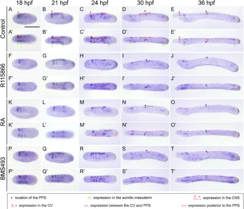
CYP26-3 likely assume the protection of the embryo from fluctuating RA levels, while CYP26-2 mediates RA-dependent developmental patterning (Carvalho et al., 2017). We hence decided to assay the expression of CYP26-2 to characterize patterning-related RA degradation activity in developing amphioxus embryos and larvae. As previously described (Carvalho et al., 2017), at 18 hpf (hours post fertilization), CYP26-2 is expressed in the rostral ectoderm and anterior endoderm, in the two anterior-most somites, in the cerebral vesicle (CV) of the CNS as well as in the posterior region, in a domain corresponding to the tail bud (Fig. 1 A-A’’). Later, at 21 and 24 hpf, inconspicuous expression of CYP26-2 is further found in the posterior-most ectoderm overlying the tail bud as well as in individual ectodermal cells along the flanks of the embryo (Fig. 1 B-C’’). At these stages, an additional CYP26-2 domain appears in the ventral region of the forming pharynx that is maintained through subsequent development (Fig. 1 B-D’’). Finally, in early larvae, at 36 hpf, the gene is no longer detectable in the anterior-most ecto-derm, but it is still expressed in all the other domains (Fig. 1 E-E’’).
Treatment with the CYP26 inhibitor R115866 generally leads to an expansion of CYP26-2 expression during development. At 18 hpf, for instance, the domain in the posterior tip of the embryo is enlarged (Fig. 1 F-F’’), and, by 21 and 24 hpf, the CYP26-2 domains in the anterior ectoderm and endoderm as well as in the CNS are expanded posteriorly (Fig. 1 G-H’’). This upregulation of CYP26-2 in anterior and posterior tissues becomes even more conspicuous at 30 hpf (Fig. 1 I-I’’), and, by 36 hpf, the signal has spread throughout most of the early larva, most noticeably in general ectoderm and CNS (Fig. 1 J-J’’). Treatments with exogenous RA generally induce a similar expansion of CYP26-2 expression in the amphioxus em-bryo (Fig. 1 K-O’’). However, compared to the CYP26 inhibitor, RA elicits a stronger response at 18 and 21 hpf (Fig. 1 F-G’’,K-L’’), a similar one at 24 and 30 hpf (Fig. 1 H-I’’,M-N’’), and a weaker one
at the 36 hpf (Fig. 1 J-J’’,O-O’’). Thus, at 18 and 21 hpf, conspicu-ous CYP26-2 staining is observed anteriorly and posteriorly as well as along almost the entire A-P axis of the general ectoderm (Fig. 1 K-L’’), while at 36 hpf, expression of the gene appears only slightly upregulated in the anterior regions of the larva and along the CNS (Fig. 1 O-O’’). In contrast, treatment with the RAR antagonist BMS493 induces no noticeable changes of CYP26-2 expression anteriorly, but abolishes the posterior signal (Fig. 1 P-T’’).
Altogether, these results show that CYP26-2 is expressed in anterior mesoderm and endoderm, the general ectoderm and CNS, most prominently in anterior and posterior territories of developing amphioxus embryos and larvae. Furthermore, the pharmacological treatments along with previous morphological studies (Carvalho et
al., 2017) suggest that CYP26 function is required for the
tissue-specific regulation of RA signaling activity during amphioxus develop-ment. To further characterize the involvement of CYP26 enzymes in RA-dependent patterning processes, we next analyzed expression of a series of tissue-specific marker genes in amphioxus embryos and larvae following pharmacological treatments.
CYP26 action is not involved in patterning the anterior am-phioxus mesoderm
Because CYP26 is conspicuously expressed in the mesoderm, we first characterized the potential action of CYP26 in this tissue. We thus assessed the expression of two mesodermal markers,
En and Pax2/5/8, in embryos and larvae treated with R115866
and compared the results to embryos and larvae treated with RA or BMS493. It should be noted that expression profiles of En and
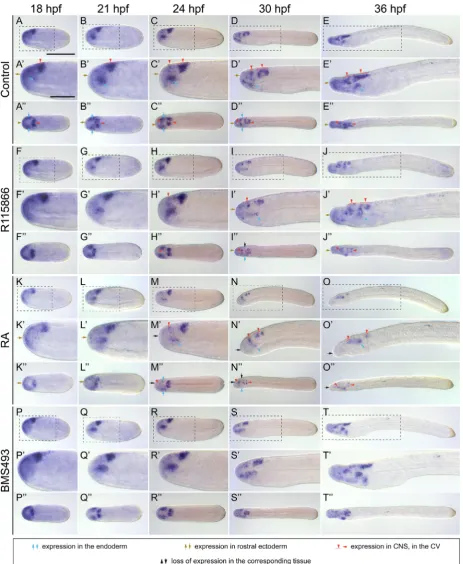
Fig. 3. Developmen-tal expression of
Branchiostoma lan-ceolatum Pax2/5/8.
(A-D’’) DMSO con-trols. (E-H’’) CYP26 inhibitor (R115866) treatments (final con-centration: 0.5 mM).
(I-L’’) all-trans retinoic acid (RA) treatments (final concentration: 0.1 mM). (M-P’’) RAR antagonist (BMS493) treatments (final con-centration: 1 mM). (A-D,E-H,I-L,M-P) Lateral views with anterior to the left and dorsal side up. (A’-D’,E’-H’,I’-L’,M’-P’) Close-up of regions of interest highlighted by dashed boxes in the lateral views. (A’’-D’’,E’’-H’’,I’’-L’’,M’’-P’’)
Dorsal views with anterior to the left. Developmental stages are given as hours post fertilization (hpf) at 19°C. For controls, tissues expressing the gene are marked as de-scribed in the legend. For treatment condi-tions, marked areas highlight expression changes (see main text for details). Black arrows indicate loss of Pax2/5/8 expression. Specifically, blue ar-rows allow an assess-ment of the relative distances between distinct endodermal expression domains. Scale bar in (A) is 100 mm and also applies to (A-D,A’’-D’’,E-H,E’’- H’’,I-L,I’’-L’’,M-P,M’’-P’’). Scale bar in (A’)
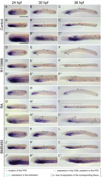
Fig. 4. Developmental expression of
Branchios-toma lanceolatum Pitx. (A-C’’) DMSO controls.
(D-F’’) CYP26 inhibitor (R115866) treatments (final concentration: 0.5 mM). (G-I’’) all-trans retinoic acid (RA) treatments (final concentration: 0.1 mM).
(J-L’’) RAR antagonist (BMS493) treatments (final concentration: 1 mM). (A-C,D-F,G-I,J-L) Lateral views with anterior to the left and dorsal side up. Pax2/5/8 during amphioxus development
have previously been described in the Florida amphioxus, B. floridae (Beaster-Jones et al., 2008; Holland et al., 1997; Holland and Hol-land, 1998; Kozmik et al., 1999), establishing that they are indeed expressed in mesodermal structures. Moreover, Pax2/5/8 expression has also been characterized in the amphioxus species used in this study, B. lanceolatum (Somorjai et al., 2008), and, following treat-ments with R115866, RA or the RAR antagonist BMS009, in developing B. floridae, albeit only at specific developmental stages (Koop et al., 2014; Schubert et al., 2006).
During B. lanceolatum development, the early mesodermal expression of En, from 18 through 24 hpf, is restricted to the posterior wall of the newly-formed somites, forming a striped pattern that progresses posteriorly with the elongation of the embryo (Fig. 2 A-C’). At 21 and 24 hpf, En expression in the anterior- and posterior-most somite pairs is conspicu-ous, while the En signal in the intermediate somite pairs is much less widespread (Fig. 2 B-C’). At 30 and 36 hpf, contrary to what has previously been described for the Florida amphioxus (Holland et al., 1997), we were able to identify En expression in newly-formed somites derived from the tail bud (Fig. 2 D-E’). Regarding Pax2/5/8, in contrast to En, its mesodermal expression during B. lanceolatum development is detectable starting at about 21 hpf in a thickening of the mesothelial wall of the anterior-most somite on the left side (Fig. 3 A-A’’). During subsequent development, this mesodermal Pax2/5/8 signal, which cor-responds to Hatschek’s nephridium, expands slightly until 36 hpf (Fig. 3 B-D’’). Intriguingly, despite the conspicuous expression of
CYP26-2 in anterior mesoderm, treatments with
R115866 or RA do neither affect the expression of En in the somitic mesoderm (Fig. 2 F-O’), nor the mesodermal expression of Pax2/5/8 (Fig. 3 E-L’’). Similarly, BMS493 does not in-duce any marked changes in the mesodermal
(A’-C’,D’-F’,G’-I’,J’-L’) Dorsal views with anterior to the left. (A’’-C’’,D’’-F’’,G’’-I’’,J’’-L’’) Close-up of regions of interest highlighted by dashed boxes in the dorsal views.Developmental stages are given as hours post fertilization (hpf) at 19°C. For controls, tissues expressing the gene are marked as described in the legend. For treatment conditions, marked areas highlight expression changes (see main text for details). Black circles and arrowheads indicate loss of Pitx expression. Specifically, for each specimen, blue arrows show the anterior (vertical arrow on the left) to posterior (horizontal arrow on the right) extent of the endodermal expression domain. Scale bar in (A) is 100 mm and also applies to (A-C,A’-C’,D-F,D’-F’,G-I,G’-I’,J-L,J’-L’). Scale bar in
expression of En (Fig. 2 P-T’) and Pax2/5/8 (Fig. 3 M-P’’). These results thus suggest that RA degradation mediated by CYP26 is not overtly involved in the developmental patterning of the mesoderm. This finding is consistent with previous reports assessing the functions of RA signaling in developing amphioxus (Bertrand et al., 2015; Carvalho et al., 2017; Koop et al., 2014; Schubert et al., 2005).
CYP26 is a key factor in the establishment of anterior endo-dermal structures in amphioxus
In previous reports, it has been suggested that CYP26 action is required for patterning the anterior amphioxus endoderm, with the inhibition of CYP26 activity leading to the loss of mouth, gill slits, endostyle, and club-shaped gland (Carvalho et al., 2017; Koop et al., 2014). To complement these results, we character-ized the impact of treatments with CYP26 inhibitor, RA or RAR antagonist on the endodermal expression of Pax2/5/8, Pitx, and
Pax6 in B. lanceolatum embryos and larvae. The developmental
expression of all three genes has previously been established in
B. floridae (Boorman and Shimeld, 2002; Glardon et al., 1998;
Kozmik et al., 1999; Soukup et al., 2015), those of Pax2/5/8 and
Pitx have also been reported in B. lanceolatum (Somorjai et al.,
2008; Soukup et al., 2015), and that of Pitx also in B. japonicum (Yasui et al., 2000). Moreover, effects of R115866, RA, and the RAR antagonist BMS009 have been assessed for Pax2/5/8 and
Pitx in B. floridae, but exclusively in mid-neurulae and early larvae
(Koop et al., 2014; Schubert et al., 2005; Schubert et al., 2006). In the endoderm, expression of B. lanceolatum Pax2/5/8 is first detectable anteriorly at 21 hpf (Fig. 3 A-A’’). Subsequently, at 24 hpf, Pax2/5/8 expression in the anterior endoderm becomes subdivided into three individual domains: the first one, localized anteriorly, is in a territory giving rise to Hatschek’s left and right diverticula, the second one is located slightly posteriorly, where the mouth will penetrate, and the third one is in the ventral endo-derm on the left side of the forming pharynx where the gill slits will form (Fig. 3 B-B’’). At 30 and 36 hpf, Pax2/5/8 expression in the amphioxus pharyngeal endodermal is detectable in dif-ferent structures: on the left side, transiently in Hatschek’s left diverticulum and where the mouth will open, on the right side, in the endostyle, and, ventrally, where the gill slits will penetrate (Fig. 3 C-D’’). Concerning Pitx, at 24 hpf, this gene is expressed exclusively on the left side in the anterior half of the endoderm (Fig. 4 A-A’’). During subsequent development, at 30 and 36 hpf, expression of Pitx becomes restricted to the left side of the pharyngeal endoderm and to the developing club-shaped gland, on both the left and the right side of the body (Fig. 4 B-C’’). This bilateral signal of Pitx in the B. lanceolatum club-shaped gland contrasts with previous reports in B. japonicum, where the club-shaped gland-associated expression of the gene seems to be limited to the left side (Yasui et al., 2000). Finally, for Pax6, the first endodermal expression of this gene is observable at 18 hpf, in anterior cells located dorsolaterally on either side of the endo-derm, marking the presumptive territories of Hatschek’s left and right diverticula (Fig. 5 A-A’’). During subsequent development,
Pax6 remains detectable in both of these endodermal pouches
without extending to any other endodermal structure (Fig. 5 B-D’’). By 36 hpf, Pax6 expression is only observable in Hatschek’s left diverticulum (Fig. 5 E-E’’).
Treatment with R115866 affects the expression of all three
marker genes, although in different ways. Thus, Pax2/5/8 expres-sion seems largely unaffected by the treatment at 21 and 24 hpf (Fig. 3 E-F’’), whereas, by 30 and 36 hpf, expression in all three pharyngeal Pax2/5/8 domains is less conspicuous (Fig. 3 G-H’’). R115866 also restricts endodermal Pitx expression along the A-P axis starting at 24 hpf in a posterior to anterior progression (Fig. 4 D-F’’). Expression in the club-shaped gland is also completely lost following CYP26 inhibition (Fig. 4 E-F’’), with only the Pitx signal in Hatschek’s left diverticulum still being discernable in the endo-derm at 36 hpf (Fig. 4 F-F’’). In contrast, treatment with R115866 does not affect endodermal Pax6 expression until 30 hpf (Fig. 5 F-H’’), when the signal is no longer observable in Hatschek’s right diverticulum and is downregulated in Hatschek’s left diverticulum, albeit not completely (Fig. 5 I-J’’). Exogenous RA treatments also reduce the endodermal expression domains of Pax2/5/8 (Fig. 3 I-L’’), Pitx (Fig. 4 G-I’’), and Pax6 (Fig. 5 K-O’’). However, in contrast to R115866 treatments, RA affects Pax2/5/8 expression already at 21 hpf, leading to a loss of most of the endodermal expression (Fig. 3 E-E’’,I-I’’). Furthermore, the domain of Pitx is more severely restricted to anterior endodermal territories by RA at 24 hpf (Fig. 4 D-D’’,G-G’’), although, at 30 and 36 hpf, this effect is more pronounced following CYP26 inhibition (Fig. 4 E-F’’,H-I’’). Likewise, the endodermal Pax6 domain is already less conspicuous at 24 hpf following RA treatment (Fig. 5 H-H’’,M-M’’). Consistent with the R115866 and RA results, BMS493 treatments induce a posterior expansion of the endodermal Pax2/5/8 (Fig. 3 M-P’’) and Pitx (Fig. 4 J-L’’) domains, particularly at early de-velopmental stages, but do not induce any significant changes in the endodermal expression of Pax6 (Fig. 5 P-T’’).
Altogether, these results thus support previous findings (Car-valho et al., 2017; Koop et al., 2014) suggesting that CYP26 is required for both regional patterning of the endoderm and the development of specific pharyngeal structures.
CYP26 plays a major role in patterning specific ectodermal sensory neuron populations
Given the role of RA signaling in patterning the amphioxus general ectoderm (Schubert et al., 2004) and the scattered expression of
CYP26-2 in this tissue (Carvalho et al., 2017), we next assessed
the effects of CYP26 inhibition in the general ectoderm by assess-ing expression of Pax6, Tlx as well as Prdm12. The expression profiles of Pax6 and Tlx have previously been described during
B. floridae development (Glardon et al., 1998; Kaltenbach et al.,
2009; Lu et al., 2012) and those of Tlx have been reported in developing B. lanceolatum (Zieger, 2016). Furthermore, the ef-fects of RA and BMS493 on Tlx expression have been assessed in B. lanceolatum (Zieger, 2016).
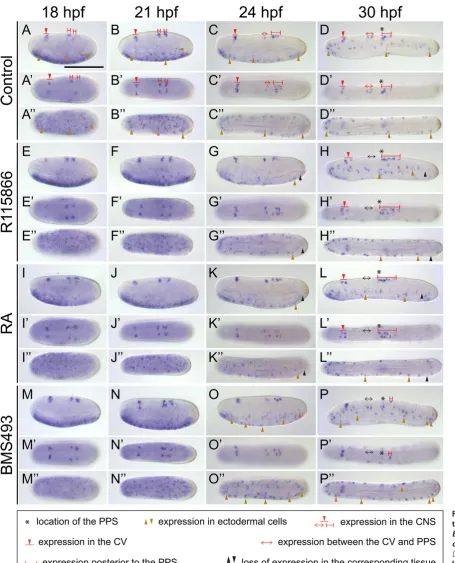
Fig. 6. Developmen-tal expression of
dispersed along the lateral ectoderm (Fig. 6 D,D’’). Similar to
Tlx, at 21 and 24 hpf, Prdm12 is expressed transiently and very
inconspicuously in individual cells of the ventral ectoderm (Fig. 7 A,A’,B,B’’). In contrast, by 30 hpf, Prdm12 expression in the ectoderm is completely downregulated (Fig. 7 C-D’).
Treatments with R115866 affect the ectodermal expression of both Pax6 and Tlx. Thus, while R115866 does not affect early expression of Pax6 in the rostral ectoderm between 18 and 24
hpf (Fig. 5 F-H’’), the signal is less conspicuous at 30 and 36 hpf (Fig. 5 I-J’’). For the ectodermal expression of Tlx, no effects of R115866 are discernable until 24 hpf (Fig. 6 E,E’’,F,F’’). At this stage, the treatments seem to shift the distribution of Tlx-positive ESN progenitors anteriorly (Fig. 6 G,G’’), a trend that is some-what more pronounced at 30 hpf (Fig. 6 H,H’’). In contrast to Tlx, we were unable to document an effect of R115866 treatments on the Prdm12-expressing ESN progenitor population (Fig. 7
Tlx-expressing ESN progenitors along the A-P axis at 24 and 30
hpf, shifting them towards more anterior levels of the ectoderm (Fig. 6 I,I’’,J,J’’,K,K’’,L,L’’). For Prdm12, it was again difficult to assess the effects of the treatments (Fig. 7 I,I’’,J,J’’). BMS493 treatments might somewhat weaken the rostral ectodermal ex-pression of Pax6, although this effect is not consistently seen in all assayed stages (Fig. 5 P-T’’). Conversely, the RAR antagonist might increase the overall number of Tlx-expressing cells at 24 and 30 hpf (Fig. 6 O,O’’,P,P’’). For Prdm12, no unequivocal ef-fect on its ectodermal expression was observed upon treatment with BMS493 (Fig. 7 M-N’).
Taken together, these results are consistent with a role for CYP26 enzymes in mediating RA-dependent patterning process-es in different regions of the developing ectoderm. Furthermore, they support previous findings suggesting an implication of RA signaling in neural specification processes in the amphioxus ectoderm (Schubert et al., 2004, Zieger, 2016).
CYP26 has a major influence on the establishment of anterior-posterior identity in the developing amphioxus central nervous system
The importance of RA signaling for patterning the developing amphioxus CNS has been well documented in the literature (Escriva
et al., 2002; Holland and Holland, 1996; Schubert et al., 2006),
but the roles for CYP26 in this process remain elusive. Given that our six marker genes, En, Pax2/5/8, Pitx, Pax6, Tlx, and Prdm12, are expressed in different regions of the developing CNS, we next studied their expression in embryos and larvae treated with R115866 and compared the resulting patterns to those obtained in embryos and larvae treated with either RA or BMS493. The CNS expression of these genes has previously been described for En, Pax2/5/8, Pitx, Pax6, and Tlx in B. floridae (Beaster-Jones
et al., 2008; Boorman and Shimeld, 2002; Glardon et al., 1998;
Holland et al., 1997; Holland and Holland, 1998; Kaltenbach et al., 2009; Kozmik et al., 1999), for En, Pax2/5/8, Pitx, and Prdm12 in
B. lanceolatum (Somorjai et al., 2008; Thélie et al., 2015) as well
as for Pitx in B. japonicum (Yasui et al., 2000). Of note, only the expression of B. floridae Pax2/5/8 has already been analyzed in the CNS of embryos and larvae following treatments with RA or the RAR antagonist BMS009 (Schubert et al., 2006).
The expression domains of the six marker genes along the A-P axis of the developing amphioxus CNS can chiefly be subdivided into three main territories: the CV, the CNS region located posterior to the CV but anterior to the primary pigment spot (PPS), and the CNS region located posterior to the PPS (Albuixech-Crespo et al., 2017; Candiani et al., 2012). In the following, we will discuss the effects of the pharmacological treatments on gene expression in each one of these three territories, in an anterior-to-posterior pro-gression. Thus, the amphioxus CV is characterized by expression of Pax6, Prdm12, En, and Tlx. Pax6 is expressed in two domains of the CV, an anterior one, first detectable at 24 hpf, and a posterior one, already discernable at 18 hpf (Fig. 5 A-E’’). Expression of
Prdm12 in the CV turns on even later, at 30 hpf, and is limited to
cells in both the anterior and posterior expression domains in the CV, with the effect on the former being observable by 24 hpf and that on the latter by 36 hpf (Fig. 5 F-J’’). Similarly, following CYP26 inhibition, the CV-associated expression of Prdm12 is much less conspicuous at 36 hpf (Fig. 7 H,H’). Of note, although expressed in the same region of the CV, the En signal is unaffected by CYP26 inhibitor treatments (Fig. 2 J,J’). Contrasting the effects on Pax6 and Prdm12, Tlx expression in the CV is expanded by R115866, with the appearance of supernumerary Tlx-positive cells in the posterior CV at 30 hpf (Fig. 6 H,H’). The effects induced by exog-enous RA are generally comparable to those obtained by CYP26 inhibition. RA thus reduces the number of stained cells both in the anterior and posterior expression domains of Pax6 in the CV, the former by 24 hpf, which is similar to R115866, and the latter by 30 hpf, which is earlier than what is observed with R115866 (Fig. 5 M-N’’). Likewise, Prdm12 expression in the CV at 36 hpf is less conspicuous in embryos treated with RA (Fig. 7 L,L’), while
En expression in the CV is generally unaffected (Fig. 2 O,O’).
Finally, like R115866, exogenous RA also induces an increase in the number of Tlx-positive cells in the CV (Fig. 6 L,L’). Contrary to the effects observed with either R115866 or RA, BMS493 treat-ments do not affect the CV-associated expression of Pax6 (Fig. 5 P-T’’), Prdm12 (Fig. 7 O-P’), En (Fig. 2 T-T’) or Tlx (Fig. 6 M-P’). These results thus suggest that normal CV patterning does not require active RA signaling and that endogenous RA levels in this anterior-most part of the developing amphioxus CNS are kept low by the activity of CYP26 enzymes.
In the second A-P CNS territory, located posterior to the CV but anterior to the PPS, Prdm12, Pax2/5/8, En, and Tlx are expressed. In this region, Prdm12 is first detectable in a few cells located just anterior to the PPS at 24 hpf (Fig. 7 B,B’). This domain is subse-quently expanded further anteriorly at 30 and 36 hpf (Fig. 7 C-D’).
Pax2/5/8 is expressed along the entire CNS, excluding the CV,
here (Fig. 3 I-L’’). The use of a significantly higher final concentra-tion of RA in this previous work (1mM compared to 0.1mM) very likely explains the different results obtained. Coming back to our study, in the territory posterior to the CV but anterior to the PPS, BMS493 treatments lead to a delayed onset of Prdm12 expres-sion, which remains undetectable until 30 hpf (Fig. 7 O,O’), and to a subsequent downregulation by 36 hpf (Fig. 7 P,P’). Surprisingly, similar to the effects induced by R115866 and RA, BMS493 treat-ments also induce a loss of the Tlx-positive cell cluster located anteriorly to the PPS (Fig. 6 P,P’). In this CNS territory, both the upregulation and downregulation of RA signaling hence induce the mispatterning of specific neural populations, suggesting that these cells require intermediary RA levels for their proper specification. In the third CNS territory, located posterior to the PPS, Pax2/5/8,
En, Pitx, Prdm12, and Tlx are expressed. While Pax2/5/8 is
conspicu-ously expressed in this region at 21 and 24 hpf, the signal weakens at 30 and 36 hpf (Fig. 3 A-D’’). In contrast, En is only detectable in a few cells just posterior to the PPS after 24 hpf (Fig. 2 C-E’). At that stage, Pitx expression is limited to a small cluster of cells located just posterior to the En-positive cells (Fig. 4 A-A’’). During subsequent development, the Pitx domain expands posteriorly, with more cells in the CNS starting to express the gene (Fig. 4 B-C’’). Tlx expression in this posterior CNS domain is limited to two bilateral clusters of cells, first detectable at 18 hpf (Fig. 6 A-A’). The overall number of cells in these two Tlx clusters does not increase significantly during subsequent development (Fig. 6 B-D’). Finally,
Prdm12 is expressed in a few cells just posterior to the PPS at 30
hpf (Fig. 7 C,C’), and this domain is expanded posteriorly at 36 hpf (Fig. 7 D,D’). Inhibition of CYP26 function does not alter the expres-sion of Pax2/5/8 (Fig. 3 E-H’’), En (Fig. 2 F-J’), Pitx (Fig. 4 D-F’’) or Prdm12 (Fig. 7 G-H’) and thus only affects Tlx in the posterior CNS: the overall number of Tlx-positive cells in the two clusters is increased at 36 hpf to between seven and eight cells (Fig. 6 H,H’). Similar to CYP26 inhibition, exogenous RA exclusively increases the number of Tlx-positive cells at 36 hpf (Fig. 6 L,L’) and does not impact the expression of Pax2/5/8 (Fig. 3 I-L’’), En (Fig. 2 K-O’),
Pitx (Fig. 4 G-I’’) or Prdm12 (Fig. 7 K-L’). Interestingly, while the
RAR antagonist also does not affect Pax2/5/8 (Fig. 3 M-P’’) and
En (Fig. 2 P-T’) expression, at 36 hpf, BMS493 treatments strongly
reduce the number of cells expressing either Tlx (Fig. 6 P,P’) or Pitx (Fig. 4 L-L’’) and completely abolish expression of Prdm12 in this posterior CNS territory (Fig. 7 P,P’). These results indicate that RA signaling is required for the specification of neural cell populations in the posterior amphioxus CNS. Given the severe effects caused by downregulation of RA signaling activity and the concurrent paucity of phenotypic changes induced by its upregulation, it is likely that this posterior region of the CNS requires high RA signaling levels for proper developmental patterning.
In sum, these data suggest that neuronal populations located at different positions along the A-P axis of the developing amphi-oxus CNS require different levels of RA signaling activity for their specification. Furthermore, our results reveal roles for CYP26 in distinctive A-P territories of the amphioxus CNS, some of which do not express CYP26 genes (Carvalho et al., 2017). It is thus conceivable that, as in vertebrates (Hernandez et al., 2007; White
et al., 2007), one of the functions of CYP26 enzymes in amphioxus
is to establish a RA gradient that is required for proper regional patterning along the A-P axis of the developing CNS.
Discussion
In the present study, we used pharmacological inhibition of CYP26 function to study the influence of CYP26 enzymes on the modulation of RA signaling activity during amphioxus development. The characterization of CYP26-2 expression, a known direct target of RA signaling in amphioxus (Carvalho et al., 2017), allowed us to identify tissues where RA degradation might be essential during development. The characterization of marker gene expression fol-lowing CYP26 inhibition subsequently revealed that CYP26 func-tion is required for patterning the anterior endoderm, the general ectoderm as well as the CNS, while it might be dispensable for mesoderm development. Although the latter notion is consistent with previous studies in amphioxus suggesting that disruption of RA signaling does not affect mesodermal patterning (Bertrand
et al., 2015; Schubert et al., 2005), further work is needed to
un-equivocally demonstrate that amphioxus mesoderm development is not regulated by RA signaling. Finally, comparisons of CYP26 inhibition with the global increase (i.e. RA treatment) or reduction (i.e. RAR antagonist treatment) of RA signaling activity yielded insights into the tissue specificity of CYP26 action in developing amphioxus embryos and larvae.
In general, CYP26 inhibition and exogenous RA induce compa-rable defects during amphioxus development, which is consistent with our current understanding of the feedback loops controlling endogenous RA levels (Carvalho et al., 2017). However, our experi-ments revealed that RA treatexperi-ments induce marker gene expression changes significantly earlier than CYP26 inhibition. This is the case, for example, for endodermal expression of Pax2/5/8, Pitx, and Pax6, for ectodermal expression of Pax6 and Tlx as well as for CNS expression of Pax6. These differences can at least partially be explained by the nature of the treatments, i.e. a local increase of RA levels by inhibition of CYP26-dependent degradation versus a global induction of RA signaling activity by exogenous RA treat-ment. Nonetheless, the developmental effects elicited by R115866 and RA are generally very similar, suggesting that in amphioxus, as in vertebrates, the CYP26-mediated creation of local RA sinks is fundamental for the establishment of global RA signaling activity in the embryo (Rydeen et al., 2015; White et al., 2007).
In vertebrates, the activity of RA synthesizing and degrading enzymes in the developing embryo have been shown to create a RA gradient that mediates RA signaling-dependent patterning along the A-P axis (Hernandez et al., 2007; Rydeen et al., 2015; Schilling et al., 2016; Shimozono et al., 2013; White et al., 2007). During amphioxus development, CYP26-2 is expressed very conspicuously in the anterior general ectoderm, CNS, endoderm, and mesoderm (Carvalho et al., 2017), while genes encoding RA production enzymes of the RALDH family are expressed in the posterior half of the embryo in endoderm and mesoderm (Sobreira
et al., 2011). Hence, comparable to the situation in vertebrates, a
RA gradient likely exists in developing amphioxus and functions in A-P patterning of the embryo. This morphogen gradient would be characterized by high endogenous RA concentrations in the region of RALDH activity posteriorly that decrease towards the anterior tip of the embryo and larva, with this decrease being ensured by the presence of 2. In addition, given that amphioxus
CYP26-2 is also expressed in the posterior-most ectoderm, albeit very
assume patterning functions, for example, in the developing brain (Schilling et al., 2016; Shimozono et al., 2013). Taken together, these observations suggest that RA-dependent regionalization by the differential interpretation of such morphogen gradients already patterned embryos and larvae of the last common ancestor of amphioxus and vertebrates.
Although future work, using, for example, in vivo imaging tools developed in vertebrates (Schilling et al., 2016; Shimozono et al., 2013), will be required to unequivocally demonstrate the existence of RA gradient(s) in amphioxus, it is nonetheless tempting to speculate, which tissues might be regulated by a morphogen gradient during amphioxus development. In general, they should be characterized by highly regionalized responses to RA along the A-P axis. While our results indicate functions for RA signaling and CYP26 enzymes in the developing CNS, ectoderm, and endoderm, we were unable to identify any in the mesoderm. This contrasts with the situation in vertebrates, where RA signaling and CYP26 enzymes are required for the proper development of different mesodermal derivatives, including, for example, the cardiovascular system and vertebrae (Rydeen and Waxman, 2014; Sakai et al., 2001). These regulatory differences are consistent with the hypothesis that, in the course of chordate evolution, mesoderm-specific functions for RA signaling and CYP26 evolved specifically in the lineage leading to extant vertebrates, after its split from the amphioxus lineage (Carvalho
et al., 2017).
RA signaling in the amphioxus endoderm is known to define the posterior limit of the pharynx by activating the expression of Hox1 in the foregut, hence limiting the expression of pro-pharyngeal genes to the pharyngeal endoderm (Schubert et al., 2005). However, it remains elusive, whether the Hox1-dependent regionalization of the amphioxus endoderm is the result of a graded response to RA, and the experimental evidence presented here did not yield additional information to clarify this issue. RA signaling, and in particular CYP26 function, has further been implicated in the development of specific structures in the pharyngeal, i.e. anterior, endoderm, including the mouth and the anterior gill slits (Carvalho et al., 2017; Koop et al., 2014). Our results are coherent with these previous reports, as they show that the pharmacological inhibition of CYP26 leads to the downregulation of genes, such as Pax2/5/8, Pitx, and Pax6, which are expressed in specific pharyngeal structures, such as the mouth, the anterior gill slits, the endostyle as well as Hatschek’s left and right diverticula. Consistently, CYP26-2 expression is particularly conspicuous in the anterior pharyngeal compartment that comprises these structures (Carvalho et al., 2017; Koop et
al., 2014). One of them, Hatschek’s left diverticulum, will give rise
to Hatschek’s pit, the likely amphioxus homolog of the vertebrate adenohypophysis (Kozmik et al., 2007). Adenohypophysis devel-opment in amphioxus might thus take place in a RA-free environ-ment, which contrasts with the situation in vertebrates, where the formation of Rathke’s pouch, i.e. of the adenohypophyseal placode (Patthey et al., 2014; Schlosser et al., 2014), requires RA signaling (Maden et al., 2007). It is likely that this inverse dependence on RA is correlated with the different embryonic origins of Hatschek’s left
established a role for RA signaling in patterning the general ecto-derm and in controlling the distribution of ESNs along its A-P axis (Schubert et al., 2004). Even though CYP26-2 is expressed in both the anterior-most and posterior-most amphioxus ectoderm (Carvalho et al., 2017), our marker gene analyses did not yield results that are indicative of a functional RA gradient in the general ectoderm. While CYP26 function is known to be required in the posterior ectoderm for tail fin development (Carvalho et al., 2017), it also seems to be involved in the regulation of Pax6 anteriorly. Of note, the Pax6-positive territory in the rostral ectoderm of amphi-oxus contains specific populations of chemosensory cells (Lacalli and Hou, 1999) and has been proposed to be homologous to the olfactory placode of vertebrates (Patthey et al., 2014; Schlosser et
al., 2014). Although RA signaling has been implicated in olfactory
placode development in vertebrates, for example in the production and maintenance of Pax6-positive progenitor cells (Paschaki et
al., 2013), a role for CYP26 in this process still remains elusive.
Finally, our data are coherent with the existence of a graded response to RA signaling in the amphioxus CNS. Indeed, at least three domains along the A-P axis of the CNS seem to react dif-ferently to alterations of endogenous RA signaling levels: (1) the CV, which represents the homolog of the forebrain and midbrain regions of vertebrates, (2) the region located posterior to the CV and anterior to the PPS that corresponds to the anterior and center of the vertebrate hindbrain, and (3) the region of the CNS located posterior to the PPS, which is the equivalent of the vertebrate posterior hindbrain and spinal cord (Albuixech-Crespo et al., 2017; Candiani et al., 2012). In the CV, which expresses CYP26-2, the upregulation of RA signaling induces the mispatterning of Pax6 and Tlx expression, while no effect is induced by RA signaling inhibition, suggesting that this domain requires very low levels of RA during development. In the intermediary region of the CNS, located posterior to the CV and anterior to the PPS, treatment with either the CYP26 inhibitor, RA or the RAR antagonist affected Tlx and Prdm12 expression, suggesting that both abnormally high and low RA signaling levels lead to mispatterning of specific neuronal populations, which in turn is consistent with an intermediate en-dogenous RA signaling activity in this CNS territory. Finally, in the domain located posterior to the PPS, RAR antagonist treatments selectively abolish Pitx as well as Prdm12 expression, while both the CYP26 inhibitor and RA do not affect these domains of Pitx and
Prdm12. The specification of these two neuronal populations, Pitx-
or Prdm12-positive, thus requires sustained RA signaling activity, which is readily provided in this territory of the CNS located in close proximity to the main source of endogenous RA in the posterior mesoderm and endoderm (Sobreira et al., 2011).
Of note, inhibition of CYP26 function affects the expression of marker genes in the CNS not only in the anterior region character-ized by conspicuous CYP26 expression, but also in both posterior territories marked by an absence of CYP26 expression (Carvalho
et al., 2017). We interpret these results as strong indications for the
CNS. A functional readout of this morphogen gradient could then be the activation of collinear Hox expression along the A-P axis of the amphioxus CNS, which is known to be under the control of RA signaling (Schubert et al., 2006). It is further conceivable that individual cells along the A-P gradient elicit different responses to RA depending on their cellular environments. The RA concentra-tion gradient might hence also establish cell type specificity in the developing amphioxus embryo, a notion that has previously been proposed for both the amphioxus CNS and general ectoderm (Schubert et al., 2004; Schubert et al., 2006).
Taken together, in this study, we provide evidence for the tissue-specific action of CYP26 enzymes during amphioxus development. Furthermore, we establish that, similar to the situation in verte-brates, amphioxus CYP26 enzymes act with other components of the RA pathway to set up a RA gradient along the A-P axis that is required for patterning at least the developing CNS. The evolu-tion of a RA morphogen gradient funcevolu-tioning during development to convey A-P patterning information might thus date back to the origin of chordates.
Materials and Methods
Amphioxus adult husbandry, embryo rearing, and pharmacological treatments
Sexually mature European amphioxus (B. lanceolatum) were collected by dredging in Argelès-sur-Mer, France, and retrieved from the sand by sieving. After transportation, animals were transferred into tanks, with about 10 to 15 adults per aquarium. The water temperature was kept constant at 16 to 17°C, and the animals were maintained under a spring-like day/night
period (14 hours of light and 10 hours of absolute darkness). Spawning was induced by a 36-hour thermal shock at 23°C, as previously described
(Fuentes et al., 2004). Following in vitro fertilization, embryos were reared in artificial seawater at 19°C in complete darkness. Pharmacological treatments
were performed at the late blastula stage (6 hpf) with the CYP26 inhibitor R115866 (at 0.5 mM) (provided by Janssen Research & Development, a
division of Janssen Pharmaceutica NV, Beerse, Belgium), all-trans RA (at 0.1 mM) (Sigma-Aldrich, Saint-Quentin Fallavier, France), and the RAR
antagonists BMS493 (at 1 mM) (Sigma-Aldrich, Saint-Quentin Fallavier,
France). The compounds were dissolved in dimethyl sulfoxide (DMSO) to obtain a 1000X stock solution and subsequently added to the cultures in a 1:1000 dilution to yield the respective final concentrations. As controls, cultures were treated with DMSO alone at a final dilution of 1:1000.
Gene cloning, in situ hybridization and imaging
Total RNA was extracted from B. lanceolatum embryos at different developmental stages according to established protocols (Yu and Hol-land, 2009), and cDNA was synthesized using the SuperScriptIII reverse transcription kit (Invitrogen, Cergy Pontoise, France). B. lanceolatum CYP26-2, En, Pax2/5/8, Pitx, Pax6, Tlx, and Prdm12 were amplified by
PCR using the gene-specific primers listed in Table 1 and cloned into commercially available vectors. The obtained clones were subsequently validated by sequencing on both strands. Gene accession numbers are as follows: CYP26-2 (KX118108), En (KP235487), Pax2/5/8 (EU685298), Pitx (EU685299), Pax6 (MF536418), Tlx (KY569298), and Prdm12 (KP235486). For in situ hybridization, following the treatments, B. lanceolatum embryos and larvae were cultured and subsequently fixed in 4% paraformaldehyde at different developmental stages, between the late gastrula and early larva stages (i.e. between 12 and 36 hpf), as previously described (Yu and Holland, 2009). Antisense riboprobe synthesis and in situ hybridiza-tion experiments were subsequently performed following the established protocols (Yu and Holland, 2009). After in situ hybridization, B. lanceolatum embryos and larvae were photographed as whole mounts using Zeiss DIC (differential interference contrast) optics and an AxioCam ERc 5s camera (Carl Zeiss SAS, Marly-le-Roi, France). Adobe Photoshop CS6 was used for the calculation of maximal projections and for image processing. For each gene, all assayed stages (from 12 to 36 hpf) were photographed and subsequently analyzed.
Acknowledgements
The authors are indebted to Hector Escriva from the Observatoire Océanologique de Banyuls-sur-Mer in Banyuls-sur-Mer, France, for pro-viding Branchiostoma lanceolatum adults and to Laurent Gilletta, Loann Gissat, and Sophie Collet for help with maintaining amphioxus adults at the Observatoire Océanologique de Mer in Villefranche-sur-Mer, France. The authors would further like to thank Janssen Research & Development, a division of Janssen Pharmaceutica NV, for providing the CYP26 inhibitor. This study was supported by a grant from the Agence Nationale de la Recherche (ANR-11-JSV2-002-01) and by funds from the Réseau André Picard (ANR-11-IDEX-0004-02, Sorbonne Universities) to Michael Schubert. João E. Carvalho was a FCT doctoral fellow (SFRH/ BD/86878/2012).
References
ALBALAT R, CAÑESTRO C (2009). Identification of Aldh1a, Cyp26 and RAR orthologs in protostomes pushes back the retinoic acid genetic machinery in evolutionary time to the bilaterian ancestor. Chem Biol Interact 178: 188–196.
ALBUIXECH-CRESPO B, LÓPEZ-BLANCH L, BURGUERA D, MAESO I, SÁNCHEZ-ARRONES L, MORENO-BRAVO JA, SOMORJAI I, PASCUAL-ANAYA J, PUELLES E, BOVOLENTA P, GARCIA-FERNÀNDEZ J, PUELLES L, IRIMIA M, FERRAN JL (2017). Molecular regionalization of the developing amphioxus neural tube challenges major partitions of the vertebrate brain. PLoS Biol 15: e2001573. BEASTER-JONES L, KALTENBACH SL, KOOP D, YUAN S, CHASTAIN R,
HOL-LAND LZ (2008). Expression of somite segmentation genes in amphioxus: a clock without a wavefront? Dev Genes Evol 218: 599–611.
BERTRAND S, ALDEA D, OULION S, SUBIRANA L, DE LERA AR, SOMORJAI I, ESCRIVA H (2015). Evolution of the role of RA and FGF signals in the control of somitogenesis in chordates. PLoS One 10: e0136587.
BLOMHOFF R, BLOMHOFF HK (2006). Overview of retinoid metabolism and func-tion. J Neurobiol 66: 606–630.
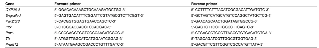
Gene Forward primer Reverse primer
CYP26-2 5’-GGACACAAAGCTGCAAAGATGCTGG-3’ 5’-CCTTTTCTTTACATCGCGACATTGATGTC-3’
Engrailed 5’-GAGTGACATTTCGGATTCGTATGCGTCTTCGGT-3’ 5’-GCTAGTCATGCATGTCCAGGCTATGCTCG-3’
Pax2/5/8 5’-CACGGTGGAGTGAACCAGCTC-3’ 5’-GAACAGCAACTGGATAGTGGCCG-3’
Pitx 5’-GTCGCAGCAGCTCCAGGAG-3’ 5’-GAGTGTTGCTTGGCCTTCAGTC-3’
Pax6 5’-CCCGAGGTGGTCGCCAAGATCGCG-3’ 5’-CTGAGCCTCCGTTAGCGTGTGACATGTGA-3’
Tlx 5’-ATGGTTGGCATCATGGAATCGGAG-3’ 5’-TAGCAGATCGTTGGCGTGGTGAG-3’
Prdm12 5’-ATAATGAAGCCGACCCTGTTTGATC-3’ 5’-GACGTTCGTTCGGTCGCCATGTTATA-3’
TABLE 1
A neurochemical map of the developing amphioxus nervous system. BMC
Neurosci 13: 59.
CARVALHO JE, SCHUBERT M (2013). Retinoic acid: metabolism, developmental functions, and evolution. In Vitamin-binding proteins: functional consequences (Eds K Dakshinamurti and S Dakshinamurti). CRC Press, Boca Raton, pp. 1–30. CARVALHO JE, THEODOSIOU M, CHEN J, CHEVRET P, ALVAREZ S, DE LERA AR, LAUDET V, CROCE JC, SCHUBERT M (2017). Lineage-specific duplication of am-phioxus retinoic acid degrading enzymes (CYP26) resulted in sub-functionalization of patterning and homeostatic roles. BMC Evol Biol 17: 24.
DOBBS-MCAULIFFE B, ZHAO Q, LINNEY E (2004). Feedback mechanisms regu-late retinoic acid production and degradation in the zebrafish embryo. Mech Dev 121: 339–350.
ESCRIVA H, HOLLAND ND, GRONEMEYER H, LAUDET V, HOLLAND LZ (2002). The retinoic acid signaling pathway regulates anterior/posterior patterning in the nerve cord and pharynx of amphioxus, a chordate lacking neural crest.
Develop-ment 129: 2905–2916.
FUENTES M, SCHUBERT M, DALFO D, CANDIANI S, BENITO E, GARDENYES J, GODOY L, MORET F, ILLAS M, PATTEN I, et al., (2004). Preliminary observa-tions on the spawning condiobserva-tions of the European amphioxus (Branchiostoma
lanceolatum) in captivity. J Exp Zoolog B 302: 384–391.
GLARDON S, HOLLAND LZ, GEHRING WJ, HOLLAND ND (1998). Isolation and developmental expression of the amphioxus Pax-6 gene (AmphiPax-6): insights into eye and photoreceptor evolution. Development 125: 2701–2710.
HERNANDEZ RE, PUTZKE AP, MYERS JP, MARGARETHA L, MOENS CB (2007). Cyp26 enzymes generate the retinoic acid response pattern necessary for hind-brain development. Development 134: 177–187.
HOLLAND LZ, HOLLAND ND (1998). Developmental gene expression in amphioxus: new insights into the evolutionary origin of vertebrate brain regions, neural crest, and rostrocaudal segmentation. Am Zool 38: 647–658.
HOLLAND LZ, HOLLAND ND (1996). Expression of AmphiHox-1 and AmphiPax-1 in amphioxus embryos treated with retinoic acid: insights into evolution and pat-terning of the chordate nerve cord and pharynx. Development 122: 1829–1838. HOLLAND LZ, KENE M, WILLIAMS NA, HOLLAND ND (1997). Sequence and
embryonic expression of the amphioxus engrailed gene (AmphiEn): the meta-meric pattern of transcription resembles that of its segment-polarity homolog in
Drosophila. Development 124: 1723–1732.
KALTENBACH SL, YU J-K, HOLLAND ND (2009). The origin and migration of the earliest-developing sensory neurons in the peripheral nervous system of amphi-oxus. Evol Dev 11: 142–151.
KOOP D, CHEN J, THEODOSIOU M, CARVALHO JE, ALVAREZ S, LERA AR de, HOL-LAND LZ, SCHUBERT M (2014). Roles of retinoic acid and Tbx1/10 in pharyngeal segmentation: amphioxus and the ancestral chordate condition. EvoDevo 5: 36. KOZMIK Z, HOLLAND ND, KALOUSOVA A, PACES J, SCHUBERT M, HOLLAND
LZ (1999). Characterization of an amphioxus paired box gene, AmphiPax2/5/8: developmental expression patterns in optic support cells, nephridium, thyroid-like structures and pharyngeal gill slits, but not in the midbrain-hindbrain boundary region. Development 126: 1295–1304.
KOZMIK Z, HOLLAND ND, KRESLOVA J, OLIVERI D, SCHUBERT M, JONASOVA K, HOLLAND LZ, PESTARINO M, BENES V, CANDIANI S (2007). Pax–Six–Eya–
Dach network during amphioxus development: conservation in vitro but context
specificity in vivo. Dev Biol 306: 143–159.
LACALLI TC, HOU S (1999). A reexamination of the epithelial sensory cells of am-phioxus (Branchiostoma). Acta Zool 80: 125–134.
LU T-M, LUO Y-J, YU J-K (2012). BMP and Delta/Notch signaling control the develop-ment of amphioxus epidermal sensory neurons: insights into the evolution of the peripheral sensory system. Development 139: 2020–2030.
MADEN M, BLENTIC A, REIJNTJES S, SEGUIN S, GALE E, GRAHAM A (2007). Retinoic acid is required for specification of the ventral eye field and for Rathke’s
JS (2015). Excessive feedback of Cyp26a1 promotes cell non-autonomous loss of retinoic acid signaling. Dev Biol 405: 47–55.
RYDEEN AB, WAXMAN JS (2014). Cyp26 enzymes are required to balance the cardiac and vascular lineages within the anterior lateral plate mesoderm.
Devel-opment 141: 1638–1648.
SAKAI Y, MENO C, FUJII H, NISHINO J, SHIRATORI H, SAIJOH Y, ROSSANT J, HAMADA H (2001). The retinoic acid-inactivating enzyme CYP26 is essential for establishing an uneven distribution of retinoic acid along the anterio-posterior axis within the mouse embryo. Genes Dev 15: 213–225.
SCHILLING TF, SOSNIK J, NIE Q (2016). Visualizing retinoic acid morphogen gra-dients. Methods Cell Biol 133: 139–163.
SCHLOSSER G, PATTHEY C, SHIMELD SM (2014). The evolutionary history of verte-brate cranial placodes II. Evolution of ectodermal patterning. Dev Biol 389: 98–119. SCHUBERT M, HOLLAND ND, ESCRIVA H, HOLLAND LZ, LAUDET V (2004).
Retinoic acid influences anteroposterior positioning of epidermal sensory neurons and their gene expression in a developing chordate (amphioxus). Proc Natl Acad
Sci USA 101: 10320–10325.
SCHUBERT M, HOLLAND ND, LAUDET V, HOLLAND LZ (2006). A retinoic acid-Hox hierarchy controls both anterior/posterior patterning and neuronal specification in the developing central nervous system of the cephalochordate amphioxus.
Dev Biol 296: 190–202.
SCHUBERT M, YU J-K, HOLLAND ND, ESCRIVA H, LAUDET V, HOLLAND LZ (2005). Retinoic acid signaling acts via Hox1 to establish the posterior limit of the pharynx in the chordate amphioxus. Development 132: 61–73.
SHIMOZONO S, IIMURA T, KITAGUCHI T, HIGASHIJIMA S, MIYAWAKI A (2013). Visualization of an endogenous retinoic acid gradient across embryonic develop-ment. Nature 496: 363–366.
SOBREIRA TJP, MARLÉTAZ F, SIMÕES-COSTA M, SCHECHTMAN D, PEREIRA AC, BRUNET F, SWEENEY S, PANI A, ARONOWICZ J, LOWE CJ, DAVIDSON B, LAUDET V, BRONNER M, OLIVEIRA PSL de, SCHUBERT M, XAVIER-NETO J (2011). Structural shifts of aldehyde dehydrogenase enzymes were instrumental for the early evolution of retinoid-dependent axial patterning in metazoans. Proc
Natl Acad Sci USA 108: 226–231.
SOMORJAI I, BERTRAND S, CAMASSES A, HAGUENAUER A, ESCRIVA H (2008). Evidence for stasis and not genetic piracy in developmental expression patterns of Branchiostoma lanceolatum and Branchiostoma floridae, two amphioxus spe-cies that have evolved independently over the course of 200 Myr. Dev Genes
Evol 218: 703–713.
SOUKUP V, YONG LW, LU T-M, HUANG S-W, KOZMIK Z, YU J-K (2015). The Nodal signaling pathway controls left-right asymmetric development in amphioxus.
EvoDevo 6: 5.
THÉLIE A, DESIDERIO S, HANOTEL J, QUIGLEY I, DRIESSCHE BV, RODARI A, BORROMEO MD, KRICHA S, LAHAYE F, CROCE J, et al., (2015). Prdm12 specifies V1 interneurons through cross-repressive interactions with Dbx1 and
Nkx6 genes in Xenopus. Development 142: 3416–3428.
WHITE RJ, NIE Q, LANDER AD, SCHILLING TF (2007). Complex regulation of cyp26a1 creates a robust retinoic acid gradient in the zebrafish embryo. PLoS Biol 5: e304. WHITE RJ, SCHILLING TF (2008). How degrading: Cyp26s in hindbrain
develop-ment. Dev Dyn 237: 2775–2790.
YASUI K, ZHANG S, UEMURA M, SAIGA H (2000). Left-right asymmetric expres-sion of BbPtx, a Ptx-related gene, in a lancelet species and the developmental left-sidedness in deuterostomes. Development 127: 187–195.
YU J-K, HOLLAND LZ (2009). Amphioxus whole-mount in situ hybridization. Cold
Spring Harb Protoc 2009: pdb.prot5286.
From the American to the European amphioxus: towards experimental Evo-Devo at the origin of chordates
Jordi Garcia-Fernàndez, Senda Jiménez-Delgado, Juan Pascual-Anaya, Ignacio Maeso, Manuel Irimia, Carolina Minguillón, Èlia Benito-Gutiérrez, Josep Gardenyes, Stéphanie Bertrand and Salvatore D’Aniello
Int. J. Dev. Biol. (2009) 53: 1359-1366 https://doi.org/10.1387/ijdb.072436jg
Evolution of CUT class homeobox genes: insights from the genome of the amphioxus, Branchiostoma floridae
Naohito Takatori and Hidetoshi Saiga Int. J. Dev. Biol. (2008) 52: 969-977 https://doi.org/10.1387/ijdb.072541nt
Peter Holland, homeobox genes and the developmental basis of animal diversity
Sebastian M. Shimeld Int. J. Dev. Biol. (2008) 52: 3-7 https://doi.org/10.1387/ijdb.072394ss
5 yr ISI Impact Factor (2016) = 2.421
Developmental expression of the High Mobility Group B gene in the amphioxus, Bran-chiostoma belcheri tsingtauense
Xiangwei Huang, Lifeng Wang and Hongwei Zhang Int. J. Dev. Biol. (2005) 49: 49-46
http://www.intjdevbiol.com/web/paper/041915xh
Cell morphology in amphioxus nerve cord may reflect the time course of cell differentiation
T C Lacalli
Int. J. Dev. Biol. (2000) 44: 903-906
http://www.intjdevbiol.com/web/paper/11206331
Embryonic development of heads, skeletons and amphioxus: Edwin S. Goodrich revisited
P W Holland
Int. J. Dev. Biol. (2000) 44: 29-34
http://www.intjdevbiol.com/web/paper/10761843
Amphioxus Hox genes: insights into evolution and development
J Garcia-Fernàndez and P W Holland Int. J. Dev. Biol. (1996) 40: S71-S72