R E S E A R C H
Open Access
Differences in risk factors between patterns of
recurrence in patients after curative resection for
advanced gastric carcinoma
Yoshitsugu Nakanishi
1,2*, Masanori Ohara
1, Hiromitsu Domen
1,2, Toshiaki Shichinohe
2, Satoshi Hirano
2and Masanori Ishizaka
1Abstract
Background:Recurrence patterns in patients who have undergone curative gastrectomy for advanced gastric carcinoma can be classified as peritoneal, hematogenous, or lymphatic. The aim of this study was to clarify differences in risk factors between these different types of recurrence pattern.
Methods:Postoperative courses, including sites of recurrence and periods between surgery and recurrence, of patients who had undergone curative gastrectomy for advanced gastric carcinoma (more than pT2 invasion) were surveyed in detail. Clinicopathological factors were examined as potential independent risk factors for each recurrence pattern, based on recurrence-free survival, using multivariate analysis.
Results:Multivariate analysis identified depth of tumor invasion (pT4 vs. pT2/3; hazard ratio (HR), 7.05;P< 0.001), number of lymph node metastases (pN2/3 vs. pN0/1; HR, 4.02;P= 0.001), and histological differentiation (G3/4 vs. G1/2; HR, 2.22;P= 0.041) as independent risk factors for peritoneal metastasis. The number of lymph node metastases (HR, 26.21;P< 0.001) and venous vessel invasion (HR, 5.09;P= 0.001) were identified as independent risk factors for hematogenous metastasis. The number of lymph node metastases (HR, 6.00;P= 0.007) and depth of tumor invasion (HR, 4.70;P= 0.023) were identified as independent risk factors for lymphatic metastasis.
Conclusions:This study clarified differences in risk factors between various patterns of recurrence. Careful examination of risk factors could help prevent oversight of recurrences and improve detection of recurrences during follow-up. The number of lymph node metastases represents an independent risk factor for all three patterns of recurrence; thus, patients with multiple lymph node metastases warrant particular attention.
Keywords:Gastric carcinoma, Patterns of recurrence, Prognosis, Risk factor
Background
Even after performing curative surgical resection, death from recurrence is frequent among patients with ad-vanced gastric carcinoma. However, early detection of recurrence sites is sometimes difficult. One reason for this is that recurrence can show various patterns. Recur-rence patterns in patients who have undergone curative surgical resection for advanced gastric carcinoma can be classified as peritoneal, hematogenous, or lymphatic
metastases. Clarification of the differences in risk factors between these patterns of recurrence may be helpful in postoperative follow-up to ensure that recurrences are not missed and to allow additional therapy, including chemo- or radiotherapy, to be initiated early in the re-currence phase.
The aim of this study, therefore, was to clarify differ-ences in risk factors between these three recurrence pat-terns among patients who had undergone curative resection for advanced gastric carcinoma.
* Correspondence:[email protected] 1
Department of Surgery, National Hospital Organization, Hakodate Hospital, 18-16 Kawahara-cho, Hakodate 041-8512, Japan
2
Second Department of Gastroenterological Surgery, Hokkaido University Graduate School of Medicine, North 15, West 7 Kita-ku, Sapporo 060-8638, Japan
Methods Patients
Patients with synchronous primary neoplasms of other organs or who had undergone neoadjuvant chemother-apy were excluded from the study. A total of 132 pa-tients (87 men, 45 women) who had undergone surgical curative resection and had been pathologically diagnosed with advanced gastric carcinoma (defined as carcinoma extending more deeply than the muscularis propria) be-tween April 1999 and December 2011 at the National Hospital Organization at Hakodate Hospital, Hakodate, Japan, were registered in the study. All these patients showed negative results on intra-operative peritoneal cy-tology. The median age at the time of surgery was 69 years (range, 30 to 92 years). Surgical procedures for these patients involved total gastrectomy for 53 patients, distal gastrectomy for 70, proximal gastrectomy for 6, and pancreaticoduodenectomy for 3. The extent of lymph node dissection was D2 level in 71 patients and below D2 in 61, according to the 2010 Japanese gastric cancer treatment guidelines [1]. Adjuvant treatment after surgical resection was administered at the discre-tion of the individual surgeon. A total of 61 patients (including 3 of 19 patients in stage 1, 15 of 51 in stage 2, and 43 of 61 in stage 3 according to theTNM
Classifica-tion of Malignant Tumors[2]) received oral administra-tion of S-1 or UFT for approximately 1 year, or until side effects became too strong to tolerate.
Postoperative follow-up
Most patients received regular follow-up sessions every 3 months. At each visit, a clinical examination, hemato-logical analysis (including tumor marker assays for carcinoembryonic antigen and carbohydrate antigen 19-9), and chest and abdominal radiography were performed. Digestive endoscopy was performed annu-ally. Follow-up ended in March 2012. The median survival period for all patients was 32 months (range, 1 to 157 months).
Computed tomography of the abdomen was per-formed every 6 months or on suspicion of clinical recur-rence, including when an increase in tumor markers above pathological levels was seen. Bone scintigraphy was used for suspected bone metastasis. If an intestinal obstruction was not improved by long tube insertion, the patient was examined for peritoneal dissemination and underwent surgery if necessary.
Clinicopathological factors
This study examined eight clinicopathological factors as candidate risk factors for recurrence after curative resec-tion of advanced gastric carcinoma: extent of the primary tumor (pT2/3 vs. pT4); number of metastatic lymph nodes (pN0/1 vs. pN2/3); histopathological
grading (G1/2, including papillary carcinoma, vs. G3/4, including signet ring cell carcinoma, mucinous
adeno-carcinoma, in accordance with the TNM Classification
of Malignant Tumors [2]); venous invasion; lymphatic vessel invasion; sex; age (<70 years vs. ≥70 years); and extent of systematic lymphadenectomy (D2 or less than D2, according to Japanese gastric cancer treatment guidelines 2010 [1]). In this study, performance of adju-vant chemotherapy was not examined as a candidate risk factor of recurrence, because this factor correlated with other factors (pT4 and pN2/3).
Prognostic factors for overall survival
Risk factors for overall survival were examined using univariate and multivariate analysis to compare them for each pattern of recurrence.
Examinations of risk factors according to patterns of recurrence
The type of recurrence was classified on the basis of im-aging studies or intra-operative and biopsy findings in patients who underwent re-operation. The incidence of recurrence depends on the time from surgical resection. We therefore examined for risk factors associated with the time of recurrence-free survival (RFS):
1. RFS was defined as the interval between completion of surgery and recurrence.
2. For patients with two or three recurrence patterns detected asynchronously, RFS for all recurrence patterns was defined as the interval between surgery and the first recurrence pattern.
3. Patients with two or three recurrence patterns detected simultaneously were classified as showing all the recurrence patterns detected.
4. In an examination for one pattern of recurrence, data from patients with only the other recurrence patterns were censored as of the date of occurrence of the other recurrence patterns.
5. Data for patients who did not experience recurrence were censored as of the date of the final observation. 6. Data for patients who died without recurrence were
censored as of the date of death.
Statistical analysis
Results
Recurrence patterns
Among the 132 patients who underwent curative resec-tion for advanced gastric carcinoma, 66 were alive with-out recurrence and 6 were alive with recurrence of
gastric carcinoma, as of March 2012, while 21 patients had died of other diseases without evident recurrence of gastric carcinoma and 39 had died of recurrent gastric carcinoma.
Of the 45 patterns of recurrence, peritoneal-only, hematogenous-only, lymphatic-only, all three patterns combined, hematogenous with lymphatic, peritoneal with hematogenous, and peritoneal with lymphatic pat-terns were seen in 21, 8, 2, 4, 5, 2, and 3 patients, respectively. Overall survival curves after surgical resec-tion with the three recurrence patterns are shown in Figure 1. The median overall survival period for periton-eal, hematogenous, and lymphatic metastasis patterns was 22.6 months (range, 7 to 115 months), 32.5 months (8 to 72 months), and 40.5 months (8 to 72 months), respectively. No statistical difference was seen between the three recurrence patterns (P= 0.939).
Prognostic factors in overall survival
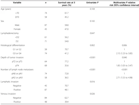
The impacts of clinicopathological variables on overall survival in all 132 patients are shown in Table 1. Histo-logical differentiation, depth of tumor invasion, number
Figure 1Overall survival curves of patient with recurrence in three recurrence patterns.
Table 1 Univariate and multivariate analyses of overall survival
Variable n Survival rate at 5
years (%)
UnivariateP MultivariatePrelative risk (95% confidence interval)
Age (years) 0.104
<70 74 61.7
≥70 58 45.2
Sex 0.165
Male 87 58.8
Female 45 47.0
Lymphadenectomy 0.647
<D2 61 56.2
D2 71 54.8
Histological differentiation 0.002 0.006
G1 or G2 58 70.7 1
G3 or G4 74 41.2 2.15 (1.23 to 3.85)
Depth of tumor invasion <0.001 0.040
pT2 or pT3 64 77.2 1
pT4 68 35.6 1.85 (1.03 to 3.47)
Number of lymph node metastases <0.001 <0.001
pN0 or pN1 74 72.8 1
pN2 or pN3 58 36.5 2.71 (1.53 to 4.98)
Lymphatic invasion 0.016
Negative 45 70.7
Positive 87 48.1
Venous invasion 0.026
Negative 84 62.7
of lymph node metastases, lymphatic vessel invasion, and venous vessel invasion were identified as prognostic factors for overall survival on univariate analysis (P = 0.020, P < 0.001, P < 0.001, P = 0.016, and P = 0.026, respectively). On multivariate analysis, histological differentiation, depth of primary tumor invasion, and number of lymph node metastases were identified as independent factors affecting overall survival (P= 0.006,
P= 0.040, andP< 0.001, respectively).
Risk factors for recurrence patterns
Peritoneal metastasis
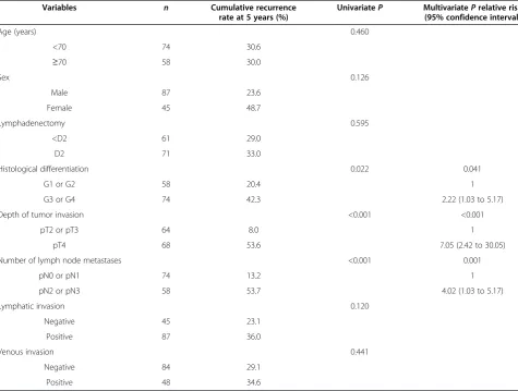
The median RFS for the 30 patients with peritoneal me-tastasis was 14.5 months (range, 3.4 to 64.2 months). The impacts of clinicopathological variables on the RFS of peritoneal recurrence are shown in Table 2. On uni-variate analysis, histological differentiation, depth of tumor invasion, and number of lymph node metastases were identified as risk factors for peritoneal metastasis (P = 0.022, P < 0.001, and P < 0.001, respectively). On multivariate analysis, histological differentiation, depth of tumor invasion, and number of lymph node
metas-tases represented independent risk factors associated with peritoneal metastasis (P = 0.041, P < 0.001, and
P= 0.001, respectively).
Hematogenous metastasis
Recurrence sites in the 19 patients with hematogenous recurrence were the liver in ten patients (52.6%), bone in four (21.1%), pleura in three (15.8%), lungs in three (15.8%), brain in two (10.5%), and intramural residual stomach, non-resected stump or site of anastomosis, in two (10.5%). Some patients had recurrence in more than one site.
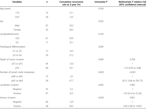
The median RFS of the 19 patients with peritoneal metastasis was 14.2 months (range, 2.1 to 59.8 months). The impacts of clinicopathological variables on RFS for hematogenous metastasis are shown in Table 3. On uni-variate analysis, depth of tumor invasion, number of lymph node metastases, lymphatic vessel invasion, and venous invasion were identified as risk factors for
hematogenous metastasis (P = 0.009, P < 0.001,
P = 0.004, and P < 0.001, respectively). On multivariate analysis, the number of lymph node metastases and
Table 2 Univariate and multivariate analyses of recurrence-free survival for peritoneal metastasis
Variables n Cumulative recurrence rate at 5 years (%)
UnivariateP MultivariatePrelative risk (95% confidence interval)
Age (years) 0.460
<70 74 30.6
≥70 58 30.0
Sex 0.126
Male 87 23.6
Female 45 48.7
Lymphadenectomy 0.595
<D2 61 29.0
D2 71 33.0
Histological differentiation 0.022 0.041
G1 or G2 58 20.4 1
G3 or G4 74 42.3 2.22 (1.03 to 5.17)
Depth of tumor invasion <0.001 <0.001
pT2 or pT3 64 8.0 1
pT4 68 53.6 7.05 (2.42 to 30.05)
Number of lymph node metastases <0.001 0.001
pN0 or pN1 74 13.2 1
pN2 or pN3 58 53.7 4.02 (1.03 to 5.17)
Lymphatic invasion 0.120
Negative 45 23.1
Positive 87 36.0
Venous invasion 0.441
Negative 84 29.1
venous vessel invasion represented independent risk
factors for hematogenous metastasis (P < 0.001 and
P= 0.001, respectively).
Lymphatic metastasis
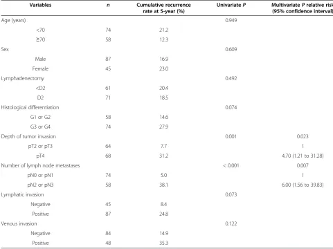
The median RFS for the 14 patients with lymphatic metastasis (including 3 patients with lymphangiosis carcinomatosa) was 15.3 months (range, 4.2 to 59.8 months). The impacts of clinicopathological variables on RFS for lymphatic metastasis are shown in Table 4. On univariate analysis, depth of tumor invasion and number of lymph node metastases were identified as risk factors
for lymphatic metastasis (P = 0.001, and P < 0.001,
respectively). On multivariate analysis, depth of tumor invasion and number of lymph node metastases repre-sented independent risk factors for lymphatic metastasis (P= 0.023 andP= 0.007, respectively).
Discussion
This study examined differences in risk factors between various patterns of recurrence in patients who under-went surgical curative resections for advanced gastric
carcinoma. As a result, independent risk factors for each recurrence pattern were identified as follows. For peri-toneal metastasis, depth of tumor invasion, number of lymph node metastases, and histological differentiation were identified. For hematogenous metastasis, number of lymph node metastases, and venous vessel invasion were identified. For lymphatic metastasis, depth of tumor invasion, and number of lymph node metastases were identified.
Seeding of cancer cells into the abdominal cavity rep-resents the first step in peritoneal metastasis. This means that pT4 can reasonably be considered an inde-pendent risk factor for peritoneal metastasis, as previ-ously reported [3-6]. Histological differentiation was also detected as a risk factor for peritoneal metastasis in some reports [4,6-9]. Although some reports have de-scribed lymph node metastasis as an independent risk factor for peritoneal metastasis, as in our result [4,5,10-12], the role of lymph node metastasis in peri-toneal metastasis has been unclear. However, because peritoneal recurrence occurred in patients with cancer limited to the gastric mucosa or submucosa but with
Table 3 Univariate and multivariate analyses of recurrence-free survival for hematogenous metastasis
Variables n Cumulative recurrence rate at 5-year (%)
UnivariateP MultivariatePrelative risk (95% confidence interval)
Age (years) 0.309
<70 74 27.1
≥70 58 13.5
Sex 0.505
Male 87 20.2
Female 45 28.3
Lymphadenectomy 0.234
<D2 61 20.9
D2 71 25.1
Histological differentiation 0.094
G1 or G2 72 14.5
G3 or G4 59 31.2
Depth of tumor invasion 0.009 0.708
pT2 or pT3 64 13.0 1
pT4 68 33.1 1.23 (0.43 to 4.08)
Number of lymph node metastases <0.001 <0.001
pN0 or pN1 74 2.0 1
pN2 or pN3 58 47.7 26.21 (3.66 to 581.73)
Lymphatic invasion 0.004 0.982
Negative 45 3.3 1
Positive 87 32.5 1.03 (0.14 to 22.26)
Venous invasion <0.001 0.001
Negative 84 12.9 1
lymph node metastasis, invasion of the lymphatic system by cancer cells has been suggested as the mechanism underlying peritoneal recurrence [13,14]. Moreover, in-jury to the lymphatic system during operative proce-dures in patients with highly extensive metastatic lymph nodes may allow the spread of viable cancer cells into the peritoneal cavity [12].
The first step in hematogenous metastasis is invasion of cancer cells into the lumen of the venous circulation. Our finding of vessel invasion as an independent factor for hematogenous metastasis is reasonable. The same re-sult has been reported from other institutions [15,16]. However, vessel invasion is not incorporated as a factor in the Union for International Cancer Control (UICC) staging criteria or the Japanese classification of gastric carcinoma. Attention should thus be given to hemato-genous recurrence in patients showing vessel invasion, even if the tumor stage is otherwise comparatively low. Conversely, the number of lymph node metastases might be an independent risk factor for hematogenous metas-tasis because of the connection of lymphatic channels to the systemic circulation via the thoracic duct. Noguchi
et al. [16] reported venous invasion and lymph node me-tastasis as risk factors for liver meme-tastasis. Kodera et al. [17] reported lymph node metastasis as a risk factor for bone metastasis.
With regard to lymphatic metastasis, the number of lymph node metastases and depth of tumor invasion represented independent risk factors. In this study, how-ever, lymphatic vessel invasion was not identified as a risk factor for lymphatic metastasis, perhaps for the fol-lowing reasons. First, cancer cells flow through lymph-atic vessels to distant vessels. Cancer cells invading lymphatic vessels are therefore sometimes not detected in resected specimens. Second, lymphatic vessels are sometimes difficult to distinguish from the venous vas-culature. In addition, some lymphatic vessels are thought to be destroyed by invasion of cancer cells, so patholo-gists cannot always detect lymphatic vessel invasions correctly. Third, if the number of cancer cells invading lymphatic vessels is small, the invasion might not be reflected in the patient prognosis. In addition, quantify-ing the grade of lymphatic vessel invasion objectively is difficult. However, the number of lymph node
Table 4 Univariate and multivariate analyses of recurrence-free survival for lymphatic metastasis
Variables n Cumulative recurrence rate at 5-year (%)
UnivariateP MultivariatePrelative risk (95% confidence interval)
Age (years) 0.949
<70 74 21.2
≥70 58 12.3
Sex 0.609
Male 87 16.9
Female 45 23.0
Lymphadenectomy 0.492
<D2 61 20.4
D2 71 18.5
Histological differentiation 0.074
G1 or G2 58 14.6
G3 or G4 74 27.9
Depth of tumor invasion 0.001 0.023
pT2 or pT3 64 7.7 1
pT4 68 31.2 4.70 (1.21 to 31.28)
Number of lymph node metastases < 0.001 0.007
pN0 or pN1 74 5.0 1
pN2 or pN3 58 38.1 6.00 (1.56 to 39.83)
Lymphatic invasion 0.073
Negative 45 8.4
Positive 87 24.8
Venous invasion 0.122
Negative 84 14.9
metastases reflects outflow of cancer cells into lymphatic vessels. In our study, the number of lymph node metas-tases was stratified into N0/N1 or N2/N3. This stratifica-tion of lymph node metastasis may be considered to reflect the amount of cancer cells invading lymphatic networks better than the presence or absence of lymph-atic vessel invasion.
As with our result, some reports have described the number of lymph node metastases as a risk factor for lymphatic metastasis [12,14]. The identification of pT4 as an independent factor for lymphatic metastasis might reflect cancer cell invasion into the entire subserosal layer through the abundant lymphatic vessels.
The status of lymph node metastasis has been identi-fied as the most important prognostic factor in patients undergoing gastrectomy [3,4,7,18-21]. This is reflected in the fact that the number of lymph node metastases represented an independent prognostic factor for all three patterns of recurrence in the present study. As mentioned previously, the number of lymph node me-tastases might reflect the amount of cancer cells in lymphatic channels in the peritoneum and both the greater and lesser omentum. Preoperative neoadjuvant chemotherapy for patients with a strong indication of lymph node metastases might therefore be acceptable to reduce seeding of cancer cells into the abdominal cavity as the result of surgical procedures. However, no ran-domized studies have yet addressed the survival benefits of this approach [1]. Randomized controlled trials of neoadjuvant therapy for patients with lymph node metas-tasis are thus needed to clarify means of achieving better prognosis in patients undergoing curative resection.
Conclusions
Risk factors for recurrence after curative gastrectomy for advanced gastric carcinoma differ between patterns of recurrence. By paying more attention to the specific risk factors of recurrence present in patients, the likelihood of missing sites of recurrence could be decreased and recur-rences identified earlier. This would allow appropriate treatment to be initiated more quickly for patients with re-currence. In addition, the status of lymph node metastasis contributed to all patterns of recurrence, even peritoneal metastasis. For patients in whom lymph node metastasis is suspected preoperatively, neoadjuvant therapy might be utilized to achieve better treatment outcomes.
Abbreviations
HR:Hazard ratio; RFS: Recurrence-free survival; UICC: Union for International Cancer Control.
Competing interests
The authors declare that they have no competing interests.
Authors’contributions
MO, HD, and MI carried out surgical resections. MO, TS and SH revised the manuscript for important intellecutual content. YN carried out surgical resections and collected clinical data and designed this study and drafted the manuscript. All authors read and approved the final manuscript.
Received: 20 October 2012 Accepted: 9 May 2013 Published: 17 May 2013
References
1. Japanese Gastric Cancer Association:Japanese gastric cancer treatment guidelines 2010 (ver. 3).Gastric Cancer2011,14:113–123.
2. Sobin LH, Gospodarowicz MK, Wittekind C:(Eds): TNM Classification of Malignant Tumours.7th edition. New York: Wiley-Blackwell; 2009. 3. Roukos DH, Lorenz M, Karakostas K, Paraschou P, Batsis C, Kappas AM:
Pathological serosa and node-based classification accurately predicts gastric cancer recurrence risk and outcome, and determines potential and limitation of a Japanese-style extensive surgery for Western patients: a prospective with quality control 10-year follow-up study. Br J Cancer2001,84:1602–1609.
4. Roviello F, Marrelli D, de Manzoni G, Morgagni P, Di Leo A, Saragoni L, De Stefano A:Prospective study of peritoneal recurrence after curative surgery for gastric cancer.Br J Surg2003,90:1113–1119.
5. Schwarz RE, Zagala-Nevarez K:Recurrence patterns after radical gastrectomy for gastric cancer: prognostic factors and implications for postoperative adjuvant therapy.Ann Surg Oncol 2002,9:394–400.
6. D’Angelica M, Gonen M, Brennan MF, Turnbull AD, Bains M, Karpeh MS: Patterns of initial recurrence in completely resected gastric adenocarcinoma.Ann Surg2004,240:808–816.
7. Yoo CH, Noh SH, Shin DW, Choi SH, Min JS:Recurrence following curative resection for gastric carcinoma.Br J Surg2000,87:236–242.
8. Marrelli D, Roviello F, de Manzoni G, Morgagni P, Di Leo A, Saragoni L, De Stefano A, Folli S, Cordiano C, Pinto E:Different patterns of recurrence in gastric cancer depending on Lauren’s histological type: longitudinal study.World J Surg2002,26:1160–1165.
9. Zheng HC, Zheng YS, Xia P, Xu XY, Xing YN, Takahashi H, Guan YF, Takano Y:The pathobiological behaviors and prognosis associated with Japanese gastric adenocarcinomas of pure WHO histological subtypes. Histol Histopathol2010,25:445–452.
10. Aoyama T, Yoshikawa T, Hayashi T, Kuwabara H, Mikayama Y, Ogata T, Cho H, Tsuburaya A:Risk factors for peritoneal recurrence in stage II/III gastric cancer patients who received S-1 adjuvant chemotherapy after D2 gastrectomy.Ann Surg Oncol2012,19:1568–1574.
11. Otsuji E, Yamaguchi T, Sawai K, Sakakura C, Okamoto K, Takahashi T: Regional lymph node metastasis as a predictor of peritoneal carcinomatosis in patients with Borrmann type IV gastric carcinoma. Am J Gastroenterol1999,94:434–437.
12. Kunisaki C, Shimada H, Nomura M, Matsuda G, Otsuka Y, Ono H, Akiyama H: Surgical outcome of serosa-negative advanced gastric carcinoma. Anticancer Res2004,24:3169–3175.
13. Lee HJ, Kim YH, Kim WH, Lee KU, Choe KJ, Kim JP, Yang HK:
Clinicopathological analysis for recurrence of early gastric cancer.Jpn J Clin Oncol2003,33:209–214.
14. Saka M, Katai H, Fukagawa T, Nijjar R, Sano T:Recurrence in early gastric cancer with lymph node metastasis.Gastric Cancer2008, 11:214–218.
15. Ikeguchi M, Katano K, Oka A, Tsujitani S, Maeta M, Kaibara N:Relationship between hematogenic metastasis of gastric cancer and the maximum extent of venous invasion by cancer cells in the gastric wall. Hepatogastroenterology1995,42:660–665.
16. Noguchi Y:Blood vessel invasion in gastric carcinoma.Surgery1990, 107:140–148.
17. Kodera Y, Ito S, Mochizuki Y, Yamamura Y, Misawa K, Ohashi N, Nakayama G, Koike M, Fujiwara M, Nakao A:The number of metastatic lymph nodes is a significant risk factor for bone metastasis and poor outcome after surgery for linitis plastica-type gastric carcinoma.World J Surg2008, 32:2015–2020.
19. Saito H, Tsujitani S, Oka S, Kondo A, Ikeguchi M, Maeta M, Kaibara N: Prediction of survival period for patients with postoperative recurrence after curative resection for advanced gastric carcinoma.
Hepatogastroenterology2001,48:290–293.
20. Nozoe T, Iguchi T, Egashira A, Adachi E, Matsukuma A, Ezaki T:Pathological prognostic score as a simple criterion to predict outcome in gastric carcinoma.J Surg Oncol2010,102:73–76.
21. Deng J, Liang H, Sun D, Pan Y:The prognostic analysis of lymph node-positive gastric cancer patients following curative resection. J Surg Res2010,161:47–53.
doi:10.1186/1477-7819-11-98
Cite this article as:Nakanishiet al.:Differences in risk factors between patterns of recurrence in patients after curative resection for advanced gastric carcinoma.World Journal of Surgical Oncology201311:98.
Submit your next manuscript to BioMed Central and take full advantage of:
• Convenient online submission
• Thorough peer review
• No space constraints or color figure charges
• Immediate publication on acceptance
• Inclusion in PubMed, CAS, Scopus and Google Scholar
• Research which is freely available for redistribution