Analysis of DNA - ligand interaction in
a parallel force-based assay
Katja Maria Limmer
Dissertation
an der Fakult¨
at f¨
ur Physik
der Ludwig–Maximilians–Universit¨
at
M¨
unchen
vorgelegt von
Katja Maria Limmer
aus M¨
unchen
Zusammenfassung v
Zusammenfassung
Biomolekulare Wechselwirkungen, zum Beispiel zwischen DNA und Proteinen, sind sehr vielf¨altig. Ihre Charakterisierung erfordert hochgradig parallele und universal einsetzbare Untersuchungs-methoden. Kraftmessungen an einzelnen Molek¨ulen k¨onnen die Wechselwirkungen unterschiedlicher molekularer Komplexe auch in dichten Medien quantifizieren ohne die molekulare Bindung durch eine Farbstoffmarkierung zu beinflussen. In den meisten F¨allen haben diese Methoden allerdings einen sehr geringen Durchsatz. Die Technik des Molecular Force Assay (MFA) umgeht diesen Nachteil, indem zwei molekulare Bindungen miteinander verglichen werden. So k¨onnen tausende molekulare Wechselwirkungen parallel untersucht werden, ohne dass die Messung den Einzel-molek¨ul-Charakter verliert. Ziel dieser Arbeit war eine Weiterentwicklung des MFA, um dessen Anwendbarkeit auf aktuelle biologische Fragestellungen zu erweitern.
Als erstes wird ein Aptamer-Sensor f¨ur die Charakterisierung kleiner Molek¨ule beschrieben. Aptamere sind Oligonukleotide, die spezifisch einen bestimmten Liganden binden. Eine Ap-tamersequenz als Teil einer der beiden molekularen Komplexe f¨uhrt zu dessen Stabilisierung durch die Bindung mit dem Liganden, so dass das bindende Molek¨ul auch in komplexen Fl¨ussigkeiten analysiert werden kann. Dieser kraftbasierte Nachweis der Aptamer-Ligand Wechselwirkung erm¨oglicht auch eine Charakterisierung von kleinen oder nur schwach bindenden Molek¨ulen, da auf zus¨atzliche Techniken zur Signalverst¨arkung oder Waschprozeduren verzichtet werden kann. Die Dissoziationskonstante kann dank des Parallelformats in einer einzigen Messung bestimmt werden. Ein kraftbasierter Aptamer-Sensor ist somit ein vielversprechendes Instrument f¨ur eine schnelle Charakterisierung kleiner Molek¨ule in dichten Medien.
Des Weiteren wird gezeigt, dass sich der MFA zur Suche RNA-bindender Molek¨ule eignet, die gleichzeitig das Protein Dicer inhibieren. Dicer spielt bei der RNA Interferenz, ein Mechanismus der Genregulation mit Hilfe kurzer St¨ucke nichtkodierender RNA, eine entscheidende Rolle, in-dem er lange, doppelstr¨angige RNA in kurze Segmente von 19-22 Basenpaaren schneidet. Diesen Vorgang gezielt zu verhindern ist ein vielversprechender Ansatz bei der Entwicklung neuer medi-zinischer Therapien. Der MFA kann sowohl verl¨asslich die Dicer-Aktivit¨at messen als auch die Dissoziationskonstante des RNA-Liganden bestimmen. Werden die molekularen Komplexe des MFA mit dem RNA-bindenden Molek¨ul und Dicer inkubiert, kann eine gezielte Hemmung von Dicer beobachtet werden, die mit der Konzentration des Liganden korreliert. Dies zeigt, dass der MFA eine effektive Methode zur Suche von RNA-bindenden Molek¨ulen ist, die gleichzeitig das Protein Dicer an der Produktion kleiner RNA Segmente f¨ur die RNA Interferenz hindern.
Als drittes wird die Eignung des MFA zur Quantifizierung von Protein-DNA Wechselwirkung-en untersucht. Um dWechselwirkung-en komplexWechselwirkung-en Vorgang der Transkription zu verstehWechselwirkung-en, ist es unabdingbar, die Wechselwirkungen der beteiligten Proteine mit der DNA im Nukleus genau zu bestimmen. Durch die große Zahl von Wechselwirkungen sind Methoden mit hohem Durchsatz unerl¨asslich. In einer Weiterentwicklung des MFA kann die Bindungsst¨arke eines Zinkfinger-Proteins mit drei verschiedenen DNA-Motiven durch den Vergleich mit zwei unterschiedlichen Referenzen in einer einzigen Messung quantifiziert werden. Dieses Experiment zeigt eindrucksvoll das Potential des MFA, zum Verst¨andnis des komplexen Mechanismus der Transkription beizutragen.
Summary vii
Summary
Biomolecular interactions like protein-DNA binding are very diverse. Their analysis requires highly parallel assays that are easily adaptable for the characterization of various molecular complexes. Single-molecule force measurements can quantify the interaction of different molecules even in complex ambients without disturbing the molecular bond by labeling, but are in most cases low throughput. The technique of the Molecular Force Assay (MFA) overcomes this drawback by comparing two molecular bonds with each other. It allows the analysis of thousands of molecular interactions in parallel without losing the single-molecule character. The objective of this thesis was to adapt the technique of the MFA for new applications that have the potential to contribute significantly to the solution of current biological problems.
First, a force-based aptamer sensor for the characterization of small molecules is described. Aptamers are oligonucleotides that are selected for their ability to bind a certain ligand with high specificity. Implementing an aptamer into one of the bonds of the MFA enables the detection of its ligand even in complex fluids by a strengthening of this bond upon ligand binding. The force-based detection of the interaction between aptamer and ligand enables the characterization of low affinity binders or small molecules without relying on signal amplification or stringent washing requirements. The parallel format allows the determination of the dissociation constant in a single measurement. Thus, a force-based aptamer sensor is a promising analytical tool for a fast characterization of analytes in complex fluids.
Second, the application of the MFA as a screening tool for RNA binders that selectively inhibit the protein Dicer is demonstrated. Dicer is a fundamental part of the RNA interference mechanism that regulates gene expression with small non-coding RNA sequences. Dicer matures the small RNAs by cutting the precursor molecules into pieces of 19-22 base pairs. The selective inhibition of Dicer by a ligand that binds specifically to a certain precursor molecule has a great potential for future medical therapeutics. The MFA can reliably detect Dicer activity and characterize possible RNA binders by determining the dissociation constant. If both the ligand and Dicer are added to a MFA sample, Dicer is clearly inhibited upon ligand binding in relation to the ligand concentration. Hence, the technique of the MFA is a valuable tool to screen for RNA binders that selectively hinder Dicer from maturing the precursor molecule upon ligand binding.
Third, the MFA is applied to quantify DNA-protein interactions by measuring their binding strength. In order to understand transcription, identifying how strong a protein binds to the genomic DNA is essential. Due to the huge number of DNA-protein interactions that participate in transcriptional regulation, high throughput methods for the analysis of these interactions are highly desirable. As a proof-of-principle, the MFA quantifies the interactions of a zinc finger protein with three DNA motifs by comparing them to various reference bonds within a single measurement. Further miniaturization can easily extend the setup’s capacity in terms of mul-tiplexing. This illustrates the potential of the MFA to quantify protein-DNA interactions in a highly parallel format in order to contribute to the understanding of transcriptional regulation.
Contents
Zusammenfassung v
Summary vii
1 Introduction 1
2 Biological Background 7
2.1 Very short introduction into gene expression . . . 7
2.2 Oligonucleotides . . . 8
2.2.1 DNA . . . 8
2.2.2 DNA as a force sensor . . . 10
2.2.3 RNA . . . 12
2.2.4 Aptamers . . . 12
2.3 Transcriptional Regulation by Proteins in Eukaryotes . . . 15
2.4 The role of the protein Dicer in the regulation of gene expression . . . 16
3 The Molecular Force Assay 21 3.1 Basic principle . . . 21
3.2 Contact and separation process . . . 22
3.3 Analysis . . . 22
4 Results 27 4.1 DNA as a force sensor in an aptamer-based biochip for adenosine . . . 27
4.2 Sequence specific inhibition of Dicer measured with a force-based microarray for RNA ligands . . . 30 4.3 A force-based, parallel assay for the quantification of protein-DNA interactions 35
A Publications 43
A.1 Publication 1: DNA as a Force Sensor in an Aptamer-Based Biochip for Adenosine . . . 43 A.2 Publication 2: Sequence-specific inhibition of Dicer measured with a
force-based microarray for RNA ligands . . . 53 A.3 Publication 3: Quantitative Detection of Small Molecule/DNA Complexes
Employing a Force-Based and Label-Free DNA-Microarray . . . 67 A.4 Publication 4: Force-Driven Separation of Short Double-Stranded DNA . . 87
B Invited Publications 109
B.1 Publication 5: Molekularer Kraftsensor: Quantifizierung biomolekularer Wechselwirkungen im Parallelformat . . . 109
C Manuscripts 111
C.1 Manuscript M1: A force-based, parallel assay for the quantification of protein-DNA interactions . . . 111
Bibliography 135
List of Figures
2.1 Double-Stranded DNA . . . 9
2.2 Two geometries to melt a DNA helix . . . 11
2.3 Structural differences between DNA and RNA molecules . . . 12
2.4 The SELEX Process . . . 13
2.5 Schematics of the protein Dicer . . . 17
2.6 Small RNA biogenesis . . . 18
3.1 Molecular setup of the MFA for DNA duplexes . . . 23
3.2 Macroscopic setup of the MFA . . . 24
4.1 Detection of the dissociation constant of ATP to its aptamer . . . 29
4.2 Determination of Dicer activity and of the dissociation constant of paro-momycin to its aptamer . . . 31
4.3 The molecular setup for the detection of the selective inhibition of Dicer activity . . . 33
4.4 Dicer hindrance upon paromomycin binding . . . 34
Chapter 1
Introduction
Biological processes are very complex due to the multitude of involved molecules and molecular interactions. Examples are cell division, photosynthesis or the interplay of cells in multicellular organisms. Signals from the environment are absorbed and converted into actions like movement in a certain direction or the synthesis of certain proteins to metabolise a new nutrient. Understanding these processes and, one step further, influencing them is the aim of biological sciences. A combination of techniques and perspectives, biological, chemical, but also physical or computational, offer the greatest chance to achieve this aim. This has already been proven in the past with the human genome project as the most prominent example. Between 1990 and 2003, several institutes and reasearch groups from multiple countries and with different scientific background combined their efforts and techniques to decipher the human DNA code. In April 2003, a consortium presented 99 % of the sequence of the 20600 human genes. Additionally, the human genome project deciphered the genomes of several model organisms and, importantly, led to the development of new and the improvement of existing sequencing techniques (for more information see [1]). Some of these methods and developments are based on physical principles, for instance gel electrophoresis or fluorescence-based sequencing techniques. The application of physical knowledge in biology has a far-reaching tradition of several decades. A prominent example is the discovery of the helical structure of double-stranded DNA molecules by x-ray diffraction [2][3][4]. Today, structural analysis of biological molecules like proteins by x-ray diffraction or nuclear magnetic resonance is a standard method and has helped to elucidate the binding properties of many molecular complexes.
3
molecular complexes cannot be accessed by other methods. Hence, force-based techniques that are compatible with high-throughput systems are highly desirable.
A possible realization for a parallel, force-based technique is the Molecular Force Assay (MFA). In the MFA, the macroscopic force transducer, the cantilever or bead, is replaced by a molecular bond, in most cases a well-characterized DNA duplex which acts as refer-ence bond. This referrefer-ence bond is connected in series with the sample bond, the molecular complex in question. If a force is applied to this whole construct of sample and reference bond, the weaker one will rupture with a higher probability and a fluorophore attached to the linker polymer between the two bonds will indicate the intact molecular complex. Thus, the MFA does not record forces or extensions, but compares the two molecular bonds with each other. As the binding strength of the two complexes are of the same order of magnitude, very small differences in molecular stability can be resolved. A skillful choice of sample and reference complex enables a good characterization of the sample bond. The molecular construct of sample and reference bond is connected to two surfaces that are separated in order to apply a force to the bonds. Since every sample bond is only compared to its own reference without being disturbed by other molecules attached nearby to the surfaces, thousands of molecular constructs can be tested within several square microme-ters. Hence, the statistics of a single measurement already allow to draw valid conclusions. Depending on the technical implementation, several sample bonds can be tested against multiple references so that a single experiment enables the characterization of the molecule under investigation. So far, the MFA was applied to detect single nucleotide polymorphism [20], study differences in antibody/antigen interactions [21], investigate the chiral binding selectivity of small peptides [22][23] and analyse the interaction of the proteins EcoR1 and p53 with different DNA sequences [24][25].
the ligand concentration allowed the determination of the dissociation constant in a single MFA measurement. Techniques that can capture analytes in complex fluids and quantify their concentration are highly desirable for diagnostic purposes. P1 describes a proof-of-principle for the detection and characterization of a small molecule by an aptamer in a force-based assay. The parallel design allowes to extend the assay easily for the analysis of multiple small molecules by the integration of additional aptamers.
The RNA interference mechanism is the motivation for publication P2. Small RNA duplexes are matured by the protein Dicer that cuts double-stranded RNA into pieces of 19-22 base pairs independently of their sequence. One of the matured RNA strands is integrated in a protein complex known as RNA-induced-silencing-complex that binds to messengerRNA at least partly complementary to the small RNA strand and, in most cases, hinders protein translation [26]. Several classes of small RNAs exist that differ in their origin and function, for instance short-interfering RNA (siRNA) or microRNA (miRNA). Number and kind of the naturally occuring miRNAs in a cell depend on the stage of the cell cycle and health status. The quantity of some miRNAs is greatly increased in connection with severe illnesses like cancer [27]. Hence, the selective inhibition of single microRNA precursors might be a first step in the development of a new generation of medical therapeutics. Here, the application of the MFA comprised a screening assay for RNA ligands that selectively inhibit Dicer from cleaving and, thus, maturing the small RNA precursor. The activity of the protein Dicer can easily be detected in a force-based assay as it greatly destabilizes the sample bond by cutting off about 20 basepairs in comparison to a DNA reference that Dicer cannot process. As a proof-of-principle, a RNA aptamer for the aminoglycoside paromomycin was integrated into the RNA duplex and a selective inhibition of Dicer processing upon ligand binding was detected by means of the MFA. Additionally, the dissociation constant of paromomycin to its aptamer was measured. Hence, the MFA can be applied to screen for and analyze miRNA ligands that selectively hinder Dicer from cleaving. The parallel format of the MFA offers the possibility to screen for several ligands or multiple miRNAs within a single experiment.
ma-5
jority of transcription factors with genomic DNA is of utmost importance. But not only the gigantic number of interactions is a challenge. Low affinity binders might easily be missed, especially by methods that rely on stringent washing procedures. Furthermore, affinity or thermodynamic constants might not be the best quantities to characterize protein-DNA interaction in a nuclear environment, where a transcription factor will probably always be bound to some DNA sequence due to the high concentration of DNA inside the nucleus. Specificity that comprises the discrimination between high and low or no binding sequences is better suited to characterize the binding properties of a protein. Binding strength is a quantity analogous to the specificity and is accessible in force measurements. Manuscript M1 describes an adaptation of the MFA which enables the quantification of the binding properties of a six zinc finger construct against three sample sequences, a high affinity, a low affinity and a no binding DNA sequence in comparison to two reference DNA duplexes in a single measurement. Weak interactions can also be detected without problems since the force measurement is performed under equilibrium conditions. Furthermore, the par-allel format of the MFA allows its extension to the testing of additional DNA sequences or proteins within a single experiment. Thus, with further miniaturization and paralleliza-tion, the MFA has a great potential to contribute to the understanding of transcriptional regulation.
Chapter 2
Biological Background
Gene expression can be summarized in the phrase ”from DNA to RNA to protein”. After a very short introduction into the mechanism of gene expression, this chapter describes the molecules of interest for this thesis, especially the oligonulceotides DNA, RNA and aptamers, and the cellular processes that gave the motivation for the experiments of this thesis.
2.1
Very short introduction into gene expression
in chromatin structures caused by histone modifications change the accessibility of certain DNA sequences. After transcription, modulation of capping, splicing and polyadenylation of the messengerRNA as well as the nuclear export rate or sequestration of the RNA tran-script alter the rate of gene expression. Thus, gene expression is an extremely complex mechanism that requires a multitude of molecular interactions.
2.2
Oligonucleotides
The oligonucleotides DNA (deoxyribonucleic acid) and RNA (ribonucleic acid) are also known as the molecules of life and are crucial for the transmission, expression and con-servation of genetic information. DNA as the carrier of the genetic information has to be very robust against external influences. RNA is the bridge between the genome and the functional protein. The information stored in the DNA is transcribed into messengerRNA (mRNA), which in turn is translated into a protein. Additionally, non-coding RNA plays a vital role in the processes that regulate transcription and translation.
2.2.1
DNA
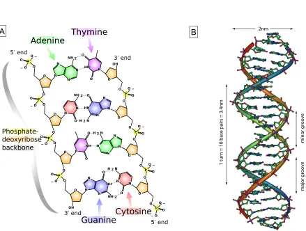
A DNA molecule is a polymer chain consisting of nucleotides. A nucleotide is composed of the pentose sugar 2-deoxy-D-ribose, a phosphate residue and one of four different nitrogen heterocyclic bases. Cytosine and thymine are monocyclic pyrimidine derivatives, while adenine and guanine are bicyclic purines (see Figure 2.1 A). Nucleotides are covalently linked together by phosphodiester bonds between the phosphate group attached to the 5’ hydroxyl group of the pentose sugar molecule and the 3’ hydroxyl group of the next pentose. Hence, a DNA polymer has a 5’ end and a 3’ end (see [30] for additional information). Typically, two antiparallel DNA strands compose a double helix as was shown in 1953 by J. Watson and F. Crick [4]. The negatively charged sugarphosphate groups constitute the backbone of the helix, while the opposing nucleobases form pairs in the core of the helix (see Figure 2.1 B). In the most stable configuration adenine binds thymine via two hydrogen bonds and guanine pairs with cytosine via three hydrogen bonds. This is known as Watson-Crick base pairing.
irreg-2.2 Oligonucleotides 9
2nm
1 tur
n = 10 base pairs = 3.4nm
minor gr
o
ov
e
major gr
o
ov
e
A B
ularities in the double helical structure are assumed to play an important role in protein recognition and binding or in other interactions involved in the process of transcription and replication [30]. Non-Watson-Crick base pairing and base mismatches also alter the double-strand structure and are especially relevant for synthesized oligonucleotides and aptamers described in detail in Chapter 2.2.4.
The specific sequence of the four nucleobases codes the genetic information. This system is very simple but also very versatile since a DNA molecule ofnnucleotides can consist of 4n
different sequences. Additionally, the double-stranded nature and the backbone structure account for the robustness of a DNA molecule. These properties, specificity, simplicity and robustness, render the DNA so special and are the reason for its usage in nanotechnological applications. Its ability to build programmable sequence-specific duplexes by self-assembly enables the construction of two and three-dimensional structures, DNA origami, [32][33] and molecular nanodevices [34][35]. Furthermore, DNA duplexes can be applied as force sensors in force-based techniques like the Molecular Force Assay (see Chapter 3).
2.2.2
DNA as a force sensor
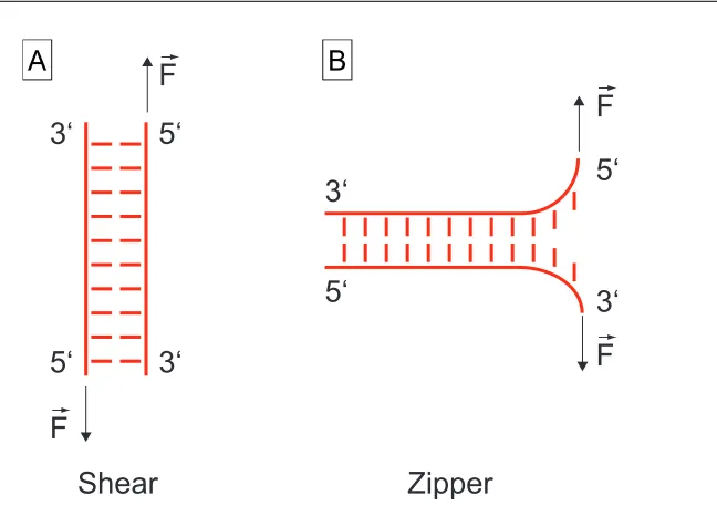
Several factors influence the thermal stability of a DNA duplex. Watson-Crick base pairing and stacking interactions [36] between the nucleobases stabilize the DNA structure. Hence, sequences with a majority of CG pairs melt at higher temperatures compared to strands with a majority of AT pairs. Mismatches and bulges disrupt the double-stranded structure and weaken the complex. The negatively charged phosphate backbones reject each other depending on the salt concentration of the buffer system and can have a destabilizing effect. Furthermore, two entropic effects weaken the DNA duplex. First, double-stranded DNA has a persistence length of 50nm at a [N a+] concentration of around 0.1M [37] and is, thus, rather rigid. Single-stranded DNA is considerably more flexible and environmental conditions influence its mechanical properties significantly stronger. As a consequence, the persistence length of single-stranded DNA varies from of 0.75nm at high salt concentra-tions to 10nm at low ionic strength [35]. Second, the entropy of the solvent also has a destabilizing effect since the hydration shell of a DNA duplex is greater than of two single strands. Both effects counteract the formation of a DNA duplex and reduce its stability. Summarizing, all these effects influence the thermal stability of a DNA duplex and have to be taken into account when analyzing the behavior of DNA under force.
2.2 Oligonucleotides 11
5‘
3‘
5‘
3‘
Shear
3‘
3‘
5‘
5‘
Zipper
F
F
F
F
B
A
Figure 2.2: Two geometries to melt a DNA helix. (A) In shear geometry the force F~ is applied in the direction of the longitudinal axis of the duplex and the entire helix is loaded. (B) In zipper geometry, the base pairs are melted separately from one end to the other.
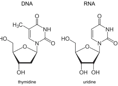
thymidine uridine
DNA RNA
Figure 2.3: Structural differences between DNA and RNA molecules. DNA is composed of 2-deoxy D-ribose, while RNA is built of D-ribose molecules. The base thymine in DNA is replaced by uracil in RNA (adapted from [42]).
2.2.3
RNA
On the molecular level, ribonucleic acid (RNA) differs from DNA in two aspects. Instead of 2-deoxy-D-ribose RNA is composed of the pentose sugar D-ribose and the base thymine in DNA is replaced by uracil in RNA, which, like thymine, forms Watson-Crick base pairs with adenine. Figure 2.3 displays the nucleotides of RNA and DNA in a direct comparison. These structural differences cause RNA to be more susceptible to hydrolysis but are also responsible for a greater diversity in RNA secondary structure, since the extra hydroxyl group can be an additional binding site for forming branched polynucleotides. While for DNA Watson-Crick base pairing and the double-stranded helix inside the cell dominates, base pairing in RNA structures is very diverse. A good overview of these structures is given by Leontis et al. [41]. RNA molecules in the cell are mostly single-stranded, but can be found to form double helices, for which the A-form predominates [30]. Additionally, other structures like bulges, hairpins, loops or pseudoknots appear and are determined by the function of the particular RNA molecule. These conformations can be as diverse as the different tasks RNA can perform.
2.2.4
Aptamers
2.2 Oligonucleotides 13
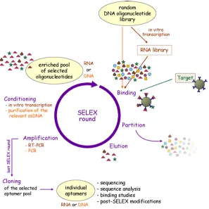
[45]. Tuerk and Gold developed a new method named SELEX - systematic evolution of ligands by exponential enrichment - to select RNA oligonucleotides from a large library with the property to bind a certain ligand tightly and with high specificity. Andrew D. Ellington and Jack W. Szostak used a similar technique to find RNA molecules that bind specifically to a variety of organic dyes and termed these oligonucleotides aptamers after the Latin expression ”aptus” meaning ”fitting” [44]. Shortly after, single-stranded DNA aptamers were discovered [46]. The SELEX technique is an iterative process to find ap-tamers for all kinds of ligands under different conditions (see Figure 2.4) . The selection process starts with a large, random DNA library (about 1015 different sequences) that can
2.3 Transcriptional Regulation by Proteins in Eukaryotes 15
2.3
Transcriptional Regulation by Proteins in
Eukary-otes
ribonucleotides to the RNA strand that are complementary to the DNA template strand. Elongation factors prevent the RNA polymerase from dissociation before the end of the gene and assist in unwinding of the DNA. Beside producing the RNA molecule, RNA poly-merase assembles proteins on its tail that can process the RNA into the functional mRNA. Examples are capping, which means the modification of the 5’ end of the RNA molecule for the discriminaton of mRNA from other RNAs, and splicing that removes the introns, non-coding sequences, from the pre-mRNA. Transcription termination is encoded in the DNA so that the transcribed RNA can recruit proteins that end transcription and modify the RNA 3’ end if necessary (for details see [48]). Summing up, every step in transcription is regulated by proteins that can activate, accelerate or terminate transcription and thus enable the cell to react very flexibly and rapidly to its needs. In this work, a force-assay is presented that characterizes protein-DNA specificity via the binding strength (see Chap-ter 4.3). This assay has the potential to contribute to the understanding of the complex network of protein- DNA interactions that govern transcriptional regulation.
2.4
The role of the protein Dicer in the regulation of
gene expression
The enzyme Dicer is a multidomain RNAse III-related endonuclease of about 250kDAand is responsible for cleaving double-stranded RNA into pieces of 19-22 base pairs. Dicer has been found in the cytoplasm of all eukaryotes studied to date [49], sometimes in several variants with different tasks. The L-shape of the protein seems to be well-conserved for all variants. Recognition of dsRNA by a PAZ-domain occurs in the head of Dicer, which is separated from the two RNAse III-domains by a ruler domain (see Figure 2.5). The base of the L is formed by a helicase, whose function is not totally understood [49]. Dicer cleaves long and short (more than 30 nucleotides) dsRNA strands with equal efficiency, whereas duplexes of 21 nucleotides or less are not processed in vitro. A 3’ 2-nucleotide long overhang increases Dicers efficiency compared to blunt ends [50].
2.4 The role of the protein Dicer in the regulation of gene expression 17
RNAse III Paz
Ruler
RNAse III Helicase
dsRNA
Figure 2.5: Schematics of the protein Dicer. The L-shape of the protein seems to be well-conserved for all variants. The PAZ-domain in the head part recognizes dsRNA. The two RNAse III domains are separated from the PAZ head by a ruler domain and cut the two RNA strands. A helicase, whose function is not totally understood, forms the base of the L.
2.4 The role of the protein Dicer in the regulation of gene expression 19
Chapter 3
The Molecular Force Assay
This chapter describes the technique of the Molecular Force Assay (MFA) that enables the parallelization of force measurements by comparing the mechanical stability of two molecular bonds.
3.1
Basic principle
Two molecular bonds, a sample and a reference bond, are connected in series via a thymine-linker to which a fluorophore is conjugated. The lower complex is covalently bound to a functionalized glass slide, while the upper complex is attached to a soft PDMS surface. The two surfaces are separated by a constant velocity so that the two molecular bonds are loaded with a force until the weaker one ruptures. An alternative view is that the force greatly enhances the off-rate of the bonds under investigation and reduces the otherwise extremely long spontaneous dissociation times towards seconds [57]. The fluorophore conjugated to the linker sequence indicates the intact bond. Hence, the ratio of the fluorescence intensity before and after the force probe allows to determine the mechanical stability of the sample bond in comparison to the reference bond. Every molecular complex under load corresponds to a single molecule force experiment since the stability of every sample bond is measured against its own reference bond. This enables about 104 parallel force measurements per µm2 so that a single MFA experiment already provides a valid result supported with very good statistics.
an aldehyde functionlized glass slide. Five HEGL (hexaethylene glycol) polymer units and a poly-T stretch act as spacer between the surface and the molecular complex. Sample and reference bond are also separated by a poly-thymine sequence to which a fluorophore is conjugated. The uppermost strand is functionalized with a biotin that binds to the streptavidin-coated PDMS stamp during the contact process. Figure 3.1 displays the details of the molecular setup for two DNA duplexes.
3.2
Contact and separation process
Macroscopically, the contact and separation process is performed as described in the follow-ing. The functionalized stamp adheres upside-down to a glass block glued to a closed-loop piezoelectric actuator and a DC motorized translation stage (see Figure 3.2) . The slide with the molecular constructs is fixed beneath the stamp on a stainless steel stage with permanent magnets so that every stamp pillar meets a spot of 1mm diameter of molec-ular complexes on the glass slide. The whole contact device is mounted on an inverted microscope with an xy-DC motorized high-accuracy translation stage. Contact is made by means of the piezo and care is taken that each individual pillar is not compressed more than 3µm. The planar adjustment of stamp and slide as well as the contact process are controlled by reflection interference contrast microscopy [58]. The contact between stamp and slide is maintained for 10 minutes to ensure that the molecular complexes are coupled to the surfaces. If DNA or RNA duplexes form sample and reference bond, the molecular complex is completely built up on the glass slide before the contact and separation pro-cess and the biotin modification of the uppermost strand binds to the streptavidin-coated PDMS surface during the contact time. The piezo retracts the stamp with a constant velocity of 1µm/s, and a force builds up in the molecular complexes until the weaker one breaks with higher probability. In order to quantify the number of intact lower bonds in relation to the total number of molecular constructs, images of the fluorescence intensity on the glass slide are taken before and after the contact process.
3.3
Analysis
3.3 Analysis 23
B B
B
?
B B
?
B
PDMS
PDMS
Glass Glass
Cy3
Cy5 Cy5
Cy3 reference
bond
sample bond
PDMS surface fluorescence
glass surface fluorescence
Glass
Piezo
Stamp
Objective 10x
stamp with 16 pads
100 x 100µm pad diameter = 1,1mm
Figure 3.2: Macroscopic setup of the MFA. The contact and separation unit consists of a closed-loop piezo to which the PDMS stamp is attached via a glass block. Current technical implementation allows the measurement of 16 molecular constructs in parallel corresponding to 16 pillars of the stamp and the 4x4 pattern on the glass slide. Every stamp pillar is microstructured with pads of 100µm×100µm separated by 41µm wide and 5µm deep rectangular trenches enabling the drainage of liquid during the contact and separation process. The contact unit is mounted on an inverted microscope.
hIStarti and after the separation process hIF inali. The quotient is given by
Fintact lower bonds =
hIF inali
hIStarti. (3.1)
3.3 Analysis 25
fluorescence intensities are calculated from a single image. Due to the microstructure of the PDMS stamp, every fluorescence image taken after the contact and separation process will always show both contacted and non-contacted areas. Thus, the ratio of molecular complexes that did not couple to the total number of molecular constructs is determined by dividing the mean fluorescence intensity values of contacted to non-contacted areas
Fnon−coupled =
hIcontactedi
hInon−contactedi. (3.2)
This number is subtracted from Fintact lower bonds and the result is normalized to the total
number of molecules that have been under load, yielding the Normalized Fluorescence NF
N F = Fintact lower bonds−Fnon−coupled 1−Fnon−coupled
. (3.3)
Thus, the Normalized Fluorescence is the mean number of intact lower bonds to the total number of molecular complexes that have been tested. The data of the publications P1 (see Chapter 4.1) and P3 were analyzed according to this method.
An alternative method for the determination of molecular constructs that have not coupled to the PDMS surface is the labeling of the uppermost strand with a fluorophore at the lower end that constitutes a FRET-pair (F¨orster-Resonance-Energy-Transfer [59]) with the fluorophore attached to the middle strand (see Figure 3.1) . For this thesis, the cyanine dyes Cy3 and Cy5 were chosen as FRET-pair. Cy5 is conjugated to the middle strand and indicates the number of intact lower bonds on the glass slide, while Cy3 is attached to the uppermost strand so that a FRET-signal is only measured for molecular constructs of intact sample and reference bond. This configuration enables the calculation of the Normalized Fluorescence by analyzing the images pixel-by-pixel. In addition to the images of the Cy5 fluorescence intensity that indicate the intact lower bonds, the FRET signal is also detected before and after the contact and separation process. After background subtraction, the fluorescence images of the Cy5 signal as well as of the FRET signal are divided and corrected for bleaching, yielding
FCy5 = ICyF inal5 IStart Cy5
. (3.4)
and
FF RET =
IF inal F RET
IStart F RET
An image of the Normalized Fluorescence is calculated according to equation 3.3 in the form of
N F = FCy5−FF RET 1−FF RET
. (3.6)
Chapter 4
Results
The objective of this thesis was to adapt the MFA for new forms of application that contribute to the solution of current biological problems. The main results are shortly presented in the following. Additional information can be found in publications P1 and P2 and manuscript M1. The utilization of aptamers in a force-based biochip for the capture of small molecules is described in Chapter 4.1 and enables the detection of a broad range of ligands that include toxins and low affinity binders. Due to the label-free detection, complex fluids do not cause problems. The protein Dicer is one of the major protagonists in the RNA interference pathway so that a selective inhibition of Dicer activity has a great potential for the development of future medical therapeutics. Chapter 4.2 describes the application of the MFA for the screening of small molecules that selectively bind a RNA sequence and hinder Dicer from processing. Furthermore, the MFA can directly quantify the binding strength of DNA-protein interactions. Chapter 4.3 describes the necessary changes in the molecular setup as well as in the analysis and demonstrates in a proof-of-principle experiment the potential of this application of the MFA.
4.1
DNA as a force sensor in an aptamer-based biochip
for adenosine
that are selected to specifically bind their ligand with high affinity and specificity (see Chapter 2.1.4). They are similar to antibodies but surpass them in terms of small molec-ular weight, ease of modification and ability to detect toxins [61]. As a proof-of principle, a split aptamer selective for adenosine [62][63][64] was introduced into the upper, sample bond. Ligand binding stabilized the sample bond and led to a higher rupture probability for the reference bond compared to a molecular complex without ligand. This resulted in a decrease of the NF for increasing concentrations of ATP (adenosine triphosphate) and is clearly visble in Figure 4.1 A. In order to determine the dissociation constant, every spot of molecular complexes bound to the surface was incubated with a different concentration of ATP ranging from 0µM to 2000µM. Fitting the ATP titration data to the Hill equation [65]
θ = L
n
Ln+K D
(4.1) with θ the fraction of occupied binding sites to all possible binding sites, [L] the ligand concentration, KD the dissociation constant and n = 2, yielded a half maximal effective
4.1 DNA as a force sensor in an aptamer-based biochip for adenosine 29
101 102 103
0.35 0.40 0.45
0.50 ATP in FBS
ATP
101 102 103
0.65 0.70 0.75 0.80 0.85
ATP ATP in FBS GTP
100% 30%
1 mM ATP 0 mM ATP
100%
0% 1 mM ATP
0 mM ATP
Normalized Fluorescence
concentration [µM]
Normalized Fluorescence
concentration [µM]
A
B
4.2
Sequence specific inhibition of Dicer measured with
a force-based microarray for RNA ligands
RNA interference describes a mechanism for the regulation of gene expression at the level of translation by small RNAs. The essential role of the protein Dicer is based on its task to mature precursor molecules like hairpin structures or long RNA duplexes into functional small RNAs. Dicer cleaves the precursors into pieces of 19-22 base pairs so that the matured small RNAs can subsequently influence translation in combination with other proteins. microRNAs (miRNA) are one class of small RNAs and an endogenous means in eukaryotic cells to fine-tune gene expression. They are critical for many cellular processes like developmental timing or cell proliferation. Many severe diseases are accompanied by miRNA dysregulation. Thus, a selective inhibition of single miRNAs precursors might be a good approach for the development of a new generation of medical therapeutics. Therefore, parallel screening formats for RNA binders that, in addition, assess their potential for the hindrance of Dicer activity are highly desirable. As part of this thesis, it was demonstrated that the MFA can be utilized for both the screening of RNA ligands that inhibit Dicer cleavage and the characterization of the ligand by the determination of the dissociation constant. The results are published in publication P2.
As a proof-of-principle, a RNA duplex of 35 base pairs incorporating an aptamer against the aminoglycoside paromomycin [66][67] in zipper geometry was used as sample bond and compared against a DNA duplex. Again, measurements were performed in both configura-tions with the RNA duplex constituting either the lower (RNA down) or upper bond (RNA up). Dicer cleavage should be easily detectable by the MFA since Dicer cuts off around 20 base pairs from the sample bond, thus greatly destabilizing the RNA duplex. This is clearly visible in Figure 4.2 A. Dicer activity caused a destabilization of the lower RNA duplex so that the values of the Normalized Fluorescence decreased compared to the ini-tial value at t= 0min. Dicer processes the RNA duplex in multiple enzymatic turnovers. Consequently, the Normalized Fluorescence declined further with increasing incubation time. The experimental design provided Dicer with an excess of substrate, dsRNA, so that the substrate concentration could be assumed constant and the reaction rate of Dicer was solely limited by the amount of Dicer present. Thus, a linear relation of the Normalized Fluorescence to Dicer processing time was expected and verified. The slope of the fit was used to quantify the rate of Dicer processing.
concen-4.2 Sequence specific inhibition of Dicer measured with a force-based
microarray for RNA ligands 31
0.01 0.1 1 10 100 1000 10000
0.20 0.25 0.30 0.35 0.40 0.45
kd=2.551±2.182 M
c(Paromomycin) [ M]
No
rm
al
iz
ed
F
lu
or
es
ce
nc
e
0 100 200 300
0.70 0.72 0.74 0.76 0.78 0.80 0.82
Time [min]
No
rm
al
iz
ed
F
lu
or
es
ce
nc
e
B
A
tration of the aptamer ligand paromomycin allowed to determine the dissociation constant by fitting the data with the Hill equation as described in chapter 4.1. Measurements in the RNA up configuration yielded a dissociation constant of 2.55±2.18mM. Literature reports values of 0.2mM−1mM depending on the technique [67][68], in agreement with the results presented in this thesis (see Figure 4.2 B). Measurements with the RNA duplex attached to the glass slide resulted in dissociation constants of about 100±70mM (data not shown), which deviated by a factor of 50 from the other measurements with the inverted geometry. As non-specific binding of the ligand to the surfaces or molecular complexes would af-fect the measurements in both configurations, this increase in dissociation constant might be attributed to the proximity of the RNA construct to the glass slide. Notwithstand-ing the passivation of the glass slide, the RNA duplex as lower bond in zipper geometry presumably stretches across the surface. This might reduce the accessibility of the RNA aptamer binding pocket for the ligand, resulting in an apparent increase of the dissociation constant. Consequently, this configuration with the ligand-binding part integrated in the lower complex in the zipper geometry does not seem suited for the characterization of a RNA-binding ligand. In contrast, providing the ligand-binding sequence with a spacer and removing it from the surface by implementing it in the upper RNA duplex yielded reliable values for the dissociation constant.
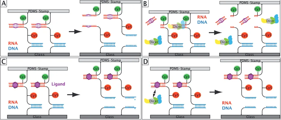
In the next step, the two measurements were combined to demonstrate Dicer inhibition upon ligand binding. Figure 4.3 shows the single steps of the experiment. First, the initial value of the Normalized Fluorescence was measured without the addition of Dicer nor ligand. Next, the sample was incubated for a specified amount of time (1h) with 2.5µl
4.2 Sequence specific inhibition of Dicer measured with a force-based
microarray for RNA ligands 33
Glass PDMS-Stamp PDMS-Stamp Glass PDMS-Stamp A B Cy3 RNA DNA PDMS-Stamp Glass Glass RNA DNA RNA DNA PDMS-Stamp Glass PDMS-Stamp C Ligand Glass PDMS-Stamp Glass D RNA DNA PDMS-Stamp Glass Dicer Dicer Dicer Dicer Dicer Cy3
Cy5 Cy5 Cy5
Cy5 Cy3 Cy3
Cy3 Cy3
Cy5
Cy5 Cy5 Cy5
Cy3 Cy3 Cy3 Cy3 Cy5 Cy5 Cy5 Cy5 Cy3 Cy3 Cy3 Cy3 Cy5 Cy5 Cy5 Cy5 Cy3 Cy3
Start
Value 2.5 l Dicer Paromomycin1mM 1mM Paro +2.5 l Dicer
0.7 0.8 0.9 1.0 No rm al iz ed F lu or es ce nc
e RNA down
Start
Value 2.5 l Dicer Paromomycin1mM 1mM Paro +2.5 l Dicer
0.20 0.25 0.30 0.35 0.40 0.45 RNA up No rm al iz ed F lu or es ce nc e A B
Figure 4.4: Dicer hindrance upon paromomycin binding. (A) In the RNA up configura-tion, the initial value of N F = 0.34±0.01 increased to N F = 0.40±0.02 due to the destabilization of the RNA duplex by Dicer cleavage. Paromomycin binding strengthened the RNA duplex and decreased the Normalized Fluorescence to 0.27±0.01. If the paro-momycin binding blocks Dicer, a NF value close to 0.27±0.01 was expected for the last data point. The measured value 0.30±0.01 confirmed the capability of the assay to inhibit Dicer by ligand binding. (B) The experiment in the RNA down configuration yielded the same results and demonstrates the consistency of the assay.
the RNA aptamer but not completely which led to values of the Normalized Fluorescence close to the ligand-only case. Furthermore, a minimum concentration of paromomycin for Dicer inhibtion was determined. For 2.5µlDicer solution in 1mlbuffer, a partial hindrance was observed at a concentration of 2.82µM paromomycin, whereas 52µM paromomycin blocked most of Dicer activity. This result agrees nicely with the measured dissociation constant of 2.55±2.18mM.
4.3 A force-based, parallel assay for the quantification of protein-DNA
interactions 35
this application of the MFA towards high throughput.
4.3
A force-based, parallel assay for the quantification
of protein-DNA interactions
Characterization of protein interactions with genomic DNA is essential to understand the intricate system of the regulation of gene expression, especially transcription. Most tech-niques can either measure protein-DNA interaction in detail but are very time consuming and not suited to analyze great numbers of proteins or DNA sequences or can determine a large amount of protein-DNA binding events but have difficulties to accurately quantify those interactions. Furthermore, affinity or thermodynamic constants might not be the best quantities to characterize protein-DNA interaction in a nuclear environment, where a DNA-binding protein will probably always be attached to some DNA sequence due to the high concentration of DNA inside the nucleus. Specificity that comprises the discrim-ination between high and low or no binding sequences is better suited to characterize the binding properties of a protein. This quantitiy can be measured in force measurements by determining the binding strength between two molecules. An adaptation of the MFA that enables the parallel quantification of protein-DNA interactions is described in the following and in manuscript M1.
Cy5 Cy5
Amino-coated Glass Slide
no binding sequence high affinity sequence reference bond stamp with 16 pads
pad diameter = 1,1mm
A
C B
Cy5
contact
Cy5
superfolderGFP with YbbR-tag PDMS StampPDMS Stamp
Cy5 Cy5
Amino-coated Glass Slide
Cy5
PDMS StampPDMS Stamp
Cy5
Amino-coated Glass Slide
Cy5 Cy5
Cy5 Cy5
PDMS StampPDMS Stamp retract
F
F
100 x 100µm
zinc finger construct
4.3 A force-based, parallel assay for the quantification of protein-DNA
interactions 37
F
transfer Cy5F
functional protein Cy5NF
A Calculation of the Normalized Fluorescence
shear
20 no shear 40 no 20 lowshear 40 lowshear 20 highshear 40 highshear
0.0 0.2 0.4 0.6 0.8
N
or
m
al
iz
ed
F
lu
or
es
ce
nc
e
N
F
C Normalized Fluorescence for different binding motifs
and reference sequences
B Histogram of the Normalized Fluorescence
and Gaussian fit to calculate the mean
0.0 0.2 0.4 0.6 0.8 1.0
0 100 200 300 400
mean(NF) = 0.632 ± 0.069
Normalized Fluorescence NF
oc
cu
rre
nc
e
Figure 4.6: Quantification of the binding strength of a six zinc finger construct to different DNA sequences. (A) In order to quantify the binding strength, the fluorescence signal rep-resenting the DNA transfer has to be normalized to the number of available protein binding sites. For this purpose, protein binding sites are saturated by adding a Cy5-labeled 40 base pair DNA duplex harboring a high affinity binding motif subsequently to the force measure-ment. The fluorescence intensity of transferred DNAFtransf er is divided by the fluorescence
signal corresponding to the saturated protein binding sites Fintact protein, yielding the
covalently attached via the ybbR-tag to a glass slide coated with Coenzyme A in a 4x4 pattern. The two double-stranded DNA complexes in series are covalently bound to the 16 pillars of a soft PDMS surface with the upper one as reference bond and the lower one as sample bond (see Figure 4.5 A ). The DNA sequences in shear geometry are separated by a linker sequence to which a Cy5 fluorophore is conjugated. Due to the macrostructure of the PDMS stamp a maximum of 16 combinations of different reference sequences as well as sample sequences can be tested within one experiment. The GFP signal of the fusion protein is used to place the protein spots below the stamp pillars functionalized with the different DNA sequences. The PDMS surface is carefully brought into contact with the glass slide so that the sample sequence is able to bind to the protein on the glass slide (Figure 4.5 B). After 10 minutes, the PDMS surface is retracted with constant velocity by the Piezo actuator. Thereby, a force is applied to the protein-sample complex as well as to the reference bond until the weaker one ruptures (Figure 4.5 C). The fluorescence Cy5 signal on the glass slide is measured by an inverted epi-fluorescence microscope and indicates the number of intact protein-DNA complexes. Thus, the protein-DNA interaction is directly probed and compared to a well-characterized DNA double-strand. In order to approximate the environment in a eukaryotic nucleus, the experiment was designed as a competition assay and the zinc finger protein was pre-incubated with low-molecular weight DNA from salmon sperm before the contact process.
4.3 A force-based, parallel assay for the quantification of protein-DNA
interactions 39
to the force probe experiment to saturate all functional proteins bound to the surface. Calibration measurements confirmed a complete saturation after 30 minutes incubation time. After removing unbound fluorophores by a washing step, the fluorescence on the glass slide was determined again. It is a measure for the maximum number of functional proteins on the slide. Since the binding density of the DNA complexes on the PDMS always exceeds the number of functional proteins on the glass slide, further corrections are not necessary. The ratio of fluorescence signal on the glass slide directly after the rupture event Ftransf er to the maximal number of functional proteins Fintact protein is defined as
20 base pair DNA double strand.
Chapter 5
Outlook
This thesis demonstrates various applications for the Molecular Force Assay that have a great potential to contribute to the solution of current biological problems. Especially its parallel format distinguishes the MFA from other force-based techniques and enables its use for investigations that were so far unsuited for force measurements. The force-based aptamer sensor (chapter 4.1) characterizes even small molecules and weak binders in buffer solution as well as complex fluids within a single measurement. Additionally, labeling of the molecules in question is unnecessary. Hence, its principle could be utilized in diagnostic tools that analyze medical samples like blood for properties indicating a certain malfunction or disease. Furthermore, the MFA can assist in the development of new medical therapeu-tics that aim at the RNA interference pathway. Chapter 4.2 describes the use of the MFA for the screening and characterization of RNA ligands that selectively inhibit the protein Dicer. The third part of this thesis illustrates an application of the MFA that directly quantifies the binding strength between proteins and DNA sequences. The specificity of a protein to all kinds of genomic DNA is of utmost importance in order to understand transcriptional regulation. The specificity correlates with the binding strength and is, thus accessible in force measurements. Chapter 4.3 describes the quantification of the binding strength of a zinc finger protein with several DNA motifs within one measurement. This clearly proves that the binding strength of multiple interactions can be determined in a single experiment.
of 25µm2 is sufficient to get valid results. Furthermore, the fabrication of DNA microarrays
is a standard procedure so that a further parallelization of the MFA seems feasible. Addi-tionally, the possibility to incubate every of these miniature spot with a different ligand or protein concentration would greatly enhance the multiplexing capabilities of the MFA. This was realized only recently by implementing the MFA in a microfluidic device [76]. Another possible development is the combination of in vitro protein expression on a microfluidic chip with the MFA. Proteins could be directly expressed on the chip without the need to store them and could be immediately attached to the surface via the ybbR-Coenzyme A technology. A button valve functionlized with the DNA sample and reference bond could come down and quantify the protein-DNA interaction similar as described by Otten et al. [76]. The current microfluidic chip format would, thus, allow the measurement of hundreds of interactions in a single experiment and could extend the potential of this application of the MFA towards high throughput.
Appendix A
Publications
A.1
Publication 1:
DNA as a Force Sensor in an
Aptamer-Based Biochip for Adenosine
Dominik Ho, Katja Falter, Philip Severin and Hermann E. Gaub;
DNA as a Force Sensor in an Aptamer-Based
Biochip for Adenosine
Dominik Ho, Katja Falter, Philip Severin, and Hermann E. Gaub*
Lehrstuhl fu¨r Angewandte Physik and Center for Nanoscience (CeNS), Ludwig-Maximilians-Universita¨t, Amalienstrasse 54, 80799 Munich, Germany, and Munich Center For Integrated Protein Science (CIPSM), Ludwig-Maximilians-Universita¨t, Butenandtstrasse 5-13, 81377 Munich, Germany
Without prior signal amplification, small molecules are difficult to detect by current label-free biochip approaches. In the present study, we developed a label-free capture biochip based on the comparative measurement of un-binding forces allowing for direct detection of small-molecule-aptamer interactions. The principle of this assay relies on increased unbinding forces of bipartite aptamers due to complex formation with their cognate ligands. The bipartite aptamers are immobilized on glass support via short DNA duplexes that serve as references to which unbinding forces can be compared. In a simple model system, adenosine is captured from solution by an adenosine-selective aptamer. Linking the molecular chains, each consisting of a short DNA reference duplex and a bipartite aptamer, between glass and a poly(dimethylsi-loxane) (PDMS) surface and subsequently separating the surfaces compares the unbinding forces of the two bonds directly. Fluorescence readout allows for quantification of the fractions of broken aptamer and broken reference bonds. The presence of micromolar adenosine concentra-tions reliably resulted in a shift toward larger fracconcentra-tions of broken reference bonds. Because of the force-based design, the interactions between the bipartite aptamer and the target, rather than the presence of the target, are detected and no washing step disturbing the equilibrium state prior to probing and no reporter aptamer or antibody is required. The assay exhibits excellent selectivity against other nucleotides and detects adenosine in the presence of a complex molecular background. Multiplexing was demonstrated by performing whole titration experiments on a single chip revealing an effective half-maximal concentration of 124.8µM agreeing well with literature values.
A current goal within the field of bioanalytical methods is the development of label-free detection formats, which probe multiple interactions simultaneously employing massively parallel assays.1
High impact of DNA biochips on the field of biology provides motivation to develop arrays for other classes of molecules,
including peptides, proteins, and small molecules.2DNA or RNA
aptamers are promising candidates for fabrication of microarray surfaces, which simply and effectively capture above-mentioned analytes from solution. Numerous reports confirm that aptamers specifically respond to all kinds of molecules3such that they are
increasingly recognized as rivals for antibodies in in vitro diag-nostics4and molecular sensor applications,5-7surpassing them
in terms of small molecular weight, ease of modification, and ability to detect toxins.8In contrast to proteins, which are difficult
to immobilize on surfaces due to their tendency to adsorb unspecifically and thus lose activity,9 standard protocols for
oligonucleotide microarrays are used for aptamer biochips. The development of such arrays has proven to be more difficult than expected. Small molecules in particular are rarely detected due to several reasons: First, their small size induces only small signals using current biochip-compatible, label-free detection techniques (e.g., surface plasmon resonance,10electrochemical,11
or cantilever bending12 based sensors). Second, background
signals are generally large in biochip assays due to the tendency of molecules to adsorb to basically all man-made surfaces.13-15
Third, aptamers developed against small molecules are generally of low affinity,16prohibiting washing steps that would increase
the signal-to-background ratio. The combination of small signal, high background, and no option for stringent washes creates an overwhelming technical hurdle for the quantification of small molecules without prior signal amplification. Here, we present a widely applicable strategy for the direct detection of
small-* To whom correspondence should be addressed. Phone:+49 89 2180 3172. Fax:+49 89 21 80 2050. E-mail: [email protected].
(1) Gurard-Levin, Z. A.; Mrksich, M.Annu. Rev. Anal. Chem.2008,1, 767– 800.
(2) Lockhart, D. J.; Winzeler, E. A.Nature2000,405, 827–836.
(3) Hermann, T.; Patel, D. J.Science2000,287, 820–825. (4) Rimmele, M.ChemBioChem2003,4, 963–971.
(5) Zhang, S.; Xia, J.; Li, X.Anal. Chem.2008,80, 8382–8388.
(6) Wang, J.; Zhou, H. S.Anal. Chem.2008,80, 7174–7178. (7) Navani, N. K.; Li, Y. F.Curr. Opin. Chem. Biol.2006,10, 272–281.
(8) Tang, J.; Xie, J.; Shao, N.; Yan, Y.Electrophoresis2006,27, 1303–1311.
(9) Kusnezow, W.; Hoheisel, J.J. Mol. Recognit.2003,16, 165–176. (10) Boozer, C.; Kim, G.; Cong, S.; Guan, H.; Londergan, T. Curr. Opin.
Biotechnol.2006,17, 400–405.
(11) Xu, D.; Xu, D.; Yu, X.; Liu, Z.; He, W.; Ma, Z.Anal. Chem.2005,77, 5107– 5113.
(12) Zhang, J.; Lang, H.; Huber, F.; Bietsch, A.; Grange, W.; Certa, U.; Mckendry, R.; Gu¨ntherodt, H.; Hegner, M.; Gerber, C.Nat. Nanotechnol.2006,1, 214–220.
(13) Haab, B. B.; Dunham, M. J.; Brown, P. O.Genome Biol. 2001, 2, 0004.10004.13.
(14) Michaud, G.; Salcius, M.; Zhou, F.; Bangham, R.; Bonin, J.; Guo, H.; Snyder, M.; Predki, P.; Schweitzer, B.Nat. Biotechnol.2003,21, 1509–1512.
(15) Ma, H.; Horiuchi, K.Drug Discovery Today2006,11, 661–668. (16) Hamula, C.; Guthrie, J.; Zhang, H.; Li, X.; Le, X.TrAC, Trends Anal. Chem.
2006,25, 681–691. Anal. Chem.2009,81,3159–3164
10.1021/ac802766j CCC: $40.752009 American Chemical Society Analytical Chemistry, Vol. 81, No. 8, April 15, 2009 3159
Published on Web 03/19/2009
Downloaded by BBWS CONSORTIA GERMANY on July 15, 2009
the mere presence of small molecules close to a sensor surface. Thereby, nonspecific adhesion, the major bottleneck in the development of next-generation biochips, is rendered insignificant. For detection of the bound analyte we applied the comparative unbinding force assay (CUFA), which operates label-free and is insensitive to nonspecifically adsorbed target molecules. CUFA has already been applied to detect single-nucleotide polymor-phisms,17to study differences of antibody/antigen interactions,18
to eliminate cross-reactions on protein microarrays,19 and to
investigate the chiral selectivity of small peptides.20 Whereas
standard single-molecule force spectroscopy experiments measure the unbinding forces of molecular complexes by a microscopic, springlike object, e.g., an atomic force microscopy (AFM) canti-lever21 or a bead in an optical trap,22CUFA reduces the force
detector to a single DNA reference duplex. Many molecular chains, each consisting of a bipartite aptamer and a DNA reference duplex, are grafted between two surfaces. The linker between the bonds is conjugated to a fluorophore. Upon separation of the surfaces, a force gradually builds up within the molecular chain and the unbinding forces of the bipartite aptamer structure are compared directly against the unbinding forces of the short DNA reference duplex. The result, i.e., the fractions of broken aptamer and broken reference bonds, is stored in a binary fluorophore distribution (fluorophore top or bottom surface). If complex formation between aptamer and ligand results in increased unbinding forces of the aptamer structure, a shift toward larger fractions of broken reference bonds is expected. Thereby, CUFA combines the advantages of fluorescence-based techniques, namely, fast and sensitive detection employing commercially available scanners or fluorescence microscopes, with the advantages of label-free methods, namely, no undesirable label interactions and the option for simultaneous detection of multiple analytes. Importantly, high specificity when investigating molecular interac-tions is a major strength of CUFA. Current fluorescent23and
label-free biochip methods do not allow for discrimination between specifically and nonspecifically adhered analytes, which may lead to false-positives. Washing steps are performed to reduce non-specific adhesion. However, if molecular interactions with equi-librium dissociation constants in the micromolar range need to be characterized, specifically but weakly interacting molecules are also removed resulting in false-negatives. CUFA overcomes these difficulties by detecting the interaction between the capture
adenosine and an adenosine-selective aptamer. This model system isinstructiveforthereasonsthattheaptameriswell-characterized,24-26,28
the interaction is very weak, and the analyte is so small that it cannot be detected directly with surface plasmon resonance.6
EXPERIMENTAL SECTION
Immobilization of Aptamer-DNA Unbinding Force Com-plex on Slides (Bottom Surface).DNA oligomers1,2, and3
were purchased HPLC grade from IBA GmbH (Go¨ttingen, Germany). Sequences and modifications of all oligonucleotides are the following:1, NH2-(hexaethyleneglycol)5-5′-TTT TTT TTT
TCG GTC TGT CGC GTA CTT GCA-3′;2, 3′-GCC AGA CAG CGC ATG AAC GTT TTT T-5′-5′-T(Cy3) TTT TTC AAC ATA CCT GGG GGA GTA TAT AAT GAC TGA CCC C-3′;3, biotin-5′-TTT TTT TTT TGG GGT CAG TCA TTA TAG CGG AGG AAG GTA TGT TG-3′. For the upside-down experiment the NH2-(hexaethyleneglycol)5(HEGL) and biotin modifications are
exchanged. The five HEGL linkers are connected via phosphate groups. DNA oligomer 1 is amine-modified, which allows covalent linkage to aldehyde-functionalized glass slides (Schott GmbH, Jena, Germany). We spotted 2µL drops of 5× SSC (saline sodium citrate; Sigma-Aldrich GmbH, Munich, Ger-many) containing 25µM oligomer1on the aldehyde slide in a 4×4 pattern and incubated the slide in a saturated NaCl ddH2O atmosphere overnight. After washing the slide with
ddH2O containing 0.2% sodium dodecyl sulfate (SDS; VWR
Scientific GmbH, Darmstadt, Germany) and thoroughly rinsing the slide with ddH2O we reduced the resulting Schiff bases
with 1% aqueous NaBH4(VWR Scientific GmbH, Darmstadt,
Germany) for 20 min. Subsequently, the slide was washed with 1×SSC and thoroughly rinsed with ddH2O. In order to reduce
nonspecific binding, the slides were blocked in 1× SSC containing 4% bovine serum albumin (BSA; Sigma-Aldrich GmbH, Munich, Germany) for 30 min. Custom-made 16-well silicone isolators (Grace-Biolabs, OR) were placed on top of the immobilized DNA oligomer1. The 0.1µM Cy3-modified oligomer 2 and 0.2 µM biotin-modified oligomer 3 were hybridized to the latter for 1 h, completing the1·2·3complex on the glass slide. The slides were washed with 1× SSC containing 0.05% SDS and thoroughly rinsed with 1×SSC. The silicone isolators stayed on the slide throughout the experi-ment, and care was taken that after hybridization the slide remained immersed in 1×SSC.
Ligand Incubation. The 16-well silicon isolators allow for incubation with different concentrations of the ligand molecule on one slide. An amount of 100µL of the respective solutions was circulated through the wells employing a self-made microf-luidic system. The latter was driven by two 16-channel peristaltic pumps (Ismatec GmbH, Wertheim-Mondfeld, Germany) pumping the different solutions through tubing and blunt needles leading into and out of the wells in a closed circuit. The tubing was
(17) Albrecht, C.; Blank, K.; Lalic-Multhaler, M.; Hirler, S.; Mai, T.; Gilbert, I.; Schiffmann, S.; Bayer, T.; Clausen-Schaumann, H.; Gaub, H. E.Science
2003,301, 367–370.
(18) Blank, K.; Mai, T.; Gilbert, I.; Schiffmann, S.; Rankl, J.; Zivin, R.; Tackney, C.; Nicolaus, T.; Spinnler, K.; Oesterhelt, F.; Benoit, M.; Clausen-Schau-mann, H.; Gaub, H. E.Proc. Natl. Acad. Sci. U.S.A.2003,100, 11356– 11360.
(19) Blank, K.; Lankenau, A.; Mai, T.; Schiffmann, S.; Gilbert, I.; Hirler, S.; Albrecht, C.; Benoit, M.; Gaub, H. E.; Clausen-Schaumann, H.Anal. Bioanal. Chem.2004,379, 974–981.
(20) Dose, C.; Ho, D.; Gaub, H. E.; Dervan, P. B.; Albrecht, C.Angew. Chem., Int. Ed.2007,46, 8384–8387.
(21) Florin, E. L.; Moy, V. T.; Gaub, H. E.Science1994,264, 415–417.
(22) Svoboda, K.; Schmidt, C. F.; Schnapp, B. J.; Block, S. M.Nature1993,
365, 721–727.
(23) Warren, C.; Kratochvil, N.; Hauschild, K.; Foister, S.; Brezinski, M.; Dervan, P.; Phillips, G., Jr.; Ansari, A.Proc. Natl. Acad. Sci. U.S.A.2006,103, 867– 872.
(24) Huizenga, D. E.; Szostak, J. W.Biochemistry1995,34, 656–665.
(25) Lin, C. H.; Patel, D. J.Chem. Biol.1997,4, 817–832.
(26) Stojanovic, M. N.; de Prada, P.; Landry, D. W.J. Am. Chem. Soc.2000,
122, 11547–11548.
(28) Jhaveri, S. D.; Kirby, R.; Conrad, R.; Maglott, E. J.; Bowser, M.; Kennedy, R. T.; Glick, G.; Ellington, A. D.J. Am. Chem. Soc.2000,122, 2469–2473.
3160 Analytical Chemistry, Vol. 81, No. 8, April 15, 2009
Downloaded by BBWS CONSORTIA GERMANY on July 15, 2009