| WORMBOOK WormMethods
CRISPR-Based Methods for
Caenorhabditis elegans
Genome Engineering
Daniel J. Dickinson1and Bob Goldstein Department of Biology and Lineberger Comprehensive Cancer Center, University of North Carolina, Chapel Hill, North Carolina 27599-3280
ABSTRACT The advent of genome editing techniques based on the clustered regularly interspersed short palindromic repeats (CRISPR)–Cas9 system has revolutionized research in the biological sciences. CRISPR is quickly becoming an indispensible experimental tool for researchers using genetic model organisms, including the nematodeCaenorhabditis elegans. Here, we provide an overview of CRISPR-based strategies for genome editing in C. elegans. We focus on practical considerations for successful genome editing, including a discussion of which strategies are best suited to producing different kinds of targeted genome modifications.
KEYWORDSCaenorhabditis elegans; CRISPR/Cas9; genome editing; WormBook
TABLE OF CONTENTS
Abstract 885
Overview of the CRISPR-Cas9 system 886
Genome engineering via double-strand break repair 887
Four basic steps for genome engineering with Cas9 888
Using Cas9 to Generate DNA Double-Strand Breaks 889
Expression of Cas9 and sgRNA in C. elegans 889
Choosing a Cas9 target site 889
Specificity: 889
Activity: 890
Proximity to the desired modification: 890
Strategies for Identifying CRISPR Modifications 890
Screening based on mutant phenotype 890
PCR screening 890
Co-CRISPR 891
Positive selectable markers 891
Construction of Repair Templates for HDR 892
Designing ssDNA oligo repair templates 892
Producing dsDNA repair templates from preexisting vectors 894
Continued
Copyright © 2016 by the Genetics Society of America doi: 10.1534/genetics.115.182162
Manuscript received August 13, 2015; accepted for publication January 12, 2016. Available freely online through the author-supported open access option.
1Corresponding author: Department of Biology and Lineberger Comprehensive Cancer Center, CB3280, University of North Carolina, Chapel Hill, NC 27599-3280.
CONTENTS,continued
Building more complex repair templates using Gibson assembly 894
Addressing Cas9 Specificity 896
Recommended Strategies for Different Types of Modifications 896
Null mutations 896
Recommendation: 897
Point mutations 897
Recommendation: 897
Fluorescent protein fusions 897
Recommendation: 899
Other changes 899
A general strategy for dissecting gene function 899
Where to Go for More Information and Detailed Protocols 899
A
fundamental goal of biological research is to understand the functions of genes. One common strategy for study-ing gene function is to observe the phenotypes of mutants to deduce the biological processes in which a gene participates and, sometimes, details of its mechanism of action. This basic idea is the foundation of classical genetics and also under-lies reverse genetic approaches including RNAi. A second strategy is to observe the localization and dynamics of a gene’s protein product within a cell or animal, either by antibody staining or by expressing a fluorescent protein (FP) fusion. Together, these two basic strategies form the backbone of much research inCaenorhabditis elegansand other model systems.The use of the clustered regularly interspaced short palin-dromic repeats (CRISPR)-Cas9 system for genome engineering (Hsuet al.2014) has greatly facilitated the study of gene func-tion inCaenorhabditis elegansand other organisms. By making precisely targeted mutations in endogenous genes, an investi-gator can examine the relationship between gene function and phenotype. By inserting coding sequence for afluorescent pro-tein, the expression and localization of endogenous proteins can be monitored. In both cases, one avoids the caveats of overexpression and silencing that are associated with conven-tional transgenes. Moreover, forfluorescent protein (FP) fu-sions, insertion of the FP into the endogenous locus allows one to use phenotypic assays to quickly determine whether the resulting fusion protein is functional. Together, these advan-tages permit more carefully controlled experiments to be done and thus allow greater confidence in the results. As an added benefit, current CRISPR-based approaches (Arribere et al. 2014; Dickinson et al. 2015; Paix et al. 2015; Ward 2015) are faster and require less labor than either conventional trans-genesis (Mello et al. 1991) or microparticle bombardment (Praitiset al.2001), and they eliminate the need for special-ized strain backgrounds that are required for these methods and those based on the Mos1 transposon (Robert and Bessereau 2007; Frøkjaer-Jensenet al.2008, 2010, 2012).
Many different CRISPR approaches have been developed forC. elegansand are being widely adopted by the research community. In general, all of these methods work well, with different strategies being best suited to different experimen-tal goals. By choosing the appropriate strategy, one can now make essentially any desired change to theC. elegansgenome in a matter of days to weeks, with,1 day of hands-on labor (Dickinsonet al.2013, 2015; Arribereet al.2014; Zhaoet al. 2014; Paixet al.2015; Ward 2015). The goal of this article is to aid users in choosing the best strategy for a given applica-tion. We provide an overview of CRISPR-based methods for C. elegans, including a discussion of which strategies are most appropriate for generating different kinds of modifications.
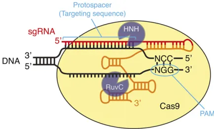
Overview of the CRISPR-Cas9 system
dinucleotide, by simply changing the sequence at the 59end of the sgRNA. It is this ease of programming that makes Cas9 such a powerful andflexible tool for genome engineering.
More recently, engineered derivatives of SpCas9 have been described that recognize alternate PAMs including NGA, NGAG, and NGCG (Kleinstiveret al.2015), and some of these have been tested and shown to be effective inC. elegans(Bell et al.2015). Cas9 homologs from bacterial species other than S. pyogeneshave also been found to recognize alternate PAMs (Ranet al.2015). Also, the unrelated CRISPR nuclease Cpf1 recognizes its targets differently from Cas9 and has been suc-cessfully used for genome editing in mammalian cells (Zetsche et al.2015). Although Cpf1 and non-Sp Cas9 homologs have not yet been tested inC. elegansto our knowledge, it seems likely that a growing collection of RNA-guided nucleases rec-ognizing a wider variety of PAMs than the conventional NGG will become available in the next few years.
It is important for a user of Cas9 to have some understand-ing of the different roles that the guide sequence and PAM play in determining Cas9 specificity. When searching for a sub-strate, Cas9first binds to the PAM and only then interrogates the adjacent DNA to look for a match to the guide sequence (Sternberget al.2014). Thus, even DNA sequences that per-fectly match the guide sequence are not recognized or cleaved if they do not contain a PAM. The requirement for an NGG PAM sequence appears fairly stringent (Jineket al. 2012; Kuscu et al.2014; Sternberg et al.2014; Wu et al. 2014), although an NAG sequence may be able to support low-efficiency cleavage in some instances (Hsuet al.2013; Jianget al.2013). In contrast to its strict requirement for the PAM sequence, Cas9 is somewhat tolerant of mismatches be-tween the guide sequence and the target, especially when they occur near the 59end of the guide sequence (i.e., distal to the PAM) (Jineket al.2012; Fuet al.2013; Hsuet al.2013; Pattanayaket al.2013; Kuscuet al.2014; Renet al.2014; Wu et al. 2014). The practical consequences of this mismatch tolerance are discussed inAddressing Cas9 Specificity, below.
Genome engineering via double-strand break repair
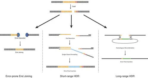
As described above inOverview of the CRISPR-Cas9 system, Cas9 can be used to generate a DNA double-strand break at a defined location in the genome. These double-strand breaks are useful because they allow an investigator to make use of endogenous cellular DNA repair machinery to generate cus-tom modifications in the genome. Three different types of DNA repair strategies have been used to produce custom modifications inC. elegans(Figure 2):
1. Error-prone repair via end joining: When Cas9 cleaves ge-nomic DNA, some of the resulting DNA double-strand breaks are repaired by an error-prone pathway that pro-duces small insertions or deletions (indels) at the site of the break. Mechanistically, these indels arise not via ca-nonical nonhomologous end joining (NHEJ) as had been widely assumed, but from an alternative end-joining path-way that requires DNA polymeraseQ(van Schendelet al. 2015). When generated in protein-coding sequence, indels can shift the reading frame, resulting in a prema-ture stop codon. Thus, error-prone repair can be used to produce loss-of-function alleles (C. Chenet al.2013; Chiu et al.2013; Choet al.2013; Friedlandet al.2013; Katic and Großhans 2013; Loet al.2013; Waaijerset al.2013). 2. Homology-directed repair: In homology-directed repair (HDR), an exogenous DNA molecule is introduced along with Cas9 and serves as a template for DNA repair. Mod-ifications present in the repair template are copied into the genome in an error-free manner. Different kinds of repair templates have been reported to yield different HDR effi-ciencies inC. elegans(Arribereet al.2014; Paixet al.2014, 2015; Dickinsonet al.2015; Ward 2015). For insertions up to1 kb, repair was most efficient when the repair template contained 30–40 bp of homology to the genome, and longer homology arms led to reduced efficiency (Paix et al.2014). On the other hand, insertions of6 kb were readily obtained using 500- to 700-bp homology arms, but occurred rarely or not at all when using 30- to 40-bp ho-mology arms (Dickinsonet al.2015). Based on these ob-servations, there appear to be two distinct HDR pathways in C. elegans, which we callshort-range HDR and long-range HDR. For convenience, we discuss these two repair pathways as if they occur via different mechanisms (as proposed in Figure 2), although the actual molecular mechanisms are not yet known.
2a. Short-range HDR is a highly local repair mechanism that occurs most efficiently within10 bp of the Cas9 cut site (Arribereet al.2014; Paixet al.2015; Ward 2015) and when the repair template carries 30- to 40-bp homology armsflanking the desired modifica-tion (Arribere et al. 2014; Paix et al. 2014; Ward 2015). Short-range HDR can be very efficient in C. elegans: in the best cases, .50% of F1 progeny that received active Cas9 and the repair template can carry short-range HDR events. Short-range HDR can be used to introduce point mutations, precise Figure 1 DNA recognition by the Cas9–sgRNA complex. Cas9 identifies
deletions, and small epitope tags by using a single-stranded DNA oligonucleotide as the repair template (Paix et al.2014; Zhao et al.2014). Larger insertions such as GFP insertion can also be made via short-range HDR, using a PCR product as the repair template (Paix et al.2014, 2015). The main advantages of short-range HDR are its high efficiency and the fact that only 30–40 bp of homology to the genome are required for efficient repair. Short-range HDR has two main limitations. First, it occurs most efficiently within 10 bp of a Cas9 cleavage site, and efficiency declines sharply at larger distances (Arribereet al.2014; Paixet al.2015; Ward 2015). This can make it challenging to isolate edits that are not located near an efficient sgRNA target site (seeChoosing a Cas9 target site, below, for a discussion of factors gov-erning sgRNA efficiency). Second, for reasons that re-main unclear, short-range HDR cannot accommodate insertions much larger than 1–2 kb (Dickinson et al. 2015; Paixet al.2015); thus, short-range HDR is suitable for GFP insertion but not for larger-scale modifications. 2b. Long-range HDR allows insertion of much larger
se-quences [at least 12 kb (Das et al. 2015)] and at a greater distance from the cut site [at least 1 kb (Dickinson et al. 2013; Das et al. 2015; Sullivan-Brown et al. 2016)]. Plasmids carrying 500–1500 bp of genomic ho-mology flanking the desired modifications are robust
substrates for this repair mechanism (Dickinson et al. 2013, 2015). On a per-F1 basis, long-range HDR is much less efficient than short-range HDR; however, be-cause it can accommodate larger inserts, long-range HDR allows use of selectable markers, which offset the lower efficiency by facilitating quick and easy iden-tification of repair events. Long-range HDR is relatively insensitive to variations in sgRNA efficiency (Dickinson et al. 2015), presumably because the repair process itself, rather than Cas9 cleavage, is the limiting factor.
The different properties of short-rangevs.long-range HDR influence both the experimental design and the types of mod-ifications that each strategy is best suited to generate, as discussed in the following sections.
Four basic steps for genome engineering with Cas9
To generate custom genome modifications with CRISPR/ Cas9 in any experimental system, one must accomplish four basic tasks: (1) introduce Cas9 and an appropriately targeted sgRNA; (2) if using HDR, supply the appropriate repair tem-plate; (3) identify the animals that carry the desired genome modification; and (4) address specificity, since Cas9 can generate off-target mutations under some conditions.
Strategies for Different Types of Modifications, we recommend approaches to generate different kinds of custom alleles with minimal time and labor.
Using Cas9 to Generate DNA Double-Strand Breaks
Expression of Cas9 and sgRNA in C. elegans
Cas9 can be easily expressed in theC. elegansgermline by inject-ing an expression plasmid (Dickinsonet al.2013; Friedlandet al. 2013) or messenger RNA (mRNA) (Chiuet al.2013; Katic and Großhans 2013; Lo et al. 2013). Alternatively, purified Cas9 protein may be reconstituted with its RNA cofactors and directly injected into the gonad of the worm (Choet al.2013; Paixet al. 2015). For plasmid-based germline expression of Cas9, theeft-3 promoter (Frøkjaer-Jensenet al.2012) has been widely used. By substituting heat-shock or tissue-specific promoters for Peft-3, it is possible to generate indels in somatic tissue, producing tissue specific loss-of-function phenotypes (Liuet al.2014; Shen et al.2014). Generally speaking, it appears that transgenic ex-pression of Cas9 can be easily achieved using the same basic approaches that are well established for other transgenes.
Similarly, sgRNA can be either expressed from a plasmid or synthesizedin vitroand injected. A third option is to feed the worms bacteria expressing sgRNA, which has low efficiency compared to other methods but may be useful for high-throughput screening (Liuet al.2014). Plasmid-based sgRNA expression constructs use a U6 promoter, which directs tran-scription by RNA polymerase III (C. Chen et al. 2013; Dickinson et al.2013; Friedlandet al. 2013). U6 snRNA is an essential component of the mRNA splicing machinery and thus would be expected to be ubiquitously expressed. Con-sistent with this prediction, PU6::sgRNAconstructs have been successfully used to produce mutations both in the germline and in somatic tissues (C. Chenet al.2013; Dickinsonet al. 2013; Friedlandet al.2013; Shenet al.2014). Note that two independently identified U6 promoters have been used in published work (Dickinson et al. 2013; Friedland et al. 2013). Although CRISPR mutations have been successfully isolated using sgRNAs expressed from both promoters, two studies have reported conflicting observations of higher effi-ciency with one promoter or the other (Farboud and Meyer 2015; Katic et al.2015), suggesting that the choice of pro-moter might influence editing efficiency in some cases. For direct RNA injection, the RNA may be synthesized or pur-chased commercially. Note that if RNA is chemically synthe-sized commercially, it is more cost-effective to purchase separate crRNA and tracrRNA rather than the longer chime-ric sgRNA, because only the crRNA is specific to a given ex-periment, while the tracrRNA sequence is constant.
The choice of whether to use plasmid-based Cas9 and sgRNA expression or direct Cas9 and RNA injection will de-pend on the needs of each individual user. Plasmid injection is simple, reliable, and familiar to mostC. elegansresearchers. However, this approach requires cloning each new guide se-quence into an expression construct, and a relatively large
number of animals (50–60 in our experience) need to be injected to consistently obtain the desired modification. Di-rect injection of Cas9 ribonucleoprotein complexes yields a higher frequency of successful injections compared to plasmid-based expression, thus reducing the number of animals that need to be injected to as few as 10 (Paixet al.2015). The trade-off is that the user must either purchase Cas9 protein and the required small RNAs or purify them in house. Pur-chasing Cas9 protein, tracrRNA, and crRNA is currently quite costly ($200 per target, with most of the cost going to the synthetic RNAs), but the cost may drop as more commercial sources become available, and the ability to inject fewer worms may justify the cost for some users.
Cas9 and sgRNA expression plasmids from several different laboratories are available from Addgene (http://www.
addgene.org/CRISPR/worm/). Escherichia coli expression
vectors for producing Cas9 protein are also available
(http://www.addgene.org/crispr/bacteria/).
Choosing a Cas9 target site
The first step in any CRISPR strategy is to choose the Cas9 target site. First, one needs to identify the general region to be targeted. To generate loss-of-function indel mutations, one should target a region close to the 59end of the coding region of the gene of interest, to maximize the chances that an indel will abolish the function of the gene. For HDR-based strate-gies, it is best to choose a site as close as possible to where the desired modification will be made.
Once the general region to be targeted has been identi-fied, the next step is to identify the actual guide sequence within the target region. Three considerations govern the choice of a guide sequence: activity, specificity, and proximity to the desired modification. The relative importance of these considerations depends on the repair mechanism and screen-ing strategy bescreen-ing used (seeStrategies for Identifying CRISPR Modifications, below, for discussion of screening strategies). For long-range HDR with a selectable marker, specificity is the primary concern; for short-range HDR, activity and proximity to the desired modification are more important.
Specificity:Ideally, one should select a guide sequence that is unique in the genome, to minimize the chances of gen-erating off-target mutations. We identify specific guide sequences, using a CRISPR design tool developed by Feng Zhang’s laboratory (Hsu et al. 2013) and available at
http://crispr.mit.edu. This tool lists all possible guide
Activity: Different guide sequences support different Cas9 cleavage efficiencies (Doenchet al.2014; Wanget al.2014; Farboud and Meyer 2015; Xuet al.2015). Whether cleavage efficiency is an important experimental consideration de-pends on the screening strategy being used (see Strategies for Identifying CRISPR Modifications, below). When using long-range HDR with a selectable marker, variations in cleav-age efficiency are of no practical consequence because the repair process, rather than Cas9 cleavage efficiency, is the limiting factor. On the other hand, for short-range HDR, cleavage activity is a critical determinant of efficiency, and so it may be worthwhile to choose a slightly less specific guide to achieve higher cleavage efficiency.
Guide sequences ending in GG (not to be confused with the NGG PAM motif) have been shown to have consistently high cleavage efficiency inC. elegans(Farboud and Meyer 2015). However, these“39GG guides”occur only once every 128 bp in random sequence (and even more infrequently in the AT-rich C. elegansgenome), so using a 39GG guide is usually not fea-sible. As an alternative, several prediction algorithms have been developed that may be useful for identifying the most active guide sequences (Doenchet al.2014; Liuet al.2015; Xu et al.2015). As of this writing, our preferred prediction tool is SSC, which is available at http://crispr.dfci.harvard.edu/ SSC/. In general, guide sequences that are rich in G residues and lack pyrimidines in the last four bases before the PAM tend to be most active. Guides containing four or more consecutive T/U bases should be avoided, as these stretches can prema-turely terminatePolIII transcription. Cleavage efficiency can also be improved by using an engineered sgRNA, termed sgRNA(F+E) (B. Chenet al.2013; Ward 2015).
Proximity to the desired modification:For short-range HDR using an oligonucleotide or PCR product repair template (see PCR screeningand Co-CRISPR), the Cas9 target site should ideally be within 10 bp of the desired modification (Arribere et al.2014; Paixet al.2014, 2015). It is sometimes necessary to choose a less specific and/or less active guide to achieve this degree of proximity. For long-range HDR with a select-able marker (see Positive selectable markers), proximity is much less important, since efficient editing can be achieved at least 1 kb from the Cas9 target site (Dickinsonet al.2013; Daset al.2015; Sullivan-Brownet al.2016).
Once a guide sequence has been selected, it must be either cloned into an appropriate sgRNA vector (a U6 promoter vector for plasmid-based expression inC. elegansor a T7 pro-moter vector forin vitro transcription) or synthesized com-mercially for direct injection. The U6 promoter requires a G residue as the first base of the sgRNA sequence to initiate transcription, while for the T7 promoter, the sgRNA should typically begin with GG. If these guanine residues are not present in the chosen guide sequence, they can either be substituted for the most 59residues in the guide, since mis-matches at these positions are well tolerated (Jinek et al. 2012; Fu et al. 2013; Hsu et al. 2013; Pattanayak et al. 2013; Kuscu et al.2014; Renet al.2014; Wuet al.2014),
or simply appended to the 59end of the guide, since exten-sions of the sgRNA beyond 20 bp do not affect cleavage ac-tivity (Ran et al.2013; Farboud and Meyer 2015). Both of these approaches have succeeded in our laboratory.
Strategies for Identifying CRISPR Modifications
Choosing an appropriate selection or screening approach is perhaps the most critical step in planning a new CRISPR genome modification. Different strategies have been devised that vary greatly in their applicability, efficiency, and difficulty. Each approach has strengths that are appropriate for different applications. We summarize each strategy here; Recom-mended Strategies for Different Types of Modificationsprovides recommendations for which strategy to use for different applications.
Screening based on mutant phenotype
The first demonstrations of Cas9-induced mutations in C. elegansinvolved simple visual screening for obvious mu-tant phenotypes such asDpyorUnc, benomyl resistance con-ferred by ben-1 mutations, or loss of fluorescence from a bright GFP transgene (C. Chenet al.2013; Chiuet al.2013; Cho et al.2013; Friedlandet al.2013; Katic and Großhans 2013; Loet al.2013; Waaijerset al.2013). While these were useful proof-of-principle experiments, many genome modi-fications that are of biological interest do not confer a visible plate-level phenotype. Nevertheless, phenotype-based screen-ing for edits at one locus can be used to enrich for edits at a second locus in“co-CRISPR”approaches (described in Co-CRISPRsection).
There have also been reports of isolation of GFP knock-in strains by visually screening forfluorescence of the introduced GFP (Kimet al.2014; Paixet al.2014, 2015). Although fluo-rescence-based screening can clearly be effective in these reported cases, it requires that the gene being tagged be expressed at a high enough level that the GFP fusion protein is easily visible on a dissecting microscope at reasonably low magnification. Fluorescence-based screening is also greatly facilitated when the pattern of protein expression is known in advance. In our experience, the majority ofC. elegansgenes do not meet these criteria, and so screening for knock-ins based on visual examination offluorescence is not an advis-able strategy in general. It is possible in principle that dimmer knock-ins could be isolated using a flow-sorting system (Pulak 2006) or another automated system, but we are un-aware of any reports of such an approach.
PCR screening
then singled and the process is repeated to identify homozy-gotes. Direct PCR screening has now been essentially replaced by co-CRISPR (seeCo-CRISPRsection), which greatly reduces the number of animals that need to be screened.
Primer design for PCR screening depends on the nature of the genome modification (Paixet al.2014). HDR insertions large enough to accommodate a PCR primer can be detected using a primer inside the insertion and a second primer out-side the homology arm. Large deletions can be detected with flanking primers. For small indels or point mutations, it is best if the mutation introduces (or deletes) a unique restriction site, which enables screening by restriction fragment length polymorphism (RFLP). When performing HDR, a restriction site can often be introduced into the repair template by mak-ing silent substitutions in addition to the mutation of interest. If RFLP is not possible, thefinal choice is to screen by looking for a mobility shift of PCR products on polyacrylamide gels (Kim et al. 2014) or by using a nuclease that detects mis-matches when wild-type and mutant PCR products are annealed (Conget al.2013; Ward 2015).
Co-CRISPR
Co-CRISPR (Arribereet al.2014; Kimet al.2014; Ward 2015) is a screening strategy that uses a visible phenotype at one locus to help identify edits at a second locus. Two loci are edited simultaneously: the locus of interest and an unlinked marker locus that produces a visible phenotype (Figure 3). The marker mutation is used to identify F1animals derived from oocytes that received active Cas9. Among all F1progeny of injected animals, those that received active Cas9 are most likely to carry the desired modification (Arribereet al.2014; Kimet al.2014; Ward 2015). By restricting PCR screening to these animals, co-CRISPR can substantially reduce the num-ber of animals that need to be screened [to only a few dozen in the best cases (Farboud and Meyer 2015; Paix et al. 2015)]. Co-CRISPR is the screening strategy of choice for modifications generated using short-range HDR.
For co-CRISPR to work well, the desired modification needs to occur with high efficiency relative to the marker mutation; if the marker mutation is efficient but the desired modification is inefficient, most marked F1’s will lack the mutation of interest. Thus, co-CRISPR is best suited to gen-erating modifications that are (1) produced by short-range HDR, which is efficient on a per-F1basis; (2) induced by a highly active sgRNA; and (3) introduced as close as possible to the cut site.
Several different marker mutations have been tested for co-CRISPR applications (Arribereet al.2014; Kimet al.2014; Ward 2015). The most effective of these are the gain-of-function dpy-10(cn64)andsqt-1(e1350)mutations (Figure 3A) (Arribere et al.2014) or rescue of the temperature-sensitive lethal pha-1(e2123) mutation (Figure 3B) (Ward 2015). Since these marker mutations produce dominant phenotypes, they can be recognized in the F1progeny of the injected animals, which are then screened by PCR (see PCR screening section) to identify animals carrying the desired modification. Then, F2
progeny of successfully edited animals are genotyped to iden-tify homozygotes. During this F2screening step, the dpy-10(cn64)orsqt-1(e1350)marker mutations can be eliminated by picking wild-type animals (Figure 3A), provided the desired edit and marker mutation are unlinked. When usingpha-1for co-CRISPR, the marker “mutation”is the wild-type allele of pha-1, which must be genotyped along with the desired mu-tation to identify homozygotes (Figure 3B).
A unique advantage of co-CRISPR compared to other screening strategies reported to date is the ability to multiplex: that is, to simultaneously generate edits at two different loci (Paixet al.2015; Ward 2015) or two different edits at a single locus (Paixet al.2014) from one batch of injections. Although one can also obtain doubly edited worms by editing two loci sequentially (for example, Arribereet al.2014) or by gener-ating two alleles separately and then crossing them together, multiplexing may save time in some cases.
Positive selectable markers
To identify genome modifications produced by long-range HDR, a selectable marker is typically introduced into the genome along with the desired modifications. Selection al-lows one to interrogate all progeny from a batch of injections (on the order of 10,000 in a typical experiment) without PCR screening, and thus it is the least labor-intensive strategy for identifying relatively rare long-range HDR events. Selection has a very high success rate in our experience (.95% of projects have succeeded in producing the desired edit, with 80% of these succeeding on the first batch of injec-tions, for.50 different loci targeted in our laboratory). The high success rate is probably due to at least two factors. First, selection-based strategies use the long-range HDR mecha-nism, which is insensitive to variations in sgRNA efficiency (Dickinsonet al.2015) and to distance from the cut site up to at least 1 kb (Dickinsonet al.2013; Daset al.2015; Sullivan-Brown et al.2016; and our unpublished results). Second, selection allows recovery even of rare edits.
We and others have generated publicly available vectors in which SEC is placed within a synthetic intron of a fluores-cent protein tag. This creates an FP–SEC module that can be inserted at any desired location in the genome, and after SEC removal, the remaining LoxP site is left in a synthetic intron within thefluorescent protein tag. Thus, no residual sequence is left in the genome outside of thefluorescent protein. These vectors also includeccdBnegative selection markers for effi-cient insertion of homology arms (seeProducing dsDNA repair templates from preexisting vectors, below). Taking the design principles of SEC as a starting point, it should be straightfor-ward to substitute other markers for the hygromycin resistance gene andsqt-1(d)marker used in our vectors.
Because SEC contains transcriptional terminators, in-sertion of afluorescent protein–SEC module at the 59end produces a loss-of-function mutation that is also a tran-scriptional reporter. The resulting allele converts to an N-terminalfluorescent protein tag after SEC removal. Thus, this approach can be used to generate a loss-of-function mutation, a promoter fusion, and a protein fusion in a single injection step.
Construction of Repair Templates for HDR
HDR is used to produce precise genome edits, in contrast to the random indels that are generated by error-prone repair. HDR
can be performed using either single-stranded DNA oligonu-cleotides or double-stranded DNA molecules as homologous repair templates (Figure 2). Single-stranded DNA (ssDNA) repair templates are used to produce small, precise edits (e.g., point mutations), while double-stranded DNA (dsDNA) repair templates can be used to produce larger modifications. Linear repair templates with 30- to 40-bp homology arms are substrates for short-range HDR, while plasmid repair tem-plates with 500- to 1500-bp homology arms are used for long-range HDR. Design considerations for each type of re-pair template are discussed separately.
Designing ssDNA oligo repair templates
ssDNA repair templates consist of the genome modification(s) of interestflanked by 30-80 nt of unmodified DNA sequence (Paixet al.2014; Zhaoet al.2014; Ward 2015). The longest commercially available ssDNA oligonucleotides available as of this writing are Ultramer oligos from Integrated DNATech-nologies, which can be up to 200 nt in length. Thus, ssDNA repair templates can in principle be used to produce inser-tions or substituinser-tions up to140 nt in size (200 nt minus 30 nt for each homology arm) or precise deletions of at least several kilobases (Paixet al.2014).
A published protocol (Paixet al.2014) includes a detailed set of instructions for designing ssDNA repair templates. In brief, an ssDNA repair template has four parts:
1. The homology arms are designed similarly regardless of the modification being made and comprise 30–80 nt of unmodified sequence at each end of the ssDNA oligo. 2. One needs to ensure that Cas9 cannot cut the modified
locus; otherwise, after HDR occurs, repeated rounds of cleavage and repair will ultimately lead to the formation of an indel or random mutation rather than the precise genome modification desired. In some cases, the desired mutation already disrupts the Cas9 target site (for exam-ple, an insertion or deletion can disrupt or eliminate the target sequence). If the desired mutation leaves the Cas9 target site intact, then additional mutations must be in-troduced to block Cas9 cleavage. In these cases, it is best to select a Cas9 target site that resides within a protein-coding sequence, since silent (synonymous) substitutions can be introduced to block the Cas9 cleavage without otherwise affecting the activity of the gene of interest. Where possible, the simplest approach is to mutate the PAM, since a single substitution in the PAM is sufficient to completely block cleavage. If the PAM cannot be mutated without introduc-ing an amino acid substitution, then the next best choice is to make multiple synonymous substitutions in the guide sequence. We generally make as many mutations as possi-ble, and we consult a codon usage table (Carboneet al. 2003) to ensure that the mutations we make minimally perturb the codon optimality of the target sequence. 3. If one intends to screen by RFLP (see PCR screening,
above), then a unique restriction site must be included. 4. Finally, the repair template must include the desired
ge-nome modification.
PAGE purification of repair oligos is not essential but has been reported to increase efficiency (Ward 2015).
Producing dsDNA repair templates from preexisting vectors
To produce insertions or substitutions.140 bp in length, a double-stranded homologous repair template is required. PCR products carrying 30- to 60-bp homology arms are effi-cient substrates for short-range HDR (Paixet al.2014, 2015), while long-range HDR requires homology arms 500–1500 bp that are typically cloned into a plasmid (C. Chenet al.2013; Dickinsonet al.2013, 2015; Arribereet al.2014; Kimet al. 2014). In either case, the repair template must include mu-tations to prevent Cas9 cleavage (seeDesigning ssDNA oligo repair templates).
Fluorescent protein insertion (the most common applica-tion that requires dsDNA repair templates) can be accom-plished via either short-range or long-range HDR (Dickinson et al. 2013, 2015; Paixet al.2014, 2015). For short-range HDR, homology arms can be incorporated into PCR primers that amplify the DNA to be inserted, and the resulting PCR product can be purified and used as the repair template (Paix et al.2014, 2015).
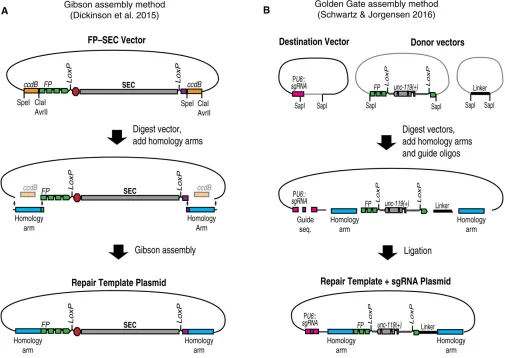
For long-range HDR, homology arms must be cloned into a vector to produce a plasmid repair template (Dickinson
et al. 2015). To simplify the process of cloning homology arms, we developed a cloning procedure based onccdB neg-ative selection (Figure 5) (Dickinson et al. 2015). Vectors carrying differentfluorescent protein–SEC modulesflanked byccdBmarkers are available via Addgene. To insert homol-ogy arms into one of these constructs, the vector is first digested with restriction enzymes to release the ccdB markers. Then, homology arms are inserted in place of the ccdB markers, using Gibson assembly (Gibson et al. 2009). Because ccdB is toxic to standard cloning strains of E. coli, only clones in which the ccdB markers have been replaced by the homology arms will grow. These clones can be identi-fied by direct sequencing, without screening for inserts.
Since any sequence can be cloned in place of the ccdB markers (Figure 5), this same basic cloning strategy can be used to build a repair template for any genome engineering project that will utilize SEC selection. To include additional modifications beyond the built-in FP tag, one simply needs to insert a larger DNA fragment comprising both the homology arm and any additional modifications in place of the ccdB marker. In general, any sequence located between the Cas9 cleavage site and the selectable marker is guaranteed to be cop-ied into the genome. Therefore, when designing complex ge-nome modifications, choose the Cas9 target site in such a way that the desired modifications lie between the cut site and the selectable marker.
An alternative, high-throughput method for assembling repair template plasmids was recently described by Schwartz and Jorgensen (2016; Figure 5B). Their approach, referred to as “SapTrap,” is based on the Golden Gate assembly method (Engleret al. 2008), which allows multiple DNA frag-ments to be joined together in a single reaction tube. SapTrap adds homology arms to pre-existing building blocks that can include various FP and epitope tags, selectable markers, and modules for sophisticated applications such as conditional tag-ging. The SapTrap destination vector also contains a second acceptor site for the guide sequence, eliminating the need to clone a separate sgRNA expression plasmid. A significant advantage of the modular SapTrap approach is that it allows a large variety of different repair constructs to be built by simply selecting the appropriate building blocks for a given application. The original SapTrap publication did not incorporate SEC selec-tion (Schwartz and Jorgensen 2016), but a SapTrap-compatible SEC module is under construction as of this writing.
Building more complex repair templates using Gibson assembly
In this cloning method, linear DNA fragments (most commonly PCR products) that overlap by 20–30 bp at their ends are co-valently joined together. The requisite 20- to 30-bp overlaps are easily incorporated into the PCR primers that are used to gen-erate the individual fragments. We prefer Gibson assembly over other cloning methods for two reasons:first, Gibson assembly does not require the addition of any extra sequences such as restriction sites or recombination targets; and second, up to six fragments can be assembled in a single step.
For researchers who are new to Gibson assembly, the following tips may be helpful:
1. We obtain the highest rates of successful assembly using fragments that overlap by 30 bp.
2. We often use PCR to amplify the vector backbone and include it as one of the fragments in the assembly. The most common cause of failure with this approach is large amounts of parent vector that carry through to the transformation. To avoid this, treat the vector PCR prod-uct withDpnI and gel purify it. If vector background still
persists, reduce the amount of plasmid template used in the PCR reaction that generates the vector backbone. 3. When gel purifying DNA fragments for use in a Gibson
assembly reaction, avoid using ethidium bromide or sim-ilar stains to visualize bands, since both ethidium bromide and UV radiation cause DNA damage that can significantly reduce cloning efficiencies. Instead, add 8mg/ml crystal violet to the agarose gel, which allows DNA bands to be visualized under ambient light without UV exposure. 4. Since Gibson assembly joins DNA fragments covalently,
1ml of a completed Gibson assembly reaction can be used as template for PCR to amplify the assembled product. We sometimes get better results by amplifying an assembled product and then ligating it into a vector, rather than in-cluding the vector directly in the assembly reaction. 5. If a multifragment Gibson assembly fails, try a sequential
Addressing Cas9 Specificity
The ability of Cas9 to recognize a specific target in the context of a complex genome is remarkable. Nevertheless, this specificity is not absolute; in mammalian systems andin vitro, Cas9 has been observed to cleave substrates that do not perfectly match the guide sequence (Jineket al.2012; Fuet al.2013; Hsuet al.2013; Pattanayaket al.2013). These results call for an appropriate degree of caution when using Cas9 as an experimental tool.
Two studies have examined Cas9 specificity inC. elegans via whole-genome sequencing of mutant animals (Chiuet al. 2013; Paixet al.2014), and two additional studies looked for evidence of off-target activity of Cas9 by sequencing candidate loci that closely matched the guide sequence (Dickinsonet al. 2013; Friedland et al.2013). None of these experiments de-tected any evidence ofbonafideoff-target mutations induced by Cas9, suggesting that inC. elegans, off-target mutations gen-erated by Cas9 are uncommon. However, the two whole-genome sequencing studies both found evidence of other
“passenger”mutations in CRISPR strains, at sites with no sequence similarity to the Cas9 target site. These second-site lesions are most likely spontaneous mutations that arose before or during strain construction. To avoid confounding effects of these passenger mutations on subsequent experi-ments, it should be standard practice to outcross mutant alleles and to isolate and characterize at least two indepen-dent alleles of every experimental genome modification. The ease and efficiency of Cas9-based approaches are such that isolating multiple alleles of each modification does not represent a significant burden. As variant Cas9 proteins rec-ognizing different PAMs become available (Bellet al.2015; Kleinstiver et al. 2015; Ranet al. 2015), the specificity of these enzymes will need to be carefully characterized.
When performing HDR, a second potential confounding issue is the incomplete or incorrect copying of the repair template into the genome. Rearrangements have been reported during homologous recombination from dsDNA templates (Berezikov et al.2004; Frøkjaer-Jensenet al.2008; Dickinsonet al.2013, 2015). Using plasmid repair templates, the frequency of rearrangements is5–10% that of recombinant alleles in our experience. With short-range HDR, the repair template may be incompletely copied into the genome. Partial copying appears to occur stochastically (Arribere et al. 2014; Ward 2015) but is more frequent at larger distances from the cut site. Point mutations can occur in the repair template, due to mis-takes in oligo synthesis (for the ssDNA oligo repair template), PCR errors (for dsDNA templates generated by PCR), or the DNA repair machinery responsible for HDR. Again, a straight-forward solution to all of these issues is to isolate and character-ize multiple independent alleles for each genome modification.
Recommended Strategies for Different Types of Modifications
In this section, we provide our recommendations for gener-ating common types of genome edits, taking into account all of
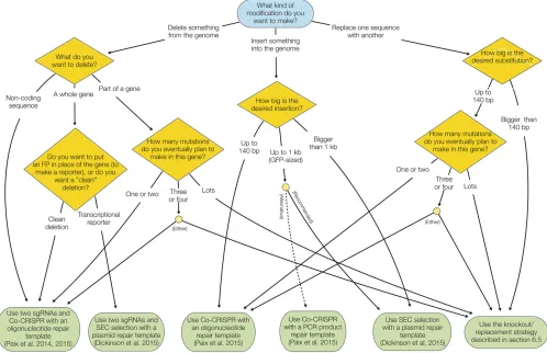
the information from the preceding sections. These recom-mendations are based on published information, but also reflect our personal preferences to some extent. There are now multiple valid strategies to generate most kinds of edits, and these recommendations should be taken only as our suggestions for“what to tryfirst.”Figure 6 shows aflow chart summarizing the recommendations.
Null mutations
A null mutation is a useful starting point for the analysis of almost any gene. By ascertaining the null phenotype of a gene, one establishes a basis for comparison when making targeted mutations later on. In addition, it is valuable to know the null phenotype of a gene when evaluating fluorescent protein knock-in strains: if a knock-in strain exhibits a phenotype similar to the null, this indicates that fusion to thefluorescent protein compromises the gene’s function. At least four differ-ent strategies can produce null (or strong loss-of-function) mutations:
1. Error-prone end-joining repair can be used to produce indels near the 59end of a gene, resulting in frameshift and early termination. This approach is useful for gener-ating tissue-specific phenotypes (Shen et al. 2014) and might be adaptable to high-throughput screening based on feeding (Liu et al. 2014). However, an end-joining event leaves the majority of the gene’s coding sequence intact, and thus it is difficult to guaranteea priorithat an indel mutation will be a bona fide null allele. Random indel mutations are also more difficult to screen for by PCR than HDR mutations (which can incorporate a restric-tion site to facilitate RFLP). Therefore, as a general rule, we prefer to use HDR to produce null mutations in which the entire coding sequence of a gene is deleted.
2. Paix et al.(2014, 2015) showed that gene-sized dele-tions could be generated by end joining, by using two sgRNAs that cut at opposite ends of the region to be deleted. More precise deletions could be generated by adding an ssDNA oligo with homology to the two ends of the deletion. In either case, the entire coding sequence of the gene is eliminated, which formally eliminates the possibility that any gene products will be produced. The same approach can also be used to delete portions of genes.
3. When afluorescent protein–SEC module is inserted at the 59end of a gene of interest, the SEC separates the pro-moter from the protein-coding sequence of the gene, resulting in a loss-of-function allele (Dickinsonet al. 2015). This loss-of-function allele is a useful intermediate in the construction of an N-terminal protein tag. However, N-terminalfluorescent protein–SEC insertions are not true genetic null mutations, in part because spontaneous SEC excision (resulting in expression of the gene of interest) occurs in certain tissues (Dickinsonet al.2015).
sequence of a gene with thefluorescent protein–SEC mod-ule. We have generated deletions of up to 9 kb using a single sgRNA and selection, but using two sgRNAs (one at each end of the region to be deleted) is expected to in-crease efficiency. The visible phenotype conferred by SEC makes it trivial to maintain the null allele as a heterozy-gote, which facilitates isolation and subsequent balanc-ing of null mutations in essential genes. SEC can be used to facilitate mutant isolation and balancing and then eliminated once a stable strain is in hand, yielding an allele in which the coding sequence of the gene of inter-est is replaced by a fluorescent protein. The resulting allele functions both as a null mutation and as a tran-scriptional reporter (promoter fusion).
Recommendation:Use two sgRNAs and an oligonucleotide repair template, with co-CRISPR screening, when a clean de-letion of a gene (or part of a gene) without insertion of any exogenous sequence is desired (Paix et al. 2015). Use the SEC-based strategy when insertion of afluorescent protein in place of the gene’s coding sequence is desired (Dickinson et al.2015).
Point mutations
By “point mutations”we mean substitutions, insertions, or deletions of one or a few amino acids that can be easily templated by an ssDNA oligo. Although a selection-based strategy with long-range HDR can produce point mutants (Dickinsonet al.2013), this approach is overkill since short-range HDR with co-CRISPR can efficiently produce point mu-tations with minimal need for PCR screening (Kim et al. 2014; Arribere et al. 2014). Also, the co-CRISPR strategy allows one to make substitutions in the middle of genes, where integration of a selectable marker could be problem-atic. Finally, a co-CRISPR approach could allow multiple point mutations to be produced simultaneously (Paixet al. 2015; Ward 2015).
Recommendation: For point mutations, use ssDNA-based HDR, with dpy-10, sqt-1, or pha-1 co-CRISPR and RFLP for screening (Arribereet al.2014; Paixet al.2015; Ward 2015).
Fluorescent protein fusions
HDR with a PCR product repair template and co-CRISPR (Arribere et al.2014; Kimet al.2014; Paixet al.2015). In our experience, these two approaches are very similar in terms of both the total time and hands-on labor required. The SEC strategy requires more work up front because of the need to clone homology arms in to the SEC vector, but the actual isolation of knock-in animals is easier. The co-CRISPR approach is quicker initially because the repair template is a PCR product and no cloning is required, but isolating knock-ins takes more work because PCR screening of 50–100 animals is needed. Thus, which strategy one chooses is largely a matter of personal preference. We prefer the SEC strategy in most cases, for two reasons:first, when used to generate N-terminal tags, the SEC-based strategy produces both a fluorescent protein fusion and a function mutation from a single injection step. The loss-of-function intermediate is useful because one can quickly determine whether the tagged protein is functional by com-paring the loss-of-function phenotype to the phenotype of the fluorescent protein fusion. Second, because it employs long-range HDR, the SEC strategy is insensitive to sgRNA effi-ciency and can produce insertions at a greater distance from the cut site, allowing moreflexibility in experimental design.
Recommendation:Use the SEC-based strategy forfluorescent protein fusions (Dickinsonet al.2015). We suggest making an N-terminal fusion unless there is a specific reason to choose a C-terminal fusion instead, because the process of generating an N-terminal fusion also yields a useful loss-of-function intermediate.
Other changes
Although the kinds of modifications above are the most common, they only scratch the surface of what is possible using Cas9-triggered homologous recombination. For exam-ple, simultaneous cutting on two chromosomes can produce custom translocations that function as balancer chromo-somes (Chenet al.2015). We can also imagine, for example, insertingLoxPor FLP recombinase target (FRT) sites to gen-erate conditional alleles or replacement of whole genes by their homologs from other species to probe evolutionary questions. The methods one chooses to use for these or other kinds of experiments will depend on the details of the experiment, but as a general rule, we suggest that short-range HDR with co-CRISPR screening be used for all modifications that can be templated by an ssDNA oligo, while long-range HDR with SEC selection is best suited for making larger changes. SEC or other selectable markers can be incorporated into custom repair templates, using Gibson assembly.
A general strategy for dissecting gene function
A common task for any protein of interest is to determine how different domains, binding sites, sequence motifs, or other features contribute to the function of the protein as a whole. Often this involves making many different mutants in a gene
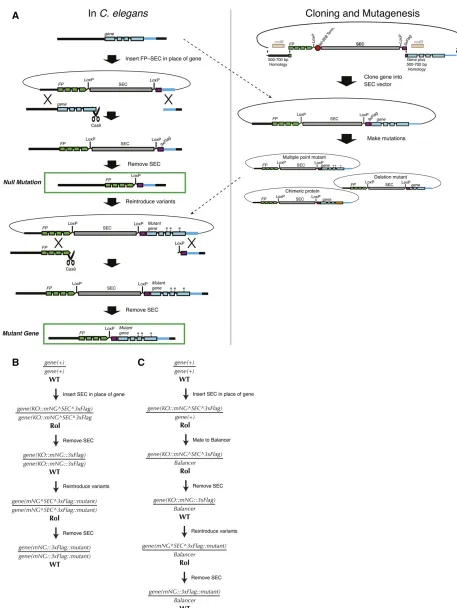
of interest and assaying their function.C. elegansis especially well suited to such“structure–function”analysis because of its short generation time, rich cell biology, defined lineage, and now with CRISPR, the ease of generating many mutants in the endogenous locus, without the need for overexpression. We have devised a simple, general strategy for performing structure–function analysis of C. elegans genes, which we demonstrated in Daset al.(2015) and describe here.
Briefly, we begin by making a null mutation and then reinsert either a wild-type or a mutant version of the gene of interest at the endogenous locus (Figure 7). The advantage of this strategy is that one can generate variants of the gene of interest in vitro, using standard cloning procedures, rather than designing a new CRISPR approach to produce each var-iant. Once the initial null allele is in hand, the same sgRNA, homology arms, and screening strategy can be used to reinsert each variant back into the endogenous locus. The phenotype of each variant can then be examined and compared directly to the null.
We generate the null mutation by inserting afluorescent protein–SEC module in place of the coding sequence (see Null mutationssection, strategy 4) and then removing SEC. In principle, the null allele could also be generated by co-CRISPR with two sgRNAs and an oligonucleotide repair tem-plate. In parallel, we clone the genomic sequence of the gene of interest into an SEC-containing vector to generate a rescue construct. Mutations can be made to this rescue construct, using standard cloning techniques such as site-directed mu-tagenesis. If the gene of interest is nonessential, the (possibly mutated) rescue construct can be introduced directly into the homozygous null mutant in a second homologous recombi-nation step. To generate multiple variant versions of a gene, one needs only to repeat the second recombination step for each variant. Figure 7, A and B, shows a detailed schematic of this procedure.
For essential genes, the workflow requires only one simple modification (Figure 7C). After generating the null allele, we mate it to a balancer and then remove the SEC. Variant versions of the gene are then introduced directly into the bal-anced null mutant background. By using an sgRNA targeting thefluorescent protein present in the null allele, we ensure that recombination occurs only on the null chromosome and not on the balancer. The resulting variant, like the parent null allele, is immediately balanced; any phenotypic assays are done using the fraction of progeny that have lost the balancer and are homozygous mutant.
Where to Go for More Information and Detailed Protocols
Acknowledgments
We thank the members of the Goldstein laboratory, espe-cially Ariel Pani, Jennifer Heppert, and Christopher Higgins, for helpful discussions. We are also grateful to Geraldine Seydoux for sharing results prior to publication and to Jordan Ward for helpful discussions. We thank Amy Maddox, Geraldine Seydoux, and Jordan Ward for com-ments on the manuscript. Our work on CRISPR methods development was made possible by National Institutes of Health (NIH) grant T32 CA009156 and a Howard Hughes postdoctoral fellowship from the Helen Hay Whitney Foun-dation (to D.J.D.) and by NIH grant R01 GM083071 and National Science Foundation grant IOS 0917726 (to B.G.).
Literature Cited
Arribere, J. A., R. T. Bell, B. X. H. Fu, K. L. Artiles, P. S. Hartman
et al., 2014 Efficient marker-free recovery of custom genetic modifications with CRISPR/Cas9 in Caenorhabditis elegans. Genetics 198: 837–846.
Bell, R. T., B. X. H. Fu, and A. Z. Fire, 2015 Cas9 variants expand the target repertoire inCaenorhabditis elegans. Genetics 202: 381–388.
Berezikov, E., C. I. Bargmann, and R. H. A. Plasterk,
2004 Homologous gene targeting in Caenorhabditis elegans
by biolistic transformation. Nucleic Acids Res. 32: e40. Carbone, A., A. Zinovyev, and F. Képès, 2003 Codon adaptation
index as a measure of dominating codon bias. Bioinformatics 19: 2005–2015.
Chen, B., L. A. Gilbert, B. A. Cimini, J. Schnitzbauer, W. Zhanget al., 2013 Dynamic imaging of genomic loci in living human cells by an optimized CRISPR/Cas system. Cell 155: 1479–1491. Chen, C., L. A. Fenk, and M. de Bono, 2013 Efficient genome
editing in Caenorhabditis elegans by CRISPR-targeted homolo-gous recombination. Nucleic Acids Res. 41: e193.
Chen, X., M. Li, X. Feng, and S. Guang, 2015 Targeted chromo-somal translocations and essential gene knockout using CRISPR/Cas9 technology in Caenorhabditis elegans. Genetics 201: 1295–1306.
Chiu, H., H. T. Schwartz, I. Antoshechkin, and P. W. Sternberg, 2013 Transgene-free genome editing inCaenorhabditis elegans
using CRISPR-Cas. Genetics 195: 1167–1171.
Cho, S. W., J. Lee, D. Carroll, J.-S. Kim, and J. Lee, 2013 Heritable gene knockout in Caenorhabditis elegansby direct injection of Cas9-sgRNA ribonucleoproteins. Genetics 195: 1177–1180.
Cong, L., F. A. Ran, D. Cox, S. Lin, R. Barretto et al.,
2013 Multiplex genome engineering using CRISPR/Cas
systems. Science 339: 819–823.
Das, A., D. J. Dickinson, C. C. Wood, B. Goldstein, and K. C.
Slep, 2015 Crescerin uses a TOG domain array to
regu-late microtubules in the primary cilium. Mol. Biol. Cell 26: 4248–4264.
Dickinson, D. J., J. D. Ward, D. J. Reiner, and B. Goldstein,
2013 Engineering the Caenorhabditis elegans genome using
Cas9-triggered homologous recombination. Nat. Methods 10: 1028–1034.
Dickinson, D. J., A. M. Pani, J. K. Heppert, C. D. Higgins, and
B. Goldstein, 2015 Streamlined genome engineering with
a self-excising drug selection cassette. Genetics 200: 1035–1049.
Doench, J. G., E. Hartenian, D. B. Graham, Z. Tothova, M. Hegde
et al., 2014 Rational design of highly active sgRNAs for
CRISPR-Cas9–mediated gene inactivation. Nat. Biotechnol. 32: 1262–1267.
Engler, C., R. Kandzia, and S. Marillonnet, 2008 A pot, one-step, precision cloning method with high throughput capability. PLoS One 3: e3647.
Farboud, B., and B. J. Meyer, 2015 Dramatic enhancement of
genome editing by CRISPR/Cas9 through improved guide RNA design. Genetics 199: 959–971.
Friedland, A. E., Y. B. Tzur, K. M. Esvelt, M. P. Colaiácovo, G. M. Churchet al., 2013 Heritable genome editing in C. elegans via a CRISPR-Cas9 system. Nat. Methods 10: 741–743.
Frøkjaer-Jensen, C., M. W. Davis, C. E. Hopkins, B. J. Newman, J. M. Thummelet al., 2008 Single-copy insertion of transgenes in Caenorhabditis elegans. Nat. Genet. 40: 1375–1383. Frøkjaer-Jensen, C., M. W. Davis, G. Hollopeter, J. Taylor, T. W.
Harriset al., 2010 Targeted gene deletions in C. elegans using transposon excision. Nat. Methods 7: 451–453.
Frøkjaer-Jensen, C., M. W. Davis, M. Ailion, and E. M. Jorgensen,
2012 Improved Mos1-mediated transgenesis in C. elegans.
Nat. Methods 9: 117–118.
Fu, Y., J. A. Foden, C. Khayter, M. L. Maeder, D. Reyon et al.,
2013 High-frequency off-target mutagenesis induced by
CRISPR-Cas nucleases in human cells. Nat. Biotechnol. 31: 822–826.
Gibson, D. G., L. Young, R.-Y. Chuang, J. C. Venter, C. A. Hutchison
et al., 2009 Enzymatic assembly of DNA molecules up to sev-eral hundred kilobases. Nat. Methods 6: 343–345.
Greiss, S., and J. W. Chin, 2011 Expanding the genetic code of an animal. J. Am. Chem. Soc. 133: 14196–14199.
Hsu, P. D., D. A. Scott, J. A. Weinstein, F. A. Ran, S. Konermann
et al., 2013 DNA targeting specificity of RNA-guided Cas9 nucleases. Nat. Biotechnol. 31: 827–832.
Hsu, P. D., E. S. Lander, and F. Zhang, 2014 Development and applications of CRISPR-Cas9 for genome engineering. Cell 157: 1262–1278.
Jiang, W., D. Bikard, D. Cox, F. Zhang, and L. A. Marraffini,
2013 RNA-guided editing of bacterial genomes using
CRISPR-Cas systems. Nat. Biotechnol. 31: 233–239.
Jinek, M., K. Chylinski, I. Fonfara, M. Hauer, J. A. Doudna et al.,
2012 A programmable dual-RNA-guided DNA endonuclease in
adaptive bacterial immunity. Science 337: 816–821.
Katic, I., and H. Großhans, 2013 Targeted heritable mutation and gene conversion by Cas9-CRISPR in Caenorhabditis elegans. Genetics 195: 1173–1176.
Katic, I., L. Xu, and R. Ciosk, 2015 CRISPR/Cas9 genome editing inCaenorhabditis elegans: evaluation of templates for homology-mediated repair and knock-ins by homology-independent DNA repair. G3 5: 1649–1656.
Kim, H., T. Ishidate, K. S. Ghanta, M. Seth, D. Conteet al., 2014 A co-CRISPR strategy for efficient genome editing inCaenorhabditis elegans. Genetics 197: 1069–1080.
Kleinstiver, B. P., M. S. Prew, S. Q. Tsai, V. V. Topkar, N. T. Nguyen
et al., 2015 Engineered CRISPR-Cas9 nucleases with altered PAM specificities. Nature 523: 481–485.
Kuscu, C., S. Arslan, R. Singh, J. Thorpe, and M. Adli, 2014 Genome-wide analysis reveals characteristics of off-target sites bound by the Cas9 endonuclease. Nat. Biotechnol. 32: 677–683.
Liu, H., Z. Wei, A. Dominguez, Y. Li, X. Wang et al.,
2015 ERA: a comprehensive design tool for
CRISPR-mediated gene editing, repression and activation. Bioinformatics 31: 3676–3678.
Liu, P., L. Long, K. Xiong, B. Yu, N. Changet al., 2014 Heritable/ conditional genome editing in C. elegans using a CRISPR-Cas9 feeding system. Cell Res. 24: 886–889.
Lo, T.-W., C. S. Pickle, S. Lin, E. J. Ralston, M. Gurling et al.,
CRISPR/Cas9 to engineer precise insertions and deletions in evolutionarily diverse nematode species. Genetics 195: 331– 348.
Mello, C. C., J. M. Kramer, D. Stinchcomb, and V. Ambros, 1991 Efficient gene transfer in C.elegans: extrachromosomal maintenance and integration of transforming sequences. EMBO J. 10: 3959–3970.
Norris, A. D., H.-M. Kim, M. P. Colaiácovo, and J. A. Calarco, 2015 Efficient genome editing inCaenorhabditis eleganswith a toolkit of dual-marker selection cassettes. Genetics 201: 449– 458.
Paix, A., Y. Wang, H. E. Smith, C.-Y. S. Lee, D. Calidas et al., 2014 Scalable and versatile genome editing using linear DNAs with microhomology to Cas9 sites in Caenorhabditis elegans. Genetics 198: 1347–1356.
Paix, A., A. Folkmann, D. Rasoloson, and G. Seydoux, 2015 High efficiency, homology-directed genome editing inCaenorhabditis elegans using CRISPR-Cas9 ribonucleoprotein complexes. Genetics 201: 47–54.
Pattanayak, V., S. Lin, J. P. Guilinger, E. Ma, J. A. Doudnaet al., 2013 High-throughput profiling of off-target DNA cleavage
reveals RNA-programmed Cas9 nuclease specificity. Nat.
Biotechnol. 31: 839–843.
Praitis, V., E. Casey, D. Collar, and J. Austin, 2001 Creation of low-copy integrated transgenic lines in Caenorhabditis elegans. Genetics 157: 1217–1226.
Pulak, R., 2006 Techniques for analysis, sorting, and dispensing of C. elegans on the COPASflow-sorting system. Methods Mol. Biol. 351: 275–286.
Ran, F. A., P. D. Hsu, C.-Y. Lin, J. S. Gootenberg, S. Konermann
et al., 2013 Double nicking by RNA-guided CRISPR Cas9 for enhanced genome editing specificity. Cell 154: 1380–1389. Ran, F. A., L. Cong, W. X. Yan, D. A. Scott, J. S. Gootenberget al.,
2015 In vivo genome editing using Staphylococcus aureus
Cas9. Nature 520: 186–191.
Ren, X., Z. Yang, J. Xu, J. Sun, D. Mao et al., 2014 Enhanced specificity and efficiency of the CRISPR/Cas9 system with opti-mized sgRNA parameters in Drosophila. Cell Rep. 9: 1151–1162. Robert, V., and J.-L. Bessereau, 2007 Targeted engineering of the Caenorhabditis elegans genome following Mos1-triggered chro-mosomal breaks. EMBO J. 26: 170–183.
Schwartz, M. L., and E. M. Jorgensen, 2016 SapTrap, a Toolkit for
High-Throughput CRISPR/Cas9 Gene Modification in
Caeno-rhabditis elegans. Genetics DOI: genetics.115.184275.
Shen, Z., X. Zhang, Y. Chai, Z. Zhu, P. Yiet al., 2014 Conditional knockouts generated by engineered CRISPR-Cas9 endonuclease reveal the roles of Coronin in C. elegans neural development. Dev. Cell 30: 625–636.
Sternberg, S. H., S. Redding, M. Jinek, E. C. Greene, and J. A.
Doudna, 2014 DNA interrogation by the CRISPR RNA-guided
endonuclease Cas9. Nature 507: 62–67.
Sullivan-Brown, J. L., P. Tandon, K. E. Bird, D. J. Dickinson, S. C. Tintori et al., 2016 Identifying regulators of morphogenesis common to vertebrate neural tube closure and Caenorhabditis elegansgastrulation. Genetics 202: 123–139.
van Schendel, R., S. F. Roerink, V. Portegijs, S. van den Heuvel, and M. Tijsterman, 2015 PolymeraseQis a key driver of genome evolution and of CRISPR/Cas9-mediated mutagenesis. Nat. Commun. 6: 7394.
Waaijers, S., V. Portegijs, J. Kerver, B. B. L. G. Lemmens, M. Tijsterman
et al., 2013 CRISPR/Cas9-targeted mutagenesis inCaenorhabditis elegans. Genetics 195: 1187–1191.
Wang, T., J. J. Wei, D. M. Sabatini, and E. S. Lander, 2014 Genetic screens in human cells using the CRISPR-Cas9 system. Science 343: 80–84.
Ward, J. D., 2015 Rapid and precise engineering of theCaenorhabditis elegansgenome with lethal mutation co-conversion and inac-tivation of NHEJ repair. Genetics 199: 363–377.
Wu, X., D. A. Scott, A. J. Kriz, A. C. Chiu, P. D. Hsu et al.,
2014 Genome-wide binding of the CRISPR endonuclease
Cas9 in mammalian cells. Nat. Biotechnol. 32: 670–676.
Xu, H., T. Xiao, C.-H. Chen, W. Li, C. A. Meyer et al.,
2015 Sequence determinants of improved CRISPR sgRNA
design. Genome Res.25:1147–1157.
Zetsche, B., J. S. Gootenberg, O. O. Abudayyeh, I. M. Slaymaker, K. S. Makarovaet al., 2015 Cpf1 is a single RNA-guided en-donuclease of a class 2 CRISPR-Cas system. Cell 163: 759–771. Zhao, P., Z. Zhang, H. Ke, Y. Yue, and D. Xue, 2014 Oligonucleotide-based targeted gene editing in C. elegans via the CRISPR/Cas9 system. Cell Res. 24: 247–250.