Cationic Peptides as Feed Additives during the First Week Posthatch
Michael H. Kogut,aKenneth J. Genovese,aHaiqi He,aChristina L. Swaggerty,aYiwei Jiangb
USDA-ARS, SPARC, College Station, Texas, USAa; My Galaxy LLC, Ft. Worth, Texas, USAb
We have been investigating modulation strategies tailored around the selective stimulation of the host’s immune system as an
alternative to direct targeting of microbial pathogens by antibiotics. One such approach is the use of a group of small cationic
peptides (BT) produced by a Gram-positive soil bacterium,
Brevibacillus texasporus
. These peptides have immune modulatory
properties that enhance both leukocyte functional efficiency and leukocyte proinflammatory cytokine and chemokine mRNA
transcription activities
in vitro
. In addition, when provided as a feed additive for just 4 days posthatch, BT peptides significantly
induce a concentration-dependent protection against cecal and extraintestinal colonization by
Salmonella enterica
serovar
En-teritidis. In the present studies, we assessed the effects of feeding BT peptides on transcriptional changes on proinflammatory
cytokines, inflammatory chemokines, and Toll-like receptors (TLR) in the ceca of broiler chickens with and without
S
.
Enteriti-dis infection. After feeding a BT peptide-supplemented diet for the first 4 days posthatch, chickens were then challenged with
S
.
Enteritidis, and intestinal gene expression was measured at 1 or 7 days postinfection (p.i.) (5 or 11 days of age). Intestinal
expres-sion of innate immune mRNA transcripts was analyzed by quantitative real-time PCR (qRT-PCR). Analysis of relative mRNA
expression showed that a BT peptide-supplemented diet did not directly induce the transcription of proinflammatory cytokine,
inflammatory chemokine, type I/II interferon (IFN), or TLR mRNA in chicken cecum. However, feeding the BT
peptide-supple-mented diet primed cecal tissue for increased (
P
<
0.05) transcription of TLR4, TLR15, and TLR21 upon infection with
S
.
Enteri-tidis on days 1 and 7 p.i. Likewise, feeding the BT peptides primed the cecal tissue for increased transcription of
proinflamma-tory cytokines (interleukin 1
[IL-1], IL-6, IL-18, type I and II IFNs) and inflammatory chemokine (CxCLi2) in response to
S
.
Enteritidis infection 1 and 7 days p.i. compared to the chickens fed the basal diet. These small cationic peptides may prove useful
as alternatives to antibiotics as local immune modulators in neonatal poultry by providing prophylactic protection against
Sal-monella
infections.
R
esearch efforts in our laboratory have focused on developing
immunoprophylactic strategies (
1–4
) and selective genetics
(
5
) that prevent or control intestinal
Salmonella enterica
organ
and intestinal colonization in poultry. Specifically, our research
has concentrated on upregulating the innate immune response in
chickens during the immunologically inefficient first week
post-hatch (
6
).
Recently, a novel Gram-positive bacterium,
Brevibacillus
tex-asporus
(ATCC PTA-5854), was isolated from the soil and found
to produce BT, a group of structurally related cationic peptides
(
7
). BT peptides were found to be highly efficacious against a
natural outbreak of colibacillosis in broiler chickens based on
im-proved performance and reduced mortality in comparison with
unmedicated birds at a level (12 ppm) that was below the MIC for
Escherichia coli
(
8
).
In vitro
, BT displays efficient bactericidal
ac-tivity against Gram-positive bacteria (MIC of 1 ppm) but a
re-duced efficacy against Gram-negative bacteria (MIC of
⬎
20
ppm). Interestingly, orally delivered BT seems to completely lack
direct antibacterial activities (12 ppm) (
8
). In addition, chickens
given BT as a feed additive for the first 4 days posthatch provided
protection against both cecal colonization and extraintestinal
Sal-monella enterica
serovar Enteritidis infections in a
concentration-dependent manner and induced the upregulation of peripheral
blood heterophil (the avian equivalent to the mammalian
neutro-phil) and monocyte functional activities (
4
,
9
).
The exact mechanisms of interaction between BT peptides and
the cecum in chickens have not been determined. Therefore, the
objective of the present experiments was to assess the effects of
feeding BT peptides for the first 4 days posthatch on
transcrip-tional changes on proinflammatory cytokines, inflammatory
chemokines, and Toll-like receptors (TLR) in the ceca of broiler
chickens with or without infection with
S
. Enteritidis.
MATERIALS AND METHODS
Experimental animals.Experiments were conducted according to the regulations established by the U.S. Department of Agriculture Animal Care and Use Committee. Broiler chickens used in this study were ob-tained from a commercial breeder and were all of the same genetic back-ground. Chicks were placed in floor pens containing wood shavings, pro-vided supplemental heat, water, and a balanced, unmedicated corn and soybean meal-based chick starter dietad libitumthat met or exceeded the levels of critical nutrients recommended by the National Research Coun-cil (10).Salmonellawas not detected in the feed or from the paper tray liners.
Partial purification of BT.B. texasporusE58 cells were grown in 1 liter of lysogeny broth (LB) in an air shaker at 37°C for 3 days. The culture was spun in a clinical centrifuge at 3,000⫻gfor 15 min. The supernatant was collected, and 500 g of ammonium sulfate was added and dissolved. The
Received15 May 2013Returned for modification12 June 2013
Accepted12 July 2013
Published ahead of print17 July 2013
Address correspondence to Michael H. Kogut, [email protected].
Copyright © 2013, American Society for Microbiology. All Rights Reserved.
doi:10.1128/CVI.00322-13
on August 17, 2020 by guest
http://cvi.asm.org/
sample was spun in the clinical centrifuge at 3,000⫻gfor 15 min. The pellet was dissolved in 200 ml of distilled water. The solution was then boiled for 15 min and cooled on ice. The sample was filtered with a 0.2-mm-pore-size filter (Nalgene Inc., Rochester, NY). The filtrate was mixed with 0.2 liters of chloroform at room temperature for 20 min with a stir bar. The mixture was separated into two phases by centrifugation in the clinical centrifuge at 3,000⫻gfor 15 min. The organic phase was collected and dried in a vacuum evaporator as described previously (7).
Premix preparation.The dried material was dissolved in ethanol, and the BT concentration was analyzed for anti-Staphylococcus aureusactivity by standard microdilution methods as described previously (MIC⫽0.8 g/ml) (7). The solution was sprayed onto cornmeal and then dried. The resulting material was used as a premix for the chicken studies.
S. Enteritidis challenge.An isolate ofS. Enteritidis was obtained from NVSL (Ames, IA) (ID 9711771, part 24). The isolate was selected for resistance to novobiocin and carbenicillin (NO-CN) and was maintained in tryptic soy broth or tryptic soy agar at 40°C. Brilliant green agar (BGA), a selective culture medium forSalmonella, was used to culture the resis-tant isolate in experimental studies and contained 100 mg/ml CN and 25 mg/ml NO to inhibit growth of other bacteria (BGA–NO-CN). Inoculum for challenge was prepared from 18- to 24-h-grown tryptic soy broth, and NO-CN cultures were maintained at 39°C and diluted in sterile phos-phate-buffered saline (PBS) (pH 7.2). A stock solution (1⫻109CFU/ml) was prepared, and bacterial concentration was determined spectrophoto-metrically using a standard curve at a reference wavelength of 625 nm.
Experimental challenge design.One-day-old broiler chickens were randomly distributed into four experimental groups (group 1, control diet, uninfected; group 2, control diet, infected withS. Enteritidis; group 3, BT-supplemented diet, uninfected; group 4, BT-supplemented diet, infected withS. Enteritidis). Each group contained 30 birds fed a bal-anced, unmedicated corn and soybean meal-based diet that contained either 0 (control) or 24 ppm BT for 4 days. On the fifth day after hatch, all BT feed was removed and replaced with control diet feed for the remain-der of the experiment. In addition, on the fourth day after hatch, all chick-ens were orally challenged with either 5⫻106CFU/mlS. Enteritidis or mock challenged with sterile PBS. One and 7 days after challenge (5 and 11 days after hatch), 15 chickens from each group were killed by cervical dislocation, cecal contents were analyzed forS. Enteritidis colonization, and cecal tonsils were collected for quantitative real-time PCR (qRT-PCR). All experiments were conducted three times. Therefore, the ceca from a total of 45 chickens for each of the 4 groups (15 chickens each in 3 experiments) were used to prepare the mRNA for the qRT-PCR assays described below. RNA from each bird (n⫽45) was isolated and assayed separately and not pooled. Each RNA sample was replicated 3 times per immune gene per experiment).
Sample collection for bacterial counts.The ceca from each chicken was removed aseptically, and the contents (0.25 g) were serially diluted to 1:100, 1:1,000, or 1:10,000 and spread onto BGA–NO-CN plates. The plates were incubated at 37°C for 24 h, and the number of NO-CN-resis-tantS. Enteritidis cells per gram of cecal contents was determined. The data from each experimental group were pooled from three separate trials for statistical analysis.
Sample collection for mRNA. Chickens from each experimental group were euthanized at either 1 or 7 days postchallenge. A 25-mg piece of tissue was removed from the cecal tonsils. The tissue was washed in PBS and placed in a 2-ml microcentrifuge tube with 1 ml of RNAlater (Qiagen Inc., Valencia, CA) and stored at⫺20°C until processed.
RNA isolation.Tissues were removed from RNAlater and processed using the RNeasy minikit (Qiagen Inc.) according to the manufacturer’s protocol, where tissues were placed in 600 ml of buffer RLT and homog-enized using a handheld TissueRuptor (Qiagen Inc.), and total RNA was eluted in 50l of DNase-free water and stored at⫺80°C. RNA was quan-tified using a spectrophotometer (NanoDrop Products, Wilmington, DE). The data from these three repeated experiments were pooled for
presentation and statistical analysis. Total RNA (300 ng) from each sam-ple was prepared.
Quantitative real-time PCR.Primer and probe sets for the cytokines and 28S rRNA were designed using the Primer Express software program (PE Applied Biosystems, Foster City, CA).
Cytokine and chemokine mRNA expression was quantitated using a well-described method. Primers and probes for cytokines, chemokines, and 28S rRNA-specific amplification have been described (11,12) and are provided inTable 1. The qRT-PCR was performed using the TaqMan fast universal PCR master mix and one-step RT-PCR master mix reagents. Amplification and detection of specific products were performed using the Applied Biosystems 7500 Fast real-time PCR system with the follow-ing cycle profile: one cycle of 48°C for 30 min and 95°C for 20 s and 40 cycles of 95°C for 3 s and 60°C for 30 s. Quantification was based on the increased fluorescence detected by the 7500 Fast sequence detection sys-tem due to hydrolysis of the target-specific probes by the 5=nuclease activity of the rTthDNA polymerase during PCR amplification. Normal-ization was carried out against 28S rRNA, which was used as a housekeep-ing gene. To correct for differences in RNA levels between samples within the experiment, the correction factor for each sample was calculated by dividing the mean threshold cycle (CT) value for 28S rRNA-specific prod-uct for each sample by the overall meanCTvalue for the 28S rRNA-specific product from all samples. The corrected cytokine mean was cal-culated as follow: average of each replicate⫻cytokine slope/28S slope⫻ 28S correction factor.
Statistical analysis.The mean and standard error of the mean were calculated for 40CTvalues for each of the 4 treatment groups. Differences between group 1, group 2, group 3, and group 4 were determined by analysis of variance. Significant differences were further separated using Duncan’s multiple range test. Fold changes in RNA levels were calculated from mean 40 CTvalues by the formula 2(40CTin groups 2, 3, or 4⫺40CTin group 1). APvalue of ⬍0.05 was considered statistically significant.
RESULTS
Proinflammatory cytokine mRNA expression.
The effect of
feed-ing BT peptides to young chickens on the expression of
proinflam-matory cytokine mRNA expression in the cecum on day 5
post-hatch (1 day after removal of BT peptide-supplemented feed) is
shown in
Fig. 1A
.
S
. Enteritidis infection in chickens on the
con-trol diet (SE
⫹/BT
⫺) induced a small but significant (
P
ⱕ
0.05)
upregulation of IL-1

, IL-6, and IL-15. BT-supplemented feed
(SE
⫺/BT
⫹) had no direct effect on proinflammatory cytokine
mRNA expression in the cecum. BT-supplemented diet appeared
to “prime” the intestine for a significant synergistic upregulation
of all proinflammatory cytokines except for IL-15 following
infec-tion with
S
. Enteritidis (SE
⫹/BT
⫹).
The effect of feeding BT peptides to young chickens on the
expression of proinflammatory cytokine mRNA expression in the
cecum on day 11 posthatch (7 days after removal of BT
peptide-supplemented feed) is shown in
Fig. 1B
.
S
. Enteritidis cecal
colo-nization for 7 days in chickens on the control diet (SE
⫹/BT
⫺)
stimulated a significant (
P
ⱕ
0.05) upregulation of IL-1

, IL-6,
and IL-18. BT-supplemented feed (SE
⫺/BT
⫹) had no direct effect
on proinflammatory cytokine mRNA expression in the cecum.
BT-supplemented diet maintained a priming effect in the intestine
for at least a week after removal, as evidenced by the significant
upregulation of all proinflammatory cytokines during a persistent
infection with
S
. Enteritidis (SE
⫹/BT
⫹).
Inflammatory chemokine mRNA expression.
The effect of
feeding BT peptides to young chickens on inflammatory
chemo-kine mRNA expression in the cecum on days 5 or 11 posthatch (1
or 7 days after removal of BT peptide-supplemented feed,
respec-tively) is shown in
Fig. 2A
and
B
. BT-supplemented feed (SE
⫺/
on August 17, 2020 by guest
http://cvi.asm.org/
BT
⫹) had no direct effect on chemokine mRNA expression in the
cecum.
S
. Enteritidis infection in chickens on the control diet
(SE
⫹/BT
⫺) induced a significant (
P
ⱕ
0.05) upregulation of
CXCLi2 at 1 and 7 days after infection (days 5 and 11 posthatch).
However, in
S
. Enteritidis infection in chickens on the control diet
(SE
⫹/BT
⫺), no effect was observed on the expression of CXCLi1
(
Fig. 2A
), but there was a small but significant increase in CXCLi1
by 7 days after infection (
Fig. 2B
). Likewise, BT-supplemented
diet had a priming effect on the cecum, resulting in a significant
upregulation of CXCLI2 1 and 7 days after infection with
S
.
En-teritidis (SE
⫹/BT
⫹) (
Fig. 2B
). BT-supplemented diet had a
prim-ing effect on CXCLi1 only on day 7 postinfection (
Fig. 2B
).
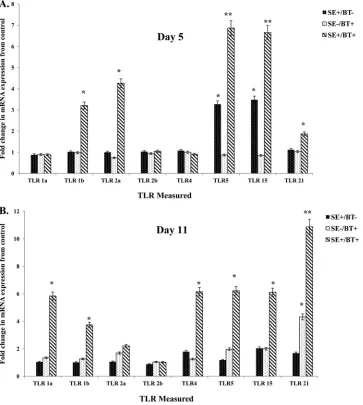
Toll-like receptor mRNA expression.
The effect of BT
pep-tides on TLR expression in cecum 5 days posthatch (1 day after
removal of BT peptide-supplemented feed) is shown in
Fig. 3A
.
S
.
Enteritidis infection in chickens on the control diet (SE
⫹/BT
⫺)
induced a significant upregulation in expression of TLR5 and
TLR15. BT-supplemented feed (SE
⫺/BT
⫹) had no significant,
di-rect effect on TLR mRNA expression in the cecum.
BT-supple-mented diet appeared to “prime” the intestine for a significant
(
P
ⱕ
0.05) synergistic upregulation of TLR1b, TLR2a, TLR5,
TLR15, and TLR21 following infection with
S
. Enteritidis (SE
⫹/
BT
⫹).
The effect of feeding BT peptides to young chickens on the
expression of TLR mRNA in the cecum on day 11 posthatch (7
days after removal of BT peptide-supplemented feed) is shown in
Fig. 3B
.
S
. Enteritidis cecal colonization for 7 days in chickens on
the control diet (SE
⫹/BT
⫺) did not upregulate the expression of
TLR mRNA. BT-supplemented feed (SE
⫺/BT
⫹) had no direct
ef-fect on TLR mRNA expression in the cecum except for TLR21.
BT-supplemented diet induced a priming effect in the cecum for
at least a week after removal, as evidenced by the significant
up-regulation of all TLR mRNA (except TLR2a and TLR2b) during a
persistent infection with
S
. Enteritidis (SE
⫹/BT
⫹).
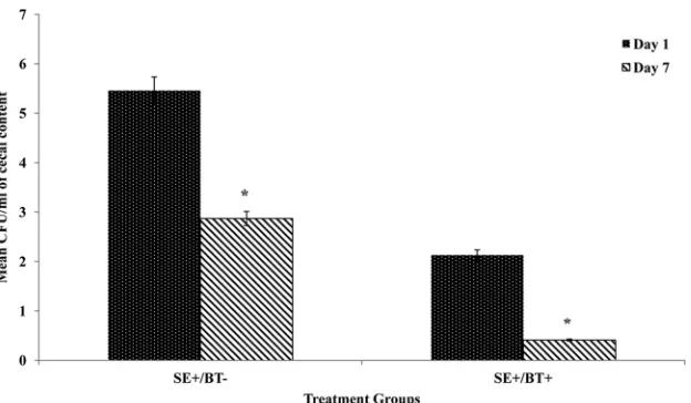
S
. Enteritidis cecal colonization.
The effect of BT peptide on
S
.
Enteritidis cecal colonization of chickens 1 and 7 days
postchal-lenge is shown in
Fig. 4
. The number of
S
. Enteritidis CFU/ml in
the cecal contents of chickens in BT-fed groups was significantly
less (
P
ⱕ
0.01) than in the control diet-fed group at both 1 and 7
days postchallenge. Similarly, the percentage of
S
. Enteritidis cecal
culture-positive chickens among the BT-fed birds was
signifi-cantly less than that among the control diet-fed chickens (10%
TABLE 1Real-time quantitative RT-PCR probes and primers for proinflammatory cytokines, inflammatory chemokines, and type I and II interferons
RNA target Sequence typeb Probe/primer sequencec Accession no.a
28S Probe 5=-(FAM)-AGGACCGCTACGGACCTCCACCA-(TAMRA)-3= X59733
F 5=-GGCGAAGCCAGAGGAAACT-3=
R 5=-GACGACCGATTGCACGTC-3=
IL-1 Probe 5=-(FAM)-CCACACTGCAGCTGGAGGAAGCC-(TAMRA)-3= AJ245728
F 5=-GCTCTACATGTCGTGTGTGATGAG-3=
R 5=-TGTCGATGTCCCGCATGA-3=
IL-6 Probe 5=-(FAM)-AGGAGAAATGCCTGACGAAGCTCTCCA-TAMRA)-3= AJ250838
F 5=-GCTCGCCGGCTTCGA-3=
R 5=-GGTAGGTCTGAAAGGCGAACAG-3=
IL-15 Probe 5=-(FAM)-CCACCCAATCCAGGAAATGTTAACCCA-(TAMRA)-3= NM_204571.1
F 5=-AGCTGAACTGCTGCCACATTT-3=
R 5=-TTTCCTCTGTTCTTCTTTGTCTGAATC-3=
IL-18 Probe 5=-(FAM)-CCGCGCCTTCAGCAGGGATG-(TAMRA)-3= AJ416937
F 5=-AGGTGAAATCTGGCAGTGGAAT-3=
R 5=-ACCTGGACGCTGAATGCAA-3=
IFN-␥ Probe 5=-(FAM)-TGGCCAAGCTCCCGATGAACGA-(TAMRA)-3= YO7922
F 5=-GTGAAGAAGGTGAAAGATATATCATGGA-3=
R 5=-GCTTTGCGTGGATTCTCA-3=
IFN-␣ Probe 5=-(FAM)-CTCAACCGGATCCACCGCTACACG-(TAMRA)-3= U07868
F 5=-GACAGCCAACGCCAAAGC-3=
R 5=-GTCGCTGCTGTCCAAGCATT-3=
CXCLi1 Probe 5=-(FAM)-CCACATTCTTGCAGTGAGGTCCGCT-(TAMRA)-3= AF277660
F 5=-CCAGTGCATAGAGACTCATTCCAAA-3=
R 5=-TGCCCATCTTTCAGAGTAGCTATGAACT-3=
CXCLi2 Probe 5=-(FAM)-CTTTACCAGCGCGTCCTACCTTGCGACA-(TAMRA)-3= AJ009800
F 5=-GCCCTCCTCCTGGTTTCAG-3=
R 5=-TGGCACCGCCAGCTCATT-3=
a
For the genomic DNA sequence.
bF, forward; R, reverse. c
FAM, 5-carboxyfluorescein; TAMRA, N,N,N,N=-tetramethyl-6-carboxyrhodamine.
on August 17, 2020 by guest
http://cvi.asm.org/
versus 59%, respectively). All nonchallenged chicks were
S
.
Enter-itidis negative regardless of the diet administered (data not
shown).
DISCUSSION
Both viral and bacterial diseases remain a threat to the poultry
industry, and countermeasures to prevent and control them are
needed due to food safety issues and production losses. The design
of new immunological interventions or therapeutic
antimicrobi-als to reduce microbial pathogens in poultry is now, more than
ever, required. Based on the data provided herein and in previous
experiments (
4
,
8
,
9
), the use of BT peptides as a feed additive for
poultry may be an important component of an on-the-farm
pro-gram for the control of food safety pathogens, including
Salmo-nella
.
We have targeted the innate immune responses for the
devel-opment of immunomodulatory and/or antimicrobial compounds
for prevention or treatment of bacterial infections in neonatal
poultry (
2
,
3
,
13–17
). Since the innate immune response is not
pathogen specific, the ability to stimulate the response in birds is a
FIG 1Effect of feeding BT peptide-supplemented ration on the expression of proinflammatory cytokine mRNA (IL-1, IL-6, IL-12, IL-18, IL-15, IFN-␣, IFN-␥) in the ceca from experimental chickens with or without infection withS. Enteritidis. One-day-old broiler chickens were randomly distributed into two experimental groups. Each group contained 25 birds fed a balanced, unmedicated corn and soybean meal-based diet that contained either 0 (control) or 24 ppm BT for 4 days. On the fourth day after hatch, all BT feed was removed and replaced with the control diet feed for the remainder of the experiment, and all chickens were orally challenged with 5⫻106CFU/mlS. Enteritidis. (A) Ceca collected 5 days posthatch (1 day after removal of BT peptide-supplemented diet); (B) ceca collected 11 days posthatch (7 days after removal of BT peptide-supplemented diet). Data represent the means⫾standard errors of the means (SEM) from three independent experiments. Data are presented as the fold change in mRNA expression relative to the noninfected, normal ration-fed control chickens (SE⫺/BT⫺). Columns with asterisks are significantly different at eitherPvalues ofⱕ0.05 (*) orPvalues ofⱕ0.01 (**) from SE⫺/BT⫺chickens.
on August 17, 2020 by guest
http://cvi.asm.org/
promising approach of increasing resistance to a variety of
patho-gens. The one characteristic of the avian innate response that we
have exploited is the ability of the response to be modulated
dur-ing the first week after hatch (
3
,
15
,
18
,
19
). We have shown that
the stimulated response results in an increased resistance to
Sal-monella
infections with concomitant increases in heterophil
func-tional activity (
2
,
3
,
13–17
). Further, we found that this
stimula-tion was self-limiting, lasting 3 to 5 days after administrastimula-tion of
the immune modulator (
4
,
16
). These results indicate that the
innate immune responses can be augmented and that natural
products can potentially be used as antimicrobial compounds.
Cationic antimicrobial peptides encompass a large group of
small peptides that are widely distributed in most forms of life,
from microbes to invertebrates to plants to humans (
20–22
).
Functionally, these peptides modulate the immune response
and/or direct antimicrobial activity (
23–28
). In mammals, most of
these cationic peptides have little direct antimicrobial activity but
do enhance the innate immune responses without the harmful
inflammatory responses (
24
,
26–29
). The BT peptides used in the
present studies have similar characteristics to these “host defense
peptides” in contrast with the “antimicrobial peptides” that have
direct antibiotic-like activity on pathogens (
25
). In addition,
pre-FIG 2Effect of feeding BT peptide-supplemented ration on the expression of inflammatory chemokine mRNA (CxCLi2, CXCLi1) in the ceca from experimental chickens with or without infection withS. Enteritidis. One-day-old broiler chickens were randomly distributed into two experimental groups. Each group contained 25 birds fed a balanced, unmedicated corn and soybean meal-based diet that contained either 0 (control) or 24 ppm BT for 4 days. On the fourth day after hatch, all BT feed was removed and replaced with the control diet feed for the remainder of the experiment, and all chickens were orally challenged with 5⫻ 106CFU/mlS. Enteritidis. (A) Ceca collected 5 days posthatch (1 day after removal of BT peptide-supplemented diet); (B) ceca collected 11 days posthatch (7 days after removal of BT peptide-supplemented diet). Data represent the means⫾SEM from three independent experiments. Data are presented as the fold change in mRNA expression relative to the noninfected, normal ration-fed control chickens (SE⫺/BT⫺). Columns with asterisks are significantly different at eitherP values ofⱕ0.05 (*) orPvalues ofⱕ0.01 (**) from SE⫺/BT⫺chickens.
on August 17, 2020 by guest
http://cvi.asm.org/
liminary experiments have shown a growth-promoting effect
(im-proved weight gains and feed conversion) in broiler chickens fed
BT peptides continuously for a 42-day grow out (
8
). To date, we
have observed no obvious toxic effects normally associated with
an uncontrolled inflammatory response in chickens provided BT
peptides as a feed additive. Thus, because of its low MIC but strong
innate immune modulatory activities, we suggest that BT peptides
may be considered host defense peptides when provided as a feed
additive to chickens.
Activation of the innate immune response is initiated
follow-ing a series of rigorous, rapid, and precise discriminatory steps
that differentiate between “self” and “nonself” based on the
rec-ognition of broadly conserved molecular patterns by germ
line-encoded pattern recognition receptors (PRRs) (
30
), which
in-clude the families of Toll-like receptors (TLRs) and NOD-like
receptors (NLRs). PRRs are crucial regulators of intestinal innate
immunity and are thus essential to the control of host defense by
maintaining mucosal homeostasis. They play key roles in
recog-nition and sensing of nonpathogen danger signals, inhibition of
invasion of facultative/obligate pathogens and other threats,
in-duction of antimicrobial effector pathways, and control of
adap-tive immune responses, by acting through a series of
interdepen-dent signaling events (
31
,
32
). Environmental factors, including
dietary components, may alter PRR function (
30
).
Our findings that increased expression of TLR mRNA
follow-ing
S
. Enteritidis infection of BT peptide-fed birds suggest that BT
peptides and
S
. Enteritidis pathogen-associated molecular
pat-terns (PAMPs) may interact at the level of TLR recognition and
FIG 3Effect of feeding BT peptide-supplemented ration on the expression of TLR mRNA (TLR1a, TLR1b, TLR2a, TLR2b, TLR4, TLR5, TLR15, TLR21) in the ceca from experimental chickens with or without infection withS. Enteritidis. One-day-old broiler chickens were randomly distributed into two experimental groups. Each group contained 25 birds fed a balanced, unmedicated corn and soybean meal-based diet that contained either 0 (control) or 24 ppm BT for 4 days. On the fourth day after hatch, all BT feed was removed and replaced with the control diet feed for the remainder of the experiment, and all chickens were orally challenged with 5⫻106CFU/mlS. Enteritidis. (A) Ceca collected 5 days posthatch (1 day after removal of BT peptide-supplemented diet); (B) ceca collected 11 days posthatch (7 days after removal of BT peptide-supplemented diet). Data represent the means⫾SEM from three independent experiments. Data are presented as the fold change in mRNA expression relative to the noninfected, normal ration-fed control chickens (SE⫺/BT⫺). Columns with asterisks are significantly different at eitherPvalues ofⱕ0.05 (*) orPvalues ofⱕ0.01 (**) from SE⫺/BT⫺chickens.
on August 17, 2020 by guest
http://cvi.asm.org/
signaling (
33
). The priming effect of the BT
peptide-supple-mented diet resembles “endogenous TLR ligands” that are
recog-nized as PAMP-sensitizing molecules (
34
). These
PAMP-sensitiz-ing molecules have been categorized as released intracellular
proteins, modified lipids, and other soluble mediators. Like the
BT peptides used here, these molecules may serve a beneficial
purpose by enhancing (priming) the sensitivity of host tissues to
potential microbial challenge. Alternatively, BT peptides may play
a specialized role in local intestinal innate responses. For example,
it is possible that the BT peptides may act as TLR accessory
mole-cules (
35
,
36
). TLR accessory molecules, such as the human host
defense peptide LL-37, are used by TLR for microbial recognition,
signaling, and regulation of innate immune responses (
36
,
37
).
LL-37 converts nonstimulatory self-DNA into a potent stimulator
of dendritic cells (
37
). Further experiments are in progress to fully
characterize this potential functional mechanism of the BT
pep-tides.
The present results demonstrate that BT peptides play a
spe-cialized regulatory role in the local intestinal innate responses.
Contact between the
Salmonella enterica
serovars Enteritidis or
Typhimurium and intestinal epithelium in 1- to 4-day-old
chick-ens resulted in the upregulated expression of proinflammatory
cytokines (IL-1

and IL-6) (
38–43
) and the inflammatory
chemo-kines CXCLi1 and CXCLi2 (
38–45
). Feeding the BT
peptide-sup-plemented feed primes the local mucosal to respond
transcrip-tionally in a qualitative manner to a stimulus. The differential
expression of CXCLi2 and CXCLi1 during acute response to
S
.
Enteritidis (1 day postinfection) illustrates the direct recruitment
of heterophils to the intestine early during a
S
. Enteritidis
tion, thus increasing the intestinal mucosa’s ability to limit
infec-tion. Moreover, the increased expression of the chemokines,
CXCLi1 and CXCLi2, 7 days after a
S
. Enteritidis infection is
in-dicative of a functional switch of the intestinal mucosa from a
defensive nature to one that is more regulatory/homeostatic (
46
).
It is important to note that we have observed no pathological
effects in the intestine due to this increased expression of the
in-flammatory chemokines.
Multiple inflammatory mediators are involved in modulating
the cellular response to an infection. Inflammatory mediators that
function in this modulating role are known as priming agents.
Priming has been defined as the potentiation of the phagocyte
activation process by previous exposure to priming (
47
). Priming
has a direct effect on cell shape, integrin/selectin expression, and
longevity of the phagocyte and thus has a profound effect on the
chemotactic, adhesive, and survival properties of the host innate
cells (
47
,
48
). Characteristically, the priming agent does not
in-duce a direct functional response (
47
). The priming activity of BT
peptides on the transcription of chicken intestinal
proinflamma-tory cytokines, inflammaproinflamma-tory chemokines, type I/II IFNs, and
TLRs was verified in the present experiments. Although BT
pep-tide priming modulated the expression of multiple innate
im-mune response genes in the cecum during an infection with
S
.
Enteritidis, the BT peptide-supplemented diet, by itself, did not
directly induce gene expression of any of the immune response
genes. By definition, this represents true priming of the local cecal
innate immune response.
In summary, when challenged with
Salmonella enterica
serovar
Enteritidis, chickens fed a BT peptide-supplemented diet for the
first 4 days posthatch had a significant increase in mRNA
tran-scripts for TLR4, TLR15, TLR21, proinflammatory cytokines, and
inflammatory chemokines (CxCLi1, CxCLi2) 1 and 7 days p.i. The
BT peptide-supplemented diet alone did not directly induce an
increase in expression of any of the innate immune response in
chicken cecum. Practically speaking, the ability to orally deliver
the cationic peptides in the diet to chickens at a time of
immuno-logic inefficiency and increased susceptibility to bacterial
infec-tions (first week posthatch) is of utmost importance to the poultry
industry. Our findings describe a new strategy to induce a
protec-tive immune response with TLR/proinflammatory activity.
FIG 4Effect of feeding BT peptide-supplemented ration on cecal colonization byS. Enteritidis. One-day-old broiler chickens were randomly distributed into two experimental groups. Each group contained 25 birds fed a balanced, unmedicated corn and soybean meal-based diet that contained either 0 (control) or 24 ppm BT for 4 days. On the fourth day after hatch, all BT feed was removed and replaced with the control diet feed for the remainder of the experiment, and all chickens were orally challenged with 5⫻106CFU/mlS. Enteritidis. One or 7 days after challenge (5 or 10 days after hatch), all chickens were killed and cecal contents were analyzed forS. Enteritidis colonization. Data represent means and standard deviations from three independent experiments. An asterisk indicates differences among treatments are statistically significant (Pⱕ0.05).
on August 17, 2020 by guest
http://cvi.asm.org/
REFERENCES
1.Kogut MH.2002. Dynamics of a protective avian inflammatory response: the role of an IL-8-like cytokine in the recruitment of heterophils to the site of organ invasion bySalmonella enteritidis. Comp. Immunol. Micro-biol. Infect. Dis.25:159 –172.
2.Lowry VK, Farnell MB, Ferro PJ, Swaggerty CL, Bahl A, Kogut MH.
2005. Purified-glucan as an abiotic feed additive upregulates the innate immune response in immature chickens againstSalmonella enterica sero-var Enteritidis. Int. J. Food Microbiol.98:309 –318.
3.He H, Lowry VK, Swaggerty CL, Ferro P, Kogut MH.2005.In vitro activation of chicken leukocytes andin vivoprotection againstSalmonella enteritidisorgan invasion and peritonealS. enteritidisinfection-induced mortality in neonatal chickens by immunomodulatory CpG oligodeoxy-nucleotide. FEMS Immunol. Med. Microbiol.43:81– 89.
4.Kogut MH, Genovese KJ, He H, Li MA, Jiang YW.2007. The effects of the BT/TAMUs 2032 cationic peptides on innate immunity and suscepti-bility of young chickens to extraintestinalSalmonella entericaserovar En-teritidis infection. Int. Immunopharmacol.7:912–919.
5.Swaggerty CL, Pevzner IY, He H, Genovese KJ, Nisbet DJ, Kaiser P, Kogut MH.2009. Selection of broilers with improved innate immune responsiveness to reduce on-farm infection by food-borne pathogens: a review. Foodborne Path. Dis.6:772–783.
6.Wells LL, Lowry VK, DeLoach JR, Kogut MH.1998. Age dependent phagocytosis and bactericidal activities of the chicken heterophil. Dev. Comp. Immunol.22:103–109.
7.Wu X, Ballard J, Jiang YW.2005. Structure and biosynthesis of the BT peptide antibiotic fromBrevibacillus texasporus. Appl. Environ. Micro-biol.71:8519 – 8530.
8.Jiang YW, Sims MD, Conway DP.2005. The efficacy of TAMUS 2032 in preventing a natural outbreak of colibacillosis in broiler chickens in floor pens. Poult. Sci.84:1857–1859.
9.Kogut MH, He H, Genovese KJ, Jiang Y.2010. Feeding the BT cationic peptides to chickens at hatch reduces cecal colonization bySalmonella entericaserovar Enteritidis and primes innate immune cell functional ac-tivity. Foodborne Path. Dis.7:23–30.
10. National Research Council.1994. Nutritional requirements of poultry, 9th edition. National Academy Press, Washington, DC.
11. Kaiser P, Rothwell L, Galyov EE, Barrow PA, Burnside J, Wigley P.
2000. Differential cytokine expression in avian cells in response to inva-sion bySalmonella typhimurium,Salmonella enteritidisandSalmonella gallinarum. Microbiology146(Part 12):3217–3226.
12. Kogut MH, Rothwell L, Kaiser P.2003. Differential regulation of cyto-kine gene expression by avian heterophils during receptor-mediated phagocytosis of opsonized and nonopsonizedSalmonella enteritidis. J. In-terferon Cytokine Res.23:319 –327.
13. Crippen TL, Bischoff KM, Lowry VK, Kogut MH.2003. P33 activates antimicrobial functions of heterophils from chicken peripheral blood. J. Food Prot.66:787–792.
14. He H, Genovese KJ, Swaggerty CJ, Nisbet DJ, Kogut MH.2007.In vivo priming heterophil innate immune functions and increasing resistance to Salmonella enteritidisinfection in neonatal chickens by immune stimula-tory CpG oligodeoxynucleotides. Vet. Immunol. Immunopathol.117:
275–283.
15. Genovese KG, He H, Lowry VK, Nisbet DJ, Kogut MH.2007. Dynamics of the avian inflammatory response toSalmonellafollowing administra-tion of the Toll-like receptor 5 agonist, flagellin. FEMS Immunol. Med. Microbiol.51:112–117.
16. MacKinnon KM, He H, Swaggerty CL, McReynolds JL, Genovese KJ,
Duke SE, Nerren JR, Kogut MH. 2009.In ovotreatment with CpG
oligodeoxynucleotides decreases colonization ofSalmonella enteriditisin broiler chickens. Vet. Immunol. Immunopathol.127:371–375. 17. Swaggerty CL, He H, Genovese KJ, Duke SE, Kogut MH.2012.
Loxor-ibine pretreatment reducesSalmonellaEnteritidis organ invasion in 1-day-old chickens. Poult. Sci.91:1038 –1042.
18. McGruder ED, Kogut MH, Corrier DE, DeLoach JR, Hargis BM.1995. Comparison of prophylactic and therapeutic efficacy ofSalmonella enter-itidis-immune lymphokines againstSalmonella enteritidisorgan invasion in neonatal Leghorn chicks. Avian Dis.39:21–27.
19. Kogut MH, McGruder ED, Hargis EM, Corrier DE, DeLoach JR.
1995.In vivoactivation of heterophil function in chickens following injection withSalmonella enteritidis-immune lymphokines. J. Leukoc. Biol.57:56 – 62.
20. Brogden KA, Ackerman M, McCray PB, Jr, Tuck BF.2003. Antimicro-bial peptides in animals and their role in host defenses. Int. J. Antimicrob. Agents22:465– 478.
21. Vizioli J, Salzet M.2002. Antimicrobial peptides from animals: focus on invertebrates. Trends Pharmacol. Sci.23:494 – 496.
22. Linde A, Ross CR, Davis EG, Dib L, Blecha F, Melgarejo T.2008. Innate immunity and host defense peptides in veterinary medicine. J. Vet. Intern. Med.22:247–265.
23. Ganz T.2003. Defensins: antimicrobial peptides of innate immunity. Rev. Immunol.3:710 –720.
24. Bowdish DME, Davidson DJ, Lau YE, Lee K, Scott MG, Hancock REW.
2005. Impact of LL-37 on anti-infective immunity. J. Leukoc. Biol.77:
451– 459.
25. Hancock REW, Sahl H-G.2006. Antimicrobial and host-defense peptides as new anti-infective therapeutic strategies. Nat. Biotechnol.24:1551– 1557.
26. Mookherjee N, Hancock REW. 2007. Cationic host defense peptides: innate immune regulatory peptides as a novel approach for treating infec-tions. Cell. Mol. Life Sci.64:922–933.
27. Scott MG, Dullaghan E, Mookherjee N, Glavas N, Waldbrook M,
Thompson A, Wang A, Lee K, Doria S, Hamill P, Yu JJ, Li Y, Donin O,
Guarns MM, Finlay BB, North JR, Hancock REW. 2007. An
anti-infective peptide that selectively modulates the innate immune response. Nat. Biotechnol.25:465– 472.
28. Hancock REW, Nijnik A, Philpott DJ.2012. Modulating immunity as a therapy for bacterial infections. Nat. Rev. Microbiol.10:243–254. 29. Finlay BB, Hancock REW.2004. Can innate immunity be enhanced to
treat microbial infections? Nat. Rev. Microbiol.2:497–504.
30. Creagh EM, O’Neill LA.2006. TLRs, NLRs, and RLRs: a trinity of patho-gen sensors that co-operate in innate immunity. Trends Immunol.27:
352–357.
31. Kawai T, Akira S.2007. TLR signaling. Sem. Immunol.19:24 –32. 32. Henao-Mejia J, Elinav E, Strowig T, Flavell RA.2012. Inflammasomes:
far beyond inflammation. Nat. Immunol.13:321–324.
33. Kogut MH, Chiang HS, Swaggerty CL, Pevzner IY, Zhou H.2012. Gene expression analysis of Toll-like receptor pathways in heterophils from genetic lines that differ in their susceptibility toSalmonella enteritidis. Front. Genetics3:121.
34. Erridge C.2010. Endogenous ligands of TLR2 and TLR4: agonists or assistants? J. Leukoc. Biol.87:989 –999.
35. Lee CC, Avalos AM, Ploegh HL.2012. Accessory molecules for Toll-like receptors and their function. Nat. Rev. Immunol.12:168 –179. 36. Akashi-Takamura S, Miyake K.2008. TLR accessory molecules. Curr.
Opin. Immunol.20:420 – 425.
37. Lande R, Gregorio Facchinetti JV, Chatterjee B, Wang Y-H, Homey B, Cao W, Wang Y-H, Su B, Nestle FO, Zal T, Mellman I, Schroder J-M, Liu Y-J, Gilliet M.2007. Plasmacytoid dendritic cells sense self-DNA coupled with antimicrobial peptide. Nature449:564 –569.
38. Setta A, Barrow PA, Kaiser P, Jones MA. 2012. Immune dynamics following infection of avian macrophages and epithelial cells with typhoi-dal and non-typhoityphoi-dalSalmonella entericaserovars: bacterial invasion and persistence, nitric oxide and oxygen production, differential host gene expression, NF-B signaling and cell cytotoxicity. Vet. Immunol. Immu-nopathol.146:212–224.
39. Beal RK, Powers C, Wigley P, Barrow PA, Smith AL.2004. Temporal dynamics of the cellular, humoral, and cytokine responses in chickens during primary and secondary infection withSalmonella entericaserovar Typhimurium. Avian Pathol.17:571–588.
40. Berndt A, Wilhelm A, Jugert C, Pieper J, Sachse K, Methner U.2007. Chicken cecum immune response toSalmonella entericaserovars of dif-ferent levels of invasiveness. Infect. Immun.75:5993– 6007.
41. Haghighi HR, Abdul-Careem MF, Dara RA, Chambers JR, Sharif S.
2008. Cytokine gene expression in chicken cecal tonsils following treat-ment with probiotics andSalmonellainfection. Vet. Microbiol.126:225– 233.
42. Withanage GS, Kaiser P, Wigley P, Powers C, Mastroeni P, Brooks H, Barrow P, Smith A, Maskell D.2004. Rapid expression of chemokines and proinflammatory cytokines in newly hatched chickens infected with Salmonella entericaserovar Typhimurium. Infect. Immun.72:2152–2159. 43. Shaughnessy RG, Meade KG, Cahalane S, Allan B, Reiman C, Callahan JJ, O’Farrelly C.2009. Innate immune gene expression differentiates the early intestinal responses betweenSalmonellaandCampylobacter. Vet. Immunol. Immunopathol.132:191–198.
on August 17, 2020 by guest
http://cvi.asm.org/
44. Fasina YO, Holt PS, Moran ET, Moore RW, Conner DE, McKee SR.
2008. Intestinal cytokine response of commercial source broiler chicks to Salmonella typhimuriuminfection. Poult. Sci.87:1335–1346.
45. Cheeseman JH, Levy NA, Kaiser P, Lillehoj HS, Lamont SJ. 2009. Salmonella enteritidis-induced alteration of inflammatory CXCL chemo-kine messenger-RNA expression and histologic changes on the ceca of infected chicks. Avian Dis.52:229 –234.
46. Kaiser P, Hardt W.2011.Salmonella typhimuriumdiarrhea: switching the
mucosal epithelium from homeostasis to defense. Curr. Opin. Immunol.
23:456 – 463.
47. Rohn TT, Nelson LK, Sipes KM, Swain SD, Jutila KL, Quinn MT.1999. Priming of human neutrophils by peroxynitrite: potential role in enhance-ment of the local inflammatory response. J. Leukoc. Biol.65:59 –70. 48. Condliff AM, Kitchen E, Chilvers ER.1998. Neutrophil priming:
patho-physiological consequences and underlying mechanisms. Clin. Sci.94:
461– 471.