Scholarship@Western
Scholarship@Western
Electronic Thesis and Dissertation Repository
4-13-2016 12:00 AM
The Role of Sirtuin 6 in Maintaining Vascular Integrity
The Role of Sirtuin 6 in Maintaining Vascular Integrity
Sharon Z. Leung
The University of Western Ontario
Supervisor
Dr. J. Geoffrey Pickering
The University of Western Ontario Graduate Program in Biochemistry
A thesis submitted in partial fulfillment of the requirements for the degree in Master of Science © Sharon Z. Leung 2016
Follow this and additional works at: https://ir.lib.uwo.ca/etd
Part of the Biochemistry Commons
Recommended Citation Recommended Citation
Leung, Sharon Z., "The Role of Sirtuin 6 in Maintaining Vascular Integrity" (2016). Electronic Thesis and Dissertation Repository. 3676.
https://ir.lib.uwo.ca/etd/3676
This Dissertation/Thesis is brought to you for free and open access by Scholarship@Western. It has been accepted for inclusion in Electronic Thesis and Dissertation Repository by an authorized administrator of
Oxidative stress is an underlying cause for vascular pathologies including inflammation,
hypertension, and atherosclerosis. Sirtuins (SIRTs) are a family of NAD+ dependent
deacetylases with pronounced roles in cellular metabolism and aging. SIRT6 is expressed in
vascular smooth muscle cells (SMCs) and may offer protection from oxidative stress-induced
damage. To study the role of SIRT6 in SMCs, we created a novel strain of SMC-specific
SIRT6-deficient (SIRT6KO) mice with Cre-lox technology. Because no defects were observed
in the aortas of SIRT6KO mice, they were then infused with angiotensin II (Ang II) to induce
oxidative stress. Compared with vehicle controls, SIRT6KO mice developed aortitis, aortic
hemorrhage, and aneurysms in response to Ang II. Therefore, we propose that SIRT6 has an
anti-inflammatory role in aortic SMCs that is necessary for maintaining vessel wall integrity
in the presence of oxidative stress.
Keywords
ii
Acknowledgments
My thanks to everyone in the Pickering Lab and my advisory committee, Dr. Caroline
Schild-Pouter and Dr. David O’Gorman, for their help and support throughout this project.
To Dr. Geoffrey Pickering, thank you for envisioning the project and sharing with me your
passion for research, and particularly, the excitement that comes from looking down the
microscope and appreciating the beauty of smooth muscle cells. I am very grateful for the
opportunity to observe the logical way you approach problem solving in all projects in the lab.
To Dr. Hao Yin, thank you for your patience in teaching me all the lab techniques I had to
acquire when I first started my project. Your guidance and encouragement has been invaluable
in such a way that only other graduate students from the Pickering Lab can understand. Thank
you Zengxuan Nong and Caroline O’Neil for the histological preparation of this mouse project.
Also, thanks to Dr. Rob Gros for assisting with Ang II implantation surgeries. Thank you
Alanna Watson, Brittany Balint, John-Michael Arpino, Sina Ghoreshi, Joshua Samsoondar,
and my partner in crime, Krista Hawrylyshyn for your support and friendship during this whole
experience.
Thanks to my parents, Gideon Leung, Serena Hsu, and all my friends from London and Toronto
iii
Table of Contents
Abstracts ... i
Acknowledgments... ii
Table of Contents ... iii
List of Figures ... vi
List of Abbreviations ... viii
Chapter 1 ... 1
1 Introduction ... 1
1.1 General Introduction ... 1
1.2 Vasculature and Cellular Composition of the Vessel Wall ... 1
1.2.1 Layers of the Arterial Wall ... 2
1.3 Oxidative Stress in the Vascular Wall ... 5
1.3.1 Angiotensin II Induces Oxidative Stress... 6
1.3.2 Effects of Oxidative Stress in Vascular Inflammation and Atherosclerosis 7 1.3.3 Oxidative Stress-Induced DNA Damage in SMC ... 8
1.4 The Role of NAD+ Consuming Enzymes in Managing Oxidative Stress ... 9
1.4.1 NAD+ Biosynthesis and Consumption Pathways ... 9
1.4.2 PARPs ... 10
1.4.3 Sir2 and Sirtuin family ... 11
1.4.4 Sirtuin 1 ... 12
1.4.5 Sirtuin 2 ... 14
1.4.6 Sirtuin 3 ... 15
1.4.7 Sirtuin 4 ... 18
1.4.8 Sirtuin 5 ... 19
iv
1.5.1 SIRT6 Knockout and Transgenic Animal Models ... 21
1.5.2 SIRT6 in Lipid and Glucose Metabolism ... 22
1.5.3 SIRT6 in Genomic Stability... 25
1.5.4 SIRT6 in Inflammation ... 25
1.5.5 SIRT6 in Cardiovascular Disease ... 26
1.6 Aims and Hypothesis ... 28
Chapter 2 ... 30
2 Materials and Methods ... 30
2.1 Animals ... 30
2.2 Induction of SIRT6 Deficiency and Angiotensin II Treatment ... 32
2.2.1 Cre-Lox Recombination Induced by Tamoxifen ... 32
2.2.2 Angiotensin II Treatment ... 32
2.2.3 Blood Pressure Measurements ... 34
2.3 Qualitative Analysis of Aortic Media DNA ... 34
2.4 Quantitative Real-Time PCR Analysis for Gene Abundance ... 36
2.5 Immunoblotting... 36
2.6 Histology ... 37
2.6.1 Characterization of Cell Infiltration and Aortic Wall Destruction ... 39
2.7 Immunohistochemistry ... 39
2.8 Statistical Analysis ... 39
Chapter 3 ... 41
3 Results ... 41
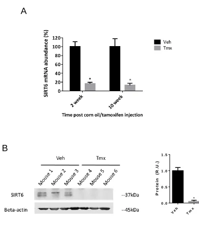
3.1 Confirmation of SIRT6 Knockdown in Aortic Medial DNA, mRNA, and Protein in Novel Mouse Strain ... 41
v
SIRT6-Deficient Mice ... 49
3.3.1 Saline-Infused Corn Oil and Tamoxifen Injected Mice have Healthy Aortas ... 49
3.3.2 Ang II-Infusion causes Aortic Petechial Hemorrhage in SMC-specific SIRT6-Deficient Mice ... 51
3.3.3 Ang II-Infusion causes Aortic Aneurysms in SMC-specific SIRT6-Deficient Mice ... 54
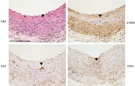
3.3.4 Evidence of an Inflammatory Cell Infiltrate in Ang II-Infused SIRT6-Deficient Mice and Minor Cell Infiltration in the Vehicle Control ... 57
3.3.5 Ang II-Induced Hypertensive Response in Vehicles and SIRT6KO Mice62 4 Discussion ... 64
4.1 Oxidative Stress Causes Aortic Wall Destruction in the Absence of SIRT6 ... 64
4.1.1 Petechial Hemorrhage and Aneurysm ... 64
4.1.2 Aortitis ... 66
4.1.3 Hypertension ... 68
4.1.4 Protective Effect of SIRT6 in SMCs ... 68
4.2 Reflections on Vascular Inflammation in Ang II-Infused Vehicle Controls and Cre-lox Technology ... 69
4.2.1 Hypotheses for Aortic Phenotype in Vehicle Controls ... 69
4.2.2 Creating SIRT6f/f Cre-negative Controls ... 71
4.3 Conclusion ... 71
References ... 72
Appendices ... 93
Curriculum Vitae ... 94
vi
List of Figures
Figure 1.1 Schematic of the three layers in the arterial wall: intima, media, adventitia. ... 4
Figure 1.2 SIRT6 is expressed in cultured SMCs and may play a role in resistance to oxidative stress ... 29
Figure 2.1 SMC-specific SIRT6KD Mouse Breeding Strategy... 31
Figure 2.2 Schematic of an inducible smooth muscle cell-specific SIRT6 knockdown using the Cre-Lox System. ... 33
Figure 2.3 Experimental timeline for Angiotensin II-induced aortic stress. ... 35
Figure 2.4 Schematic diagram of aortic regional segments. ... 38
Figure 2.5 Grading criteria for degree of cell infiltration and aortic wall destruction. ... 40
Figure 3.1 Evidence of recombination allele present in the smooth muscle of SMMHC-CreERT2;SIRT6f/f mice. ... 42
Figure 3.2 SIRT6 knockdown in mRNA expression and protein in aortic SMCs. ... 44
Figure 3.3 Gross aortic morphology, aortic morphometry, and aortic histology appear normal after induction of SIRT6 knockdown. ... 48
Figure 3.4 Aortic morphometry and histology appear normal in the saline-infused in SIRT6KD mouse. ... 50
Figure 3.5 Ang II-infusion causes petechial hemorrhage in SMC-specific SIRT6KO aortas. 53 Figure 3.6 Ang II-infusion causes aortic aneurysms in SMC-specific SIRT6KO mice. ... 56
Figure 3.7 Ang II-infusion of SIRT6KO mouse shows greater degree of inflammatory cell infiltration and aortic wall destruction compared to vehicle-control. ... 61
vii
viii
List of Abbreviations
Abbreviation Meaning
ACE Angiotensin-converting enzyme
ADP Adenosine diphosphate
Ang II Angiotensin II cADP Cyclic ADP
AMPK AMP-activated protein kinase
ATM Ataxia telangiectasia mutated
ATR Ataxia telangiectasia and Rad3-related protein
AT1R and AT2R Angiotensin type 1 and 2receptor
BAT Brown adipose tissue
BER Base excision repair
CPS1 Carbamoyl phosphate synthetase 1
CtIP C-terminal binding protein interacting protein
CVD Cardiovascular disease
DDR DNA damage response
DNA Deoxyribonucleic acid
DNA-PKcs DNA-dependent protein kinase catalytic subunit
DSB Double-strand breaks
EC Endothelial cell
ECM Extracellular matrix
EGF Epidermal growth factor
eNOS Endothelial nitric oxide synthase
FAO Fatty acid oxidation
FOXO Forkhead box protein O
G6P Glucose-6-phosphatase
GAPDH Glyceraldehyde 3-phosphate dehydrogenase
GCN5 General control nonrepressed protein
GDH Glutamate dehydrogenase
GH Growth hormone
GK Glucokinase
ix
HIF1α hypoxia-inducible factor 1α
H2O2 Hydrogen peroxide
HMGCS2 3-hydroxy-3-methylglutaryl-CoA synthase 2
HR Homologous recombination
ICAM-1 Intercellular cell adhesion molecule-1
IDE Insulin-degrading enzyme
IDH2 Isocitrate dehydrogenase 2
IGF1 Insulin-like growth factor 1
IL1 Interleukin-1
IL1B Interleukin-1 beta
IL6 Interleukin-6
IFNγ Interferon-γ
LCAD Long-chain acyl coenzyme A dehydrogenase
LDH Lactate dehydrogenase
LDL Low-density lipoprotein
LPK Liver pyruvate kinase
MCD Malonyl CoA decarboxylase
MEF Mouse embryonic fibroblast
MMP-9 Matrix metalloproteinase-9
NAM Nicotinamide
NAD+ Nicotinamide adenine dinucleotide
NAD(P)H Nicotinamide adenine dinucleotide (phosphate)
NAMPT Nicotinamide phosphoribosyltransferase
NBS1 Nijmengen Breakage Syndrome-1
NDUFA9 NADH dehydrogenase [ubiquinone] 1 alpha
subcomplex subunit 9
NHEJ Non-homologous end joining
NF-κB Nuclear factor kappa B
NO Nitric oxide
NR Nicotinamide riboside
x
O2•- Superoxide anion
OTC Ornithine transcarbamoylase
PAF53 Polymerase-associated factor 53
PAR Poly(ADP-ribose)
PARP Poly (ADP-ribose) polymerase
PCK1 Phosphoenolpyruvate carboxykinase
PCSK9 Proprotein convertase subtilisn kexin type 9
PDC Pyruvate dehydrogenase complex
PDGF Platelet-derived growth factor
PDHA1 Pyruvate dehydrogenase alpha 1
PDP Pyruvate dehydrogenase phosphatase
PFK-1 Phosphofructokinase-1
PGC-1α Peroxisome proliferator-activated receptor gamma
coactivator 1-alpha
PI3K Phosphoinositide 3-kinase
Pol I RNA polymerase I
PPAR α Peroxisome proliferator-activated receptor alpha
PPAR-γ Peroxisome proliferator-activated receptor gamma
rDNA Ribosomal DNA
ROS Reactive oxygen species
SA-β-gal Senescence-associated beta-galactosidase
SdhA Succinate dehydrogenase flavoprotein
SIPS Stress-induced premature senescence
SIR Silent information regulator
SIRT Sirtuin
SMC Smooth muscle cell
SMMHC Smooth muscle-myosin heavy chain
SOD Superoxide dismutase
SREBP1/2 Sterol regulatory element binding proteins 1 and 2
SSB Single-strand breaks
xi
TGF- β Transforming growth factor-β
TNF-α Tumor necrosis factor-α
TPI Triose phosphate isomerase
UBF Upstream binding factor
VCAM-1 Vascular cell adhesion molecule-1
WAT White adipose tissue
Chapter 1
1
Introduction
1.1
General Introduction
Cardiovascular disease (CVD) remains the leading cause of death worldwide, a fact that
drives ongoing research towards its prevention and intervention. To many, CVD such as
coronary artery disease, hypertension, congestive heart failure, and stroke have become
household terms. Overall cardiovascular well-being is dependent on the condition of our
blood vessels, which ultimately points toward the health of cells comprising the
vasculature, including endothelial cells (ECs), vascular smooth muscle cells (SMCs), and
other cell types resident in the vascular wall. It is fundamental to understand the biology
of the vascular wall and the cellular constituents for the prevention and treatment of CVDs.
1.2
Vasculature and Cellular Composition of the Vessel
Wall
The vasculature is an intricate network of blood vessels that transports blood between the
heart and peripheral parts of our body. Oxygen, nutrients, and other circulating factors,
such as hormones, are carried through the blood. Of the three main types of vessels—
arteries, veins, and capillaries—arteries carry oxygenated blood away from the heart, veins
carry deoxygenated blood to the heart, and capillaries are responsible for delivering all the
aforementioned components of blood flow to end organs. Exceptions are found in the
pulmonary vasculature with pulmonary arteries carrying deoxygenated blood away from
the heart and pulmonary veins carrying oxygenated blood to the heart.
The vessel wall is made up of three distinct layers that are comprised of different cell types
and have unique characteristics that contribute to the overall functionality of the blood
vessel. The innermost layer, tunica intima, is made up of a single layer of endothelial cells.
The middle layer, tunica media, has many concentric layers of vascular SMC and elastin
fibers. The outer and most diverse layer, tunica adventitia, is comprised of a collagen-rich
extracellular matrix (ECM) containing fibroblasts, progenitor cells, nerves, adipocytes,
Although veins and arteries have the same three layers, arterial walls have a substantially
thicker media to withstand direct pressure from cardiac output as well as high oxygen
content. An overall decrease in wall thickness, elastin, and collagen result in veins that are
less stiff than arteries [2]. In contrast, capillaries are comprised of a one cell layer thick
endothelium wrapped with pericytes. There are three types of capillary EC structure:
continuous, fenestrated, and discontinuous. These differences are indispensable for
simultaneously producing continuous capillaries in the blood brain barrier to block out
toxic substances and fenestrated capillaries in the glomerulus to allow for the filtration of
blood [3].
1.2.1
Layers of the Arterial Wall
1.2.1.1
Intima
Vascular ECs in the intima regulate blood flow through the production of nitric oxide (NO),
a vasodilator that controls the tone of vascular SMC [4]. Direct contact with the lumen
allows ECs to act as border guards that control wall permeability. ECs prevent unwanted
plasma proteins from moving into the wall through their tight junctions as well as facilitate
leukocyte extravasation in cases of injury or damage [4].
1.2.1.2
Media
In large arteries such as the aorta, the media is by far the thickest layer, therefore, making
vascular SMCs the major constituents of the aortic wall and the cell type of interest for this
project. In mice, there are 3-6 concentric layers of SMCs and elastin fibers depending on
which aortic region is under observation. The greatest number of layers are found in aortic
root media with progressively fewer layers the more distal it is to the heart.
The media exclusively consists of SMCs and the associated ECM during embryonic
development (i.e. collagen, elastin fibers, proteoglycans). The primary role of SMCs is to
mediate contraction and relaxation of the arterial wall, which is indispensable for efficient
regional distribution of blood flow into different tissues. Compared to smaller arteries,
higher quantities of elastin give the aorta the ability to simultaneously stretch and also
withstand the highest levels of blood pressure coming directly out of the heart [5].
Of greater interest to this project, the other main responsibility of SMCs is its response to
injury and repair of the vessel wall. Both roles are realized due to the profound phenotypic
plasticity of SMCs, setting them apart from striated (skeletal or cardiac) muscle cells.
Normal “contractile” SMCs are characterized by a very low rate of proliferation and
expression of proteins and signaling molecules necessary for contraction. These include
proteins such as SM α-actin, smooth muscle-myosin heavy chain (SMMHC), h1-calponin, SM22α, and smoothelin [6]. Morphologically, they appear long and spindle-shaped [7].
Being the only protein found exclusively in SMCs, SMMHC is currently the most specific
marker protein for identifying contractile SMC. Other contractile proteins mentioned
previously are also expressed in cardiomyocytes, myofibroblasts, and endothelial cells
during vascular remodeling [6,7]. In the event of vascular damage, SMCs respond to either
acute or chronic stimuli with a phenotypic change in protein expression. The switch is
facilitated via ligand-receptor interactions and epigenetic modifications, respectively.
Platelet-derived growth factor-BB (PDGF-BB) is a key repressor of vascular SMC-specific
gene expression while TGF-β a major promoter of it [8]. These rhomboid-shaped
“synthetic” SMCs have an increased ability to synthesize ECM components, proliferate,
and migrate [6].
1.2.1.3
Adventitia
Because the adventitia is home to such a variety of cell types, its function is expectedly
complex. The adventitia serves as a storehouse for many types of progenitor cells (e.g.
endothelial, mesenchymal, SMC, pericyte) [1]. A large population of fibroblasts,
macrophages, and dendritic cells allows the adventitia to have one of the first inflammatory
The intima and media are comprised of endothelial cells and smooth muscle cells, respectively. The adventitia is host to a variety of cell types and the wall’s main blood source, the vasa vasorum. (Image adapted from Stenmark 2013 [1])
1.3
Oxidative Stress in the Vascular Wall
Of the various insults that can threaten the integrity of the vascular wall, oxidative stress is
a common denominator among vascular diseases. Vascular cells naturally produce reactive
oxygen species (ROS) such as hydroxyl radicals (OH•), superoxide anion (O2•-), and
hydrogen peroxide (H2O2). During aerobic metabolism, low levels of ROS are formed as a
byproduct from the reduction of oxygen and used to regulate SMC contraction [9]. Oxidant
enzyme systems that facilitate these reactions include nicotinamide adenine dinucleotide
phosphate hydrogen (NAD(P)H) oxidase, xanthine oxidase (XO), myeloperoxidase, and
endothelial nitric oxide synthase (eNOS) [10]. ROS generation as a consequence of
oxidative phosphorylation also makes the mitochondria a major source of ROS [11].
However, the aforementioned enzymes have been implicated in the development of
vascular pathologies that make them of particular interest to this project. In particular,
NAD(P)H oxidase accounts for the majority of ROS in all vascular cells [9]. Different
isoforms of this multi-subunit protein are found in ECs, vascular SMCs, and adventitial
fibroblasts [10] and activated by vascular injury (e.g. cytokines, hormones, and
hemodynamic forces) [12]. Consequently, an increase in intracellular ROS results in
impaired vessel tone; an exaggerated inflammatory response; and SMC hypertrophy,
hyperplasia, and apoptosis [13]. To protect against accumulation of ROS, endogenous
antioxidants such as superoxide dismutase (SOD) convert superoxide radicals into H2O2
[10]. From that point on, catalase and glutathione peroxidase further catalyze H2O2 into
water and oxygen [14].
Whether caused by increased oxidase activity or deficient removal by antioxidants, excess
ROS exert their detrimental effects through uncontrolled oxidation of deoxyribonucleic
acid (DNA), proteins, and lipids [10]. Structural changes in such biological molecules
result in defective DNA replication and transcription, decreased enzymatic activity, and
loss of membrane permeability. Accordingly, oxidative stress is the term given for
collective ROS-induced damage in cells. In vascular cells, oxidative stress has a role in the
advancement of various cardiovascular pathologies that will be discussed in the following
1.3.1
Angiotensin II Induces Oxidative Stress
A diverse range of molecules activate NAD(P)H oxidase and increase ROS production:
growth factors (e.g. platelet-derived growth factor (PDGF), epidermal growth factor
(EGF), transforming growth factor-β (TGF- β), thrombin), cytokines (e.g. angiotensin II
(Ang II), interferon-γ (IFNγ), interleukin-1 (IL1), tumor necrosis factor-α (TNF-α)), lipids
(e.g. low-density lipoprotein (LDL) cholesterol, oxidized LDL), and even ROS (i.e. H2O2)
[14].
Ang II is one of the most potent and well-studied stimuli of NAD(P)H oxidase. It is the
most important effector of the blood pressure-regulating renin angiotensin system.
Through a series of cleavage reactions, angiotensinogen is converted to angiotensin I,
which is then converted to Ang II through angiotensin-converting enzyme (ACE). Ang II
can then be further cleaved by ACE2 into Ang-(1-7) [15]. As a result, ACE and ACE2 are
potential targets for diminishing the potential harmful effects of Ang II by controlling the
production and degradation of the peptide. This peptide hormone carries out its effects
through angiotensin type 1 and 2 receptor, AT1R and AT2R, respectively [16]. However,
Ang II’s harmful effects are predominantly carried out through AT1Rs expressed in liver,
adrenals, brain, lung, kidney, heart, and vasculature. In vascular SMCs, Ang II binding to
AT1R initiates a G-protein-dependent signaling pathway that triggers an influx of Ca2+
from the sarcoplasmic reticulum into the cell [17]. This increases the interaction of actin
and myosin filaments, resulting in vasoconstriction and elevated blood pressure over time.
Prolonged Ang-II infusion was shown to induce hypertension in mice, rats, rabbits, and
humans [18].
The AT1R-initiatied G-protein-dependent pathway also serves to activate NAD(P)H
oxidase [16]. This is mediated through intracellular signaling molecules upstream of
NAD(P)H (e.g. phospholipase D, protein kinase C, c-Src, phosphoinositide 3-kinase
(PI3K), Rac) [15]. Moreover, Ang II increases the abundance of essential NAD(P)H
oxidase subunits (e.g. gp91phox, p22phox, p47phox) [19]. There are many studies that
confirm the association between Ang II, NAD(P)H, oxidative stress, and hypertension. Ang
II-infusion increased both blood pressure and vascular O2•- levels in mice. p47phox
Another mouse study generated similar decreases in Ang II-induced hypertension and O2•-
production using a peptide that inhibited interaction between NAD(P)H subunits,
gp91phox and p47phox [21]. Two different rat models of hypertension showed that
increased NAD(P)H activity was correlated with elevated Ang II levels [22] or was brought
down by treatment with an AT1R antagonist [23].
1.3.2
Effects of Oxidative Stress in Vascular Inflammation and
Atherosclerosis
Atherosclerosis is a disease characterized by chronic inflammation that begins as a local
response to endothelial damage. As the disease progresses, oxidized LDLs, inflammatory
cells, and SMCs form a plaque comprised of a lipid core protected by a fibrous cap [24].
The presence of increased ROS and upregulation of NAD(P)H oxidase subunits in all
layers of the diseased wall indicate a positive correlation between oxidative stress and
atherosclerosis [14]. One of the initial effects of ROS is the oxidization of LDL particles
that have invaded the subendothelium. A proinflammatory environment is created as ECs
secrete signals such as TNF-α and Ang II. Both cytokines induce the expression of vascular
cell adhesion molecule-1 (VCAM-1) and intercellular cell adhesion molecule-1
(ICAM-1), thereby drawing leukocytes into the vascular wall [13].
Once in the wall, monocytes mature into macrophages that generate even more ROS [24].
Macrophages that take up oxidized LDL become foam cells which eventually aggregate
and form the core of an atherosclerotic plaque. In addition to those processes, O2
•-inactivates EC-secreted NO; therefore, disrupting its important vasodilatory and
anti-inflammatory effects of on both ECs and vascular SMCs [13]. In the course of plaque
development, SMCs proliferate and are recruited in an attempt to stabilize the lesion with
ECM that they produce. PDGF triggers SMC proliferation and migration in a
ROS-dependent manner [10].
In summary, ROS are extensively involved in all phases of atherosclerosis. Oxidative stress
is crucial in initiating the inflammatory response as well as propagating it throughout the
1.3.3
Oxidative Stress-Induced DNA Damage in SMC
In addition to attacking proteins and lipids, free radicals have the ability to oxidize DNA.
Because hydroxyl radicals react readily with both purines and pyrimidines, it has the
potential to form DNA adducts with all bases. This leaves DNA susceptible to single strand
breaks (SSBs), double-strand breaks (DSBs), and base modifications [25]. An example of
a common site of hydroxyl radical attack is carbon 8 on guanine (8-oxo-guanine). This
base modification has received special attention because of its ability to avoid detection by
the DNA repair system and high mutagenicity (i.e. 8-oxo-G mis-pairs with adenine;
therefore, causing a guanine to thymine mutation) [26]. The consequences of DNA damage
in a cell can be devastating: mis-pairings of bases during replication, interruption of
transcription, and overall increased mutagenesis [26].
Accumulation of oxidative stress-induced DNA damage can cause cellular senescence, a
state of permanent cell cycle arrest. Cells naturally lose the ability to divide as they age and
telomere lengths shorten from multiple rounds of replication [27]. As telomeres are tandem
repeats of nucleotide sequences at the ends of chromosomes, they protect the cell from
losing important coding and noncoding sequences during replication. Therefore, shortened
telomeres signal cell cycle arrest as a defensive measure against the malignant
transformation that is seen in cancer cells [28]. Cells remain viable until eventually
succumbing to apoptosis through activation of the tumor suppressor, p53 [27].
Aside from replicative senescence, cells exposed to external stimuli such as irradiation and
oxidative stress undergo stress-induced premature senescence (SIPS) that is largely
independent of telomere status [27]. The senescence is considered premature because cells
of similar age that are not exposed to such insults have normal proliferation rates. DNA
damage from ROS triggers the DDR mentioned earlier; therefore, causing the cell to stop
dividing until repairs can be made. An unsuccessful attempt results in senescence and
inevitably, apoptosis through p53 signaling [29]. Relatedly, ROS are implicated in
telomere-based senescence because of their ability to induce DNA strand breaks in
telomeres [30]. This can exacerbate the DDR already triggered by telomere shortening
The association of oxidative stress with both telomere-based senescence and SIPS
demonstrates the connection between the two different types of cellular senescence.
Cultured aortic SMCs from aged mice produced higher levels of ROS with decreased
proliferation compared to cells from younger mice [31]. Another study showed that
expression of telomerase, an enzyme that facilitates telomere extension, delayed SMC
senescence in vitro [32]. Furthermore, vascular SMCs in an in vivo atherosclerotic model
show this overlap particularly well. Plaque vascular SMC showed a positive correlation
between markers of senescence (i.e. senescence-associated beta-galactosidase (SA-β-gal)
and p16) and oxidative stress-induced DNA damage (i.e. 8-oxo-guanine) and had shorter
telomere lengths compared to normal vessel SMC [32].
1.4
The Role of NAD+ Consuming Enzymes in
Managing Oxidative Stress
Nicotinamide adenine dinucleotide (NAD+) and its reduced form NADH were first
discovered as electron carriers in cellular oxidation/reduction reactions. For many decades,
NAD+ was mainly known as a cofactor for enzymes in energy metabolism pathways such
as glycolysis and oxidative phosphorylation [33]. In contrast to its usage in redox reactions
where it can be recycled over and over again, NAD+ has only recently been discovered to
be consumed as a cosubstrate in biochemical reactions.
1.4.1
NAD+ Biosynthesis and Consumption Pathways
There are three major classes of enzymes that deplete cellular NAD+ levels: cyclic
adenosine diphosphate (cADP)-ribose synthases, adenosine diphosphate (ADP)-ribose
transferases, and sirtuins (SIRTs) [34]. Therefore, NAD+ deficiency disrupts the many
crucial pathways that these enzymes participate in. cADP-ribose synthase is involved in
calcium signaling. It uses NAD+ to make cADP-ribose, a second messenger that triggers
the release of calcium from intracellular stores [35]. The poly (ADP-ribose) polymerase
(PARP) family of proteins are the most prevalent ADP-ribose transferases. PARPs detect
DNA damage, bind to those sites, and form poly (ADP)-ribose (PAR) chains using NAD+.
These chains recruit other DNA repair factors to initiate a cascade of DNA repair events
consumers to be discovered. This family of deacetylases requires NAD+ to catalyze the
removal of acetyl groups from proteins. SIRTs have been considered regulators of
mammalian healthspan because of their impact on energy metabolism, cellular stress
resistance, genomic stability, aging, and tumorigenesis [36].
Naturally, discovery of NAD+ consumption pathways stemmed renewed interest in
enzymes of NAD+ biosynthesis. NAD+ is synthesized de novo from the amino acid
L-tryptophan that comes from dietary intake. The majority of NAD+, however, is generated
from the salvage pathway which uses other NAD+ precursors from our diet (e.g.
nicotinamide (NAM), nicotinamide riboside (NR), nicotinic acid) [34]. Nicotinamide
phosphoribosyltransferase (NAMPT) is a key enzyme in the salvage pathway because it
catalyzes the first, rate-limiting step in the conversion of NAM into NAD+ [34]. The ability
of NAMPT to influence PARP and SIRT activity underscores their reliance on NAD+ [37].
In cultured human SMCs, overexpression of NAMPT protected cells from
NAD+-dependent oxidative stress-induced damage [38]. Decreased expression of NAMPT
prompted premature senescence while conversely, its overexpression postponed
senescence. A corresponding increase in SIRT1 activity indicates a role for SIRTs in
resisting oxidative stress [38]. This study by our lab demonstrates the association between
NAD+ bioavailability and oxidative stress-induced damage.
1.4.2
PARPs
DNA lesions from free radicals or other sources of damage are often categorized as either
SSBs or DSBs because of the distinct repair mechanisms associated with them. Base
excision repair (BER) and nucleotide excision repair mend SSBs, while homologous
recombination (HR) and non-homologous end joining (NHEJ) process DSBs [39].
Common to all repair mechanisms is the DDR, a cascade of damage sensing and signal
amplifying enzymes (e.g. PI3K-related kinase family, PARPs, MRE11-RAD50-NBS1
complex) that regulate recruitment of DNA repair factors [40].
The PARP family, most notably PARP1, plays an important role in sensing DNA damage
and is recruited prior to all SSB and DSB repair mechanisms [41]. Although PARP1 in
catalyzes the addition of adenosine diphosphate (ADP)-ribose units onto a target protein
(e.g. histones H1 and H2B, PARP1 itself) [42]. This ADP-ribosylation reaction involves
NAD+ hydrolysis and produces poly(ADP-ribose) (PAR) chains that are linked via
glycosidic ribose-ribose bonds [43]. Although there is some baseline PARP1 activity, DNA
breaks increase the level of PAR chains by 10- to 500-fold. At more than 200 PAR units
on each target protein, this makes PARP1 a major consumer of NAD+ [44]. PAR chains
continue to advance the DDR by recruiting ataxia telangiectasia mutated (ATM) and ataxia
telangiectasia and Rad3-related protein (ATR), major signaling factors in the PI3K-related
kinase family [41]. Additionally, the presence of PAR chains on histones interferes with
transcription factors and loosens chromatin structure [42]. This further facilitates the DDR
or activates apoptosis in cases of damage too extensive to be repaired efficiently.
1.4.3
Sir2 and Sirtuin family
Through mutation studies, the silent information regulator (SIR) family of genes were first
discovered as negative regulators of mating type loci in Saccharomyces cerevisiae [45–
48]. In addition, SIR gene products are required for silencing at telomeres and ribosomal
DNA (rDNA) [49–53]. SIR2 is distinct from the remaining family members in two notable
areas: it is highly conserved from lower organisms like S. cerevisiae and Caenorhabditis
elegans to humans [54]; its gene product, Sir2, is necessary for all three areas of silencing
previously mentioned. Enzymatically, Sir2 was initially identified as an
ADP-ribosyltransferase, an enzyme that uses NAD+ to transfer an ADP-ribose group to a protein
carrier [55]. However, further studies showed that Sir2’s role as a chromatin silencer is
predominately mediated through its role as a NAD+-dependent histone deacetylase
(HDAC) [56]. While a classical HDAC mechanism uses metal ions and water molecules,
Sir2 couples NAD+ hydrolysis with deacetylation [57]. This reactions results in the
production of a deacetylated substrate and NAM. Interestingly, the expected ADP-ribose
was replaced by O-acetyl-ADP ribose (OAADPr), a novel product comprised of the
ADP-ribose moiety of NAD+ and the transferred acetyl group [58]. Interest in Sir2 took a big
leap when it was found to promote longevity in S. cerevisiae by repressing rDNA
rDNA circles that cause aging in yeast [59]. As a result, more effort was put into
understanding the role of Sir2-like proteins in mammals.
Sir2-like proteins are found in prokaryotes and eukaryotes and given the name, sirtuins.
There are seven mammalian SIRTs, SIRT1-7, that have conserved catalytic core domains
with Sir2 and varying N- and C-terminals [60,61]. Although SIRTs are phylogenetically
classified into four classes, they are more practically organized by their typical cellular
localizations: SIRT1, 6, 7 in the nucleus, SIRT2 in the cytoplasm, and SIRT3-5 in the
mitochondria [61,62]. SIRT mRNA is ubiquitously expressed in all human organs tested,
although each SIRT has its own unique expression profile [62]. SIRTs make up the class
III family of HDACs based on their unique mechanism of deacetylation as described
previously with Sir2. All SIRTs are deacetylases with the exception of SIRT4 which only
functions as an ADP-ribosyltransferase [63]. Deacetylation targets are unique to each
SIRT—with some overlap—which give rise to different physiological functions.
1.4.4
Sirtuin 1
Since its discovery, the majority of sirtuin-related studies have focused on SIRT1, the
family member with the greatest sequence similarity to SIR2 [61]. Although predominantly
located in the nucleus [62], it can also travel to and from the cytoplasm [64].
SIRT1-deficient mice models generated different phenotypes based on varying strain
backgrounds. Inbred 129/Sv SIRT1KO mice showed gross signs of developmental defects
(i.e. eye abnormalities), were consistently smaller than the WT, and seldom survived
postnatally [65,66]. In an outbred background, SIRT1KO mice survive to adulthood but
are sterile and smaller than WT [66]. As SIR2’s closest homolog, there was great interest
in a possible role for SIRT1 in lifespan extension. Lack of SIRT1 in mice decreased their
median lifespan [67]. Though, transgenic mice overexpressing SIRT1 exhibited delayed
progression of age-associated diseases (e.g. cancer, metabolic syndrome), overall longevity
was not extended [68]. Interestingly, brain-specific SIRT1 overexpression increased
lifespan through SIRT1-dependent activation of the hypothalamus. This resulted in
enhanced physiological functions such as physical activity, body temperature, oxygen
As a major regulator of metabolism, SIRT1 deacetylates important metabolic targets in
skeletal muscle and the liver including peroxisome proliferator-activated receptor alpha
(PPARα), peroxisome proliferator-activated receptor gamma coactivator 1-alpha (PGC-1α). PPARα belongs to a family of lipid-sensing receptors that activate genes involved in
fatty acid homeostasis [70]. Particularly activated during prolonged periods of fasting,
PPARα acts to conserve energy via its role in fatty acid catabolism (i.e. ketogenesis).
SIRT1 stimulates fatty acid oxidation (FAO) by deacetylating and activating hepatic
PPARα [71]. Another layer of SIRT1-dependent FAO regulation comes from activation of the PPARα transcriptional coactivator PGC-1α in skeletal muscle and the liver [71,72].
Interaction with PGC-1α also gives SIRT1 control over the initiation of gluconeogenesis
during fasting [73]. In white adipose tissue (WAT), SIRT1 deacetylation of PPARγ
represses its function as a promoter of adipogenesis [74]. In addition, deacetylation of
PPARγ at Lys268 and Lys 293 allows for recruitment of coactivators that simultaneously
induce brown adipose tissue (BAT) genes and repress WAT genes in white adipocytes
[75]. This “browning” of WAT is thought to increase energy expenditure. Therefore,
SIRT1-dependent PPARγ deacetylation in adipocytes is hypothesized to protect against
obesity-related diseases.
1.4.4.1
Sirtuin 1 in Vascular Smooth Muscle Cells
Our lab was one of the first groups to study the importance of SIRT1 in vascular SMCs.
We found that NAMPT overexpression extends lifespan in SMCs by increasing SIRT1
deacetylation of p53 [38]. SMCs overexpressing SIRT1 are senescence-resistant, provided
there are adequate NAD+ levels to sustain SIRT1 activity [76]. Furthermore, SIRT1 levels
decline with age, making SMCs more vulnerable to genomic stress and cellular senescence
[77]. SIRT1’s role in DNA repair has been a focal point for its importance in
atherosclerosis. In the event of oxidative stress, SIRT1 deacetylates and activates the DNA
repair protein Nijmengen Breakage Syndrome-1 (NBS1). As a result of reduced SIRT1
levels in atherosclerotic plaques, these diseased vascular SMCs have increased rates of
DNA damage and apoptosis [78]. SIRT1’s beneficial effects also extend toward possible
complications of atherosclerosis such as calcification and neointima formation. SIRT1
with increased cellular senescence [79]. Using carotid artery ligation and wire injury
models, SIRT1 overexpression was shown to reduce neointima thickening. SIRT1
inhibited SMC proliferation and migration via downregulation of cyclin D1 and matrix
metalloproteinase-9 (MMP-9) [80].
In vitro and in vivo Ang II models have been used to elucidate SIRT1’s role in vascular
SMCs. One of the many negative effects of Ang II is increased ROS levels in the
vasculature. In particular, Ang II activation of the NAD(P)H oxidase isoform, Nox1,
induces vascular hypertrophy. Accordingly, SIRT1 inhibits hypertrophy in SMCs by
suppressing Nox1 mRNA expression, and thereby, reducing oxidative stress [81]. As
discussed previously, Ang II-induction of hypertension and oxidative stress is
predominantly mediated via AT1R. In vascular SMCs, overexpression of SIRT1
downregulated both AT1R mRNA expression and AT1R signaling via ERK
phosphorylation. Resveratrol, an activator of SIRT1, reduced aortic AT1R protein
abundance and reduced Ang II-induced hypertension in mice [82]. A separate group
created SMC-specific SIRT1 transgenic mice that were also resistant to Ang II-induced
hypertension. Aortas from these mice had reduced ROS levels, inflammation, and collagen
accumulation. These beneficial effects of SIRT1, however, were not because of changes in
AT1R levels. It was proposed that SIRT1 decreases TGF-β1 expression via inhibition of
p65/RelA binding on its promoter [83]. Lastly, a recent SMC-specific SIRT1KO mouse
model demonstrated the protective effect of SIRT1 against aortic dissection. After a
14-day course of Ang II infusion, mice lacking SIRT1 in aortic SMCs had increased mortality
rates. The manifestation of aortic dissection was attributed to increased MMP2 and MMP9
expression in SMCs. Interestingly, aortic dissection was not a result of increased
hemodynamic stress because SMC-specific SIRT1-deficiency attenuated Ang II-induced
hypertension [84].
1.4.5
Sirtuin 2
SIRT2 is deacetylase that is ubiquitously expressed in mouse tissues and predominantly
cytoplasmic, but also found in the nucleus [85]. The first deacetylation target identified
was α-tubulin [86], important in regulating oligodendrocyte cytoskeleton myelination and
maintain genomic stability [85]. SIRT2 regulation of chromatin condensation is associated
with histone H4 lysine 16 deacetylation [88]. Interestingly, SIRT2’s role in cell cycle
control via deacetylation of the mitotic checkpoint kinase, BubR1, links SIRT2 with aging.
Overexpression of SIRT2 increases BubR1 abundance, which was shown to increase
median lifespan in BubR1-deficient male mice [89]. SIRT2-dependent genomic stability is
also believed to be the reason why SIRT2-deficient mice develop tumorigenesis [90].
SIRT2 is also involved in lipogenesis, glucose metabolism, and inflammation. Forkhead
box protein O1 (FOXO1) is a transcription factor that initiates transcription of
gluconeogenic genes (i.e. glucose-6-phosphatase (G6P)) and inhibits peroxisome
proliferator-activated receptor gamma (PPAR-γ), a nuclear receptor that activates
adipocyte maturation [91]. SIRT2 deacetylation of FOXO1 results in its activation;
therefore, promoting gluconeogenesis and preventing adipogenesis [92,93]. Nuclear factor
kappa B (NF-κB) is a major transcription factor that induces expression of inflammatory
genes. SIRT2 exhibits an anti-inflammatory role by directly deacetylating NF-κB,
hindering transcription, and weakening any resultant inflammatory response [94].
SIRT2-deficient mice are more susceptible to dextran sulfate sodium-induced colitis and have
increased levels of pro-inflammatory cytokines (e.g. TNF-α, interleukin-1 beta (IL1B),
interleukin 6 (IL-6)) compared to wildtype (WT) mice [95].
SIRT2 also contributes towards oxidative stress resistance via interactions between
FOXO3a and SOD. Deacetylation of FOXO3a increases FOXO3a binding and activation
of SOD, thus, decreasing ROS levels [96]. However, one study found contradicting results
where inhibition of SIRT2 protected ECs from H2O2-induced cell death [97]. SIRT2 is also
necessary in ECs, mediating Ang II-induced cell migration. SIRT2 deacetylates tubulin in
the aortic intima of mice and promotes microtubule reorganization. Therefore, SIRT2
mediates Ang II-induced vascular remodeling [98].
1.4.6
Sirtuin 3
SIRT3 is a mitochondrial SIRT expressed in all tissues with higher levels in the liver,
kidney, and heart [99,100]. SIRT3KO mice are healthy with no observable differences
major mitochondrial protein deacetylase [101]. Because SIRT3 target proteins are involved
in almost all mitochondrial functions, loss of SIRT3 has been shown to accelerate a wide
range of diseases.
With respect to energy homeostasis, SIRT3 regulates mitochondrial oxidative
phosphorylation by deacetylating proteins involved in the electron transport chain. SIRT3
deacetylates and activates the Complex I subunit, NADH dehydrogenase [ubiquinone] 1
alpha subcomplex subunit 9 (NDUFA9), and the Complex II subunit, succinate
dehydrogenase flavoprotein (SdhA) [99,102]. Consequently, SIRT3KD causes a decrease
in basal ATP levels and oxygen consumption in vitro [99,103]. In nitrogen metabolism,
SIRT3 activates glutamate dehydrogenase (GDH), a mitochondrial enzyme that converts
glutamate into α-ketoglutarate [104]. The nitrogen from glutamate is released as the
byproduct ammonia and is removed via the urea cycle. The second step in this cycle is
catalyzed by ornithine transcarbamoylase (OTC), another SIRT3 protein target. SIRT3
deacetylation and activation of OTC promotes amino acid catabolism and ammonia
detoxification [105].
SIRT3’s role in glucose metabolism is a mechanism through which it acts as a tumor
suppressor. An early clue came from oncogene expressing SIRT3KO MEFs that had
increased metabolic activity from sources besides mitochondrial oxidative phosphorylation
[106]. Indeed SIRT3 regulates the Warburg effect, typical metabolic reprogramming in
cancer cells that favors aerobic glycolysis, through deacetylation of hypoxia-inducible
factor-1α (HIF1α). SIRT3 suppresses the Warburg effect by destabilizing HIF1α, a
transcription factor that upregulates glycolytic genes [107]. Pyruvate dehydrogenase
phosphatase (PDP) and pyruvate dehydrogenase alpha 1 (PDHA1), a subunit of the
pyruvate dehydrogenase complex (PDC), are also deacetylation targets of SIRT3. These
actions work to activate PDC and promote oxidative phosphorylation; thereby, possibly
oppose the Warburg effect [108].
SIRT3’s capacity as a tumor suppressor via glucose metabolism regulation is dependent on
its ability to modulate ROS levels. SIRT3 deacetylation reduces oxidative stress by
112]. The previously discussed SOD2 neutralizes O2•-,while IDH2 supports regeneration
of glutathione, the antioxidant that breaks down H2O2 [110]. In a study mentioned earlier,
increased ROS levels were observed in SIRT3KO MEFs and supplementing SOD2 was
enough to stop immortalization of SIRT3-deficient MEFs expressing an oncogene [106].
SIRT3 attenuates HIF1α activity via reducing ROS production [107,113].
1.4.6.1
Sirtuin 3 in Cardiovascular Disease
As the most well-defined mitochondrial SIRT, studies have revealed protective roles for
SIRT3 in cardiovascular disease. SIRT3 drives the FAO pathway by deacetylating and
activating long-chain acyl coenzyme A dehydrogenase (LCAD). Under fasting conditions,
SIRT3-deficient mice displayed metabolic stress symptoms consistent with defective FAO
such as lower ATP levels and intolerance to cold exposure [114]. SIRT3KO mice on a
high-fat diet demonstrate an accelerated metabolic syndrome that coincides with global
mitochondrial hyperacetylation. Diet-induced obesity, insulin resistance, hepatic steatosis,
and hyperlipidemia are all exacerbated in SIRT3-deficient mice compared to WT [115].
In cardiomyocytes, SIRT3 levels increase in response to stress and is required for cell
viability [116]. SIRT3 suppresses cardiac hypertrophy by regulating the activation of
mitochondrial permeability transition pores. In addition to cardiac hypertrophy, SIRT3KO
mice develop fibrosis and have increased signs of aging in their hearts [117]. SIRT3 also
inhibits cardiac hypertrophy by lowering cellular ROS levels. By deacetylating and
stabilizing FOXO3, SIRT3 facilitates upregulation of FOXO3-dependent mitochondrial
antioxidant enzymes, SOD2 and catalase [118]. The anti-oxidant properties of SIRT3 also
play a role in protecting ECs. In response to hypoxic stress, SIRT3 mediates ROS
detoxification through the same FOXO3 pathway [119]. SIRT3-null hypercholesterolemic
(i.e. LDL receptor-KO) mice fed a high-fat diet had elevated ROS levels, accelerated
weight gain, and impaired metabolic adaptation to changes in nutrient intake. Surprisingly,
SIRT3 deficiency did not exacerbate advanced atherosclerotic lesions when compared to
controls [120]. Lastly, SIRT3-dependent mitochondrial function is necessary for
pulmonary artery SMCs and preventing spontaneous pulmonary arterial hypertension in
1.4.7
Sirtuin 4
This second mitochondrial SIRT is expressed in all tissues with higher levels in the kidney,
heart, brain, liver, and pancreatic β cells [63]. Little is known about SIRT4’s enzymatic
function; it is the only SIRT that cannot carry out NAD+-dependent deacetylation. Initial
studies implicate SIRT4 in glucose metabolism, cancer, and lipid metabolism.
Though SIRT4KO mice were developmentally normal and did not show any gross defects
compared to WT littermates, SIRT4KO pancreatic β cells had higher GDH activity. SIRT4
was first described to ADP-ribosylate, and thus, inhibit GDH activity [63]. In addition to
its role in nitrogen waste disposal, GDH metabolizes glutamate to generate more ATP and
ultimately promote insulin secretion [122]. So, SIRT4 blocks insulin secretion in pancreatic
β cells. Another study affirmed SIRT4 expression in β cells and proposed negative
regulation of insulin through interactions with insulin-degrading enzyme (IDE) [123].
Because some cancer cells need glutamine to survive [124], inhibition of glutamine
metabolism by SIRT4 gives it a tumor-suppressive role. With genomic instability being a
characteristic of all cancers, DNA damage was shown to induce SIRT4 expression [125].
SIRT4 repressed both tumor proliferation in vitro and tumor formation in vivo [125,126].
SIRT4 expression was found to be significantly lower in human bladder, breast, colon,
gastric, ovarian, and thyroid carcinomas compared to normal tissue [127]. Lastly, SIRT4
has a role in hepatic lipid metabolism that contrasts the functions of other SIRTs. SIRT4
represses FAO in primary mouse hepatocytes and in vivo [128,129]. Malonyl -oA
decarboxylase (MCD) deacetylation by SIRT4 results in elevated levels of malonyl-CoA
[129]. This metabolite’s levels are closely regulated because of its important role in
simultaneously promoting fat synthesis and inhibiting FAO [129]. Interestingly, this effect
of SIRT4 seems to be dependent on SIRT1-repression [128] .
With respect to vascular health, SIRT4 appears to have an anti-inflammatory role in
protecting ECs from cigarette smoke-induced inflammatory responses. Overexpression of
SIRT4 in the cigarette smoke-activated endothelium helped decrease monocyte adhesion
to ECs by inhibiting adhesion molecules VCAM-1 and E-selectin. Moreover, SIRT4
1.4.8
Sirtuin 5
SIRT5 is primarily located in the mitochondria but also found in the cytosol [131]. In mice,
SIRT5 protein is expressed in all tissues with higher levels in brain, heart, liver, and kidney
[132]. Upon gross inspection, SIRT5-deficient mice are normal and healthy [101,133,134].
SIRT5 was originally identified as a deacetylase with carbamoyl phosphate synthetase 1
(CPS1) as its target in vitro and in vivo [132,135]. SIRT5 deacetylation activates CPS1, the
rate-limiting step in the urea cycle that is necessary for removing potentially toxic buildup
of ammonia from the body [132]. Though this finding established SIRT5’s role in ammonia
detoxification, the underlying mechanism was brought into question when SIRT5 was
shown to remove succinyl and glutaryl moieties from CPS1 in a NAD+-dependent manner
[136,137]. The physiological function of SIRT5-dependent deacetylation is unclear
because the catalytic efficiencies of desuccinylation and demalonylation were 29- to
>1000-fold higher than deacetylation [136]. SIRT5 also desuccinylates and inhibits
glutaminase, an enzyme that generates ammonia. By lowering glutaminase activity, SIRT5
protects against ammonia-induced autophagy and mitophagy [138].
SIRT5 also targets enzymes involved in mitochondrial metabolism. High percentages of
proteins in the amino acid degradation pathway, tricarboxylic acid (TCA) cycle, and fatty
acid metabolism were succinylated. Specifically, SIRT5-dependent succinylation of
pyruvate dehydrogenase complex (PDC) and succinate dehydrogenase suppresses their
activity in MEFs [139]. SIRT5 may play a role in FAO, the pathway with the highest
percentage of succinylation target proteins and SIRT5 target proteins [133]. SIRT5 is also
proposed to regulate ketone body production because there was evidence of
3-hydroxy-3-methylglutaryl-CoA synthase 2 (HMGCS2) desuccinylation under fasting conditions in
vivo [133]. A recent study highlighted a new role for SIRT5 in glucose metabolism.
Pathway analysis revealed glycolysis as the foremost SIRT5-regulated pathway via
demalonylation of a plethora of proteins including, glyceraldehyde 3-phosphate
dehydrogenase (GAPDH) [140]. Though SIRT5 is associated with many different
metabolic pathways, the biological significance of its various enzymatic functions remains
unclear. Multiple normal SIRT5-deficient mouse lines imply the dispensable nature of
only relevance to cardiovascular health is an association between single nucleotide
polymorphisms in the Sirt5 gene and risk of carotid plaques [141].
1.4.9
Sirtuin 7
SIRT7 is one of the least studied SIRT and uniquely located in the nucleolus [62]. It is
found in all mouse tissue with higher expression in metabolically active tissues (e.g. liver,
spleen, testis) and lower expression in non-proliferating tissues (e.g. muscle, heart, brain)
[142]. With rDNA transcription as the foremost activity of the nucleolus [143], SIRT7 has
been found to increase rRNA synthesis by promoting RNA polymerase I (Pol I)-mediated
transcription [142]. SIRT7 is involved via direct interaction with the rDNA transcription
factor, upstream binding factor (UBF) [144]), and deacetylation of Pol I subunit,
polymerase-associated factor 53 (PAF53) [145]. Following the pattern of other SIRTs,
SIRT7 is a specific histone deacetylase. SIRT7-mediated H3K18 deacetylation represses
transcription at promoters of tumor suppressor genes [146]. The physiological significance
of this interaction is that SIRT7 is required for maintaining a cell’s cancerous phenotype. SIRT7’s potential as an oncogene is helped by evidence of its upregulation in all cancers
that have been studied so far (e.g. thyroid, breast, bladder, colorectal) [143]. With respect
to metabolic regulation, two separate SIRT7-deficient models revealed contrasting results.
One study reported that SIRT7 prevents the development of hepatic steatosis by
suppressing endoplasmic reticulum stress [147]. Another study showed that
SIRT7-deficient mice were resistant to high-fat diet-induced fatty liver. SIRT7 promoted hepatic
steatosis via regulation of the ubiquitin-proteasome pathway [148].
Thus far, only one study has described a role for SIRT7 in cardiovascular health. In the
first SIRT7KO model, SIRT7 was shown to protect mice from a decreased lifespan due to
heart hypertrophy and inflammatory cardiomyopathy. SIRT7-deficient myocardium
revealed inflammatory infiltrations paralleled with higher levels of cytokines 12 and
IL-13. SIRT7 also protects cardiomyocytes from apoptosis by deacetylating, and therefore,
1.5
Sirtuin 6
SIRT6’s core domain is flanked by an N-terminal important for histone deacetylation and
chromatin association and a C-terminal necessary for nuclear localization [150]. Mouse
mRNA and protein expression is highest in the brain, heart, and liver [151]. Primarily
characterized as an NAD+-dependent HDAC, SIRT6 uses the same mechanism as Sir2 to
remove acetyl groups from lysines [152,153]. Solving its crystal structure led to the
discovery that SIRT6 is uniquely able to bind NAD+ in absence of an acetylated substrate
[153]. SIRT6 targets histones H3K9, H3K56, and also directly deacetylates proteins [154–
157]. In addition to deacetylation, SIRT6 has deacylase and weak mono-ADP-ribosylase
activity [151,158]. Surprisingly, in vitro measurements of deacetylase activity were
300-fold lower than deacylation of a myristoyl group [158]. In vivo, this low intrinsic
deacetylation is activated by long-chain fatty acids [159] and SIRT6’s association with the
nucleosome complex [160]. In addition to fatty acid activation, only a few mechanisms of
SIRT6 regulation have been discovered. At the transcriptional level, c-FOS binds the
promoter to induce expression [161]. SIRT6 is negatively regulated post-transcriptionally
via micro RNA (miR)-766, -33a, and -33b [162,163]. Post-translationally, the ubiquitin
ligase CHIP ubiquitinates SIRT6 to increase protein stability and prevent
proteasome-mediated degradation [164].
1.5.1
SIRT6 Knockout and Transgenic Animal Models
Three week-old 129Sv SIRT6-deficient mice developed an acute degenerative aging-like
phenotype and died shortly after postnatal day 24. Defects included reduced body size,
severe lymphopenia, hypoglycemia, acute loss of subcutaneous fat, lordokyphosis, and
osteopenia. At the cellular level, MEFs from these mice showed enhanced sensitivity to
DNA damage agents. SIRT6KO-induced genomic instability was attributed a role in base
excision repair [165]. A separate SIRT6KO mouse line demonstrated a similar premature
aging phenotype, hypoglycemia, and early lethality in 60% of mice [166]. Interestingly,
early lethality was circumvented by feeding mice with glucose containing water; therefore,
identifying hypoglycemia as the main cause of death. This was due to enhanced insulin
dependent and independent glucose uptake from increased levels of both glucose
to develop chronic liver inflammation starting at 2 months of age [167]. In addition to
SIRT6KO models, transgenic mice overexpressing SIRT6 were also created. Lifespan was
extended in male, but not female, SIRT6 transgenic mice. This male-specific phenomenon
was associated with lower insulin-like growth factor 1 (IGF1) signaling in WAT, an
observation that has been linked to prolonged lifespan in rodent models [168]. Transgenic
mice overexpressing SIRT6 are also less prone to high fat diet-induced metabolic damage
[169].
Tissue-specific SIRT6KO models have also been created for brain, liver, and heart.
Neural-specific deletion of SIRT6 led to growth retardation at 4 weeks of age [170]. However, not
only did the mutant mice eventually reach normal size, they showed increased adiposity
and became obese by 6-8 months of age. Modified growth hormone (GH)/IGF1 signaling
and obesity-related neuropeptides were the likely causes of this phenotype [170].
Liver-specific SIRT6KO mice have fatty livers from an accumulation of triglycerides (TGs)
[171]. With respect to heart-specific models, both SIRT6KO and transgenic SIRT6
overexpressing mice were created. Both models showed that SIRT6 protects against
cardiac hypertrophy and heart failure [172].
In conclusion, SIRT6 mouse KO models have been crucial in elucidating SIRT6’s various
biological functions: glucose and lipid metabolism, genomic stability/DNA repair, and
inflammation. The following sections will further discuss the mechanisms behind these
functions and how they impact disease.
1.5.2
SIRT6 in Lipid and Glucose Metabolism
SIRT6 protects against the physiological damage of obesity through the regulation of TG
and cholesterol homeostasis. SIRT6 silences glycolytic and lipogenic genes in the liver via
H3K9 deacetylation [171]. In the absence of hepatic SIRT6, genes involved in TG
synthesis were overexpressed while expression of genes for β-oxidation were reduced.
Liver-specific SIRT6KO mice developed fatty livers as a result of TG accumulation [171].
SIRT6 also represses sterol regulatory element binding proteins 1 and 2 (SREBP1/2)
expression by deacetylating H3K56 at their promoters [163,173]. SREBP1/2 are
cholesterol synthesis, respectively. In addition to reducing their transcription levels, SIRT6
regulates SREBP1/2 via two other mechanisms: inhibiting the formation of their active
cleaved forms and increasing the inactive phosphorylated form of SREBP. SIRT6
promotes the latter event by activating another enzyme with a major role in inhibiting TG
and cholesterol synthesis, AMP-activated protein kinase (AMPK) [163]. SIRT6 also
lowers LDL-cholesterol levels by suppressing proprotein convertase subtilisin kexin type
9 (PCSK9)-dependent degradation of LDL-receptor. Similar to SREBP1/2, deacetylation
of H3K56 at the PCSK9 promoter decreases its expression [163]. As mentioned previously,
transgenic SIRT6 mice were protected from fat accumulation, impaired glucose tolerance,
and impaired insulin secretion. That study found reduced expression of PPARγ-regulated
genes and DGAT1, an important enzyme in TG synthesis [169]. Neural-specific SIRT6KO
triggered obesity in adult mice by reducing hypothalamic expression of Pomc, Sim1, Bdnf.
In humans, decrease in any one of those neuropeptides can results in obesity [170]. With
such a large range of targets, SIRT6 is an important regulator of lipid metabolism.
Lethal hypoglycemia in SIRT6-deficient mice was the first piece of evidence that indicated
an important role for SIRT6 in glucose metabolism [165,166]. As discussed, this resulted
from increased insulin dependent (GLUT4) and independent (GLUT1) glucose uptake
[166]. Since then, SIRT6 has been described as a central regulator of glycolysis and
gluconeogenesis [36]. Hypoxia-inducible factor 1 alpha (HIF1α) is a transcription factor
that mediates the shift from aerobic to anaerobic metabolism in cells under hypoxic or low
nutrient environments. SIRT6 directly interacts with HIF1α and represses transcription at
the promoters of HIF1α target genes. In addition, SIRT6 directly inhibits expression of
important glycolytic genes such as lactate dehydrogenase (LDH), triose phosphate
isomerase (TPI), adolase, and phosphofructokinase-1 (PFK-1), glucokinase (GK), and liver
pyruvate kinase (LPK) [171,174]. With respect to gluconeogenesis, SIRT6 directly
deacetylates general control nonrepressed protein (GCN5) to enhance its activity. GCN5
in return acetylates and deactivates PGC-1α, a potent stimulator of gluconeogenic enzymes
including FOXO1 [157]. FOXO1 plays a prominent role in gluconeogenesis by
upregulating expression of the rate-limiting enzymes phosphoenolpyruvate carboxykinase
(PCK1) and glucose-6-phosphatase (G6P). SIRT6 exerts yet another layer of control as it
a negative regulator of hepatic glucose production. Furthermore, SIRT6 overexpression in
mice prevented a high caloric diet-induced hyperglycemia and glucose intolerance.
Transgenic mice had enhanced insulin sensitivity in skeletal muscle and liver, making
SIRT6 a possible target for treatment of type 2 diabetes mellitus [176].
1.5.2.1
Cancer
The interaction here between SIRT6 and HIF1α was an early clue concerning a possible
role for SIRT6 in cancer. SIRT6-deficient cells favored glycolysis and lactate production
in the presence of oxygen, a metabolic shift usually reserved for anaerobic conditions
[174]. This finding was reminiscent of the “Warburg effect” in cancer and exceedingly
proliferative cells. Also known as aerobic glycolysis, cancer cells undergo metabolic
reprogramming that generates a surplus of glycolytic intermediates for cell growth and
proliferation [177]. Further studies showed that indeed, SIRT6 suppresses tumor formation
in vivo by repressing aerobic glycolysis. SIRT6 also co-represses c-Myc, a key regulator
of cell proliferation that is often constitutively activated in cancer cells [178].
The tumor-suppressing ability of SIRT6 has been observed in different cancers. In
hepatocellular carcinoma, c-Fos induces SIRT6, which then reduces expression of the
tumor initiation protein survivin. Expression of SIRT6 is enough to prevent liver
tumorigenesis in wild-type mice compared to mice carrying a malfunctioning SIRT6 [161].
SIRT6 inhibition of survivin also suppresses endometrial cancer by inducing apoptosis and
suppressing proliferation [179]. The breast cancer drug Trastuzumab inhibits cancer cell
proliferation through the induction of SIRT6. In addition, the oncoprotein AKT1 promotes
proteasome degradation of SIRT6 in breast cancer cell lines [180]. Recently, eight point
mutation in SIRT6 were observed to be selected for in a variety of tumor types including
non-small-cell lung cancer, renal clear cell carcinoma, cervical carcinoma, and melanoma.
The mutations inhibited SIRT6 deacetylase activity, affecting its repression of glycolytic