N A N O E X P R E S S
Open Access
Biocompatible Fluorescent Core-Shell
Nanoconjugates Based on Chitosan/Bi
2
S
3
Quantum Dots
Fábio P. Ramanery
1, Alexandra A. P. Mansur
1, Herman S. Mansur
1*, Sandhra M. Carvalho
1,2and Matheus C. Fonseca
3Abstract
Bismuth sulfide (Bi2S3) is a narrow-bandgap semiconductor that is an interesting candidate for fluorescent biomarkers, thermoelectrics, photocatalysts, and photovoltaics. This study reports the synthesis and characterization of novel Bi2S3quantum dots (QDs) functionalized using chitosan (CHI) as the capping ligands via aqueous“green”route at room temperature and ambient pressure. Transmission electron microscopy (TEM), UV-visible (UV-vis) spectroscopy, photoluminescence (PL) spectroscopy, dynamic light scattering (DLS), and zeta potential (ZP) analysis were used to characterize the hybrids made of biopolymer-functionalized Bi2S3semiconductor nanocrystals. The results demonstrated that the CHI ligand was effective at nucleating and controlling the growth of water-soluble colloidal Bi2S3nanoparticles. The average sizes of the Bi2S3nanoparticles were significantly affected by the molar ratio of the precursors but less dependent on the pH of the aqueous media, leading to the formation of nanocrystals with average diameters varying from 4.2 to 6.7 nm. These surface-modified Bi2S3nanocrystals with CHI exhibited photoluminescence in the visible spectral region. Moreover, the results of in vitro MTT (3-(4,5-dimethylthiazol-2yl)-2,5-diphenyltetrazolium bromide) assay with human osteosarcoma cells (SAOS) cell line demonstrated no
cytotoxic response of the nanoconjugates.
Furthermore, the results indicated that the Bi2S3QD–CHI nanoconjugates showed HEK293T cell uptake; therefore, they can be potentially used as novel fluorescent nanoprobes for the in vitro bioimaging of cells in biomedical applications.
Keywords:Nanoparticles, Nanoconjugates, Chitosan, Nanomaterial, Core-shell nanostructure
Background
In recent years, the field of colloidal semiconductor nanocrystals, also referred to as colloidal quantum dots (QDs), has grown rapidly. The developments are the result of significant advances in nanoscience and nano-technology that predominantly focus on biomedical and environmental applications [1]. The interdisciplinary contributions from several areas such as materials science, chemistry, and physics, combined with biology, pharmaceutics, medicine, and environmental science
have created a fascinating new class of hybrid nano-materials or nanoconjugates. These nanonano-materials can be designed and engineered with almost any property or to carry out almost any function [2]. Basically, these nanosized conjugates combine the intrinsic functions of inorganic semiconductor nanomaterials and the versatile organic biointerfaces offered by polymers (e.g., chitosan, PVA, PEG) and biomolecules (e.g., amino acids, peptides, proteins, DNA) [3, 4]. In the realm of inorganic low-dimensional materials for producing nanoconjugates and nanostructures, QDs have been the major choice because of their unique combination of optical, electronic, magnetic, and chemical proper-ties, which can be tuned via the modification of the nanoparticle size below the threshold value, named the Bohr radius [1, 5, 6]. In particular, the interest in * Correspondence:[email protected]
1
Center of Nanoscience, Nanotechnology and Innovation - CeNano²I, Department of Metallurgical and Materials Engineering, Federal University of Minas Gerais, Av. Antônio Carlos, 6627-Escola de Engenharia, Bloco 2-Sala 2233, Belo Horizonte, Minas Gerais 31.270-901, Brazil
Full list of author information is available at the end of the article
narrow-bandgap materials such as bismuth chalcogen-ides (e.g., Bi2X3, X = S, Se, Te) nanocrystals has
intensi-fied in recent years [7, 8]. Studies from around the world suggest that these materials are realistic pros-pects for applications including solar cells, infrared optoelectronics (e.g., lasers, optical modulators, photo-detectors, and photoimaging devices), low-cost/large-format microelectronics, and biological imaging and biosensor systems [7, 9].
However, due to their extremely low dimensions at the nanoscale and exceptionally high surface-area-to-volume ratio, these fluorescent nanocrystals must be stabilized by capping agents during their synthesis to restrict the growth of formed nuclei [10]. Hence, QDs have been produced using numerous processes such as in the pion-eer studies entrapped in glasses [11, 12] or molecular films [13], encapsulated in polymer nanoparticles [14], dispersion in organic solvents [15], and colloidal disper-sions [16, 17].
Nevertheless, despite almost three decades of ad-vances in QD synthesis, the majority of the reported methods rely on organometallic processing routes that employ toxic solvents at high temperature and that lead to the formation of nanocrystals with hydrophobic surfaces [1, 5, 18]. Thus, synthesis of semiconductor QDs using an aqueous colloidal process is an attractive alterna-tive to organometallic routes, which have received increas-ing concern because of their use of chemical processes that can be harmful to humans and to the environment [17, 19, 20]. In support of this, new environmentally friendly processes for producing QDs have been reported recently in the literature. Most studies employ the use of water-based colloidal routes using environmentally friendly and biocompatible reagents and precursors at low temperatures [17, 21]. In addition, colloidal chemistry pro-vides a flexible platform for the surface functionalization of the QDs by applying an appropriate capping ligand, which in turn can simultaneously stabilize the inorganic semiconductor core of the nanoparticles and form an organic shell with biochemical functionalities for further applications [19].
Among several alternatives for capping agents, bio-polymers, such as chitosan and its derivatives, have recently been proposed as a greener nanoplatform for producing water-soluble quantum dots. These polymers are intrinsically biocompatible, and they can be directly used as ligands for stabilizing QDs in aqueous media [22]. Chitosan is a natural biopolymer that is commonly produced from the alkaline deacetylation of chitin, which is mostly extracted from the exoskeleton of mar-ine crustaceans. As a biopolymer, it has been broadly used in numerous biomedical and environmental ap-plications due to its biocompatibility, biodegradability, commercial availability, and worldwide abundance
associated with its eco-friendly properties [20]. Sur-prisingly, few studies have reported the preparation of nanomaterials based on bismuth sulfide (Bi2S3), such
as quantum dots [7] and nanorods [9, 23], but no re-search investigating Bi2S3–chitosan nanoconjugates
was found in the consulted literature.
Thus, in this study, new carbohydrate-based nanocon-jugates combining chitosan with Bi2S3 semiconductor
QDs were designed and synthesized via a single-step “green” aqueous colloidal process at room temperature. The results demonstrated that chitosan was an effective polymer ligand for nucleating and stabilizing ultra-small Bi2S3 QDs, forming colloidal core-shell nanostructures
in aqueous dispersions. In addition, it was verified that variation of pH and molar ratio of precursors during the synthesis affected the physico-chemical properties and morphological aspects of the nanostructures. Moreover, these water-soluble nanoconjugates were photoluminescent under light irradiation and biocompatible toward SAOS cell culture, which can be potentially used as narrow-bandgap fluorophores in biomedical and pharmaceutical applications using an environmentally friendly process.
Methods Materials
All of the reagents and precursors, including bismuth chloride (Aldrich, USA, ≥98%, BiCl3), sodium sulfide
(Synth, Brazil, > 98 %, Na2S·9H2O), sodium hydroxide
(Merck, USA, ≥99 %, NaOH), and acetic acid (Synth, Brazil, ≥99.7 %, CH3COOH) were used as received.
Chitosan (Aldrich Chemical, USA, catalogue # 419419; high molecular weight, MW= 310 to > 395 kDa; degree
of deacetylation DD≥75.0 %; viscosity 800–2000 cP, 1 wt% in 1 % acetic acid) was used as the reference polysaccharide ligand. Unless otherwise indicated, deionized water (DI water, Millipore Simplicity™) with a resistivity of 18 MΩ cm was used to prepare the solu-tions, and the procedures were conducted at room temperature (RT, 23 ± 2 °C).
Methods
Synthesis of Bi2S3/Chitosan Nanoconjugates
Bi2S3 nanoparticles were synthesized via an aqueous
colloidal route in a reaction flask at room temperature. Precursors with three different molar ratios, [Bi3+]/[S2−], were evaluated: 0.33 (excess of sulfur), 0.67 (stoichiomet-ric), and 1.33 (excess of bismuth). The synthesis of the Bi2S3 nanoparticles was carried out as follows: 2 mL of
chitosan (CHI) solution (1 % w/v in 2 % v/v aqueous solu-tion of acetic acid) and 45 mL of DI water were added to the flask reaction vessel. Under moderate magnetic stir-ring,XmL (X= 6 mL for 0.33;X= 3 mL for 0.67 and 1.33) of S2−precursor solution (Na2S·9H2O, 1.0 × 10⁻²mol L−1)
of Bi3+precursor solution (BiCl3, 1.0 × 10⁻²mol L−1in acetic
acid) were added to the flask and stirred for 60 min. During the addition of Bi3+solution, the pH was measured and ad-justed to 2.5 ± 0.1 or 3.5 ± 0.1 with NaOH (1.0 mol L−1). The Bi2S3QDs suspensions produced were referred to as
QD_CHI[Bi3+]/[S2−]_pH, where [Bi3+]/[S2−] was 0.33, 0.67, or 1.33 and the pH was 2.5 or 3.5, as a function of the molar ratio of precursors and the pH of quantum dots synthesis.
Characterization of Bi2S3/Chitosan Nanoconjugates
UV-visible (UV-vis) spectroscopy measurements were con-ducted using Perkin-Elmer equipment (Lambda EZ-210) in transmission mode with a quartz cuvette. Measurements were taken over a wavelength range of 1100 to 190 nm.
The morphological and structural features of the quantum dots were characterized using transmission electron microscopy (TEM, Tecnai G2-20-FEI micro-scope, 200 kV) coupled to an energy dispersive X-ray (EDX) microprobe and using selected area electron dif-fraction (SAED) analysis. QD sizes and distribution data were obtained based on the TEM images by measuring at least 100 randomly selected nanoparticles using an image processing freeware program (ImageJ, version 1.49, public domain, National Institutes of Health).
X-ray photoelectron spectra (XPS) analysis was per-formed on an Amicus spectrometer (Shimadzu, Japan) using Mg-Kαas the excitation source. All peaks positions were corrected based on C 1s binding energy (284.6 eV).
Dynamic light scattering (DLS) and zeta potential (ZP) measurements were performed in the QD colloidal dispersions using a ZetaPlus instrument with the laser light diffusion method (Brookhaven Instruments).
Photoluminescence (PL) characterization of the nano-hybrids was conducted based on spectra acquired at room temperature using a violet diode laser module at λexc= 405 nm (150 mW, Roithner LaserTechnik, GmbH)
coupled to a USB4000 VIS-NIR spectrophotometer (Ocean Optics).
In addition, the QD colloidal solutions were placed inside a “darkroom-chamber” where they were illumi-nated by a UV radiation emission bulb (λexcitation=
365 nm, 6-W, Boitton Instruments). Digital color images were collected of the fluorescence of the QDs in the visible range of the light spectrum. Quantum yield (QY) was measured according to established procedure by using Rhodamine 6G (Sigma, USA) in ethanol as the standard atλexcitation= 405 nm [24].
Cytotoxicity Assay by MTT
Culture of Human Sarcoma Cell Line Culture (SAOS)
The immortalized human osteosarcoma-derived (SAOS)
cells were provided by Prof. A. Goes of the Department of Immunology and Biochemistry, Universidade Federal de Minas Gerais (UFMG). SAOS cells are broadly ac-cepted as a model cell line for the preliminary assess-ment of biocompatibility of materials and devices. The SAOS cells were cultured in DMEM (Dulbecco’s modi-fied eagle medium) with 10 % fetal bovine serum (FBS), streptomycin sulfate (10 mg mL−1), penicillin G sodium (10 units mL−1), and amphotericin-b (0.025 mg mL−1), all of them were supplied by Gibco BRL (NY, USA), using a humidified atmosphere of 5 % CO2at 37 °C. The
cells were used for experiments on passage 23.
All of the biological tests were performed according to ISO standards 10993-5:1999 (Biological evaluation of medical devices; part 5: tests for in vitro cytotoxicity). All experiments were performed using the direct contact methodology.
Method SAOS cells were plated (3 × 10⁵ cells/well) in
96-well plates. Cell populations were synchronized in serum-free media for 24 h. After this period, the media volume was suctioned and replaced with media contain-ing 10 % FBS for 24 h. The samples of Bi2S3/polymer
nanoconjugates were added to individual wells at a con-centration of 3.0 %. Controls had been used with cells and DMEM medium with 10 % FBS, positive control Triton X-100 (1 %) from Sigma-Aldrich (St. Louis, MO, USA) and as negative control chips sterile polypropylene Eppendorf (1 mg mL−1, Eppendorf, Hamburg, Germany). After 24 h, all media was aspirated and replaced with 60μL culture medium with serum to each well and photo-graphed using an inverted optical microscope (Leica DMIL LED, Germany). MTT (3-(4,5-dimethylthiazol-2yl)-2,5-diphenyltetrazolium bromide) (5 mg mL−1, Sigma-Aldrich, St. Louis, MO, USA) were added to each well and incubated for 4 h in an oven at 37 °C and 5 % CO2. After,
they were placed on a 40-μL SDS solution/4 % HCl, with incubation for 16 h in an oven at 37 °C and 5 % CO2.
Then, 100 μL were removed from each well and trans-ferred to a 96-well plane, and quantifying of the absorb-ance was taken into a Thermo Plate (TP-READER) with a 595-nm filter. The values obtained were expressed as per-centage of viable cells according to Eq. 1. It is attributed that the values of controls (wells with cells, and no sam-ples) has 100 % cell viability.
Cell viability¼ Abs of sample and cells Abs of control
100% ð1Þ
method, with p< 0.05 considered statistically significant. The experiments were performed in triplicate (n= 3).
Cellular Uptake of Bi2S3Quantum Dot/Chitosan
Nanoconjugates
Kidney Cell Line of a Human Embryo Culture
(HEK293T Cells) The human embryonic kidney cell
line (HEK293T) was kindly provided by Prof. M.F. Leite of the Department of Physiology and Biophysics, UFMG. The cells were cultured in DMEM with 10 % FBS, penicillin G sodium (10 units mL−1), streptomycin sulfate (10 mg mL−1), and amphotericin-b (0.025 mg mL−1), in a humidified atmosphere of 5 % CO2 at 37 °C. The
HEK293T cells were used for the experiments on passage 7.
Confocal Laser Scanning Microscopy The HEK293T
cells were plated (5 × 10⁴ cells/well) in 24-well plates. The cells were incubated for 4 days in 5 % CO2at 37 °C
and synchronized for 24 h. The QD_CHI0.67_2.5 sample containing 50 % of the medium solution was added to the HEK293T cells. Next, the cells were incubated in 5 % CO2at 37 °C for 1 h and washed with
phosphate-buffered saline (PBS, Gibco BRL, NY, USA). After wash-ing, the cells were fixed with paraformaldehyde (4 %) for 30 min and washed three times with PBS, and cover slips were mounted with Hydromount (Fisher Scientific Ltd., Leicestershire, UK). Confocal laser scanning fluor-escence microscopy (Zeiss LSM Meta 510, Carl Zeiss, Germany) was used to detect the fluorescence of the cells using a 488-nm argon laser irradiation to excite the QD–chitosan nanoconjugates. The emissions were collected in the range between 505 and 530 nm. For the control, HEK293T cells were incubated with only DMEM medium with 10 % FBS (immunofluorescence).
Results and Discussion
Physico-chemical Characterization of Chitosan/Bi2S3
Nanoconjugates
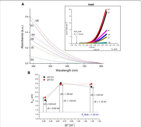
Figure 1a shows the UV-vis absorption curves of the Bi2S3 nanoparticles stabilized using chitosan as the
capping ligand. An absorbance onset was observed at approximatelyλ=800 nm. In general, the UV-vis spectra did not present clearly defined excitonic peaks (i.e., broad excitonic transitions), which may be associated with the relative dispersity of QD size characteristic of aqueous colloidal processing routes compared to or-ganometallic methods [1]. The bandgap energy (EQD) of
the synthesized nanoparticles was estimated using the linear form of TAUC relation for direct bandgap semiconductors (Eq. 2) [25] that relates the absorption coefficient (α) and the photon energy (hν) using a band form parameter (B).
h
ð Þ2¼B h‐E QD
ð Þ ð2Þ
The direct bandgap value of Bi2Si3QDs was calculated
from the plots of (αhν)2 versus hν extrapolating the straight portion of the graph (inset of Fig. 1a) toαhν= 0. The obtainedEQDvalues (Fig. 1b) were higher than the
corresponding bulk value (1.33 eV) for bismuth sulfite [26] as a consequence of the size quantization character-istics of the nanoparticles produced. The “blue-shift” (ΔE=EQD−Eg) determined for the QDs compared to
the bulk value is presented in Fig. 1b.
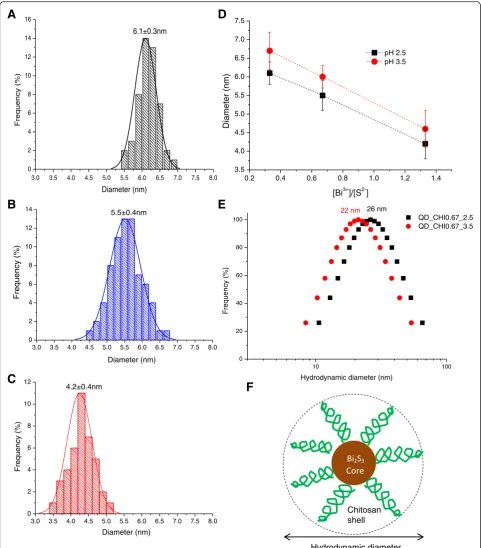
Typical TEM image obtained for the synthesized samples are shown in Fig. 2a, b, revealing that Bi2S3
QDs were spherical and homogenously dispersed. It can be noted that the nanoparticle sizes were below Bohr’s radius (aB~24 nm) for Bi2S3[26], confirming that
nano-particles were in the“quantum confinement regime”and properly stabilized by chitosan as the polymer capping ligand. SAED patterns (inset of Fig. 2b) indicated a lat-tice parameter of 0.29 ± 0.1 nm compatible with the (221) plane of bismuthinite (JCPDS-43-1471). The EDX spectrum clearly indicated Bi and S chemical elements in addition to the peaks related to chitosan, the grid, and the detector (i.e., C, Si, O). The average Bi2S3
nanoparti-cle sizes prepared at pH 2.5 were evaluated using TEM images. Histograms of the diameter distribution for the samples with three molar ratios of [Bi3+]/[S2−] = 0.33, 0.67, and 1.33 are shown in Fig. 3a–c, respectively. Based on the results, the molar ratio of the precursors consid-erably influenced the nanoparticle size of QDs with the highest content of bismuth ([Bi3+]/[S2−] = 1.33, i.e., excess of Bi) leading to the formation of the smallest nanocrystal size. The same trend was observed for the syntheses performed at pH 3.5 (Fig. 3d). Therefore, dif-ferences in the pH did not considerably alter the Bi2S3
QD size. However, it is important to point out that the size of Bi2S3 nanoparticles for higher molar ratios (i.e.,
[Bi3+]/[S2−] > 1.33), which were not synthesized in this study cannot be directly predicted or extrapolated based on the results displayed in Fig. 3d of the three molar ratios of [Bi3+]/[S2−] (0.33, 0.67, 1.33). Essentially, there are several complex thermodynamics and kinetics aspects involved in the colloidal system, regarding the nucleation/growth of these ultra-small nanocrystals in aqueous media using chitosan as a pH-dependent and multidentate polymeric ligand. It can be expected that the dimension of the Bi2S3nanocrystals will not reduce
The effect of the molar ratio ([Bi3+]/[S2−]) on nanoparti-cle size may be explained as follows: at pH = 2.5 and 3.5, all of the amine groups of chitosan are protonated [27] and negatively charged sulfides (S2−) interact with–NH3+
groups as represented in Eqs. 3–5. When Bi3+ ions were added to the reacting vessel, they acted as nucleation sites for Bi2S3 particle formations (Eq. 6). The excess of
bismuth at a molar ratio of [Bi3+]/[S2−] = 1.33 favored the stabilization of the Bi2S3 nanocrystals at smaller
dimen-sions due to the increase in the number of nucleation sites (“seeds”). In parallel, the lower ratio of [Bi3+]/[S2−] pro-duced relatively larger Bi2S3 QDs, as the S2− species in
solution were stabilized by the protonated amine groups in the chitosan chain reducing the nucleation kinetics.
CHI‐NH2 aqð Þ þ Hþð Þ→aq CHI‐NH3þð Þaq ð3Þ
Na2Sð Þ→aq 2Naþð Þaq þS2−ð Þaq ð4Þ
2CHI‐NH3þð Þaq
þ S2−ð Þaq →2 CHIð ‐NH3þÞ−−− S2−
aq
ð Þ ð5Þ
2 CHIð ‐NH3þÞ−−− S2−
aq
ð Þ
þ Bi3þ→Bi2S3 sð Þ þ CHI‐NH3þ ð6Þ
DLS results (Fig. 3e) revealed that the hydrodynamic diameter (HD) of the core-shell (Bi2S3–chitosan)
nanocon-jugates were approximately 22 and 26 nm for the systems QD_CHI0.67_2.5 and QD_CHI0.67_3.5, respectively.
0.30 0.45 0.60 0.75 0.90 1.05 1.20 1.35 1.50
1.0 1.2 1.4 1.6 1.8 2.0 2.2 2.4 2.6 2.8 3.0
E = 0.64 eV
EQD
)
V
e(
[Bi3+]/[S2-]
pH 2.5 pH 3.5
E
g Bulk = 1.33 eV
E = 0.52 eV
E = 1.29 eV
E = 1.23 eV
E = 1.25 eV
E = 1.12 eV
1.00 1.25 1.50 1.75 2.00 2.25 2.50 2.75 3.00 3.25 3.50 3.75 4.00 0
4 8 12 16 20
Bi2S3 bulk
Eg = 1.33 eV
(c) (d)
(a)
(b) (e)
(f)
(
h)
2 (eV.cm -1)
2
h (eV)
2.56 eV 2.62 eV
400 500 600 700 800
0.0 0.2 0.4 0.6
).
u.
a(
e
c
n
a
br
o
s
b
A
Wavelength (nm)
(a)
(b) (c)
(d)
(e)
(f)
A
B
Inset
[image:5.595.57.541.87.519.2]These values are associated with the interactions between chitosan and aqueous medium (Fig. 3f ). As expected, the HD values are higher than the sizes evaluated by TEM
technique, which were only related to dimension of the inorganic core of Bi2S3nanocrystals.
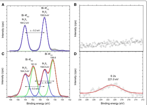
To perform a more in-depth characterization of the Bi2S3 QDs at the nanoscale order and to identify the
chemical states of the elements at the surfaces, the nanoconjugates were evaluated by XPS. Figure 4a shows typical XPS spectra obtained directly at the surface of the Bi2S3/chitosan nanoconjugate. The peaks at 158.9
and 164.2 eV correspond to the Bi 4f7/2 and Bi 4f5/2
levels, respectively, which are generally assigned to Bi–S bonding in Bi2S3 [28, 29]. After etching (Fig. 4c, Ar+,
1 cycle, 60 s, emission current 10 mA, and beam voltage 0.5 kV), the Bi 4f7/2consisted of two binding energies of
156.6 and 158.9 eV, whereas the binding energies values for Bi 4f5/2 were centered at 161.9 and 164.2 eV. The
peaks at 156.6 and 161.9 eV were assigned to bismuth under-coordinated and reduced species due to the etch-ing by argon ion beam [28, 29]. The peaks at 158.9 and 164.2 eV indicate the presence of Bi3+in the Bi2S3phase,
as already observed at the surface prior to etching. In addition, the XPS spectra at the QD surface in the S 2s region (Fig. 4b) indicated no detectable signal from sul-fur. However, after etching, the surface showed a broad peak approximately at 221.0 eV, which can be assigned to the binding energy of S2− state in Bi2S3 [30]. This
0.0 1.0 2.0 3.0 10.0 12.0 14.0
Bi
Energy (keV) Bi
Si S
C
O Cu
Inset
A
B
C
20 nm
[image:6.595.61.538.87.530.2]result indicated that Bi-rich surfaces of Bi2S3 quantum
dots were produced, which is consistent with metal-chalcogenide nanocrystals synthesized in the presence of coordinating ligands bound covalently to cations [7].
Zeta potential (ζ) measurements for QD_CHI0.67_2.5 and QD_CHI0.67_3.5 were ζ= +56 ± 1 mV and ζ= +37 ± 3 mV, respectively. Under the highly acidic conditions of both pH values investigated, all amine groups of
3.0 3.5 4.0 4.5 5.0 5.5 6.0 6.5 7.0 7.5 8.0
0 2 4 6 8 10 12 14 16
)
%(
y
c
n
e
u
q
er
F
Diameter (nm) 6.1±0.3nm
3.0 3.5 4.0 4.5 5.0 5.5 6.0 6.5 7.0 7.5 8.0
0 2 4 6 8 10 12 14
)
%(
y
c
n
e
u
q
er
F
Diameter (nm) 5.5±0.4nm
3.0 3.5 4.0 4.5 5.0 5.5 6.0 6.5 7.0 7.5 8.0
0 2 4 6 8 10 12
)
%(
y
c
n
e
u
q
er
F
4.2±0.4nm
Diameter (nm)
0.2 0.4 0.6 0.8 1.0 1.2 1.4
3.5 4.0 4.5 5.0 5.5 6.0 6.5 7.0 7.5
)
m
n(
r
et
e
m
ai
D
[Bi3+]/[S2-]
pH 2.5 pH 3.5
10 100
0 20 40 60 80
100 QD_CHI0.67_2.5
QD_CHI0.67_3.5
)
%(
y
c
n
e
u
q
er
F
Hydrodynamic diameter (nm)
Hydrodynamic diameter Chitosan shell
22 nm 26 nm
A
B
C
D
E
F
[image:7.595.57.539.87.636.2]chitosan are protonated, but at pH 2.5, there are more H+ions in solution repelling and exposing more –NH3+
groups at the nanohybrid surface (i.e., the outermost surface of the organic shell). At pH 3.5, the molar ratio of the precursors also influenced the ZP values. At a molar ratio [Bi3+]/[S2−] = 0.33, ZP was measured to be ζ= +31 ± 3 mV, which is lower than the value obtained for the stoichiometric system where [Bi3+]/[S2−] = 0.67 (i.e., Bi2S3, ratio 2/3 = 0.67). This difference may be due
to the excess S2− ions forming complexes with proton-ated amines reducing the positive charge at the double layer. For the system QD_CHI1.33_3.5, ZP wasζ= +33 ± 11 mV. The relatively higher statistical standard devi-ation may be attributed to the excess of Bi3+ species that increases the repulsion between the–NH3+ groups
in the chitosan chains, thus disrupting the balanced core-shell nanostructure. In addition, ZP results indi-cated that the QDs were mostly electrostatically stabi-lized (ζ> +30 mV), preventing close contact between nanoparticles, compatible with the homogenously dis-persed features of nanoparticles observed in TEM
images. As a general trend, irrespective to the molar ra-tio of Bi:S used in the synthesis, all nanoconjugates pre-sented positiveζ-potential values, which was attributed to the protonation of amine groups of the chitosan polymeric shell under the acidic conditions at pH = 2.5 and pH = 3.5. Clearly, it is worth mentioning that, as far as the potential applications of these nanoconjugates in biomedical and environmental fields are concerned, these colloidal systems need to be previously buffered to raise the pH of the media to the range of 6.0 to 8.0, to render them biocompatible.
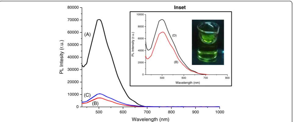
All of the systems presented luminescent behavior under excitation, with the predominant PL emission band appearing in the spectral range of 450 to 650 nm (green-orange) (Fig. 5 and inset). The results of photolu-minescence of Bi2S3 nanoconjugates indicated that the
excitonic emission is not predominant, because the max-ima wavelengths of the PL spectra were statistically simi-lar (λPL= 504 ± 1 nm) despite the different absorptions
in UV-vis spectroscopy. Thus, the emission is the overall contribution of radiative recombination from QDs and/
A
C
B
D
Fig. 4XPS spectra of Bi2S3nanoconjugate (QD_CHI0.67_3.5): Bi 4f region (a) before and (c) after Ar +
[image:8.595.60.540.86.430.2]or electronic transitions from Bi3+, usually the back-ward radiative transition3P1→1S0[31]. Based on Fig. 5,
the luminescence intensity was reasonably dependent on the molar ratio of precursors: the highest lumines-cence response was measured for the 0.33 ratio, which is associated with the biggest nanocrystal size. This be-havior is usually observed due to the density of surface defects (e.g., energy trap stages) and increase in the non-radiative pathways as the quantum dot size de-creases [1, 32]. In addition, the QY of the Bi2S3
nano-conjugates was estimated to be approximately 1.0 %, in good agreement with the reports published of QDs synthe-sized using aqueous colloidal routes at low temperatures,
which is usually smaller than of QDs prepared at high temperature in organic process [1].
Cytotoxicity Assay by MTT of Chitosan/Bi2S3
Nanoconjugates
The biocompatibility of Bi2S3 nanomaterials has been
reported in the literature [33–35]. In this study, the assessment of the cytotoxicity of core-shell Bi2S3/chitosan
nanoconjugates was performed using the enzymatic-based MTT assay, with samples synthesized at pH = 3.5, (QD_CHI0.67_3.5) using SAOS cell line. This method is considered superior to other similar methods
500 600 700 800 900 1000
0 10000 20000 30000 40000 50000 60000 70000 80000
).
u.r
(
yti
s
et
nI
L
P
Wavelength (nm) (A)
(B) (C)
Inset
500 600 700 800
0 2000 4000 6000 8000 10000
).
u.
r(
yti
s
n
et
nI
L
P
Wavelength (nm) (D)
(B)
Fig. 5PL spectra of Bi2S3nanoconjugates: (a) QD_CHI0.33_3.5, (b) QD_CHI0.67_3.5, (c) QD_CHI1.33_3.5, and (d) QD_CHI0.67_2.5 (λexcitation= 405 nm). Inset: effect of pH in PL emission and green luminescence of Bi2S3quantum dots under UV excitation (“dark chamber”). A two-column figure
Cell control Control + Control -
Bi
2
S3
/chitosan nanoconjugates
Inset
[image:9.595.59.540.87.287.2] [image:9.595.58.540.523.703.2]because it is safe, easy to use, has a high reproduci-bility, and is broadly performed for both cell viability and cytotoxicity tests.
Thus, the results of SAOS cells in contact with the Bi2S3/chitosan nanoconjugate samples evidenced no
difference in the cell viability and non-toxic effect com-pared to the control group, within the statistical range of variation (Fig. 6). The nanoconjugates showed cell viability response of over 90 % (i.e., non-cytotoxic), which can be essentially assigned to the approach used in this research. That means, these nanoconjugates were designed and produced with a “cadmium-free” nanocore (Bi2S3) and surface functionalized with the
chitosan polysaccharide shell aiming at rendering them
biocompatible and, therefore, theoretically safer for bio-logical applications. The schematic representation of interactions of the nanoconjugates with the SAOS cell membranes is depicted in Fig. 6 (inset). Moreover, these novel biocompatible and water-soluble nanoconjugates were developed using a facile eco-friendly aqueous pro-cessing route.
Cell Uptake of Bi2S3/Chitosan Nanoconjugates Biomarkers
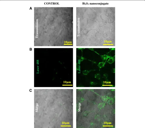
Endocytosis is the biological process responsible for the internalization of nanoparticles by cells. This process depends on energy and functional coordination of lipids and plasma membrane. Therefore, studies that allow
A
B
C
Laser 488
Laser 488
n
oi
ss
i
ms
n
ar
T
Transmission
CONTROL Bi2S3nanoconjugate
e
gr
e
M
Merge
[image:10.595.59.538.280.703.2]the visualization of internalization of nanomaterials are important tools for the development and biological characterization of nanoparticles [36, 37]. Thus, in this study, confocal fluorescence microscopy was used to determine the cell uptake of the Bi2S3 nanoconjugates
in HEK293T cells. Figure 7 shows fluorescent images of QD_CHI0.67_3.5 internalization in comparison with the control sample (autofluorescence). These results demonstrated the biocompatibility and bioimaging properties of the novel Bi2S3 QD–chitosan
nanoconju-gates. They can be used for intracellular targeting as biomarker or for drug-delivery because these nanocon-jugates presented efficient translocation across the cell plasma membrane [38].
Conclusions
In summary, nanoconjugates were designed and synthe-sized with chitosan as the biopolymer shell and Bi2S3
semiconductor quantum dot as the fluorescent inorganic core. These nanohybrids were produced using a single-step eco-friendly aqueous colloidal processing route at room temperature. The results demonstrated that chito-san behaved as an effective ligand for nucleating and stabilizing ultra-small Bi2S3 QDs, leading to the
forma-tion of colloidal core-shell nanostructures in aqueous dis-persions. These nanoconjugates were cytocompatible evaluated by the MTT assay and exhibited photolumines-cence under light excitation. Furthermore, the results of cell studies show that the QD–chitosan conjugates display good HEK293T cell uptake in the absence of non-specific binding to the cell membrane and, therefore, they can be potentially used as fluorescent nanosized bioprobes to label cells in vitro for cell bioimaging applications.
Competing Interests
The authors declare that they have no competing interests.
Authors’Contributions
HSM carried out the experimental design and analysis and drafted the manuscript. AAPM carried out the characterization and analysis and drafted the manuscript. FPR participated in the synthesis, characterization, and analysis of quantum dots. SMC and MCF designed, performed, and analyzed the biological assays of nanoconjugates regarding cytotoxicity and cellular bioimaging. All authors read and approved the final manuscript.
Acknowledgements
The authors acknowledge financial support from the following Brazilian research agencies: CAPES—Coordenação de Aperfeiçoamento de Pessoal de Nível Superior (PROEX-433/2010;PNPD), FAPEMIG—Fundação de Amparo à Pesquisa do Estado de Minas Gerais (PPM-00202-13;BCN-TEC 30030/12), CNPq—Conselho Nacional de Pesquisa (PQ1B-306306/2014-0; UNIVERSAL-457537/2014-0), and FINEP—Financiadora de Estudos e Projetos (CTINFRA-PROINFRA 2008/2010). The authors also express their gratitude to the staff of the Microscopy Center, UFMG, for their assistance on the TEM analysis.
Author details
1
Center of Nanoscience, Nanotechnology and Innovation - CeNano²I, Department of Metallurgical and Materials Engineering, Federal University of Minas Gerais, Av. Antônio Carlos, 6627-Escola de Engenharia, Bloco 2-Sala 2233, Belo Horizonte, Minas Gerais 31.270-901, Brazil.2Department of
Preventive Veterinary Medicine, UFMG, Belo Horizonte, Brazil.3Department of
Physiology and Biophysics, UFMG, Belo Horizonte, Brazil.
Received: 7 February 2016 Accepted: 4 April 2016
References
1. Mansur HS (2010) Quantum dots and nanocomposites. Wiley Interdiscip Rev Nanomed Nanobiotechnol 2:113–129. doi:10.1002/wnan.78
2. Chaudhuri RG, Paria S (2012) Core/shell nanoparticles: classes, properties, synthesis mechanisms, characterization, and applications. Chem Rev 112: 2373–2433. doi:10.1021/cr100449n
3. Hezinger AF, Tessmar J, Gopferich A (2008) Polymer coating of quantum dots—a powerful tool toward diagnostics and sensorics. Eur J Pharm Biopharm 68:138–152. doi:10.1016/j.ejpb.2007.05.013
4. Mansur A, Mansur H, González J (2011) Enzyme-polymers conjugated to quantum-dots for sensing applications. Sensors 11:9951–9972. doi:10.3390/ s111009951
5. Medintz IL, Uyeda HT, Goldman ER, Mattoussi H (2005) Quantum dot bioconjugates for imaging, labeling and sensing. Nat Mater 4:435–446. doi:10.1038/nmat1390
6. Yin Y, Alivisatos AP (2005) Colloidal nanocrystal synthesis and the organic– inorganic interface. Nature 437:664–670. doi:10.1038/nature04165 7. Aresti M, Saba M, Piras R, Marongiu D, Mula G, Quochi F, Mura A, Cannas C,
Mureddu M, Ardu A, Ennas G, Calzia V, Mattoni A, Musinu A, Bongiovanni G (2014) Colloidal Bi2S3nanocrystals: quantum size effects and midgap states. Adv Funct Mater 24:3341–3350. doi:10.1002/adfm.201303879
8. Kershaw SV, Susha AS, Rogach AL (2013) Narrow bandgap colloidal metal chalcogenide quantum dots: synthetic methods, heterostructures, assemblies, electronic and infrared optical properties. Chem Soc Rev 42: 3033–3087. doi:10.1039/C2CS35331H
9. Luo Y, Chen H, Li X, Gong Z, Wang X, Peng X, He M, Sheng Z (2013) Wet chemical synthesis of Bi2S3nanorods for efficient photocatalysis. Mater Lett 105:12–15. doi:10.1016/j.matlet.2013.04.006
10. Green M (2010) The nature of quantum dot capping ligands. J Mater Chem 20:5797–5809. doi:10.1039/C0JM00007H
11. Efros AL (1982) Interband absorption of light in a semiconductor. Sphere Sov Phys Semicond 16:772–775
12. Ekimov AI, Onushchenko AA (1982) Quantum size effect in the optical-spectra of semiconductor micro-crystals. Sov Phys Semicond 16:775–778 13. Mansur HS, Grieser F, Urquhart RS, Furlong DN (1995) Photoelectrochemical
behaviour of Q-state CdSxSe1-x particles in arachidic acid LB films. J Chem Soc Faraday Trans 91:3399–3404. doi:10.1039/FT9959103399
14. Tan WB, Huang N, Zhang Y (2007) Ultrafine biocompatible chitosan nanoparticles encapsulating multi-coloured quantum dots for bioapplications. J Colloid Interface Sci 310:464–470. doi:10.1016/j.jcis. 2007.01.083
15. Drbohlavova J, Adam V, Kizek R, Hubalek J (2009) Quantum dots—characterization, preparation and usage in biological systems. Int J Mol Sci 10:656–673. doi:10.3390/ijms10020656
16. Henglein A (1982) Photo-degradation and fluorescence of colloidal-cadmium sulfide in aqueous solution. Ber Bunsen-Ges Phys Chem 86:301– 305. doi:10.1002/bbpc.19820860409
17. Mansur AAP, Mansur HS, Ramanery FP, Oliveira LC, Souza PP (2014)“Green” colloidal ZnS quantum dots/chitosan nano-photocatalysts for advanced oxidation processes: study of the photodegradation of organic dye pollutants. Appl Catal B 158–159:269–279. doi:10.1016/j.apcatb.2014.04.026 18. Murray CB, Norris DJ, Bawendi MG (1993) Synthesis and characterization of
nearly monodisperse CdE (E = S and Se and Te) semiconductor nanocrystallites. J Am Chem Soc 115:8706–8715. doi:10.1021/ja00072a025 19. Lesnyak V, Gaponik N, Eychmüller A (2013) Colloidal semiconductor
nanocrystals: the aqueous approach. Chem Soc Rev 42:2905–2929. doi:10.1039/C2CS35285K
20. Mansur AAP, Carvalho SM, Mansur HS (2016) Bioengineered quantum dot/ chitosan-tripeptide nanoconjugates for targeting the receptors of cancer cells. Int J Biol Macromol 82:780–789. doi:10.1016/j.ijbiomac.2015.10.047 21. Mansur HS, Mansur AAP, Soriano-Araujo A, Lobato ZIP (2015) Beyond
22. Mansur HS, Mansur AAP, Curti E, Almeida MV (2012) Bioconjugation of quantum-dots with chitosan andN,N,N-trimethyl chitosan. Carbohyd Polym 90:189–196. doi:10.1016/j.carbpol.2012.05.022
23. Liao HC, Wu MC, Jao MH, Chuang CM, Chen YF, Su WF (2012) Synthesis, optical and photovoltaic properties of bismuth sulfide nanorods. Cryst Eng Comm 14:3645–3652. doi:10.1039/C2CE06154F
24. Eaton DF (1988) Reference materials for fluorescence. Pure Appl Chem 60: 1107–1114. doi:10.1351/pac198860071107
25. Tauc J, Menth A (1972) States in the gap. J Non Cryst Solids 8–10:569–585. doi:10.1016/0022-3093(72)90194-9
26. Cheng H, Huang B, Qin X, Zhang X, Dai Y (2012) A controlled anion exchange strategy to synthesize Bi2S3nanocrystals/BiOCl hybrid architectures with efficient visible light photoactivity. Chem Commun 48: 97–99. doi:10.1039/C1CC15487G
27. Ramanery FP, Mansur AAP, Mansur HS (2013) One-step colloidal synthesis of biocompatible water-soluble ZnS quantum dot/chitosan nanoconjugates. Nanoscale Res Lett 8:512. doi:10.1186/1556-276X-8-512
28. Zhang J, Liu S, Yu J, Jaroniec M (2011) A simple cation exchange approach to Bi-doped ZnS hollow spheres with enhanced UV and visible-light photocatalytic H2-production activity. J Mater Chem 21:14655–14662. doi:10.1039/C1JM12596
29. Ginting RT, Lee HB, Tan ST, Tan CH, Jumali MHH, Yap CC, Kang JW, Yahaya M (2016) A simple approach low-temperature solution process for preparation of bismuth-doped ZnO nanorods and its application in hybrid solar cells. J Phys Chem C 120:771–780. doi:10.1021/acs.jpcc.5b11094 30. Panigrahi PK, Pathak A (2013) The growth of bismuth sulfide nanorods from
spherical-shaped amorphous precursor particles under hydrothermal condition. J Nanopart 2013:367812. doi:10.1155/2013/367812 31. Gao G, Peng M, Wondraczek L (2014) Spectral shifting and NIR
down-conversion in Bi3+/Yb3+co-doped Zn
2GeO4. J Mater Chem C 2:8083–8088. doi:10.1039/C4TC01242A
32. Dabbousi BO, Rodriguez-Viejo J, Mikulec FV, Heine JR, Mattoussi H, Ober R, Jensen KF, Bawendi MG (1997) (CdSe)ZnS core−shell quantum dots: synthesis and characterization of a size series of highly luminescent nanocrystallites. J Phys Chem B 101:9463–9475. doi:10.1021/jp971091y 33. Mohan R (2010) Green bismuth. Nat Chem 2:336. doi:10.1038/nchem.609 34. Fang Y, Peng C, Guo R, Zheng L, Qin J, Zhou B, Shen M, Lu X, Zhang G, Shi
X (2013) Dendrimer-stabilized bismuth sulfide nanoparticles: synthesis, characterization, and potential computed tomography imaging applications. Analyst 138:3172–3180. doi:10.1039/c3an00237c
35. Rabin O, Perez JM, Grimm J, Wojtkiewicz G, Weissleder R (2006) An X-ray computed tomography imaging agent based on long-circulating bismuth sulphide nanoparticles. Nat Mater 5:118–122. doi:10.1038/nmat1571 36. Wang T, Bai J, Jiang X, Nienhaus GU (2012) Cellular uptake of nanoparticles
by membrane penetration: a study combining confocal microscopy with FTIR spectroelectrochemistry. ACS Nano 6:1251–1259. doi:10.1021/ nn203892h
37. Zhao F, Zhao Y, Liu Y, Chang X, Chen C, Zhao Y (2011) Cellular uptake, intracellular trafficking, and cytotoxicity of nanomaterials. Small 7:1322–1337. doi:10.1002/smll.201100001
38. Yameen B, Choi WI, Vilos C, Swami A, Shi J, Farokhz OC (2014) Insight into nanoparticle cellular uptake and intracellular targeting. J Control Release 190:485–499. doi:10.1016/j.jconrel.2014.06.038
Submit your manuscript to a
journal and benefi t from:
7 Convenient online submission
7 Rigorous peer review
7 Immediate publication on acceptance
7 Open access: articles freely available online
7 High visibility within the fi eld
7 Retaining the copyright to your article