N A N O E X P R E S S
Open Access
Detection of pH and Enzyme-Free H
2
O
2
Sensing Mechanism by Using GdO
x
Membrane in
Electrolyte-Insulator-Semiconductor Structure
Pankaj Kumar
1, Siddheswar Maikap
1,2,3*, Jian-Tai Qiu
4,5, Surajit Jana
1, Anisha Roy
1, Kanishk Singh
1,
Hsin-Ming Cheng
6, Mu-Tung Chang
6, Rajat Mahapatra
7, Hsien-Chin Chiu
1and Jer-Ren Yang
8Abstract
A 15-nm-thick GdOx membrane in an electrolyte-insulator-semiconductor (EIS) structure shows a higher pH sensitivity of 54.2 mV/pH and enzyme-free hydrogen peroxide (H2O2) detection than those of the bare SiO2
and 3-nm-thick GdOx membranes for the first time. Polycrystalline grain and higher Gd content of the thicker GdOx films are confirmed by transmission electron microscopy (TEM) and X-ray photo-electron spectroscopy (XPS), respectively. In a thicker GdOx membrane, polycrystalline grain has lower energy gap and Gd2+ oxidation states lead to change Gd3+ states in the presence of H2O2, which are confirmed by electron energy loss
spectroscopy (EELS). The oxidation/reduction (redox) properties of thicker GdOx membrane with higher Gd content are responsible for detecting H2O2 whereas both bare SiO2 and thinner GdOx membranes do not
show sensing. A low detection limit of 1μM is obtained due to strong catalytic activity of Gd. The reference voltage shift increases with increase of the H2O2 concentration from 1 to 200 μM owing to more generation
of Gd3+ ions, and the H2O2 sensing mechanism has been explained as well.
Keywords: Enzyme-free H2O2, pH detection, GdOx, Sensing mechanism, Catalytic, EIS structure
Background
Recently, hydrogen peroxide (H2O2) is a major inter-mediate of biological cycles which has been used as a potential biomarker for oxidative stress diagnosis as well as a major catalyst for immune sensing [1, 2]. On the other hand, it is also an essential compound of bleach industries and waste water treatment. H2O2has a major role in modulating mitochondrial function by inhibiting activities of the mitochondrial enzyme in a fully revers-ible fashion [3, 4]. The H2O2sensing assay relies on the use of the enzyme horse radish peroxidase (HRP) to oxidize its substrates and detection using spectropho-tometer [5]. H2O2sensing in a simple way, with a short
time detection with high specificity, is demanded for future disease diagnosis of the human body, and enzyme-free electro-catalytic methods have gained the attention for H2O2sensing. Therefore, various catalysts such as metal, metal oxides, and redox polymers have been reported to detect H2O2 [6–12]. Huang et al. [13] have used the glassy carbon electrode modified by Si nanowire-dispersed CuO nanoparticle. Maji et al. [14] have demonstrated an amperometric H2O2sensor based on reduced graphene oxide-coated silica modified with Au nanoparticles. Wang et al. [15] have developed a H2O2 sensor by using MoS2 nanoparticles. Sun et al. [16] have reported a dumbbell-like Pt-Pd-Fe3O4 nanoparticle-modified glassy carbon electrode which shows electro-catalytic reduction. Liu et al. [17] have reported an amperometric H2O2 sensor based on a Si substrate modified with carbon nanotube microelectrode coated by Pd nanoparticles. Kong et al. [18] have reported a non-enzymatic H2O2sensor based on a Co3O4nanowire * Correspondence:[email protected]
1
Department of Electronic Engineering, Chang Gung University (CGU), 259 Wen-Hwa 1st Rd., Kwei-Shan, Tao-Yuan 333, Taiwan
2Bio-Sensor Lab., Biomedical Engineering Research Center, Department of
Electronic Engineering, Chang Gung University, Tao-Yuan 333, Taiwan Full list of author information is available at the end of the article
grown over a reduced graphene oxide sheet. Hao et al. [19] have developed an amperometric H2O2sensor based on Fe2O3 nanoparticles. Bai et al. [20] have reported a sensor based on carbon dot-decorated multi-walled car-bon nano-composites. Silver (Ag) nanowire [21] and nanoparticle-decorated graphene [22] have been also reported for H2O2sensing. Most of the above groups have used different materials using cyclic voltammetry/ampero-metric methods to sense H2O2 (ranging from few nano-molars to millinano-molars) due to different oxidation states in the presence of H2O2. On the other hand, high-k materials such as Al2O3 [23], Ta2O5 [24], and HfO2 [25] in an electrolyte-insulator-semiconductor (EIS) structure have been reported for pH sensing only; however, the Gd2O3 materials that have been reported are few [26, 27], and even then, there is no report for enzyme-free H2O2 sensing by using a GdOx(x< 1.5) material in a simple EIS structure. In this paper, detection of a pH and enzyme-free H2O2 sensing mechanism has been investigated by using a GdOxmembrane in a simple EIS structure for the first time. Polycrystalline grain, Gd content, and oxidation states (Gd2+/Gd3+) have been confirmed by transmission electron microscope (TEM), X-ray photo-electron spec-troscopy (XPS), and electron energy loss specspec-troscopy (EELS) on grain and boundary regions. The 15-nm-thick GdOxmembrane detects H2O2whereas both 3-nm-thick GdOxand bare SiO2membranes do not sense H2O2. Due to the strong catalytic activity of Gd, a low detection limit of 1 μM is obtained. Both time- and concentration-dependent H2O2 sensing and its mechanism have been investigated.
Methods
p-type 4-in. Si (100) wafer was cleaned by the Radio Corporation of America (RCA) process. Prior to thermal growth of SiO2, HF dip was used to remove native oxide from the surface. After the cleaning process, a 40-nm-thick SiO2layer was grown as an insulating layer by dry oxidation process at 950 °C. Then, the back-side-grown SiO2layer was removed by using a buffer oxide etching (BOE) solution. To fabricate the EIS chip, a 300-nm-thick Al film was deposited on the back side of the Si wafer. The sensing membrane area was defined by standard photolithography process using a negative photoresist-SU8. Then, EIS devices were attached on a printed circuit board having copper lines. An epoxy layer was used to encapsulate the EIS structure and the cop-per line. Therefore, a sensor (S1) using SiO2membrane was fabricated. Our fabrication process of EIS structure can be found elsewhere [28]. This SiO2 sensing mem-brane was modified by deposition of 3-nm- (S2) and 15-nm-thick (S3) GdOx films. The GdOx film was depo-sited by electron beam evaporation. The Gd2O3granules were used during deposition, and the deposition rate was
6 nm/min. A schematic view of the Gd2O3- (or GdOx (x< 1.5)) modified SiO2 sensor is shown in Fig. 1. To probe the thickness and microstructure of GdOx films, low-voltage spherical aberration corrected field emission TEM (Cs-corrected FE-TEM) was performed. The model number is JEOL JEM-ARM200F with accelerating voltages of 60, 120, and 200 kV. In addition, a Cs-corrected FE-TEM Oxford energy spectrometer (energy-dispersive spectroscopy, EDS) and electron loss EDS (EELS, Model 965 QuantumERTM) were used to observe the elemental composition on polycrystalline grain and boundaries. The ambient temperature of our laboratory was 21 ± 3 °C and relative humidity was 50 ± 10 %. The elemental compo-sition was investigated by XPS analyzing chamber. The vacuum of the XPS chamber was 1 × 10⁻⁹Torr. The spec-tra were recorded by using an Al K∝ monochrome X-ray at an energy of 1486.6 eV. The scanning energy range from 0 to 1350 eV was used. All spectra were calibrated by C1sspectrum at a centered peak energy of 284.6 eV. After depositing the GdOxfilms on the SiO2/Si substrates, the samples were transferred immediately to the XPS chamber. The capacitance-voltage (C-V) measurements were performed by using Agilent 4284A LCR meter and an Ag/AgCl reference electrode was used. The measure-ment frequency was 100 Hz. The sweep voltage was ap-plied on the Ag/AgCl electrode. The reference voltage (Vr) was measured at 50 % of accumulation capacitance.
[image:2.595.305.538.505.692.2]Results and Discussion
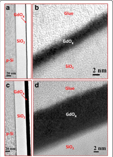
Figure 2 shows the cross-sectional TEM images of the S2 and S3 sensors. The thickness of SiO2 is 41.2 nm (Fig. 2a), and the thickness of the GdOx film is 3.3 nm (Fig. 2b). The TEM image of the S3 sensor shows that the thickness of SiO2 is 41.5 nm (Fig. 2c) and the
Fig. 1Schematic view of our pH and H2O2sensor using Gd2O3
(or GdOx(x< 1.5)) membrane and demonstration of H2O2
thickness of the GdOx film is 14.8 nm (Fig. 2d). There-fore, the thickness of SiO2 is 40 ± 2 nm and the thick-ness of GdOx is 15 ± 0.5 nm. The thicker GdOx film shows clearly polycrystalline grains and its boundary [29, 30], which will help to detect H2O2. Elemental composition of the SiO2and GdOx films is observed by XPS, which is shown in Fig. 3. The peak binding energy of Si2pspectra for the S1 sample is 103.35 eV (Fig. 3a), which is similar to the reported value of SiO2 at 103.58 eV [31]. The spectra are fitted by Shirley back-ground subtraction and Gaussian/Lorentzian functions. The Si2p spectrum shows one characteristic peak after de-convolution. Similarly, one characteristic peak of O1s centered at 531.5 eV is also observed (Fig. 3d). Lower values of full-width half-maximum (FWHM) are found to be 1.84 and 1.64 eV for the Si2pand O1sspectra, respec-tively. The ratio of O:Si is 1.84, which signifies the stoichiometric SiO2. An XPS spectrum of GdOx shows Gd3d3/2 and Gd3d5/2 doublet with binding energy of 1220.5 and 1188.3 eV, respectively (not shown here). However, peak binding energies of Gd3d3/2and Gd3d5/2 spin-orbits are reported as 1218 and 1186 eV, respectively
[image:3.595.57.291.88.412.2][32]. XPS spectra of Gd3d5/2 core-level electrons are 1189 eV for S2 (Fig. 3b) and 1188.7 eV for S3 (Fig. 3c) samples, which are identified to be Gd2O33d5/2or Gd2O3 films. Corresponding lower binding energy peaks at 1186.2 and 1185.8 eV indicate the metallic Gd3d5/2peaks for the S2 and S3 samples, respectively. The area ratios of Gd/Gd2O3are found to be 0.64:1 and 0.69:1 for the S2 and S3 samples, respectively, which show higher percen-tage of Gd in the S3 samples owing to polycrystalline grains. However, the O1s core-level spectra show three distinct peaks for the S2 (Fig. 3e) and S3 (Fig. 3f) samples. The strong peaks at 531.5 eV correspond to the oxygen in the Gd2O3film, whereas lower (O1sA) and higher (O1s B) binding energy peaks centered at 529 and 532.9 eV are attributed to the hydroxyl (OH−) and carbonate groups in Gd2O3 films, respectively [33, 34]. Moreover, the lower binding energy peak corresponds to Gd-O bonding or GdOx [35]. The area ratios of O1s A and O1s B with respect to O1s are 0.04:1 and 0.48:1 for the S2 samples whereas those values are 0.08:1 and 0.1:1 for the S2 samples, respectively. Therefore, the S2 samples show higher percentage of O1s B owing to higher carbonate groups in the GdOx films, which is insensitive to H2O2 sensing. On the other hand, the S3 samples have higher percentage of O1sA owing to higher OH−and higher Gd content in Gd2O3film, i.e., GdOxfilm. So, oxygen can be bonded loosely with Gd on a polycrystalline grain boun-dary as well as a thicker GdOx film will help to sense H2O2, which will be explained below.
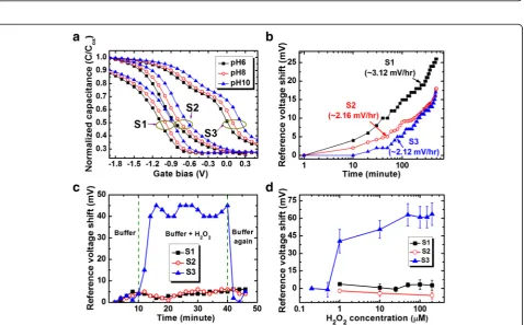
Figure 4a shows the C-V characteristics with pH values from 6 to 10 for the S2 and S3 sensors. The Vr values of the S2 sensors are−0.84,−0.75, and−0.63 V for pH 6, 8, and 10, while those values are 0.01, 0.1, and 0.23 V for the S3 sensors, respectively. The Vr values of the S3 sensor are shifted towards the positive direction and are lower than the Vr values of the S2 sensors. This is due to lower oxide charges for the thicker GdOx membrane (55 vs. 43 nm [36]) and polycrystalline grains with higher OH− ions (Fig. 3f ). The pH sensitivity values are found to be 51.2 and 54.2 mV/pH for the S2 and S3 sensors, respectively, which are higher than the pH sensitivity of approximately 35 mV/pH from pH 2 to 10 [28, 37] and 42 mV/pH from pH 6 to 10 for the S1 sensors. The pH sensitivity of a 30-nm-thick GdOx membrane is appro-ximately 51.7 mV/pH (not shown C-V curves), which is slightly lower than the S3 sensors. The pH sensitivity value of our GdOx membrane is comparable with other reported values of 48.29 mV/pH by Wang et al. [27], 64.78 mV/pH by Chang et al. [38], and 55 mV/pH by Yang et al. [39]. However, the S3 sensors show the lowest drift rate as compared to the S1 and S2 sensors (2.12 mV/h vs. 3.12 mV/h and 2.16 mV/h), as shown in Fig. 4b. The drift characteristics were measured a long time up to 500 min at pH 7 buffer solution. Considering a Fig. 2TEM images ofa3-nm-thick GdOxmembrane on 40-nm-thick
SiO2layer (S2) andbzoom in view of a. TEM images ofc15-nm-thick
GdOxmembrane (S3) anddzoom in view of c. Thicker membrane
Fig. 3XPS characteristics of Si2pforaS1,bS2, andcS3 samples. Corresponding O1sspectra ofdS1,eS2, andfS3 samples are shown. The S3 film shows higher Gd/Gd2O3ratio or oxygen deficient and higher OH group which helps to sense H2O2
[image:4.595.57.540.88.376.2] [image:4.595.61.540.406.703.2]low drift rate (2.12 mV/h), the pH detection limit of the S3 sensors is 0.039 pH, which is due to high pH sensitivity. It is interesting to note that the GdOx membrane will detect H2O2. Figure 4c shows the time-dependent re-sponse of H2O2for the S3 sensors. A negligible Vrshift is observed for pH 7 buffer solution up to 10 min. By includ-ing H2O2with a concentration of 1μM, a good Vrshift of approximately 40 mV is observed because of Gd1+, Gd2+, and Gd3+ oxidation states (https://en.wikipedia.org/wiki/ Work_function) [40]. On the other hand, both S1 and S2 sensors do not show H2O2sensing. When in contact with H2O2, the Gd2+changes to the Gd3+oxidation state and provides electrons for the reduction of H2O2. H2O as a byproduct is observed (Fig. 1). However, the pH value is unchanged by adding H2O2in the buffer solution. A short response time of <2 min is needed without enzyme. After washing out, the sensor does not show any Vrshift owing to the reduction from the Gd3+to Gd2+states. Therefore, this sensor can be used repeatedly for H2O2 sensing. Based on our knowledge, this is the first ever report of H2O2 detection with a polycrystalline GdOx membrane. Basically, the oxidation/reduction of the GdOxmaterial in contact with H2O2with buffer solutions is responsible for the Vr shifting, which is shown by chemical reactions below.
Gd↔Gd2þþ2e−↔Gd3þþ3e− ð1Þ
H2O2þe−↔OH−þOH ð2Þ
OHþe−↔OH− ð3Þ
2OH−þ2Hþ↔2H2O ð4Þ
By following the above Eqs. (1), (2), (3), and (4), the oxidation state of Gd changes from Gd2+ to Gd3+. The H+ ions are supplied by buffer solutions. The Vr shift increases with increasing H2O2concentration from 1 to 200 μM because the generation of Gd3+ ions increases (Fig. 4d). A moderate sensitivity of 0.13 mV/μM is obtained from a linear range of 1 to 200μM whereas it is 82 mV/μM from a linear range of 0.5 to 1 μM. Our detection limit of 1 μM is inferior than the published results [9–12, 15, 16, 41–43], comparable with the pub-lished results [44–47], and superior than the published results [13, 17, 18, 20, 48–52] in literature by using different sensing methods, as shown in Table 1. Further study is needed to improve the detection limit in the future. However, our sensing method’s surface potential is changed when in contact with H2O2 because of the catalytic activity of Gd. It is known that Gd2O3material is n-type and the energy difference in between the Fermi level and the conduction band (Ec) is 2.71 eV [53]. The electron affinity of Gd2O3is 1.45 eV by considering the conduction band offset of 2.6 eV with Si [54]. The work
function of Gd increases from 2.9 eV (https://en.wikipe dia.org/wiki/Work_function) to 4.16–4.76 eV [53–55] after oxidation. This suggests that the work function of GdOx is modulated by oxidation/reduction or Gd
3+
[image:5.595.305.538.110.464.2]concentration as well as the energy band bending of Si is changed. In consequence, the Vr is needed to bring Si energy bands to be flat. On the other hand, the S1 and S2 sensors do not show H2O2detection because they do not have redox properties. The thinner GdOxfilm (S2) has a smaller crystalline grain with less Gd content (Fig. 3), while the S3 sensor has larger crystalline grain (Fig. 5a) with higher Gd content. Figure 5b shows electron energy loss spectroscopy of Gd measured at polycrystalline grain (P1) and amorphous region or grain boundary (P1). The regions of P1and P2are marked on Fig. 5a. The edges of the Gd M-4 and M-5 peaks at the P1region are located at 1216.8 and 1187.5 eV, while those values at the P2region are 1216.5 and 1187 eV, respectively. Du et al. [56] have reported the M-4 and M-5 peak values of 1217 and 1185 eV for the Gd(OH)3nanorods. The edges of the O-K Table 1Comparison of linear range and detection limit of H2O2
published in literature [9–13, 15–18, 20, 41–52]
Sensing materials pH value Linear range (μM)
Detection limit (μM)
MoS2NP [15] 7.4 5–100 0.002
WS2NS [10] 7.4 – 0.002
Pt-Pd-Fe3O4[16] 7.4 0.02–0.1, 2–14,000 0.005
Pt-Pd/rGO [11] 7.0 0.1–37.6 0.01
Au NP [12] 7.0 2–5000 0.01
Pt NP [9] 7.2 3–300 0.03
rGO [41] 7.0 0.05–1500 0.05
Au/C/Pt [42] 7.0 9.0–1860, 1860–7110 0.13
Au NP [43] 6.8 3–605 0.18
Ag NP [44] 7.5 100–10,000 0.88
GS/CeO2-ZnO NP [45] 7.0 2–20,000 1.1
Pt-Pd and Pt-Ir [46] 7.4 2.5–125 1.2
Pt NP [47] 6.9 5–2000 1.23
CeO2NP/N-rGO [48] 7.0 1.8–920.8 1.3
CuO [13] 7.0 10–13,180 1.6
Ag NPs/PPy/Fe3O4[49] 7.2 5–11,500 1.7
Pd NP [17] 7.4 2–1300 2
Co3O4NW [18] 7.4 15–675 2.4
Carbon dots [20] 7.4 3–300 3
Se/Pt [50] 7.0 10–15,000 3.1
Ag NP [51] 7.0 25–500, 500–5500 10
Co-Mn [52] 7.2 100–25,000 15
GdOxin EIS structure (this work)
7.0 1–200 1
peak at both P1and P2regions are located at 538.5 eV, as shown in Fig. 5c, which is close to the reported value of 536.5 eV [56]. It is interesting to note that another peak of crystalline grain (P1) is located at 532.9 eV, which is shifted downwards to 3.9 eV. Egerton has reported the reduced energy gap of SiOx at the SiO2/Si interface with energy shift downwards to 3 eV [57]. In our case, this reduced energy gap is observed in the polycrystalline grain region. Therefore, the crystalline grain is GdOx(or Gd2+) and the amorphous region or grain boundary is Gd2O3(or Gd3+). When in contact with H2O2, the oxidation state of the S3 sensor changes from Gd2+ to Gd3+ and the crystalline
grain takes a major role, which is confirmed by EELS spec-tra. So, the thicker crystalline GdOxmembrane can sense H2O2 repeatedly which will be useful to detect human disease in the near future.
Conclusions
Higher pH sensitivity (54.2 m/pH) and the enzyme-free H2O2 sensing characteristics have been investigated by using 15-nm-thick GdOx membranes for the first time. The polycrystalline grain and thickness of the GdOx/ SiO2film have been observed by TEM image. XPS cha-racteristics of the S3 membrane show higher Gd/Gd2O3
Fig. 5aTEM image for EELS spectra of the S3 membranes. The edges ofbGd andcO-K are plotted for theP1andP2regions marked on a.
[image:6.595.57.542.84.545.2]ratio than the S2 membrane (0.69/1 vs. 0.64/1). The S3 membrane shows GdOx and higher OH content in the crystalline grain, which help to sense H2O2 whereas both S1 and S2 sensors do not show H2O2 detection. Therefore, a larger polycrystalline GdOx grain has oxida-tion/reduction properties when in contact with H2O2, which is confirmed by EELS. During oxidation, the Gd2+ changes to the Gd3+ state and the amount of Gd3+ ions increases with increasing H2O2 concentration from 1 to 200μM. A low defection limit of 1μM is obtained owing to the catalytic effect of Gd. The time-dependent response and the sensing mechanism of H2O2have been explored. Due to the short time detection of H2O2in the EIS struc-ture, this novel GdOx sensing membrane paves a way to diagnose other diseases of the human body in the near future.
Acknowledgements
This work was supported by the Ministry of Science and Technology (MOST), Taiwan, under contract numbers MOST-104-2221-E-182-075 and MOST-105-2221-E-182-002 and Chang Gung Memorial Hospital (CGMH), Linkou, under contract numbers CMRPD270021 and CMRPD2E0091. The authors are grateful to Mr. S. Chatterjee for the partial support to measure the concentration-dependent hydrogen peroxide sensing.
Authors’Contributions
PK fabricated these sensors and analyzed the data under the instruction of SM. JTQ helped to analyze the sensing mechanism and application of this sensor. SJ and AR helped to measure the pH and H2O2sensing
characteristics and checked the repeatability of these sensors. They review the papers under the instruction of SM. KS helped to check the redox characteristics and review the papers under the instruction of SM. HMC measured the XPS and analyzed the spectra. MTC helped to obtain TEM and EELS. RM helped to analyze the sensing characteristics. HCC helped to deliver the idea for deposition of sensing membrane by using electron beam evaporation. JRY analyzed the EELS spectra for oxidation and reduction. All authors contributed to the revision of the manuscript, and they approved it for publication.
Competing Interests
The authors declare that they have no competing interests.
Author details
1Department of Electronic Engineering, Chang Gung University (CGU), 259
Wen-Hwa 1st Rd., Kwei-Shan, Tao-Yuan 333, Taiwan.2Bio-Sensor Lab.,
Biomedical Engineering Research Center, Department of Electronic Engineering, Chang Gung University, Tao-Yuan 333, Taiwan.3Center for
Reliability Science and Technologies (CReST), Department of Electronic Engineering, Chang Gung University, Tao-Yuan 333, Taiwan.4Department of
Biomedical Sciences, School of Medicine, Chang Gung University (CGU), Tao-Yuan 333, Taiwan.5Division of Gyn-Oncology, Department of Obs/Gyn,
Chang Gung Memorial Hospital (CGMH), Tao-Yuan 333, Taiwan.6Material and
Chemical Research Laboratories (MRL), Industrial Technology Research Institute (ITRI), Hsinchu 195, Taiwan.7Department of Electronics and
Communications Engineering, National Institute of Technology (NIT), Durgapur 713209, India.8Department of Materials Science and Engineering,
National Taiwan University (NTU), Taipei 106, Taiwan.
Received: 13 August 2016 Accepted: 22 September 2016
References
1. Chen X, Wu G, Cai Z, Oyama M, Chen X (2014) Advances in enzyme-free electrochemical sensors for hydrogen peroxide, glucose, and uric acid. Microchim Acta 181:689
2. Wang J, Chen XJ, Liao KM, Wang GH, Han M (2015) Pd nanoparticle-modified electrodes for nonenzymatic hydrogen peroxide detection. Nanoscale Res Lett 10:311
3. Nulton-Persson AC, Szweda LI (2001) Modulation of mitochondrial function by hydrogen peroxide. J Biol Chem 276:23357
4. Veal EA, Day AM, Morgan BA, Morgan D (2007) Hydrogen peroxide sensing and signaling. Mol Cell 26:1
5. Rhee SG, Chang TS, Jeong W, Kang D (2010) Methods for detection and measurement of hydrogen peroxide inside and outside of cells. Mol Cell 29:539 6. Chen S, Yuan R, Chai Y, Hu F (2013) Electrochemical sensing of hydrogen
peroxide using metal nanoparticles: a review. Microchim Acta 180:15 7. Chen W, Cai S, Ren QQ, Wen W, Zhao YD (2012) Recent advances in
electrochemical sensing for hydrogen peroxide: a review. Analyst 137:49 8. Li L, Du Z, Liu S, Hao Q, Wang Y, Li Q, Wang T (2010) A novel nonenzymatic
hydrogen peroxide sensor based on MnO2/graphene oxide nanocomposite.
Talanta 82:1637
9. Zhang M, Liao C, Mak CH, You P, Mak CL, Yan F (2016) Highly sensitive glucose sensors based on enzyme-modified whole-graphene solution-gated transistors. Sci Rep 5:1
10. Tang J, Quan Y, Zhang Y, Jiang M, Al-Enizi AM, Kang B, An T, Wang W, Xia L, Gong X, Zheng G (2016) Three-dimensional WS2nanosheet networks for
H2O2produced for cell signaling. Nanoscale 8:5786
11. Sun Y, Zheng H, Wang C, Yang M, Zhou A, Duan H (2016) Ultrasonic-electrodeposition of PtPd alloy nanoparticles on ionic liquid-functionalized graphene paper: towards a flexible and versatile nanohybrid electrode. Nanoscale 8:1523
12. Vilian TE, Veeramani V, Chen S-M, Madhu R, Kwak CH, Huh YS, Han YK (2015) Immobilization of myoglobin on Au nanoparticle-decorated carbon nanotube/polytyramine composite as a mediator-free H2O2and nitrite
biosensor. Sci Rep 5:18390
13. Huang J, Zhu Y, Zhong H, Yang X, Li C (2014) Dispersed CuO nanoparticles on a silicon nanowire for improved performance of nonenzymatic H2O2
detection. ACS Appl Mater Interfaces 6:7055
14. Maji SK, Mandal AK, Nguyen KT, Borah P, Zhao Y (2015) Cancer cell detection and therapeutics using peroxidase-active nanohybrid of gold nanoparticle-loaded mesoporous silica-coated graphene. ACS Appl Mater Interfaces 7:9807
15. Wang T, Zhu H, Zhuo J, Zhu Z, Papakonstantinou P, Lubarsky G, Lin J, Li M (2013) Biosensor based on ultrasmall MoS2nanoparticles for electrochemical
detection of H2O2released by cells at the nanomolar level. Anal Chem
85:10289
16. Sun X, Guo S, Liu Y, Sun S (2012) Dumbbell-like PtPd–Fe3O4nanoparticles
for enhanced electrochemical detection of H2O2. Nano Lett 12:4859
17. Liu Y, Sun G, Jiang C, Zheng XT, Zheng L, Li CM (2014) Highly sensitive detection of hydrogen peroxide at a carbon nanotube fiber microelectrode coated with palladium nanoparticles. Microchim Acta 181:63
18. Kong L, Ren Z, Zheng N, Du S, Wu J, Tang J, Fu H (2015) Interconnected 1D Co3O4nanowires on reduced graphene oxide for enzymeless H2O2
detection. Nano Res 8:469
19. Hao C, Shen Y, Wang Z, Wang X, Feng F, Ge C, Zhao Y, Wang K (2016) Preparation and characterization of Fe2O3nanoparticles by solid phase
method and its hydrogen peroxide sensing properties. ACS Sustainable Chem Eng 4:1069
20. Bai J, Sun C, Jiang X (2016) Carbon dots-decorated multiwalled carbon nanotubes nanocomposites as a high-performance electrochemical sensor for detection of H2O2in living cells. Anal Bioanal Chem 408:4705
21. Zhang M, Wang Z (2013) Nanostructured silver nanowires-graphene hybrids for enhanced electrochemical detection of hydrogen peroxide. Appl Phys Lett 102:213104
22. Zhan B, Liu C, Shi H, Li C, Wang L, Huang W, Dong X (2014) A hydrogen peroxide electrochemical sensor based on silver nanoparticles decorated three-dimensional grapheme. Appl Phys Lett 104:243704
23. Oha JY, Jang HJ, Cho WJ, Islam MS (2012) Highly sensitive electrolyte-insulator-semiconductor pH sensors enabled by silicon nanowires with Al2O3/SiO2sensing membrane. Sens Actuators B 171–172:238
24. Schoning MJ, Brinkmann D, Rolka D, Demuth C, Poghossian A (2005) CIP (cleaning-in-place) suitable‘non-glass’pH sensor based on a Ta2O5- gate EIS
structure. Sens Actuators B 111–112:423
26. Yang HW, Huang CY, Lin CW, Liu HL, Huang CW, Liao SS, Chen PY, Lu YZ, Wei KC, Ma CC (2014) Gadolinium-functionalized nanographene oxide for combined drug and microRNA delivery and magnetic resonance imaging. Biomaterials 35(24):6534. doi:10.1016/j.biomaterials.2014.04.057
27. Wang JJ, Lin SR, Kao CH, Hu CH, Chen H (2015) Electrolyte-insulator-semiconductor (EIS) with Gd2O3-based sensing membrane for pH sensing
application. J New Mat Electrochem Syst 18:1
28. Kumar P, Maikap S, Prakash A, Tien TC (2014) Time-dependent pH sensing phenomena using CdSe/ZnS quantum dots in EIS structure. Nanoscale Res Lett 9:179
29. Manna S, Aluguri R, Katiyar A, Das S, Laha A, Osteen HJ, Ray SK (2013) MBE-grown Si and Si1-xGexquantum dots embedded within epitaxial Gd2O3on Si
(111) substrate for floating gate memory device.Nanotechnology 24:505709 30. Mahapatra R, Maikap S, Lee JH, Kar GS, Dhar A, Hwang NM, Kim DY,
Mathur BK, Ray SK (2003) Effects of interfacial nitrogen on the structural and electrical properties of ultrathin ZrO2gate dielectrics on partially strain
compensated SiGe/Si heterolayers. Appl Phys Lett 82:4331
31. Jana D, Dutta M, Samanta S, Maikap S (2014) RRAM characteristics using a new Cr/GdOx/TiN structure. Nanoscale Res Lett 9:680
32. Ahren M, Selegard L, Klasson A, Soderlind F, Abrikossova N, Skoglund C, Bengtsson T, Engstrom M, Kall PO, Uvda K (2010) Synthesis and characterization of PEGylated Gd2O3nanoparticles for MRI contrast
enhancement. Langmuir 26:5753
33. Majeeda S, Shivashankar SA (2014) Rapid, microwave-assisted synthesis of Gd2O3and Eu:Gd2O3nanocrystals: characterization, magnetic, optical and
biological studies. J Mater Chem B 2:5585
34. Khan SA, Gambhir S, Absar A (2014) Extracellular biosynthesis of gadolinium oxide (Gd2O3) nanoparticles, their bio-distribution and bio-conjugation with
the chemically modified anticancer drug taxol. J Nanotechnol 5:249 35. Cho HK, Cho HJ, Lone S, Kim DD, Yeum JH, Cheong IW (2011) Preparation
and characterization of MRI-active gadolinium nanocomposite particles for neutron capture therapy. J Mater Chem 21:15486
36. Duenas S, Castan H, Garcia H, Gomez A, Bailon L, Kukli K, Hatanpaa T, Lu J, Ritala M, Leskela M (2007) Electrical properties of atomic-layer-deposited thin gadolinium oxide high-k gate dielectrics. J Electrochem Soc 154:G207 37. Maikap S, Prakash A, Banerjee W, Das A, Lai CS (2010) Characteristics of pH
sensors fabricated by using protein mediated CdSe/ZnS quantum dots. Microelectron Reliab 50:747
38. Chang LB, Ko HH, Jeng MJ, Lee YL, Lai CS (2007) pH-sensitive Gd2O3∕SiO2
stacked capacitors prepared by pure water anodic oxidation. J Electrochem Soc 154:J150
39. Yang CM, Wang CY, Lai CS (2014) Characterization on pH sensing performance and structural properties of gadolinium oxide post-treated by nitrogen rapid thermal annealing. J Vac Sci Technol B 32:03D113 40. Hong M (1999) Epitaxial cubic gadolinium oxide as a dielectric for gallium
arsenide passivation. Science 283:1897
41. Zhou M, Zhai Y, Dong S (2009) Electrochemical sensing and biosensing platform based on chemically reduced graphene oxide. Anal Chem 81:5603 42. Zhang Y, Li Y, Jiang Y, Li Y, Li S (2016) The synthesis of Au@C@Pt
core-double shell nanocomposite and its application in enzyme-free hydrogen peroxide sensing. Appl Surf Sci 378:375
43. Bao S, Du M, Zhang M, Zhu H, Wang P, Yang T, Zou M (2014) Fabrication of gold nanoparticles modified carbon nanofibers/polyaniline electrode for H2O2determination. J Electrochem Soc 161:H816
44. Wu Z, Yang S, Chen Z, Zhang T, Guo T, Wang Z, Liao F (2013) Synthesis of Ag nanoparticles-decorated poly(m-phenylenediamine) hollow spheres and the application for hydrogen peroxide detection. Electrochim Acta 98:104 45. Yang X, Wu F, Ouyang Y, Hu Y, Zhang H, Ji Y (2016) Zinc oxide
nanoparticles decorated on graphene sheets@cerium oxide
nanocomposites to sensitize gold electrode for the nonenzymatic hydrogen peroxide detection. J Electrochem Soc 163:H834
46. Chen KJ, Pillai KC, Rick J, Pan CJ, Wang CH, Liu CC, Hwang BJ (2012) Bimetallic PtM (M = Pd, Ir) nanoparticle decorated multi-walled carbon nanotubes enzyme-free, mediator-less amperometric sensor for H2O2.
Biosens Bioelectron 33:120
47. Niu X, Zhao H, Chen C, Lan M (2012) Platinum nanoparticle-decorated carbon nanotube clusters on screen-printed gold nanofilm electrode for enhanced electrocatalytic reduction of hydrogen peroxide. Electrochim Acta 65:97 48. Yang S, Li G, Wang G, Liu L, Wang D, Qu L (2016) Synthesis of highly
dispersed CeO2nanoparticles on N-doped reduced oxide graphene and
their electrocatalytic activity toward H2O2. J Alloy Compd 688:910
49. Qi C, Zheng J (2015) Novel nonenzymatic hydrogen peroxide sensor based on Fe3O4/PPy/Ag nanocomposites. J Electroanal Chem 747:53
50. Li Y, Zhang JJ, Xuan J, Jiang LP, Zhu JJ (2010) Fabrication of a novel nonenzymatic hydrogen peroxide sensor based on Se/Pt nanocomposites. Electrochem Commun 12:777
51. Roof BR, Ojani R, Hasheminejad E, Rashid-Nadimi S (2012) Electrochemical synthesis of Ag nanoparticles supported on glassy carbon electrode by means of p-isopropyl calix[6]arene matrix and its application for electrocatalytic reduction of H2O2. Appl Surf Sci 258:2788
52. Kuo CC, Lan WJ, Chen CH (2014) Redox preparation of mixed-valence cobalt manganese oxide nanostructured materials: highly efficient noble metal-free electrocatalysts for sensing hydrogen peroxide. Nanoscale 6:334 53. Chiu YP, Huang BC, Shih MC, Shen JY, Chang P, Chang CS, Huang ML,
Tsai M-H, Hong M, Kwo J (2011) Atomic-scale determination of band offsets at the Gd2O3/GaAs (100) hetero-interface using scanning tunneling
spectroscopy. Appl Phys Lett 99:212101
54. Fissel A, Czernohorsky M, Osten HJ (2006) Growth and characterization of crystalline gadolinium oxide on silicon carbide for high-application superlattices. Microstruct 40:551
55. Lipp E, Shahar Z, Bittel BC, Lenahan PM, Schwendt D, Osten HJ, Eizenberg M (2011) Trap-assisted conduction in Pt-gated Gd2O3/Si capacitors.
J Appl Phys 109:073724
56. Du G, Tendeloo GV (2005) Preparation and structure analysis of Gd(OH)3
nanorods. Nanotechnology 16:595
57. Egerton RF (2009) Electron energy-loss spectroscopy in the TEM. Rep Prog Phys 72:016502
Submit your manuscript to a
journal and benefi t from:
7Convenient online submission
7Rigorous peer review
7Immediate publication on acceptance
7Open access: articles freely available online
7High visibility within the fi eld
7Retaining the copyright to your article

![Table 1 Comparison of linear range and detection limit of H2O2published in literature [9–13, 15–18, 20, 41–52]](https://thumb-us.123doks.com/thumbv2/123dok_us/8852742.935576/5.595.305.538.110.464/table-comparison-linear-range-detection-limit-published-literature.webp)

