N A N O E X P R E S S
Open Access
Silica Cladding of Ag Nanoparticles for High
Stability and Surface-Enhanced Raman
Spectroscopy Performance
Miaomiao Zhao, Hao Guo, Wenyao Liu, Jun Tang, Lei Wang, Binzhen Zhang, Chenyang Xue, Jun Liu
*and Wendong Zhang
Abstract
For high-precision biochemical sensing, surface-enhanced Raman spectroscopy (SERS) has been demonstrated to be a highly sensitive spectroscopic analytical method and Ag is considered to be the best material for SERS performance. Due to the high surface activity of Ag nanoparticles, the high stability of Ag nanostructures, especially in moist environments, is one of the key issues that need to be solved. A method for silica (SiO2) cladding of Ag nanoparticles (NPs) is demonstrated here for high sensitivity and long-term stability when putted in aqueous solution. The chemically inert, transparent, hydrophilic, and bio-compatible SiO2surface acts as the protection layer for the Ag nanoparticles, which can also enhance the Raman intensity to a certain extent. In our study, the Ag@SiO2core-shell substrate can detect crystal violet solutions with molar concentrations down to 10−12M. After 24 h of immersion, the reduction in Raman scattering intensity is about 85 % for sole Ag NP films, compared to 12 % for the Ag coated with a 10-nm SiO2layer. This thickness was found to be optimum for Ag@SiO2core-shell substrates with long-term stability and high SERS activity.
Keywords:SERS, Ag@SiO2, Long-term stability, Layer thickness
Background
As a powerful spectroscopic technique, surface-enhanced Raman spectroscopy (SERS) has shown promising applica-tions in surface adsorption, biochemical sensing, and trace-level analysis as a result of its high sensitivity, rapid response, and the advantages of nondestructive detection [1–4]. The mechanism for SERS is mainly attributed to the electromagnetic field enhancement caused by the localized surface plasmon resonance of noble metal nanoparticles (NPs). For isolated metal particles, the electromagnetic enhancement can reach up to 106–107, and when in nanogaps (so-called hotspots), it can reach up to 1010–1011, because of the electromagnetic coupling between the neigh-boring metal NPs [5, 6].
Among noble metals, Ag is considered to be one of the most promising candidates for SERS applications due to its low loss in optical frequency and high plas-monic efficiency, as well as its lower cost compared to
other noble metals [7–10]. However, Ag NPs suffer from sulfur contamination, oxidation, and agglomeration in water and the atmosphere, and the biological incompatibility of Ag is obvious, all of which limit their practical application.
Significant efforts have been devoted to improve the chemical stability of Ag NPs, and core-shell nanostructures are one of the most popular methods, which have been reported in literatures [11, 12]. Ag NPs capped with Au [13], graphene [14], and TiO2 [15] have been reported in recent years. Ma et al. [16] prepared ultrathin (~1.5 nm) Al2O3 films by the atomic layer deposition technique on Ag nanorods that can maintain robust morphologies to a temperature of 400 °C. Li et al. [17] reported the use of a single-atom-thick monolayer of graphene for the protection of Ag NPs that can function as a highly stable SERS sub-strate for nearly 1 month with ambient aerobic exposure.
The capping thickness of the protection layers can be well controlled by the fabrication technology, which has greatly extended the application of Ag-based SERS substrates in different fields. However, a defect of this
* Correspondence:[email protected]
Science and Technology on Electronic Test & Measurement Laboratory, North University of China, Taiyuan, Shanxi 030051, China
coating approach is the tremendous decrease in SERS ac-tivity, which is caused by the coating layers that separate the target molecules from the Ag NPs and by the possible morphology changes of the Ag NPs engendered during the coating process. Thus, it is vital to find ways to deposit protective layers which can cap Ag NPs at relatively low temperatures and to precisely control the coating thickness to prohibit the reduction of SERS sensitivity, while still thick enough to be robust towards moist environments.
In this study, Ag NPs were fabricated on 2-in. silicon wafers with a sputtering and vacuum annealing process. We employed inductively coupled plasma-enhanced chem-ical vapor deposition (ICPECVD) to deposit ultrathin SiO2 layers that can cap the exposed surface of Ag NPs with a deposition temperature of 60 °C. After deposition of the SiO2 layer, the SERS performance, as well as the coating influences on the stability of the Ag NPs in a water environment, were investigated. It was found that an ultrathin (10 nm) SiO2layer was thick enough to effect-ively control the distance between the particles to avoid the agglomeration and oxidation even when immersed in water for 15 days. Furthermore, we found that the core-shell structure can improve the SERS performance of Ag substrates when the layer thickness is less than 10 nm.
Methods
Fabrication of Ag NP Films
P-type silicon wafers (2 in. in diameter) were used as the substrate. Ag films were deposited in a high vacuum sys-tem equipped with a DC magnetron sputtering source (Qprep500, Mantis, UK). The purity of the Ag target was 99.99 %, and DC magnetron sputtering was performed with a DC power of 46 W, an Ar flow rate of 30 sccm and a chamber pressure of 7.5 × 10⁻³Torr. Then, the sample was annealed at 300 °C for 2 h in a high vacuum system of 5 × 10⁻⁷Torr.
Controllable Growth of Silica Layer
The controllable thickness of the SiO2coating was realized via the ICPECVD system (SI500D, SENTECH, Germany). For the deposition of the SiO2 thin films, 130.5 sccm of SiH4and mixtures of 13 sccm of oxygen plus 126 sccm of Ar were introduced to the plasma reactor. The final working pressure during deposition conditions was 1.5 × 10⁻²Torr, under a deposition temperature of 60 °C. The deposition rate of the SiO2thin film was 20 nm/min on average, and the experimental thickness was measured by a stylus profiler (P-7, KLA-Tencor, USA).
Morphology and SERS Characterization
The water contact angle against SiO2coating was measured by a contact angle measurement instrument (JGW-360A, Chenghui, China). The morphology of the Ag NPs was characterized by scanning electron microscopy (SUPRA 55
SAPPHIRE, Carl Zeiss AG, Jena, Germany), and the size and density calculations were performed with the assistance of Smile View software.
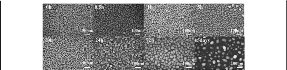
The stability analysis was conducted by soaking sub-strates, with and without the cladding layer, into deionized water for 0.5, 1, 5, 10, 24, and 72 h, as well as 15 days. For every period of time, one of the substrates was taken out for testing.
With the distinctive characteristics of Raman peaks, crystal violet (CV) is one of the most commonly used probe molecules in surface-enhanced Raman scattering. In addition, CV is listed as a banned drug of aquaculture by many countries for its high toxicity, high persistence, and cancer-causing peculiarities; thus, it is a good choice for the trace detection experiments [18, 19]. We describe it by the formula of C25H30ClN3, and the relative molecular mass is 407.99. By diluting different masses of solid CV with deionized water, different molar concentrations of CV solution were prepared. All the experiments were per-formed in a thousand level clean room with a temperature of 20 °C and a relative humidity of 60 %.
SERS characterization was performed after immersion of the samples in CV solutions. The samples were ex-cited using a 514.5-nm laser line from the Raman micro-scope system (Invia, Renishaw, UK) with an excitation power beam of 5 mW.
Results and Discussion
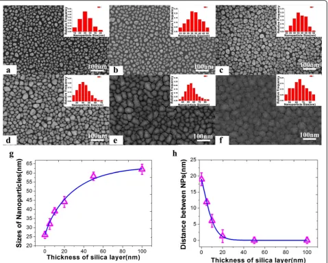
Morphology Characterization of Ag@SiO2Nanostructure Different film thickness of the SiO2layers can be achieved by controlling the SiO2deposition time, and the morph-ology of the Ag@SiO2nanostructure was characterized by scanning electron microscopy (SEM), as shown in Fig. 1 and Additional file 1: Figure S1. The corresponding energy spectrum shows the coincident results (Additional file 1: Figure S2). From the characterization results, as shown in Fig. 1g, h, the mean diameters of Ag@SiO2nanostructures were 26 ± 2.3, 32 ± 3.6, 39 ± 2, 44.1 ± 3.3, 58.2 ± 2.5, and 62.2 ± 3.2 nm, calculated with the assistance of Smile View software, and the interparticle distance decreased from 19 to 0 nm.
SERS Characterization of Ag@SiO2Core-Shell Nanostructure
and Research on the Enhancement Mechanism
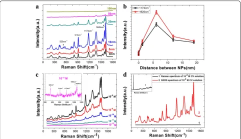
The samples were immersed into 10−6 M CV solution, which acted as the probe molecule for 30 min, and then dried in air. The SERS activity of the substrates was tested with the Raman microscope system, and the results are shown in Fig. 2.
There exist various bands for CV at around 913 cm−1, attributed to radial aromatic ring skeleton vibrating. The band at 1174 cm−1is related to the C–H bending vibra-tion, while the band at 1620 cm−1is related to the C=C stretching vibration. As shown in Fig. 2a, the thickness
of 10 nm acts as the turning point for Raman intensity, and the calculated results reveal that Ag NPs coated with the appropriate shell thickness can improve the sensitivity of SERS active substrates. The highest enhancement in Raman scattering is threefold for the 10-nm shell com-pared to the uncoated Ag NPs. Besides, the reproducibility of the Ag@SiO2core-shell structure is good by comparing the SERS performance of 12 different 10-nm SiO2-coated substrates (Additional file 1: Figure S3).
To further characterize the SERS sensitivity of
Ag@SiO2 with the SiO2 shell of 10 nm, we immersed
the substrates in different concentrations of CV solu-tion. As shown in Fig. 2c, we can find that quite high sensitivity was achieved with the Ag@SiO2 structure, which can detect CV concentrations down to 10−12 M with the signal-to-noise ratio of 6.3 dB. Finally, the en-hancement factor (EF) was used to characterize the
SERS enhancement of Ag@SiO2nanostructures. It was calculated according to the equation as Leem [20] described:
EF¼Isers=Csers
Isol=Csol ð
1Þ
whereIsersandIsol are the normalized Raman peak inten-sities of the CV molecules absorbed on the SERS substrate and the reference solution of the selected Raman peak at 1620 cm−1, respectively.Csersis the molar concentration of the CV on SERS substrates, andCsolis the molar concen-tration of the reference CV solution. According to the re-sults of our experiment, the Isers and Isol are equal to 90,000 and 136, respectively, and the Csers and Csol are equal to 10−6and 10−2M, respectively. Therefore, the EF of our Ag@SiO2substrate is 6.6 × 10⁶.
Fig. 1Morphology characterizations of the Ag nanoparticle films coated with different thickness of SiO2layers:a0 nm;b5 nm;c10 nm;d20 nm;e
50 nm;f100 nm;gvariation of nanoparticle diameter as the increase of SiO2thickness; andhvariation of the distance between Ag NPs as the increase
[image:3.595.60.540.88.473.2]We believe there were three reasons that a SiO2layer contributes to a higher SERS activity. Firstly, the high refractive index of SiO2layer can confine the light, just like the optical fiber used in the optical communication. Secondly, the multiscattering processes of the light that scattered back and forth at the two curved surfaces of SiO2 layers will contribute to a larger enhancement factor in the cavity. Thirdly, the interference of the scat-tered light from the inner and outer interfaces can di-minish the optical field inside the layer, and due to the energy conservation, a relatively larger enhanced field is focused at the outer surface of SiO2layer [21, 22]. Thus, a huge SERS enhancement especially in the cavity between coated Ag NPs can be obtained, which also have been proved by the finite-difference time-domain (FDTD) simu-lations, as shown in Fig. 3.
In addition, we believe that the high hydrophilic SiO2 coating can aggrandize the adsorption quantity of CV molecules on the surface of substrates, which will have an effect on the SERS enhancement to a certain extent [23]. We herein assessed the hydrophilic character with water contact angles. As substantiated by the shift in dynamic contact angle (Additional file 1: Figure S4), a significant increase in surface hydrophilicity of the
Ag@SiO2substrates was found. To quantitatively express the impact of the mesoporous structure and hydrophilicity of SiO2 layer on the SERS enhancement, we contrasted the Raman intensity of bare Si substrates and Si@SiO2 substrate. The enhancement factor here can be calculated according to the Eq. (1) too, and the result is 6.65.
Meanwhile, as a surface-sensitive technique, SERS performance decays exponentially according to the distance between target molecules and SERS substrates. Thus, the increase in SiO2 layers separates the probing molecules from Ag NPs, resulting in a sharp reduction in Raman intensity [24].
Stability Analysis of Ag@SiO2Core-Shell Substrates To characterize the long-term stability of Ag@SiO2 core-shell nanostructures in aqueous solution sensing applica-tions, we put the Ag NP films coated with 0, 10, and 20 nm SiO2layers into deionized water for comparison ex-periments. At the soaking times of 0.5, 1, 5, 10, 24, and 72 h and 15 days, one of the substrates was taken out and then immersed in a 10−6M CV solution for 10 min. The morphology of the NPs on three kinds of substrates after the immersion process were characterized by SEM. The SEM images (Additional file 1: Figure S5) showed that Ag Fig. 2SERS characterizations of [email protected] spectra of CV on Ag@SiO2substrates with the thickness of SiO2layers vary from 0 to
100 nm.bSpectrum intensity calculations of CV at 1174 and 1620 cm−1based on the interparticle distance.cSERS spectra of CV absorbed on
Ag@SiO2substrate after immersed in different concentrations of CV solution.dNormal Raman spectrum of 10−2M CV solution on silicon wafer (1)
and SERS spectrum of 10−6M CV solution on Ag@SiO
2substrate (2)
[image:4.595.57.540.87.365.2]NP films can be greatly destroyed when exposed to an aqueous solution. After soaking for 0.5 h, agglomeration appeared and almost one third of the NPs were removed, and after 10 h, only a few residues of the Ag NPs can be found which were also strongly agglomerated. Apparently, the stability of Ag NP films is very poor when soaking in the aqueous solution, which can greatly restrict the per-formance of the SERS activity. Inversely, Ag NPs coated with a 10 nm, as well as a 20-nm SiO2layer, can still re-main the morphology even after 24 h of immersion, and the agglomeration of NPs was not discovered after soaking for 15 days. It is obvious that the SiO2layer can protect Ag NPs with a thickness of 10 nm (Fig. 4).
The effect on the SERS activity of substrates during the immersion process was also characterized by Raman microscope system, and the result is shown in Fig. 5. From the figures, we can find that, with the increased
soaking time, the Raman intensity of CV absorbed on Ag@SiO2 substrates is stable, while for bare Ag NP films, the intensity decreasing sharply. After 24 h of immersion, the decrease in Raman spectrum intensity is about 85 % for bare Ag NP films, compared to 12 % for the Ag NP films with a 10-nm SiO2layer, and 15 % for Ag NP films with a 20-nm SiO2layer (Fig. 5d).
From the results, it can be concluded that SiO2-coated metallic NP films can greatly increase the long-term stability compared to bare Ag NP films, especially when the structures are exposed to aqueous solution.
Applications of Ag@SiO2Core-Shell Substrates
For the aqueous solution sensing applications, Ag@SiO2 substrates are further developed with better sensitivity and selectivity through chemical functionalization. We Fig. 3FDTD simulation results for the intensity trend along with the change of spacing.aAg nanoparticles coated with 5 nm SiO2, the distance
between Ag@SiO2nanostructures is 14 nm.bDistance between Ag@SiO2nanostructures is 10 nm.cDistance between Ag@SiO2nanostructures
is 4 nm.dDistance between Ag@SiO2nanostructures is 0 nm.eAg nanoparticles coated with 20 nm SiO2; here, we denoted the distance
between Ag@SiO2nanostructures as−16 nm.fVariation of the SERS intensity along with the spacing between nanoparticles
Fig. 4Morphology characterizations of the Ag@SiO2nanostructures after immersing in deionized water for 0, 0.5, 1, 5, 10, 24, and 72 h and 15 days with
[image:5.595.56.543.88.270.2] [image:5.595.56.539.585.703.2]Fig. 5Characterization of SERS-activity, Raman spectra of CV onabare Ag NP films;bAg NP films coated with 10-nm SiO2layer;cAg NP films
coated with 20-nm SiO2layer after soaking for different time; anddthe intensity curves of CV at 1174 and 1620 cm− 1
absorbed on the substrates with and without SiO2
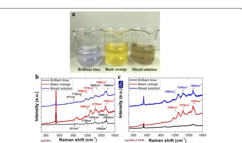
Fig. 6aThe dilute solutions of brilliant blue (BB), basic orange (BO), and the mixed solution of BB and BO, respectively.bThe Raman spectra of Ag@SiO2substrate after immersing in the three kinds of solutions.cThe Raman spectra of Ag@SiO2–COOH substrate after immersing in the three
kinds of solutions
[image:6.595.57.539.86.357.2] [image:6.595.59.538.409.693.2]found that a negatively charged surface could be achieved by−COOH functionalization.
Solutions of the food additives brilliant blue (BB) and basic orange (BO) were used. BB molecules in the solu-tion are negatively charged, and BO molecules carry two amino groups. The molarity of BB and BO solutions are 10−6M.
We immersed the Ag@SiO2as well as the Ag@SiO2–
COOH substrates in the dilute solutions of BB, BO, and the mixed solution of BB and BO, respectively. The Raman spectra of the three samples are shown in Fig. 6. From Fig. 6b, we can find that both molecules on the nonfunctionalized substrate were detected. Different phenomena were observed with the negatively charged, functionalized substrate (Fig. 6c). In the solutions of BB and the mixed solution of BB and BO, the Raman band due to BB nearly disappeared. This result indicates that the sensitivity to positively charged molecules is
improved with the negatively charged SiO2 surface
and decreased to negatively charged molecules. The high sensitivity to the specific molecule gives the potential applications of Ag@SiO2in accurate biochem-ical sensing.
Conclusions
In this study, a method for SiO2cladding of Ag NPs with long-term stability in aqueous solution was presented and demonstrated. The ICPECVD-grown SiO2layer can suppress the oxidation of Ag NPs effectively and prevent the aggregation and deformation of the particles from occurring. Furthermore, the SERS activity of Ag NP films can be increased by optimizing the thickness of the SiO2 dielectric layer that can detect CV concentrations down to 10−12M, and the EF of Ag@SiO2 substrates in our experiment can reach up to 6.6 × 10⁶. This work thus provides a straightforward and cost-effective ap-proach to fabricating Ag-based SERS substrates with un-precedented stability and gives us a reliable way to apply Ag nanostructures in the field of solution composition detection. Also, as a bio-compatible material, a SiO2 coating of the nanostructure for highly sensitive chemical sensing can be further applied for various bio-sensor applications.
Additional file
Additional file 1: Figure S1.The cross-sectional views of Ag@SiO2
nanostructures with the thickness of SiO2layers vary from 0 to 50 nm.Figure
S2.The corresponding energy spectrum of Ag@SiO2nanostructures with
the thickness of SiO2layers vary from 0 to 50 nm.Figure S3.Characterization
of the reproducibility of Ag@SiO2nanostructure (a) Raman spectra of CV on
different Ag@SiO2substrates (b). The intensity curves of CV at 1174 and
1620 cm−1absorbed on the substrates.Figure S4.A water droplet
falling on (a) the Ag film; (b) the Ag@SiO2film; (c) Raman spectra of
CV molecules on Si (1)/Si coated with 10-nm SiO2wafer (2).Figure
S5.Morphology characterizations of the Ag@SiO2nanostructures after
immersing in deionized water for 0, 0.5, 1, 5, 10, 24, and 72 h and 15 days with the thicknesses of SiO2are (a) 0 nm (b) 10 nm, and (c)
20 nm. (DOC 19191 kb)
Acknowledgements
We acknowledge the financial support from the Natural Science Foundation of China (51225504, 61127008, 61571405, and 110248-28140) and the program for the top young academic leaders of higher learning Institutions of Shanxi.
Authors’Contributions
MZ and HG contributed equally to this work, performed the most measurements, analyzed the data, and wrote the main manuscript text. JL and LW prepared the samples and performed the SEM and EDS measurements. JT and WL designed the experiment and contributed to the manuscript writing. BZ modified the manuscript. CX and WZ offered helpful suggestions in the study. All authors read and approved the final manuscript.
Competing Interests
The authors declare that they have no competing interests.
Received: 10 May 2016 Accepted: 31 August 2016
References
1. Liu J, Meng GW, Li ZB, Huang ZL, Li XD (2015) Ag-NP@Ge-nanotaper/ Si-micropillar ordered arrays as ultrasensitive and uniform surface enhanced Raman scattering substrates. Nanoscale 7:18218–18224 2. Gai SL, Yang PP, Li CX, Wang WX, Dai YL, Niu N et al (2010) Synthesis of
magnetic, up-conversion luminescent, and mesoporous core-shell-structured nanocomposites as drug carriers. Adv Funct Mater 20:1166–1172 3. Preciado FS, Wheeler DA, Tran TM, Tanaka Z, Jiang CY, Barboza FM et al
(2011) SERS spectroscopy and SERS imaging of Shewanella oneidensis using silver nanoparticles and nanowires. Chem Comm 47:4129–4131
4. Shafer P, Karen E, Haynes CL, Glucksberg MR et al (2003) Toward a glucose biosensor based on surface-enhanced Raman scattering. J Am Chem Soc 125:588–593
5. Zeman EJ, Schatz GC (1987) An accurate electromagnetic theory study of surface enhancement factors for silver, gold, copper, lithium, sodium, aluminum, gallium, indium, zinc, and cadmium. J Phys Chem 91:634–643 6. Le Ru EC, Blackie E, Meyer M, Etchegoin PG (2007) Surface enhanced Raman
scattering enhancement factors: a comprehensive study. J Phys Chem C 111:13794–13803
7. Rycenga M, Cobley CM, Zeng J, Li WY, Moran CH, Zhang Q (2011) Controlling the synthesis and assembly of silver nanostructure for plasmonic application. Chem Rev 111:669–712
8. Xia XH, Zeng J, Zhang Q, Moran CH, Xia YN (2012) Recent developments in shape-controlled synthesis of silver nanocrystals. J Phys Chem C 116:47–56 9. Hou H, Wang P, Zhang J, Li CP, Jin YD (2015) Graphene oxide-supported Ag
nanoplates as LSPR tunable and reproducible substrates for SERS applications with optimized sensitivity. ACS Appl Mater Interfaces 7:18038–18045 10. Ma Y, Qu Y (2012) A simple approach towards uniform spherical Ag-like
nanoparticles. Nanoscale 4:6–9
11. Liu K, Bai YC, Zhang L, Yang ZB, Fan QK, Zheng HQ (2016) Porous Au-Ag nanospheres with high-density and highly accessible hotspots for SERS analysis. Nano Lett 16:3675–3681
12. Liu YT, Zhou J, Wang BB, Jiang T, Ho H, Petti L et al (2015) Au@Ag core-shell nanocubes: epitaxial growth synthesis and surface-enhanced Raman scattering performance. Phys Chem Chem Phys 17:6819–6826
13. Gao CB, Lu ZD, Liu Y, Zhang Q, Chi MF, Cheng Q et al (2012) Highly stable silver nanoplates for surface plasmon resonance biosensing. Angew Chem Int Ed 51:29–33
14. Gao N, Yang T, Liu T, Zou Y, Jiang J (2015) Graphene oxide wrapped individual silver nanocomposites with improved stability for surface-enhanced Raman scattering. RSC Adv 5:55801–55807
15. Hirakawa T, Kamat PV (2005) Charge separation and catalytic activity of Ag@TiO2core-shell composite clusters under UV-irradiation. J Am Chem
16. Ma LW, Huang Y, Hou MJ, Xie Z, Zhang ZJ (2015) Silver nanorods wrapped with ultrathin Al2O3layers exhibiting excellent SERS sensitivity and outstanding
SERS stability. Sci Rep 5:12890
17. Li XY, Li J, Zhou XM, Ma YY, Zheng ZP, Duan XF et al (2014) Silver nanoparticles protected by monolayer graphene as a stabilized substrate for surface enhanced Raman spectroscopy. Carbon 66:713–719
18. Senapati S, Srivastava SK, Singh SB, Kulkarni AR (2014) SERS active Ag encapsulated Fe@SiO2nanorods in electromagnetic wave absorption
and crystal violet detection. Environ Res 135:95–104
19. Zhang KB, Zeng TX, Tan XL, Wu WD, Tang YJ, Zhang HB (2015) A facile surface-enhanced Raman scattering (SERS) detection of rhodamine 6G and crystal violet using Au nanoparticle substrates. Appl Surf Sci 347:569–573 20. Leem J, Kang HW, Ko SH, Sung HJ (2014) Controllable Ag nanostructure
patterning in a microfluidic channel for real-time SERS systems. Nanoscale 6:2895–2901
21. Liu FX, Tang CJ, Zhan P, Chen Z, Ma HT, Wang ZL (2014) Released plasmonic electric field of ultrathin tetrahedral-amorphous-carbon films coated Ag nanoparticles for SERS. Sci Rep 4:4494
22. Xu HX (2004) Theoretical study of coated spherical metallic nanoparticles for single-molecule surface-enhanced spectroscopy. Appl Phys Lett 85:5980–5982 23. Kotte L, Althues H, Mader G, Roch J, Kaskel S, Dani I et al (2013) Atmospheric
pressure PECVD based on a linearly extended DC arc for adhesion promotion applications. Surf Coat Technol 234:8–13
24. Gong JX, Li GD, Tang ZY (2012) Self-assembly of noble metal nanocrystals: fabrication, optical property, and application. Nano Today 7:564–585
Submit your manuscript to a
journal and benefi t from:
7Convenient online submission 7Rigorous peer review
7Immediate publication on acceptance 7Open access: articles freely available online 7High visibility within the fi eld
7Retaining the copyright to your article
Submit your next manuscript at 7 springeropen.com