Volume 2012, Article ID 684740,11pages doi:10.1155/2012/684740
Research Article
Induction of Mitochondria-Mediated Apoptosis in
Ca Ski Human Cervical Cancer Cells Triggered by Mollic Acid
Arabinoside Isolated from
Leea indica
Yau Hsiung Wong and Habsah Abdul Kadir
Biomolecular Research Group, Biochemistry Program, Institute of Biological Sciences, Faculty of Science, University of Malaya, 50603 Kuala Lumpur, Malaysia
Correspondence should be addressed to Habsah Abdul Kadir,[email protected] Received 22 May 2011; Revised 7 August 2011; Accepted 20 August 2011
Academic Editor: Angelo Antonio Izzo
Copyright © 2012 Y. H. Wong and H. Abdul Kadir. This is an open access article distributed under the Creative Commons Attribution License, which permits unrestricted use, distribution, and reproduction in any medium, provided the original work is properly cited.
Leea indica is a medicinal plant traditionally used to treat cancer. Through bioassay-guided approach, we isolated mollic acid
arab-inoside (MAA), for the first time from Leea indica. Here, we present the apoptosis-inducing effect of MAA on Ca Ski cervical can-cer cells. Based on DAPI staining, MAA-treated cells manifested nuclear shrinkage, condensation, and fragmentation. We further confirmed the fragmentation of DNA using TUNEL assay. During early apoptosis, MAA caused the perturbation of plasma mem-brane through externalization of PS, followed by the formation of apoptotic blebs. Prior to these events, MAA triggered rapid dis-sipation of the mitochondrial membrane potential. In the upstream, MAA increased the expression of Bax, decreased the ex-pression of Bcl-2, and augmented the Bax/Bcl-2 ratio. These findings suggested that MAA induced mitochondrial-mediated apo-ptosis in Ca Ski cells and thus provide the scientific explanation for the traditional application of this herbal medicine in cancer treatment.
1. Introduction
Apoptosis is a well-defined process that is controlled by a seq-uence of regulated events that eventually resulted in demise of the cell. Apoptosis plays a fundamental role in maintaining normal tissue homeostasis by regulating the balance between cell proliferation and cell death [1]. Characteristically, it can be defined by several morphological and biochemical chang-es that affect the nucleus, plasma membrane, and mitochon-dria, including nuclear condensation (pyknosis), nuclear fragmentation (karyorrhexis), membrane blebbing, externa-lization of phosphatidylserine, and collapse of mitochondrial membrane potential (Δψm) [2]. Induction of cell apoptosis is considered a useful approach in the treatment of cancer [3]. It has been reported that a variety of phytochemicals from natural products induced apoptosis in cancer cell lines. Indeed, many have been used as cancer chemopreventive agents [4]. In view of that, intensive efforts have been made to identify new bioactive compounds from natural products,
through isolation of apoptosis inducing agents and elucida-tion of the apoptosis mechanisms.
or rheumatoid arthritis [10]. It is also an ingredient in the herbal preparation to treat cancers [11].
In our preliminary cytotoxicity screening, the crude ethan-ol extract and fractions (ethyl acetate, hexane, and water) of the leaves were found to inhibit the growth of Ca Ski cervical cancer cells, with the strongest growth-inhibitory effects shown by the L. indica ethyl acetate fraction (LIEAF) [12]. As part of our continuing studies, LIEAF was subjected to MTT assay-directed separation by using Ca Ski cells as a model. Through the bioassay-guided approach, mollic acid arabino-side (MAA), a cycloartane triterpenoid was isolated for the first time from LIEAF. MTT studies showed that MAA was cytotoxic to Ca Ski cells (IC50of 19.21μM) after 72 h of incu-bation and much less cytotoxic to MRC5 normal cell (about 8-fold higher IC50) [13]. Recently, growing attention has been devoted to the studies on the apoptosis by cycloartane triterpenoids [14–18]. However, the apoptosis and the underlying mechanisms of MAA are still unknown. There-fore, in this study, we attempted to evaluate the apoptosis inducing effect and dissect the mechanism of apoptosis eli-cited by MAA in Ca Ski cervical cancer cells.
2. Methods
2.1. Cell Culture. The human cervical epidermoid carcinoma cell line (Ca Ski, ATCC number CRL-1550) was used in the current study. It was purchased from American Type Culture Collection (ATCC, USA). The cells were cultured as mono-layers in RPMI 1640 growth medium (Sigma), containing 10% (v/v) heat-inactivated fetal bovine serum (PAA), 100μg/mL penicillin/streptomycin (PAA), and 50μg/mL amphotericin B (PAA). The cells were maintained inside a CO2incubator (Galaxy R, RS Biotech) at 37◦C with moist at-mosphere of 5% CO2in air.
2.2. 4,6-Diamidino-2-Phenylindole (DAPI) Staining. To monitor morphological alterations typical of apoptosis, we performed DAPI nuclear staining. After MAA treatment, the cells were harvested and washed with PBS, followed by fixa-tion of cells in acetone at 4◦C for 30 min. The fixed cells were washed with PBS and stained for 30 min with 1μg/mL DAPI in the room temperature. The stained cells were then exam-ined for morphological signs of apoptosis under a Leica DM 2500 fluorescence microscope, and photographs were cap-tured by a Leica DFC 310 FX camera.
2.3. Terminal Deoxynucleotidyl Transferase (TdT) dUTP Nick End Labeling (TUNEL) Assay. To detect DNA fragmenta-tion, we used a TUNEL assay kit (Apo-BRDU, Sigma). The kit uses two dyes, FITC to label apoptotic DNA strand breaks (DSBs) and PI to stain total cellular DNA. DSBs were labeled with bromodeoxyuridine triphosphate nucleotides (BrdUTP) and detected by FITC-conjugated BrdUTP anti-body. Briefly, after MAA treatment, 1×10⁶ cells were fixed for 15 min in ice-cold 1% (w/v) paraformaldehyde in PBS. The fixed cells were stored for at least 24 h in 70% (v/v) ethanol at−20◦C. Following washing of the cells, they were incubated for 60 min with 50μL reaction buffer containing
TdT enzyme and BrdUTP at 37◦C. The incorporated BrdUTP was then labeled with 100μL FITC-conjugated BrdUTP antibody for 30 min, followed by addition of 400μL PI/RNase and further incubated for 30 min at room tempera-ture. Then, the cells were subjected to analysis by FACS Cali-bur flow cytometry using Cell Quest software (BD).
2.4. Analysis on Externalization of Phosphatidylserine (Anne-xin V/PI Staining). Anne(Anne-xin V/PI staining was assessed to measure the externalization of PS during early apoptosis [19]. After MAA treatment, the washed cells were resuspend-ed in Annexin V binding buffer (BD Biosciences) at the den-sity of 1×10⁶cells/mL. 0.1 mL (1×10⁵cells) of the cell sus-pension was then transferred to a 5 mL BD falcon round-bot-tom tube, where 5μL FITC-conjugated Annexin V (BD Bio-sciences) and 10μL PI (50μg/mL) were added. The cells were vortexed gently and incubated for 30 min at room tempera-ture. An addition of 400μL binding buffer was added to the tubes before the cells were analyzed by FACS Calibur and Cell Quest software.
2.5. Apoptotic Bleb Analysis (ScFv/PI Staining). The forma-tion of membrane blebbing in apoptotic cells was measured using an apoptotic blebs assay kit (Cayman Chemical). The kit uses a recombinant protein, which was derived from single-chain variable fragments (scFv) of an antibody, to spe-cifically recognize the autoantigens in the apoptotic blebs [20]. The recombinant protein is then detected using FITC-conjugated rabbit IgG. Briefly, after MAA treatment, 1×10⁶ cell suspensions were incubated for 30 min with scFv fusion protein at 4◦C, followed by staining for 30 min with FITC-conjugated rabbit lgG at 4◦C. After washing, the cells were stained for 10 min with PI at room temperature. The stained cells were washed twice and resuspended in 1 mL of PBS be-fore analysis on FACS Calibur and Cell Quest software.
Table 1: The primer sequence for Bcl-2, Bax, and PBGD.
Target genes Function Sequence
Bcl-2 Forward primer 5-TTGGCCCCCGTTGCTT-3
Reverse primer 5-CGGTTATCGTACCCCGTTCTC-3
Bax Forward primer 5-GTCGCCCTTTTCTACTTTGCCAG-3
Reverse primer 5-TCCAGCCCAACAGCCGCTCC-3
PBGD Forward primer 5-ACCATCGGAGCCATCTGCAAG-3
Reverse primer 5-CCCACCACACTCTTCTCTGGCA-3
2.7. Real-Time Quantitative PCR (Q-PCR). To examine the role of Bcl-2 family members in MAA-induced apoptosis, we measured the gene expression of Bax and Bcl-2 using Q-PCR. 1 ×10⁶ cells were exposed to MAA for different exposure periods. After treatment exposure times, total RNA was iso-lated using RNAqueous-4PCR kit (Applied Biosystem) based on the manufacturer’s protocol. Gene expression of Bcl-2 (B-cell CLL/lymphoma 2) and Bax (BCL2-associated X protein) was assessed by one-step SYBR green relative Q-PCR (RotorGene-6000 System, Qiagen) and normalized to PBGD (porphobilinogen deaminase) reference genes. The primer sequences were shown in Table 1. The reactions were car-ried out in a total volume of 25μL using the SensiMix One-Step Kit (Quantace). The PCR amplification for Bcl-2 was 45 cycles of 20 seconds at 95◦C, 40 seconds at 42◦C, and 10 sec-onds at 72◦C. While for Bax it was 45 cycles of 20 seconds at 95◦C, 40 seconds at 60◦C, and 10 seconds at 72◦C. The fluo-rescence threshold Ct values were calculated, and the ΔCt values were determined using the formula ΔCt = CtBax or Bcl-2−CtPBGD. TheΔΔCt values were then calculated based on formulaΔΔCt=ΔCt treated−ΔCt untreated. The expression level of Bax or Bcl-2 in the treated cells was mea-sured relatively to the level observed in the untreated cells and was quantitated using formula 2−ΔΔCT[23].
2.8. Flow Cytometric Immunofluorescence Staining. The pro-tein expression level of Bax and Bcl-2 was determined by im-munofluorescence staining using flow cytometry. This method was based on Roussi et al. [24] with some modifica-tions. Following MAA treatment, the cells were washed twice in PBS and then fixed and permeabilized using the Cytofix/ Cytoperm kit (BD Biosciences). A total of 1×10⁶cells were resuspended in 500μL of fixation/permeabilization solution and incubated for 20 min at 4◦C. The cells were washed twice with Perm/Wash buffer and incubated for 15 min in 1 mL of Perm/Wash buffer. To detect Bax or Bcl-2, the fixed and per-meabilized cells were incubated with 100μL of Perm/Wash buffer containing the antibodies. For Bcl-2 protein, the cells were stained directly for 30 min with 20μL FITC-conjugated mouse anti-human Bcl-2 monoclonal antibody (clone 124) or IgG1 isotype control (BD Biosciences) at 4◦C. For indirect Bax staining, the cells were incubated for 30 min with either rabbit anti-human Bax polyclonal antibody or IgG1 isotype control (BD Biosciences) at 4◦C. After washing, the cells were further incubated for 30 min with FITC-conjugated goat anti-rabbit F(ab)2 polyclonal secondary antibody (Abcam)
at 4◦C. The cells were then washed with Perm/Wash buffer and analyzed using FACS Calibur and Cell Quest software.
2.9. Statistical Analysis. The results are presented in means
±S.E. Statistical analysis of the results was performed using Student’st-test.P < 0.05 was considered to be significant difference.
3. Results and Discussion
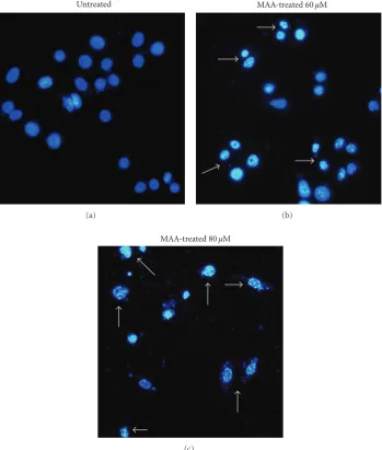
3.1. MAA Elicited Nuclear Alterations Typical of Apoptotic Cell Death in Ca Ski Cells. Apoptosis is initially characterized by morphological features, such as chromatin condensation, nuclear fragmentation, and membrane blebbing [2]. To gain an insight on the effect of MAA on nuclear alterations, cells were stained with DAPI. As shown in Figure 1, the cells underwent remarkable nuclear changes upon treatment. In the control untreated cells, the nuclei were intact, round, and uniformly stained. However, after exposed to MAA, the cells manifested two nuclear phenomena typical of apoptosis, namely pyknosis (nuclear shrinkage/condensation) and kar-yorrhexis (nuclear fragmentation). At 60μM of MAA, a number of cells exhibited nuclear shrinkage and chromatin condensation. At 80μM, the nuclear alterations were more apparent. The cells shrunk and lost its normal nuclear archi-tecture. The chromatin was compacted into dense lumps and recognized as smaller intense spots. The nuclei were frag-mented into apoptotic bodies (nuclear bodies). On the con-trary, these aberrant nuclear alterations were not seen in the control cells. These observations showed that apoptosis oc-curred in Ca Ski cells after treatment of MAA.
3.2. MAA Triggered DNA Fragmentation in Ca Ski Cells. The appearance of apoptotic nuclear alterations following MAA treatment led us to focus on another hallmark of apoptosis, DNA fragmentation. During late stage of apoptosis, endonu-cleases provoke the degradation of nuclear DNA, resulting in fragments of DNA strand breaks (DSBs) with exposed 3 -hydroxyl ends [25].
To detect the generation of DSBs in Ca Ski cells, we per-formed TUNEL assay using the Apo-BRDU kit (Sigma). In this assay, TdT enzyme catalyzed the addition of BrdUTP to the exposed 3-hydroxyl ends of DSBs. The incorporated BrdUTP was then detected with FITC-conjugated BrdUTP antibody. Simultaneously, the DNA content was measured to correlate the apoptosis with cell cycle [26]. As illustrated in
Untreated
(a)
MAA-treated 60µM
(b)
MAA-treated 80µM
[image:4.600.125.475.74.486.2](c)
Figure 1: Effect of MAA on nuclear alterations in Ca Ski cells. Cells were incubated without or with 60μM and 80μM of MAA for 24 h. Cells were stained with DAPI and visualized by fluorescence microscopy. The arrows indicate nuclear shrinkage, condensation, or fragmentation with 100x magnification.
the untreated cells showed negative TUNEL staining. After MAA treatment, a number of cells acquired TUNEL staining, indicating the occurrence of DNA fragmentation. The effect of DNA fragmentation was found intensified in a dose-de-pendent way (Figure 2(b)). The effect seemed to occur at the S phase of cell cycle. When treated with 100μM of MAA, nearly 50% of the cells underwent DNA fragmentation and was found to appear in all phases of the cell cycle.
The increase in TUNEL positivity was in parallel with the appearance of cells with apoptotic nuclear alterations. This further confirmed the DAPI morphological-based results. Thus, we can postulate that DNA fragmentation, particularly breakage of DNA strands, might contribute to the aberrant nuclear alterations. Collectively, the observations from DAPI and TUNEL assays showed that the cells underwent apop-totic DNA damage after treatment with MAA.
10 0
0
101
102
103
104
200 400 600 800 1000
10 0
0
101
102
103
104
200 400 600 800 1000 10
0
0
101
102
103
104
200 400 600 800 1000
10 0
0
101
102
103
104
200 400 600 800 1000 10
0
0
101
102
103
104
200 400 600 800 1000
10 0
0
101
102
103
104
200 400 600 800 1000 Control (untreated)
100µM 80µM
20µM 40µM
60µM
PI (DNA content) PI (DNA content) PI (DNA content)
FIT
C
B
r-dUTP
antibod
y
(DN
A
st
ra
nd
br
eaks)
FIT
C
B
r-dUTP
antibod
y
(DN
A
st
rand
br
eaks)
R3
R4
R3
R4
R3
R4
R3
R4
R3
R4
R3
R4
(a)
0 20 40 60 80 100 0
10 20 30 40 50 60
TUNEL
-positi
ve
cells
(%)
Concentration (µM)
[image:5.600.63.537.73.534.2](b)
Figure 2: Effect of MAA on fragmentation of DNA in Ca Ski cells. Cells were exposed to different concentration of MAA for 24 h and pro-ceeded to TUNEL assay as described inSection 2. (a) Representative dual parametric dot plots of PI (DNA content) on thex-axis and FITC Br-dUTP antibody (DNA strand breaks) on they-axis. The gated R3regions (red) indicate TUNEL-positive cells. (b) Bar chart shows the
percentage of TUNEL-positive cells. Values are mean±S.E. from three experiments.
apoptotic cells (Annexin V+/PI−) began to appear as earlier as 12 h of treatment and were found increased in a time-de-pendent manner till 24 h, while the late apoptotic cells (An-nexin V+/PI+) also increased after 12–24 h of exposure to MAA. Notably, a considerable accumulation of early and late apoptotic cells (approximately 20% for each) with less than 3% necrotic cells (Annexin V−PI+) was visible at 24 h. Con-sistent with the progression of apoptosis, the late apoptotic cells become dominant at later times, because we observed gradual diminution of the early apoptotic cells and incre-ment of the late apoptotic cells after 24 h (data not shown). We also challenge the cells with increasing doses of MAA at
24 h of treatment. The results clearly showed the progression from early to late apoptosis, indicating by the dose-depen-dent accumulation of the late apoptotic cells. Overall, the combined early and late apoptotic cells (Annexin V positive) were elevated in a time- and dose-dependent fashion (Figure 3(c)). It is important to note that, at every treatment dose and exposure time, the necrotic cell population was small (below 15%). This indicated that apoptosis is the preferential cell death induced by MAA in Ca Ski cells.
100 100 101 101 102 102 103 103 104 104 100 100 101 101 102 102 103 103 104 104 100 100 101 101 102 102 103 103 104 104 100 100 101 101 102 102 103 103 104 104 100 100 101 101 102 102 103 103 104 104 100 100 101 101 102 102 103 103 104 104 100 100 101 101 102 102 103 103 104 104 100 100 101 101 102 102 103 103 104 104
Annexin V-FITC Annexin V-FITC Annexin V-FITC Annexin V-FITC
0 h 12 h 18 h 24 h
0µM 60µM 80µM 100µM
P ropidium io dide (P I) P ro p idium iodide (P I) (a) 0 20 40 60 80 100 120
0 12 18 24 (h)
0 60 80 100
(µM)
Viable Time T o tal cel l p opulation (%) Dose Late apoptotic Early apoptotic Necrotic
(b) 0 20 40 60 80 100 120 Time Dose T o tal cell p opulation sho w ing ex te rnalization o f PS (%) ∗ ∗ ∗ ∗ ∗ ∗
0 12 18 24 (h)
0 60 80 100
(µM)
(c)
Figure 3: Effect of MAA on externalization of PS in Ca Ski cells. Cells were treated with 60μM of MAA for different time points. At 24 h of treatment, cells were treated with increasing concentration of MAA. At each time point, cells were collected and double stained with Annexin V-FITC and PI as described inSection 2. (a) Representative dual parametric dot plots of PI fluorescence (y-axis) versus Annexin V-FITC fluorescence (x-axis). (b) Bar charts showing the proportion of viable, early apoptotic, late apoptotic, and necrotic cells. (c) Bar charts show the percentage of Annexin V-positive cells (Annexin V+/PI−and Annexin V+/PI+). Values are mean±S.E. of three experiments. Asterisks indicate a significant difference between untreated and treated cells (∗P <0.05).
to the plasma membrane during apoptosis is the surface pro-trusion known as blebs [2]. Apoptotic cells package autoanti-gens (such as nuclear fragments and DNA) in vesicles and distributed them to the membrane surface as blebs [20]. To further explore this apoptotic phenotype, we used a commer-cial apoptotic bleb assay kit. The kit is based on the binding of scFv fusion protein to the antoantigens in apoptotic blebs, followed by detection with FITC-conjugated antibody [20]. As shown in Figures4(a)and4(b), the majority of untreated cells were not stained with both the bleb-FITC and PI. How-ever, after treatment with MAA, they were progressively stained with the bleb-FITC. We noted that most of the treated cells were double stained with both bleb-FITC and PI (PI+ bleb+). This finding was in accordance with other
reports which showed that scFv preferentially binds to cells in the later stage of apoptosis [27, 28]. This was also consistent with the fact that the surface blebs of apoptotic cells contained nuclear antigens such as DNA [20]. As shown inFigure 4(c), when compared to untreated cells, the treated cells showed significant increase of apoptotic blebs in a time- and dose-dependent manner. Similar to Annexin V/PI staining, the primary necrotic cell population (PI+ bleb−) in every treatment dose and exposure time was less than 15%. This again confirmed the proapoptotic nature of MAA in Ca Ski cells.
100 100 101 101 102 102 103 103 104 104 100 100 101 101 102 102 103 103 104 104 100 100 101 101 102 102 103 103 104 104 100 100 101 101 102 102 103 103 104 104 100 100 101 101 102 102 103 103 104 104 100 100 101 101 102 102 103 103 104 104 100 100 101 101 102 102 103 103 104 104 100 100 101 101 102 102 103 103 104 104
0 h 24 h 48 h 72 h
0µM 60µM 80µM 100µM
Bleb -FITC Bleb -FITC Bleb -FITC Bleb -FITC
P ropidium io dide (P I) P ro p idium iodide (P I) (a) 0 20 40 60 80 100 120
0 24 48 72 Time T o tal cel l p opulation (%) Dose
PI+ bleb− PI−bleb+
PI−bleb− PI+ bleb+ (h)
0 60 80 100
(µM)
(b) 0 20 40 60 80 100 120 Time Dose ∗ ∗ ∗ ∗ ∗ ∗ T o tal cell p opulation sho w ing apopt o tic b leb (%)
0 24 48 72
(h)
0 60 80 100
(µM)
(c)
Figure 4: Effect of MAA on formation of apoptotic bleb in Ca Ski cells. Cells were treated with 60μM of MAA for different time points. At 48 h of treatment, cells were treated with increasing concentration of MAA. After treatment, the cells were processed for apoptotic blebs staining and analyzed by flow cytometry as described inSection 2. (a) Representative dual parametric dot plots of PI fluorescence (y-axis) versus bleb-FITC fluorescence (x-axis). Bleb-FITC represents the binding of apoptotic blebs by scFv protein which was labeled with FITC-conjugated rabbit lgG. (b) Bar charts show the proportion of total stained cells based on flow cytometry analysis. (c) Bar charts show the percentage of total cell population with apoptotic blebs (PI−bleb+ and PI+ bleb+). Values are mean±S.E. of three experiments. Asterisks indicate a significant difference between untreated and treated cells (∗P <0.05).
which showed that Annexin V binding precedes scFv binding [20]. Taken together, the apoptosis process triggered by MAA in Ca Ski cells involved the aberrant changes to the cell membrane. During early hour (12 h) following MAA treatment, the membrane asymmetry was lost as PS was translocated from the inner to outer membrane leaflet. Long-er treatments (24–72 h) of MAA evoked the appearance of blebs.
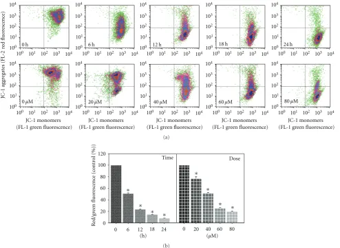
3.5. MAA Dissipated the Mitochondrial Membrane Potential (Δψm) in Ca Ski Cells. Besides PS externalization, dissipa-tion ofΔψm has also been reported to be an early apoptosis event in many different systems [29]. It is a rapid and
irreversible event preceding other manifestation of apoptosis process, such as PS externalization, nuclear DNA cleavage, and chromatin condensation [30,31].
100 100 101 101 102 102 103 103 104 104 100 100 101 101 102 102 103 103 104 104 100 100 101 101 102 102 103 103 104 104 100 100 101 101 102 102 103 103 104 104 100 100 101 101 102 102 103 103 104 104 100 100 101 101 102 102 103 103 104 104 100 100 101 101 102 102 103 103 104 104 100 100 101 101 102 102 103 103 104 104 100 100 101 101 102 102 103 103 104 104 100 100 101 101 102 102 103 103 104 104
0 h 6 h 12 h 18 h 24 h
0µM 20µM 40µM 60µM 80µM
JC-1 monomers (FL-1 green fluorescence)
JC-1 monomers (FL-1 green fluorescence)
JC-1 monomers (FL-1 green fluorescence)
JC-1 monomers (FL-1 green fluorescence)
JC-1 monomers (FL-1 green fluorescence)
re d fl uor esc enc e) JC-1 agg regat es (FL -2 (a) 0 20 40 60 80 100 120
0 6 12 18 24 (h)
0 20 40 60 80 (µM)
[image:8.600.56.551.72.432.2]Time Dose ∗ ∗ ∗ ∗ ∗ ∗ ∗ ∗ R ed/g reen fluor esc enc e (c ont rol (%)) (b)
Figure 5: Effect of MAA on mitochondrial membrane potential (Δψm) in Ca Ski cells. Cells were treated with 60μM of MAA for different time points. At 12 h of treatment, cells were treated with different concentration of MAA. After treatment, the cells were stained with JC-1, and the change inΔψm was analyzed by flow cytometry as described inSection 2. (a) Representative dot plots of JC-1 aggregates (FL-2 red fluorescence) versus JC-1 monomers (FL-1 green fluorescence). (b) Bar charts showing the ratio of red/green fluorescence, expressed as percentage of control, indicate the ratio of high/lowΔψm. Values are mean±S.E. of three experiments. Asterisks indicate a significant difference between untreated and treated cells (∗P <0.05).
fluorescence ratio (Figure 5(b)), the loss ofΔψm occurred in a time- and dose-dependent manner. According to the time course study, MAA induced a rapid (6 h) collapse ofΔψm, which preceded PS externalization (12 h). These data indi-cated that the MAA-induced apoptosis in Ca Ski cells invol-ved mitochondria dysfunction associated with dissipation of theΔψm.
3.6. MAA Increased the Expression of Bax, Decreased the Ex-pression of Bcl-2, and Augmented the Ratio of Bax/Bcl-2. The members of the Bcl-2 family play a pivotal role in the reg-ulation of the mitochondrial apoptotic pathway [32]. Among the members, the antiapoptotic Bcl-2 inhibits apoptosis, whereas the proapoptotic Bax counterbalances the Bcl-2 ef-fect and stimulates apoptosis [33]. Other studies have impli-cated that Bcl-2 maintained the mitochondrial integrity, while Bax destroyed the mitochondrial integrity and caused loss ofΔψm [34]. Consequently, the ratio between Bcl-2 and
Bax determines the susceptibility to apoptosis and thus dictates the fate of life and death of a cell [35,36].
R
elati
ve
gene
expr
ession
0
0 .5 1 1.5 2 2.5
Bax Bcl-2
0 3 6 12 0 3 6 12
(h)
(a)
Bax/Bcl-2
0 1 2 3 4 5 6
∗
∗
∗
0 3 6 12
(h)
(b)
Figure 6: Effect of MAA on gene expression level of Bax and Bcl-2 in Ca Ski cells. Cells were treated with 60μM MAA for different times. After designated exposure times, cells were harvested and the total RNA was extracted. The RNA was subjected to Q-PCR analysis as described in Section 2. (a) The relative gene expression level of Bax and Bcl-2 was compared to control, which was normalized against PBGD expression using the formula 2−ΔΔCT. (b) Bar charts show the ratio of Bax/Bcl-2, analyzed from the Q-PCR results. Values are mean±S.E. of three
experiments. Asterisks indicate a significant difference between untreated and treated cells (∗P <0.05).
200
160
40 80 120
0
100 101 102 103 104
200
160
40 80 120
0
100 101 102 103 104
200
160
40 80 120
0
100 101 102 103 104
3 h 6 h 12 h
R
elati
ve
ce
ll
n
umber
Isotype control antibody Untreated cells MAA-treated cells
Isotype control antibody Untreated cells MAA-treated cells
Isotype control antibody Untreated cells MAA-treated cells Bax (log fluorescence intensity) Bax (log fluorescence intensity) Bax (log fluorescence intensity)
(a)
200
160
40 80 120
0
100 101 102 103 104
200
160
40 80 120
0
200
160
40 80 120
0
100 101 102 103 104 100 101 102 103 104
3 h 6 h 12 h
R
elati
ve
ce
ll
n
umber
Isotype control antibody Untreated cells MAA-treated cells
Isotype control antibody Untreated cells MAA-treated cells
Isotype control antibody Untreated cells MAA-treated cells Bcl-2 (log fluorescence intensity) Bcl-2 (log fluorescence intensity) Bcl-2 (log fluorescence intensity)
(b)
and thus sensitized the cells to apoptotic stimuli. Taken to-gether, these results showed that MAA was able to induce apoptosis by altering the regulation of apoptotic genes, parti-cularly through the upregulation of Bax and downregulation of Bcl-2.
Next, we applied flow cytometric immunofluorescence staining to confirm the expression of Bax and Bcl-2 in indivi-dual cells. As seen inFigure 7, when compared to the untreat-ed cells, the MAA-treatuntreat-ed cells profiles shiftuntreat-ed to the right in the Bax histograms and to the left in the Bcl-2 histogram. Respectively, these reflected an increase and a decrease of Bax- and Bcl-2-associated immunofluorescence. This was in consistent with the Q-PCR results, in which Bax expression was reduced and Bcl-2 expression was augmented upon treatment with MAA. Altogether, the results support the idea that the MAA-induced apoptosis in Ca Ski cells was mediated by upregulation of Bax and downregulation of Bcl-2.
In the present study, we found that MAA promoted up-regulation of Bax and downup-regulation of Bcl-2 as early as 3 h after the treatment. It is plausible that this may have been contributed to the disappearance ofΔψm that occurred later. Downstream of Bax/Bcl-2, two early apoptotic phenomena were observed, namely, PS externalization andΔψm dissipa-tion. It is interesting to note that the decrease inΔψm (6 h) preceded the externalization of PS (12 h). These early apop-totic phenomena occurred before cells manifested other apoptotic phenotypes, such as nuclear condensation, DNA fragmentation, and membrane blebbing measured by DAPI, TUNEL, and apoptotic bleb assay. Based on our results, it ap-peared that Bax and Bcl-2 played a key role in execution of MAA-induced apoptosis in Ca Ski cells. We postulated that the marked increase in Bax/Bcl-2 ratio might be switching on the mitochondria-mediated apoptotic cascades. The cascade is further amplified by substantial disruption ofΔψm. This triggers activation of downstream events such as PS externa-lization, membrane blebbing, nuclear condensation, and DNA fragmentation that eventually culminates in cellular apoptosis. Collectively, our results suggested that the apop-tosis induced by MAA in Ca Ski cells was mediated via the mitochondrial intrinsic pathway since MAA targeted the mitochondria and disturbed the mitochondrial functions, as can be seen from the disruption ofΔψm and altered expres-sion of the mitochondrial-related proapoptotic and antiapo-ptotic molecules. Nonetheless, its possible role in the extrin-sic pathway still needs to be explored in the future studies.
4. Conclusion
In conclusion, these findings unambiguously demonstrated that MAA isolated from L. indica induced mitochondrial-mediated apoptotic cell death in Ca Ski cells. Our findings may also pave the way for future development as cancer chemopreventive agents.
Acknowledgments
This research was supported by the funds from University of Malaya, PS282/2009C and UMIC/HIR/MOHE/5C/02. The
authors would like to thank Mr. Yap Fon Kwei for generous supply of the raw plant material.
References
[1] G. I. Evan and K. H. Vousden, “Proliferation, cell cycle and apoptosis in cancer,” Nature, vol. 411, no. 6835, pp. 342–348, 2001.
[2] J. F. Kerr, A. H. Wyllie, and A. R. Currie, “Apoptosis: a basic biological phenomenon with wide-ranging implications in tis-sue kinetics,” British Journal of Cancer, vol. 26, no. 4, pp. 239– 257, 1972.
[3] J. A. Hickman, “Apoptosis induced by anticancer drugs,”
Can-cer and Metastasis Reviews, vol. 11, no. 2, pp. 121–139, 1992.
[4] J. M. Pezzuto, “Plant-derived anticancer agents,” Biochemical
Pharmacology, vol. 53, no. 2, pp. 121–133, 1997.
[5] W. Chuakul, P. Saralamp, and A. Boonpleng, “Medicinal plants used in the Kutchum District, Yasothon Province, Thai-land,” Thai Journal of Phytopharmacy, vol. 9, pp. 22–49, 2002. [6] D. M. A. Jayaweera, Medicinal Plants (Indigenous and Exotic)
Used in Ceylon, part 5, The National Science Council of Sri
Lanka, Colombo, Sri Lanka, 1982.
[7] A. Jain, S. S. Katewa, P. K. Galav, and P. Sharma, “Medicinal plant diversity of Sitamata wildlife sanctuary, Rajasthan, India,” Journal of Ethnopharmacology, vol. 102, no. 2, pp. 143– 157, 2005.
[8] G. Bourdy and A. Walter, “Maternity and medicinal plants in Vanuatu I. The cycle of reproduction,” Journal of
Ethnophar-macology, vol. 37, no. 3, pp. 179–196, 1992.
[9] K. Srithi, H. Balslev, P. Wangpakapattanawong, P. Srisanga, and C. Trisonthi, “Medicinal plant knowledge and its erosion among the Mien (Yao) in northern Thailand,” Journal of
Ethnopharmacology, vol. 123, no. 2, pp. 335–342, 2009.
[10] X. L. Zheng and F. W. Xing, “Ethnobotanical study on medici-nal plants around Mt.Yinggeling, Hainan Island, China,”
Jour-nal of Ethnopharmacology, vol. 124, no. 2, pp. 197–210, 2009.
[11] W. Chuakul, P. Saralamp, W. Paonil, R. Temsiririrkkul, and T. Clayton, Medicinal Plants in Thailand, vol. II, Department of Pharmaceutical Botany Faculty of Pharmacy, Mahidol Univer-sity, Bangkok, Thailand, 1997.
[12] W. Y. Hsiung and H. A. Kadir, “Leea indica ethyl acetate frac-tion induces growth inhibitory effect in various cancer cell lines and apoptosis in Ca Ski human cervical epidermoid car-cinoma cells,” Evidence-Based Complementary and Alternative
Medicine, vol. 2011, Article ID 293060, 13 pages, 2011.
[13] Y. H. Wong, H. A. Kadir, and S. K. Ling, “Bioassay-guided iso-lation of cytotoxic cycloartane triterpenoid glycosides from the traditionally used medicinal plant Leea indica,”
Evidence-Based Complementary and Alternative Medicine, vol. 2011, no.
10, Article ID 164689, pp. 881–888, 2011.
[14] Z. Z. Fang, Y. Nian, W. Li et al., “Cycloartane triterpenoids from Cimicifuga yunnanensis induce apoptosis of breast cancer cells (MCF7) via p53-dependent mitochondrial signaling pathway,” Phytotherapy Research, vol. 25, no. 1, pp. 17–24, 2011.
[15] I. Ionkova, G. Momekov, and P. Proksch, “Effects of cycloar-tane saponins from hairy roots of Astragalus membranaceus Bge., on human tumor cell targets,” Fitoterapia, vol. 81, no. 5, pp. 447–451, 2010.
[17] Z. Tian, L. Xu, S. Chen et al., “Cytotoxic activity of schisan-drolic and isoschisanschisan-drolic acids involves induction of apop-tosis,” Chemotherapy, vol. 53, no. 4, pp. 257–262, 2007. [18] D. Wang and Z. Ma, “Cytotoxic activity of cycloartane
tri-terpenoids from Sphaerophysa salsula,” Natural Product
Com-munications, vol. 4, no. 1, pp. 23–25, 2009.
[19] G. Koopman, C. P. M. Reutelingsperger, G. A. M. Kuijten, R. M. J. Keehnen, S. T. Pals, and M. H. J. Van Oers, “Annexin V for flow cytometric detection of phosphatidylserine expression on B cells undergoing apoptosis,” Blood, vol. 84, no. 5, pp. 1415– 1420, 1994.
[20] B. A. Cocca, A. M. Cline, and M. Z. Radic, “Blebs and apop-totic bodies are B cell autoantigens,” Journal of Immunology, vol. 169, no. 1, pp. 159–166, 2002.
[21] A. Cossarizza, M. Baccarani-Contri, G. Kalashnikova, and C. Franceschi, “A new method for the cytofluorimetric analysis of mitochondrial membrane potential using the J-aggre-gate forming lipophilic cation 5,5’,6,6’-tetrachloro-1,1’,3,3’-tetraethylbenzimidazolcarbocyanine iodide (JC-1),”
Biochem-ical and BiophysBiochem-ical Research Communications, vol. 197, no. 1,
pp. 40–45, 1993.
[22] S. Salvioli, A. Ardizzoni, C. Franceschi, and A. Cossarizza, “JC-1, but not DiOC6(3) or rhodamine 123, is a reliable fluores-cent probe to assessΔΨchanges in intact cells: implications for studies on mitochondrial functionality during apoptosis,”
FEBS Letters, vol. 411, no. 1, pp. 77–82, 1997.
[23] K. J. Livak and T. D. Schmittgen, “Analysis of relative gene ex-pression data using real-time quantitative PCR and the 2-ΔΔCT method,” Methods, vol. 25, no. 4, pp. 402–408, 2001. [24] S. Roussi, F. Goss´e, D. Aoud´e-Werner et al., “Mitochondrial
perturbation, oxidative stress and lysosomal destabilization are involved in 7β-hydroxysitosterol and 7β -hydroxycholes-terol triggered apoptosis in human colon cancer cells,”
Apop-tosis, vol. 12, no. 1, pp. 87–96, 2007.
[25] M. M. Compton, “A biochemical hallmark of apoptosis: inter-nucleosomal degradation of the genome,” Cancer and
Metas-tasis Reviews, vol. 11, no. 2, pp. 105–119, 1992.
[26] Z. Darzynkiewicz, X. Huang, and M. Okafuji, “Detection of DNA strand breaks by flow and laser scanning cytometry in studies of apoptosis and cell proliferation (DNA replication),”
Methods in Molecular Biology, vol. 314, pp. 81–93, 2006.
[27] L. A. Casciola-Rosen, G. Anhalt, and A. Rosen, “Autoantigens targeted in systemic lupus erythematosus are clustered in two populations of surface structures on apoptotic keratinocytes,”
Journal of Experimental Medicine, vol. 179, no. 4, pp. 1317–
1330, 1994.
[28] B. A. Cocca, S. N. Seal, P. D’Agnillo et al., “Structural basis for autoantibody recognition of phosphatidylserine-β2 glyco-protein I and apoptotic cells,” Proceedings of the National
Academy of Sciences of the United States of America, vol. 98, no.
24, pp. 13826–13831, 2001.
[29] N. Zamzami, P. Marchetti, M. Castedo et al., “Reduction in mitochondrial potential constitutes an early irreversible step of programmed lymphocyte death in vivo,” Journal of
Experi-mental Medicine, vol. 181, no. 5, pp. 1661–1672, 1995.
[30] J. D. Ly, D. R. Grubb, and A. Lawen, “The mitochondrial mem-brane potential (δψm) in apoptosis; an update,” Apoptosis, vol. 8, no. 2, pp. 115–128, 2003.
[31] P. Marchetti, M. Castedo, S. A. Susin et al., “Mitochondrial permeability transition is a central coordinating event of apo-ptosis,” Journal of Experimental Medicine, vol. 184, no. 3, pp. 1155–1160, 1996.
[32] A. Burlacu, “Regulation of apoptosis by Bcl-2 family proteins,”
Journal of Cellular and Molecular Medicine, vol. 7, no. 3, pp.
249–257, 2003.
[33] B. Antonsson, “Mitochondria and the Bcl-2 proteins in apop-tosis signaling pathways,” Molecular and Cellular Biochemistry, vol. 256, no. 1-2, pp. 141–155, 2004.
[34] J. C. Sharpe, D. Arnoult, and R. J. Youle, “Control of mito-chondrial permeability by Bcl-2 family members,” Biochimica
et Biophysica Acta, vol. 1644, no. 2-3, pp. 107–113, 2004.
[35] J. Chang, Y. Hsu, P. Kuo, Y. Kuo, L. Chiang, and C. Lin, “In-crease of Bax/ Bcl-XL ratio and arrest of cell cycle by luteolin in immortalized human hepatoma cell line,” Life Sciences, vol. 76, no. 16, pp. 1883–1893, 2005.