R E S E A R C H
Open Access
Gene functionalities and genome structure in
Bathycoccus prasinos
reflect cellular
specializations at the base of the green lineage
Hervé Moreau
1,2*, Bram Verhelst
3,4, Arnaud Couloux
5, Evelyne Derelle
1,2, Stephane Rombauts
3,4, Nigel Grimsley
1,2,
Michiel Van Bel
3,4, Julie Poulain
5, Michaël Katinka
5, Martin F Hohmann-Marriott
6, Gwenael Piganeau
1,2,
Pierre Rouzé
3,4, Corinne Da Silva
5, Patrick Wincker
5†, Yves Van de Peer
3,4†and Klaas Vandepoele
3,4Abstract
Background:Bathycoccus prasinos is an extremely small cosmopolitan marine green alga whose cells are covered with intricate spider’s web patterned scales that develop within the Golgi cisternae before their transport to the cell surface. The objective of this work is to sequence and analyze its genome, and to present a comparative analysis with other known genomes of the green lineage.
Research:Its small genome of 15 Mb consists of 19 chromosomes and lacks transposons. Although 70% of all
B. prasinosgenes share similarities with other Viridiplantae genes, up to 428 genes were probably acquired by horizontal gene transfer, mainly from other eukaryotes. Two chromosomes, one big and one small, are atypical, an unusual synapomorphic feature within the Mamiellales. Genes on these atypical outlier chromosomes show lower GC content and a significant fraction of putative horizontal gene transfer genes. Whereas the small outlier chromosome lacks colinearity with other Mamiellales and contains many unknown genes without homologs in other species, the big outlier shows a higher intron content, increased expression levels and a unique clustering pattern of housekeeping functionalities. Four gene families are highly expanded inB. prasinos, including
sialyltransferases, sialidases, ankyrin repeats and zinc ion-binding genes, and we hypothesize that these genes are associated with the process of scale biogenesis.
Conclusion:The minimal genomes of the Mamiellophyceae provide a baseline for evolutionary and functional analyses of metabolic processes in green plants.
Background
Marine phytoplankton is responsible for about half of the photosynthetic activity on the planet [1], the second half being carried out by terrestrial plants. Two major traits differentiate these two classes of organisms. First, phyto-plankton is essentially composed of unicellular organisms that have a high turnover; whereas terrestrial plants are renewed, on average, once every 9 years, the global phy-toplankton population is replaced approximately every week [1]. Second, while photosynthesis is confined to specific organs of plants, often only a minor component of the plant biomass, in phytoplankton, photosynthesis
essentially takes place in each cell. Phytoplankton popu-lations are thus highly dynamic and may be able to adapt rapidly to changing environments. Even so, a global decline of photosynthetic micro-organisms over the past century has recently been reported [2], motivating research aimed at better understanding the global diver-sity of phytoplankton and how these species adapt to changing marine environment.
Phytoplankton is usually pragmatically classified accord-ing to size, from pico- (below 3 µm), nano- (3 to 8 µm) to micro-algae (above 5 to 8 µm), although these categories have no evolutionary significance. The eukaryotic fraction of picophytoplankton accounts for a modest part of the oceanic biomass, but nevertheless contributes an important part to primary production in many oceanic waters [3,4]. Among these picoeukaryotes, environmental diversity
* Correspondence: [email protected] †Contributed equally
1CNRS, UMR 7232, Observatoire Océanologique, Banyuls-sur-Mer, France
Full list of author information is available at the end of the article
studies based on ribosomal gene sequences showed that small green algae, and notably the three genera Bathycoc-cus,MicromonasandOstreococcus, are distributed world-wide and are numerically important in coastal areas. These three genera are characterized by their small size (1 to 2 µm), their rudimentary cellular organization (one mito-chondrion and one chloroplast) and their small genomes (from 13 to 22 Mb).Micromonas[5] is a naked cell with one long flagellum whereas the two other genera are non-motile.Ostreococcus[6,7] is naked whereasBathycoccus[8] is covered with scales. The complete genome sequences of twoMicromonas[9], twoOstreococcus[10,11] and a low-light adapted strain ofOstreococcus(strain RCC809, avail-able on the Joint Genome Institute web site) have been analyzed. The three genera belong to the order Mamiel-lales, in the class Mamiellophyceae [12,13], a monophyletic group in the phylum Chlorophyta. The ancestors of these micro-organisms emerged at the base of the green lineage and knowledge about them provides a baseline for explor-ing the evolution of this lineage, which also gave rise to ter-restrial plants. Given their small cellular and genome sizes, they may reveal the‘bare limits’of life as a free-living photosynthetic eukaryotes, thus presenting a simple orga-nization with very little non-coding sequences [14].
Here we report the analysis of the genome of one Medi-terranean strain belonging to the genusBathycoccusand its comparison with Mamiellales and other green algae, allowing a survey of the genome organization at the base of the green lineage. AlthoughBathycoccuswas initially isolated from deep water (100 meters) [8], it has been fre-quently reported in various marine environments and seems an important component of the picoeukaryote com-partment [15-18]. The availability of this genome, coupled to the development of new sequencing possibilities for metagenomes [19,20] from various marine environments, opens the way for comparative studies and to a better understanding of the adaptations of this organism to its environment(s).
Results and discussion
Characterization and phylogenetic position of the
Bathycoccus prasinos RCC1105 strain
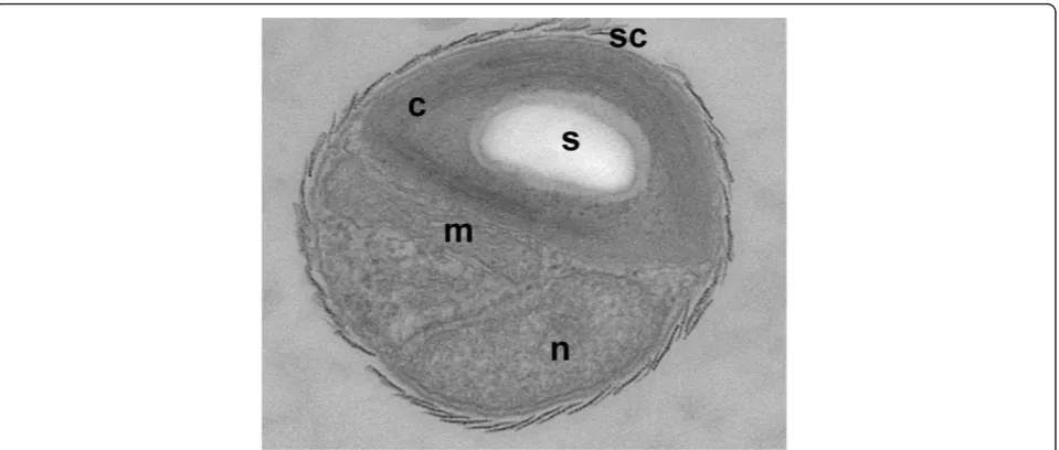
We isolated theBathycoccus prasinosstrain RCC1105 from a seawater sample from Banyuls’bay collected in January 2006. Contrary to the type strain described as Bathycoccus prasinos[8], which was isolated at a depth of 100 meters, RCC1105 was isolated from surface water (5 m). The strain RCC1105 has a typical Bathycoccus morphology with scales covering the cell (Figure 1) and we confirmed its taxonomic affiliation by PCR amplifica-tion of its 18S ribosomal gene. The complete genome of RCC1105 revealed two unlinked identical copies of the rDNA genes. Unlike the two previously reportedB. prasi-nosisolates [8,21], these two ribosomal 18S genes were
found to harbor an identical 433 bp long group I intron starting at position 551. Apart from this, the nucleotide sequence was strictly identical to the reference strain (GenBank: AY425315, FN562453). Self-splicing group I introns are widespread in nature, and have been recorded in the 18S rDNA of several other protists [22], including some within the green lineage, but, so far, not within the Mamiellales. All fourBathycoccusstrains isolated from the Mediterranean bear this intron located exactly at the same splicing site. Phylogenetic analysis based on this small ribosomal subunit and on the internal transcribed spacer (ITS) confirmed that, in contrast to the two other Mamiellales’generaMicromonasandOstreococcus, all Bathycoccusstrains isolated to date comprise only one clade [12,13]. To confirm the phylogenetic position of Bathycoccuswithin the Mamiellales, we concatenated a set of 154 single-copy genes conserved in 13 species, including plants, and aligned them over 35,431 amino acids to construct a maximum likelihood phylogenetic species tree (Figure S1 in Additional file 1, and Addi-tional files 2 and 3). The phylogeny obtained was well-supported and showed that the genus Bathycoccusis closer toOstreococcusthan toMicromonas.
Global characteristics of theBathycoccus genome
protein similarity (approximately 85%), and approximately 15% of them contain introns. Very few repeat sequences were found and no known or new transposable elements were detected (Table S1 in Additional file 1). The synteny observed between the chromosomes ofOstreococcusand Bathycoccus (Figure 2b; Figure S4 in Additional file 1) shows that the genome organization is globally better con-served between these two genera than with the genus Micromonas, in agreement with the phylogenetic analysis.
Based on the annotated gene sets of different land plants and green algae, sequence similarity searches were per-formed to group homologous genes into families (a family being defined as a set of two or more homologous genes; see Materials and methods). Subsequently, pan and core genome plots were built to quantify the number of shared and unique genes and families between different species (Figure S5 in Additional file 1). Comparing the set of core genes between different algal groups reveals that the smal-ler genome sizes of Mamiellales, as well as the lower
[image:3.595.60.540.88.292.2]number of genes, correspond both with the decrease of the average number of genes per family and with the num-ber of families conserved within a specific clade. For exam-ple, whereas the number of gene families shared between all land plants, Chlamydomonales, and Trebouxiophyceae is 2,692, this number drops to 1,959 when including all Mamiellales species. Similarly, based on a set of core gene families conserved in both land plants and algae, the aver-age gene family size is smaller for Mamiellales compared to Trebouxiophyceae or Chlamydomonales (average of 1.63, 1.78 and 1.93 genes per family, respectively). More than 500 gene families were found that were conserved between land plants and green algae but that were lost in all Mamiellales species (Figure S6 in Additional file 1). These families were enriched for functions related to zinc ion-binding and transport (ten families), UDP-glucosyl-transferase activity (six families), vitamin ion binding (eight families) and sucrose and fatty acid metabolism (eight families) (Table S2 in Additional file 1). Although Figure 1Morphology of theBathycoccus prasinosRCC1105 strain. Morphological characterization of theBathycoccusRCC1105 strain: EM picture of an exponentially growingBathycoccusRCC1105 cell. Abbreviations: c, chloroplast; n, nucleus; s, starch granule; sc, scale covering the surface of the cell.
Table 1 Nuclear genome characteristics of green algae
Family Species Genome size (Mb) G+C (%) Chromosome number Gene number
Prasinophyceae Bathycoccussp. RCC1105 151 48 19 7,847
Prasinophyceae Micromonassp. RCC299 20.9 64 17 10,286
Prasinophyceae Micromonassp. CCMP1545 21.9 65 19 10,587
Prasinophyceae Ostreococcus lucimarinusclade A 13.2 60 21 7,805
Prasinophyceae Ostreococcussp. RCC809 clade B 13.3 60 20 7,492
Prasinophyceae Ostreococcus tauriclade C 12.6 59 20 8,116
Trebouxiophyceae Chlorellasp. NC64A 46 67 12 9,791
Chlorophyceae Chlamydomonas reinhardtii 121 64 17 15,143
Chlorophyceae Volvox carterii 138 56 14 14,520
[image:3.595.58.539.598.722.2]this pattern suggests a reduction of the functional gene repertoire, we also found more than 400 gene families that are specific to Mamiellales and found in all Mamiellales species. Whereas many of these Mamiellales-specific genes have unknown functions, three families related to drug transport and ten families including genes related to zinc ion binding were found (Table S2 in Additional file 1). Although rapid sequence evolution can interfere with the accurate detection of homologs using similarity searches, the observed pattern indicates a high turnover of zinc ion binding-related genes.
Biological role and evolution of the big and small outlier chromosomes inBathycoccus and in the Mamiellales
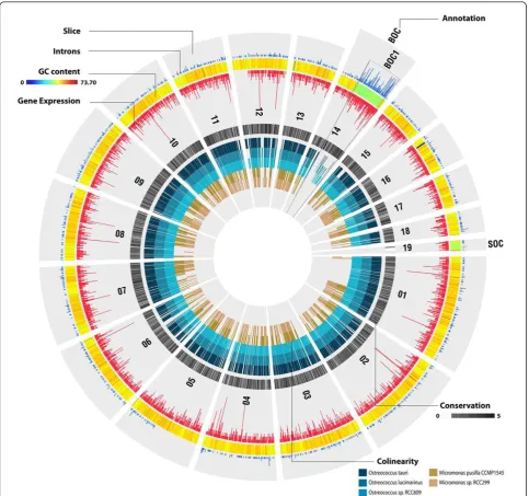
Despite the low average GC content (48%; Table 1) of the Bathycoccusgenome compared to other members of the Mamiellales (over 59%; Table 1), two outlier chromosomes were found, one‘big’(chromosome 14) and one‘small’ (chromosome 19), with lower GC content (42%) compared to the rest of the genome (Table 2; Figure S7 in Additional file 1). This kind of organization was previously reported in MicromonasandOstreococcus[9-11,23] and thus is a char-acteristic of all Mamiellales that have been sequenced so far. In all species, the atypical genomic features for the‘big’ outlier chromosomes (BOCs) are restricted to a sub-region (referred to as BOC1) of the complete chromosome,
whereas the whole length of the‘small’outlier chromo-some (SOC) shows low GC content (Figure S7 in Addi-tional file 1). However, although a BOC region was found for the‘low-light’Ostreococcussp. RCC809 genome, which is available on the Joint Genome Institute website (unpub-lished), no clear SOC could be identified (Additional file 1). Whether this observation is biologically correct or the con-sequence of the applied sequencing approach, read filter-ing, or genome assembly remains currently unclear. Similar outlier chromosomes have not been found in other green algae such asChlamydomonas,VolvoxorChlorella. In Chlorellalow GC chromosome regions were reported [24], but these were, in contrast to those in the Mamiellales, scattered throughout different chromosomes. Outlier chro-mosomes are highly diverged in terms of gene content. Whereas mostBathycoccuschromosomes share, to some extent, a conserved genome organization with the other Mamiellales, both BOC1 (217 annotated genes) and SOC (72 annotated genes) lack colinearity (Figure 3), and this pattern is largely conserved between the outliers of the three genera. Many BOC1 genes share orthologs with other Mamiellales while SOC comprises mainly unknown, species-specific genes with few introns (26% of the SOC proteins have Gene Ontology functional annotation versus 71% for BOC1 genes and 44% for the rest of the genome; Figure 3; Figure S8 in Additional file 1). Additionally,
1 2 3
1.125
825
750
610
450 825 785
450 1.020
285 1.020
365 2.200
1.600
945
680
565
225
4
[image:4.595.56.541.89.366.2]A
B
Figure 2Genome organization of theBathycoccus prasinosRCC1105 strain.(a)Pulse field electrophoresis of the genomes ofBathycoccus prasinosRCC1105 (lane 1),Micromonas pussilla(lane 2) andOstreococcus tauri(lane 3) DNA fragment length based on the chromosomes of
phylogenetic estimations of the proportions of genes lack-ing plant orthologs yielded 75% (54/72) for SOC, 16% for BOC1 and 25% for normal chromosomal regions.
The big outlier chromosome inBathycoccus
The size of theBathycoccusBOC is 663,424 bp. Fifty-two and seventy-eight percent of the BathycoccusBOC1 genes having orthologs in other species were also located in the BOC inMicromonasandOstreococus, respectively (Figure 4). In contrast, the locations of 29 BOC1 single-copy conserved gene markers (that is, genes having orthologs and located in BOC1 in all Mamiellales; Table S3 in Additional file 1) were scattered throughout the genomes inChlamydomonas,Volvox andChlorella, revealing that, despite the absence of colinearity, the clus-tering of the BOC1 genes is conserved and unique to the Mamiellales. These data suggest that BOC1 is a con-served genome property that was present in the last com-mon ancestor of the Mamiellales. Genes located in the BOC1 region are over-represented in basic housekeeping functions like primary metabolism, gene expression, photosynthesis and protein transport (Figure S8 in Addi-tional file 1; Table S3 in AddiAddi-tional file 1). To identify genomic features that are specific for the BOC1 region, the C-hunter tool (see Materials and methods) was applied to detect significant physical clustering of highly expressed genes and intron-containing genes on the differ-ent chromosomes (Table S4 in Additional file 1). C-hunter analysis revealed that the BOC1 region shows, in all spe-cies, a significant over-representation of EST-supported genes. Globally, 75% of all BOC1Bathycoccusgenes are EST supported versus 47% for non-BOC1 genes (Figure 3). After correcting for the overall 1.6-fold higher expression of BOC1 genes, BOC1 genes related to chromatin assem-bly, protein transport activity and signal transduction showed increased expression levels (Figure S8 in Addi-tional file 1). To verify whether the high expression is a property of the low GC genomic BOC1 region (for exam-ple, due to a more open chromatin structure [25]), we checked the expression level of the genes on the other low GC chromosome, SOC. We found that SOC genes had no difference in expression level compared to the genes on the 17 other chromosomes. We further investigated
whether this higher expression rate is an intrinsic property of the genes themselves, and estimated the expression levels for orthologs inChlamydomonas, Volvoxand Cocco-myxasp. C-169 (Figure S9A in Additional file 1). In all three species, BOC1 orthologs were also more highly expressed than other genes in the genome, suggesting that the higher expression of BOC1 genes in the Mamiellales is related to their function. Alternatively, this pattern might also be due to the global positive correlation, observed for all Mamiellales, between intron content and expression (Figure S9B in Additional file 1). Although the high expression of basic housekeeping BOC1 gene functions might yield increased metabolic rates and overall growth, it is not clear whether the physical clustering of BOC1 genes in the Mammiellophyceae lineage is based on adap-tive gene relocation or constrained ancestral location [26].
The BOC1 region also displays structural specificities that are absent in the rest of the genome. Besides its low GC content, genes in the BOC1 region are split by many small (40 to 65 bp) AT-rich introns [10,11]. This feature is present in all of the sequenced Mamiellales genomes (Figure S10 in Additional file 1) and absent from the gen-omes of other green algae (Figure S10 in Additional file 1). There is no universal RNA-fold for these introns and no conserved sequence motifs (for example, branch points, splice sites) could be detected. Although the only intrinsic indication from their DNA sequences that they are introns comes from their AT-richness relative to the surrounding GC rich exons, their existence is clear from EST data. Consequently, the BOC1 region includes a high propor-tion of multiple exon genes, a feature absent in the rest of the genome (Table S2 in Additional file 1). InBathycoccus, 103 of the 214 BOC1 genes harbor 330 introns, an intron content tenfold higher than in the rest of the genome (average of 1.54 and 0.15 introns per BOC1 and non-BOC1 gene, respectively).
[image:5.595.57.542.112.190.2]In conclusion, the BOC1 region in the Mamiellales has unique structural characteristics: it represent one contig-uous low(er) GC content region in the chromosome, flanked by two high(er) GC content regions at the extre-mities and carries between 193 and 633 genes depending on the species examined. The gene order within the region shows little colinearity between species and it
Table 2 Characteristics of the small outlier chromosomes forBathycoccusand oneMicromonasand oneOstreococcus species
Species Chromosome
number
Size (kb)
GC (%)
ORF number
Gene densities (bp/gene)
Identified genes
Sugar metabolism
Methylation enzymes
Other function
Bathycoccussp. 19 146 42 72 2,031 34 (47%) 17 (24%) 7 (10%) 4 (6%)
Ostreococcus lucimarinus
18 149 53 78 1,915 32 (41%) 16 (21%) 5 (6%) 11 (14%)
Micromonassp. RCC299
17 215 51 80 2,684 30 (38%) 14 (18%) 7 (9%) 9 (11%)
encodes a high proportion of often vital housekeeping genes with elevated expression levels clustered together in a pattern unique to the Mamiellales (Figure 4). The biological reason for the existence of this region remains obscure, although its structural characteristics (shuffling of genes, small introns, low GC content) concur with the
hypothesis that it may be a sex or species-barrier chro-mosome [27,28].
The small outlier chromosome inBathycoccus
[image:6.595.57.541.88.541.2]The size of theBathycoccus small outlier chromosome is 146,238 bp. compared to around 150 kb inOstreococcus
lucimarinus and 200 to 250 kb in Micromonas. The SOC average gene density inB. prasinosis slightly lower than that observed in the other chromosomes (72 genes with an average of 2.0 kb per gene in SOC compared to 1.7 kb per gene in the global genome), with a similar expression level based on EST counts. Only 44% of the
[image:7.595.59.540.88.549.2]genes in SOC have a potentially identified function compared to 77% in other chromosomes. Furthermore, up to 75% of the SOC genes have no known plant ortho-logs, in sharp contrast to most other chromosomes, where most genes share green lineage descent. Last but not least, inBathycoccus, 24 of the 34 SOC genes having Figure 4Distribution of theBathycoccusBOC1 orthologous genes in the genome of several other green alga species.(a-d)Bathycoccus
BOC1 orthologous genes in the genomes ofOstreococcus tauri(a),Micromonassp. RCC299 (b),Chlamydomonas reinhardtii(c) andCoccomyxasp. C-169 (d), each peripheral bar representing a chromosome. TheBathycoccusBOC1 region genes (lower right corner, labeled as‘Bathy_BOC1’) are connected by red lines to their orthologs (curated as best BLAST hits) on the chromosomes of other species. Where aBathycoccusgene also represents a BOC1 Mamiellales core gene (Table 3), the link is colored blue. Green bars show the BOC1 regions inBathycoccus,Micromonasand
an identified function group in two categories. The first group encodes enzymes involved in metabolism of glyco-conjugates (17 genes), mainly glycosyltransferase (12 genes), and the second is related to methyl transferases (7 genes). These features are globally similar in the other known Mamiellales SOCs, where the same two dominant gene functions were found (Table 2). However, despite their common function, no synteny and almost no ortho-logous relationships could be established between the SOCs of the different Mamiellales’species, suggesting a more functional convergence than a common phyloge-netic origin. To explain the presence of such genes in SOCs, an alien origin of these chromosomes was pro-posed, which could have yielded some selective advan-tages in cell surface processes, potentially related, for example, to defense against pathogens or other environ-mental interactions [10]. However, since SOCs and BOCs have now been found in all sequenced mamiellophycean genomes, it is likely that their lower GC composition, higher proportion of specific genes and higher evolution rates [9,29] are being maintained by the same evolution-ary pressure in all of these species. Interestingly, a paper on the cyanobacteriaProchlorococcusdescribes how vari-able genomic islands showing similar characteristics to those found in SOCs (low number of orthologs, a high level of horizontal gene transfer (HGT) and a high frac-tion of sugar-modifying enzymes, methyl transferases and membrane associated proteins) are involved in resistance to viruses [30]. The viral resistance determined by these genomic islands induced a fitness cost measured either by a reduced growth rate and/or a more rapid infection by other viruses. The three generaBathycoccus, Micro-monasandOstreococcusare the microalgae tested, which are among the most attacked by viruses [31], and viral resistance phenomena showing similar characteristics to what is reported forProchlorococcus(reduced growth rate and higher infection rate by other viruses) have been reported to occur frequently [32]. It is tempting to link this unusual high viral sensitivity and the ability to develop rapid and frequent resistance to these attacks to the presence of SOCs. Interestingly, two other Mamiel-lales species (Mamiellasp. orMantoniella squamata) were tested recently and did not show this high viral sen-sitivity (N Simon, personal communication). It can be predicted that if our hypothesis on the link between SOC and viral hypersensitivity/resistance is correct, these spe-cies should not present a SOC-like structure in their genome.
Phylogenomics suggests many horizontal gene transfers
Based on the observation that no plant homologs could be found for many annotatedBathycoccusgenes, a systematic analysis was performed to unravel their origin. Since plain sequence similarity search strategies are insufficient to
reliably trace a gene’s evolutionary history [33,34], a two-step comparative approach was applied to identify putative HGT events. After comparing eachBathycoccus protein sequence against the National Center for Biotechnology Information (NCBI) protein database, 6,550 phylogenetic trees were constructed and conflicts between the gene and organism phylogeny were determined. Whereas clustering patterns where the nearest neighbor in the tree corre-sponds with a homolog from a species outside the plant lineage were scored as HGT, in some cases ancestral gene duplication followed by differential gene loss or artifacts of phylogenetic reconstruction methods due to unusual modes of protein evolution could yield misleading results [35]. There were 428 genes (6%) that clustered with a homologous gene from a species outside the green lineage, whereas the remaining genes grouped with Viridiplantae genes (70%) or did not show any significant similarity. Among the 428 putative non-Viridiplantae genes, 80% were of non-green eukaryotic origin while 17% were bac-terial orthologs (Figure 5a). For the 354 non-green eukar-yotic genes, a high proportion came from Metazoa and Stramenopiles (42% and 28%, respectively). Gene Ontology enrichment analysis showed that around 50% of the non-Viridiplantae genes (including prokaryotic genes) were involved in metabolism. Focusing on the most enriched categories revealed genes involved in zinc ion binding (61, 6-fold enrichment), sialyltransferase activity (27, 12-fold enrichment), glycosylation (27, 11-fold enrichment) and ankyrin repeats (5-fold enrichment) (these observations are discussed further in the following section). Application of conservative selection criteria (retaining only phyloge-netic trees with bootstrap support >90% and more than 50% protein alignment coverage) yielded 79 genes with non-plant nearest neighbors (Table S5 in Additional file 1), which we propose might originate from HGTs, either from eukaryotes (82%) or prokaryotes (18%). Most of these 98 highly probable HGTs (43%) show unknown functions and the others, both originating from pro- or eukaryotes, show metabolite functions (based on similari-ties with protein domains). The absence of detectable eukaryotic HGT inArabidopsis thaliana, our negative control, suggests that this finding is not an artifact of the method. Using the same approach, previous putative large-scale HGTs have been reported in the available nuclear diatom genomes [36,37], both from bacteria (784 genes inPhaeodactylum tricornutum) or from the green lineage (>1,700 genes). However, although no other
This hypothesis seems unlikely, however, because most of the stramenopile genes found inBathycoccusare specific to this species and are not found in other Mamilelalles genomes. Alternatively, this mosaic gene repertoire could be the consequence of (i) parallel or convergent molecular evolution or (ii) the evolution through gene loss of a large ancestral genome, with massive and selective gene losses in all Mamiellales descendants, concurrent with genome
reduction. However, this scenario is less parsimonious compared to HGT and, again, seems unlikely because of the phylogenetic breadth of the selectively retained genes (bacterial and from different supergroups of the eukaryotic tree of life).
[image:9.595.62.536.82.566.2]genes contribute new functional properties to the Bathy-coccus genome. The analysis of a large DNA virus in Ostreococcus tauri suggested that the capture of host DNA in viral genomes could represent a mechanism for the transfer of genes between eukaryotic cells [39]. This idea was confirmed by the additional sequencing of four double-stranded DNA marine prasinovirus genomes (infectingBathycoccus,Micromonas, andOstreococcus), showing that these viruses encode a gene repertoire of certain amino acid biosynthesis pathways never pre-viously observed in viruses that are likely to have been acquired from lateral gene transfer from their host or from bacteria [40]. A similar eukaryotic phytoplankton-virus system was also described in Emiliania huxleyi, mediating the transfer of seven genes related to sphin-golipid biosynthesis [41].
To verify whether specific genomic regions or chromo-somes would be more likely to harbor genes arriving via HGT, we estimated the number of HGT genes per chro-mosome. We observed that transferred genes were more or less equally distributed over the different chromosomes, except for the low GC outlier chromosomes, which con-tained higher fractions of HGT genes (BOC1 and SOC contain 1.63 and 1.54 times more HGT genes compared to the genome-wide average; Figure 5b). Different possibi-lities for the increased abundance of HGT on the outliers include, for example: (1) they may have specific sequence features that can serve to integrate HGT genes that are subsequently re-arranged and embedded in other locations in the genome; (2) it may reflect a lower density of essen-tial gene functionalities in outliers, which could thus sup-port a higher density of random insertions; or (3) there
might be a lower level of recombination on these chromo-somes, reducing the rate of removal of deleterious alleles via sexual recombination. None of these scenarios are mutually exclusive.
In theBathycoccusgenome, the gene copy number is highly expanded for four specific gene families, phenom-ena not found (or at very low copy number expansion) in other Mamiellales or other algae (Table 3). Of these, two are involved in the metabolism of sialic acids, that is, sia-lyltransferases (69 gene copies) and sialidases (23 gene copies), the two others being ankyrin-repeat proteins (149 gene copies) and zinc finger proteins (48 gene copies) (Table 3). Among these 289 gene copies, 105 (36%) are represented within the 428 probable genes acquired by HGT, representing 24% of them.
Sialic acid metabolism inBathycoccus
[image:10.595.56.540.500.723.2]The two enzyme families involved in the metabolism of sialic acids are not present in other known green algae genomes, and both gene families are dispersed all along theBathycoccusgenome without evident clustering or tandem duplication. Although, on average, 15% of the genes have introns inBathycoccus, no introns (except three genes; Figure S11 in Additional file 1) were found in any gene from both families. Genes annotated as sialyl-transferases correspond to glycosylsialyl-transferases family 29 in the CAZy classification, which comprises enzymes able to transfer sialic residues during glycosylation of proteins or lipids [42]. All theBathycoccus sialyltrans-ferases showed a metazoan taxonic affiliation and none of them gave significant hits with bacteria. These enzymes are type II single pass membrane proteins
Table 3 Expanded gene families in theBathycoccusgenome
Gene familya Copy number in
Bathycoccus prasinos
Copy number in
Micromonassp. CCMP1545
Copy number in
Micromonassp. RCC299
Copy number in
Ostreococcus lucimarinus
Copy number in
Ostreococcussp. RCC809
Copy number inOstreococcus
tauri
Glycosyl transferase, family 29 (IPR001675)
78 0 1 0 2 0
HOM000519 43 0 0 0 0 0
HOM002813 10 0 0 0 0 0
HOM005062 10 0 0 0 0 0
HOM007941 6 0 0 0 0 0
Ankyrin repeats (IPR020683)
186 124 107 74 55 67
HOM000035 149 56 9 17 6 6
Sialidase/ neuraminidase (IPR011040)
23 1 0 0 0 0
HOM002557 17 0 0 0 0 0
HOM005056 5 0 0 0 0 0
Zinc finger, C2H2 53 29 35 19 3 17
HOM000293 48 5 4 1 1 1
a
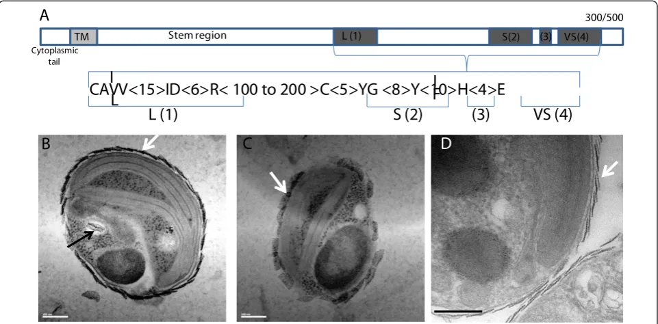
usually known to be anchored in the Golgi membranes [43,44]. A potential hydrophobic transmembrane domain was detected on the amino-terminal extremities of all the Bathycoccussialyltransferases (Figure 6a). For almost all the 69 genes (only 19 are known in human), the sialyl-transferase domain is located in the carboxy-terminal part of the protein, whereas the amino-terminal domain is composed of a highly variable stem region (Figure 6a). Although the existence of complete and active sialyl-transferases in plants is still a matter of debate [45], all four metazoan consensus motifs were found in the Bathycoccusgenes.
The second gene family includes sialidases (or neurami-nidases), which are enzymes cleaving the terminal sialic acid residues from glycoproteins or glycolipids. Again, this gene expansion is specific toBathycoccus. In contrast to the previous family, no clear domain organization could be defined in sialidases, but some key amino acids known to be involved in the catalytic activity are conserved in the Bathycoccusfamily. The taxonomic origin of the sialidases is less clear than that for the sialyltransferases discussed above, and could correspond to either metazoans or bac-teria. For sialidases, scores are globally weak and best blast hits are found mostly with hypothetical proteins either from the choanoflagellateMonosiga brevicolisor from the green algaChlorella variabilis(where only one sialidase has been annotated).
The expansion of these two enzyme families prompted us to look for specific potential‘sialic acid’metabolism in Bathycoccus. The composition of flagellar scales in Scherffelia dubia(phylum Chlorophyta, class Chloroden-drophyceae) was found to be a mix of acidic polysacchar-ides having similar structures to sialic acids [46]. Although the chemical nature of the scales covering the Bathycoccuscell membrane is unknown, it is tempting to establish a correlation between the potential biosynthetic pathway of these scales and the expansion of gene families involved in the metabolism of sialic acids. Furthermore, we confirmed previous electron microscopy studies [8,47] showing that, inBathycoccusas in other Mamiellophyceae, scale biosynthesis occurs inside intra-cellular vesicles with striking resemblance to Golgi vesi-cles (Figure 6b-d); that is, in agreement with the notion that they might be produced by sialyltransferases located at the luminal side of intracellular vesicles. Scales almost identical to those ofB. prasinosare observed in the more closely relatedMantoniella squamata[48], where they are also extruded to the surface after transport via the Golgi body [49-51].
OtherBathycoccus expanded gene families
One of the two other highly expanded gene families in theBathycoccusgenome are ankyrin-repeat proteins (149 gene copies). This family is also expanded, although to a
Stem region
TM L (1) S(2) (3) VS(4)
300/500
CAVV<15>ID<6>R< 100 to 200 >C<5>YG <8>Y<10>H<4>E
L (1)
S (2)
(3)
VS (4)
Cytoplasmic tail
A
B
C
D
L
I
I
[image:11.595.58.539.434.671.2]F
lesser extent, in theMicromonasstrain CCMP1545 (56 copies), whereas only very few copies were detected in other Mamiellales (Table 3). These genes have ankyrin repeats located in the carboxy-terminal part of the pro-tein whereas the amino-terminal part has no hit in GenBank. There are also many other ankyrin repeats containing genes inBathycoccusas in both plants and microalgae, but associated with different protein domains that often have predicted functionalities. Indeed, the ankyrin repeat is considered as one of the most common protein-protein interaction motifs in nature [52]. The 149Bathycoccus-specific genes were not distributed ran-domly among chromosomes, with the bigger chromo-somes having few copies, whereas chromochromo-somes 12 or 19 bear many tandem duplicated genes (Figure S12 in Addi-tional file 1). No obvious function can be attributed to these genes. However, by analogy with the human mem-brane-associated ankyrin, which is responsible for the attachment of the cytoskeleton to the plasma membrane, it is possible that a number of these genes might function in some way to bind extracellular scales to the plasmic membrane, although experimental evidence is lacking. It has been shown, however, by electron microscopy coupled to immunogold that scales inScherffelia dubia are linked to the membrane by glycoproteins [46]. In addition, inTetraselmis striata(Chlorodendrophyceae) some scale-associated glycoproteins may provide connec-tions between scales and the underlying flagellar mem-brane [50].
The last group of expanded genes inBathycoccusare zinc finger proteins. There are many zinc finger proteins in microalgae and in plants, but the family specifically expanded inBathycoccusis most related to the C2H2-type
zinc finger DNA-binding domain of certain integrases, which share a common alpha/beta two-layer sandwich core structure. The typical organization of the 48 copies identified in theBathycoccusgenome (Table 3) includes a short amino-terminal part (around 20 to 40 amino acids) followed by a strongly acidic region (10 to 20 amino acids) and by 2 to 6 C2H2domains. Zinc finger proteins were
originally identified as DNA-binding domains, although a growing body of evidence suggests an important and wide-spread role for these domains in protein binding. There are even examples of zinc fingers that support both DNA and protein interactions, and, globally, C2H2
protein-pro-tein interactions are proving to be more abundant than previously appreciated [53].
The most parsimonious explanation for the abundance of the four expanded gene families would be an initial single HGT event followed by expansion in the Bathycoc-cusgenome. The potential function of these four gene families and their expansion only in Bathycoccusalso suggest that they could all be involved in the biosynth-esis, exportation and fixation of the scales around the
external membrane, and possibly for protection of the cell. Several other members of the Mamiellales have mor-phologically similar scales around the cells, but they are absent in the two generaMicromonasandOstreococcus. The most parsimonious evolutionary scenario to explain these observations is that the scale synthesis pathway was acquired by the ancestor of the Mamiellales (or even before) and has been lost in the two naked genera. This scenario predicts that similar gene family expansions should be found in the genomes of other scaled Mamiel-laophyceae but not in MicromonasandOstreococcus. This is the case forMicromonasandOstreococcus, but the genome sequences of other scaled species are not yet available.
Conclusions
Mamiellophyceae, and more particularly the three gen-era Bathycoccus, Micromonas and Ostreococcus, are dominant in different marine areas, where they can play an important role in the primary biomass production. However, the ecological importance of Bathycoccushas probably been overlooked these past years, although it was sporadically mentioned in several studies [5,16-18]. The availability of this genome, coupled to the develop-ment of new sequencing possibilities for metagenomes [19,20] from various marine environments, opens the door to future comparative studies and to a better understanding of the adaptations of the organisms to their environment.
Materials and methods
B. prasinos RCC1105 genome and EST sequencing and annotation
pellets were immediately flash frozen in liquid nitrogen. The total RNA was extracted using the TriReagent (Sigma-Aldrich, Saint-Quentin, France) protocol and mRNAs purified using Poly(A)Purist (Ambion-Applied Bioystems, Saint Aubin, France). Complementary DNAs were constructed and cloned using the CloneMiner proce-dure (InvitroGen, Saint Aubin, France) with some minor modifications. EST sequences were obtained using pyrose-quencing technology developed by Roche (Boulogne-Bill-ancourt, France). A total of 253,791 EST reads were processed through the Genoscope EST pipeline. Short (<60 bp) and low complexity sequences were identified and removed. Clustering and assembly of all 251,875 filtered EST reads resulted in 8,370 EST consensus sequences.
The genome was annotated using the EuGene [56,57] gene finding system with Splice- Machine [58] signal sen-sor components trained specifically onBathycoccus data-sets. The functional annotation resulted from the synthesis of InterPro and the BLASTP hits against the non-redun-dant UniProt database. Gene Ontology assignments were derived from the InterPro results. Gene Ontology enrich-ment analysis was performed using the hypergeometric distribution with Bonferonni correction for multiple hypothesis testing and corrected P-values <0.05 were retained as significant. The resulting database is publicly available at [59] in a format that includes browse and query options and the genome has been submitted to GenBank.
Comparative sequence and expression analysis
Starting from all protein-coding genes from the included species (Table 1), only retaining the longest transcript if alternative splicing variants exist, protein sequences were used to construct gene families by applying sequence-based protein clustering. First, an all against all sequence comparison was performed using BLASTP, applying an E-value threshold of 1e-05 and retaining the best 500 hits [60]. Next, the complete sequence similar-ity graph was processed using Tribe-MCL (mclblastline, default parameters except I = 2 and scheme = 4) to identify gene families. A set of 154 single-copy core gene families was used to construct the phylogenetic tree depicted in Figure S1 in Additional file 1 (see also Additional files 2 and 3).
The boundaries of all Mamiellales BOC1 regions were manually delineated based on gene coordinates, gene family information and GC content (Table S2 in Addi-tional file 1). For non-Mamiellales, a ‘virtual’ BOC1 region was created by taking the best BLASTP hit for each B. prasinosRCC1105 BOC1 gene. Putative BOC1 Mamiellales core gene families (Figure 6, blue lines) were identified by first retaining only those families that contain at least one protein for each Mamiellales
species. Next, each family was aligned and manually curated. This was done by inspecting and correcting, if necessary, the structural and functional annotation (NCBI BLAST results plus InterProScan) of all cluster members. For Ostreococcussp. RCC809 no SOC could be identified in the current draft genome assembly (Additional file 1).
Comparative genomics
To detect co-linearity within and between species, i-ADHoRe 3.0 was used (Additional file 1) [61] and all chromosomes from all species were compared against each other and significant colinear regions were identi-fied. All gene colinearity can be browsed using the pico-PLAZA comparative genomics platform [62]. i-ADHoRe was run with the following settings: alignment_method gg, gap_size 30, cluster_gap 35, q_value 0.9, prob_cutoff 0.0001, anchor_points 5 and level_2_only false.
EST databases were retrieved from their respective public repositories and mapped on the Mamiellales gen-omes using GenomeThreader [63] with a minimum alignment score threshold of 0.95 and minimum tran-script coverage of 0.89. Only uniquely mapped ESTs were retained and assigned to genes. When an EST with no strand information overlapped with two adjacent genes, it was assigned to the gene with the highest over-lap. For the BOC expression analysis global gene, EST counts were first summarized per functional category. In a second stage, expression enrichment was determined by comparing for each functional category the fraction of BOC expressed genes against the overall fraction of BOC expressed genes (denoted‘relative BOC expression enrichment’in Figure S8 in Additional file 1).
Analysis of potential horizontal gene transfer
neighbor taxonomic information. Genes showing com-plex punctuate patterns [64] (that is, clustering with homologs from different phyla outside the Viridiplantae; labeled‘multi-kingdom’in Table S5 in Additional file 1) were excluded. Singletons refer to genes for which no phylogenetic analysis could be done because they only have a single BLAST hit based on the 20% top hits. Near-est neighbors with bootstrap support >90% and gene cov-erage of 50% or more in the multiple alignment were scored as reliable HGT genes to estimate the fraction of eukaryotic origin. Although the low number of HGT genes found inArabidopsisdoes not serve as a perfect negative control for the detection of HGT in unicellular green algae, it suggests that, when applied to a full set of proteins of a specific organism, this approach gives a con-servative estimate of putative transfer events with a low number of false positives. To verify if, for some HGT genes, homologous genes exist in other algae that were missed during the process of gene annotation, a systema-tic sequence similarity search (using tblastn, E-value threshold 1e-05against intergenic sequences ofO. tauri, O. lucimarinus,OstreococcusRCC809,M. pusillaand C. reinhardtii) revealed that, on average, no homologous locus could be found for 93% of the HGT genes. A list of all HGT genes together with protein alignment and phy-logenetic tree statistics is available in Additional file 5.
C-hunter analysis
Four functional categories (two types with two subdivi-sions each) were defined and genes were assigned to each class, if applicable. The first type of functional cate-gory describes the expression state of a gene (based on uniquely mapped ESTs; is a gene expressed (number of ESTs >0) or highly expressed (number of ESTs >2)) while the second type describes the intron content of a gene (contains an intron (number of introns >0) or con-tains a ‘lot’of introns (number of introns >2)). C-hunter [65] software was used to identify, in all genomes, signif-icant clusters of genes belonging to one of the four functional categories. The C-hunter thresholds for each category subdivision were determined by reviewing the average expression and intron content of all Mamiellales genes. C-hunter was run with the following parameters: <C-hunter categories.go genome.index genome.go 2 80 80 0.001 50 T chunter output>.
Accession numbers
Sequence data from this article (the genome ofB. prasinos RCC1105) can be found in the EMBL/GenBank data libraries under accession number [FO082258] (mitochon-drion), [FO082259] (chloroplast), [FO082278] (chromo-some 1), [FO082277] (chromo(chromo-some 2), [FO082276] (chromosome 3), [FO082275] (chromosome 4), [FO082274] (chromosome 5), [FO082273] (chromosome
6), [FO082272] (chromosome 7), [FO082271] (chromo-some 8), [FO082270] (chromo(chromo-some 9), [FO082269] (chro-mosome 10), [FO082268] (chro(chro-mosome 11), [FO082267] (chromosome 12), [FO082266] (chromosome 13), [FO082265] (chromosome 14), [FO082264] (chromosome 15), [FO082263] (chromosome 16), [FO082262] (chromo-some 17), [FO082261] (chromo(chromo-some 18), [FO082260] (chromosome19). The annotation of the genome can be found at the BOGAS web site [66]. EST data are available at the ENA database (accession number ERA148021) and raw genome sequencing data are available at the Trace archive database of the NCBI under the query: species_co-de=“BATHYCOCCUS SP. BAN7”.
Additional material
Additional file 1: Supplementary materials and methods, figures and tables.
Additional file 2: The 154 single-copy core gene families in the green plant lineage.
Additional file 3: Alignment of 154 single-copy core gene families in the green plant lineage.
Additional file 4: Statistics of the genome shotgun sequencing.
Additional file 5: Details of maximum likelihood phylogenetic trees describingB. prasinosRCC1105 HGT genes.
Abbreviations
BOC: big outlier chromosome; bp: base pair; CCMP: Center for Culture of Marine Phytoplankton; EST: expressed sequence tag; HGT: horizontal gene transfer; NCBI: National Center for Biotechnology Information; RCC: Roscoff Culture Collection; SOC: small outlier chromosome.
Acknowledgements
We are very grateful to L Fibla and L Subirana for technical help, R Cooke and M Laudié for sequencing and S Proost for technical assistance during the colinearity analysis. Sequencing of theBathycoccusgenome has been supported by the Genoscope (Evry, France) and this research was supported by the CNRS and the University Pierre et Marie Curie (UMR 7232) and the Agence Nationale de la Recherche grants PHYTADAPT n° NT09_567009 and Tara-Girus 09-PCS-GENM-218. We thank Igor Grigoriev, Brian Palenik and the Joint Genome Institute for the access to theOstreococcusstrain RCC809 genome. BV, KV and YVdP acknowledge the support of Ghent University (Multidisciplinary Research Partnership‘Bioinformatics: from nucleotides to networks’) and the Interuniversity Attraction Poles Programme (IUAP P6/25), initiated by the Belgian State, Science Policy Office (BioMaGNet).
Author details
1CNRS, UMR 7232, Observatoire Océanologique, Banyuls-sur-Mer, France. 2UPMC Univ Paris 06, UMR 7232, Observatoire Océanologique,
Banyuls-sur-Mer, France.3Department of Plant Systems Biology, VIB, Technologiepark 927, B-9052 Ghent, Belgium.4Department of Plant Biotechnology and
Bioinformatics, Ghent University, Technologiepark 927, B-9052 Ghent, Belgium.5Genoscope, CEA, Institut de Génomique, 2 rue Gaston Crémieux
CP5706, 91057 Evry cedex, France.6Department of Biotechnology, Norwegian University of Science and Technology (NTNU), 7491 Trondheim, Norway†These two authors contributed equally to this work.
Authors’contributions
Competing interests
The authors declare that they have no competing interests.
Received: 11 April 2012 Revised: 15 August 2012 Accepted: 24 August 2012 Published: 24 August 2012
References
1. Field BC, Behrenfeld MJ, Randerson JT, Falkowski P:Primary production of the biosphere: integrating terrestrial and oceanic components.Science
1998,281:237-240.
2. Boyce DG, Lewis MR, Worm B:Global phytoplankton decline over the past century.Nature2010,466:591-596.
3. Li WKW:Primary productivity of prochlorophytes cyanobacteria, and eucaryotic ultraphytoplankton: measurements from flow cytometric sorting.Limnol Oceanogr1994,39:169-175.
4. Worden AZ, Nolan JK, Palenik B:Assessing the dynamics and ecology of marine picophytoplankton: the importance of the eukaryotic component.Limnol Oceanogr2004,49:168-179.
5. Knight-Jones EW, Walne PR:Chromulina pusillaButcher; a dominant member of the ultraplankton.Nature1951,167:445.
6. Courties C, Vaquer A, Trousselier M, Lautier J, Chrétiennot-Dinet MJ, Neveux J, Machado MC, Claustre H:Smallest eukaryotic organism.Nature
1994,370:255.
7. Chrétiennot-Dinet MJ, Courties C, Vaquer A, Neveux J, Claustre H, Lautier J, Machado MC:A new marine picoeukaryoteOstreococcus tauri genet sp. nov (Chlorophyta, Prasinophyceae).Phycologia1995,4:285-292. 8. Eikrem W, Throndsen J:The ultrastructure ofBathycoccusgen nov andB.
prasinos spNov, a non-motile picoplanktonic alga (Chlorophyta, Prasinophyceae) from the Mediterranean and Atlantic.Phycologia1990,
29:344-350.
9. Worden AZ, Lee JH, Mock T, Rouzé P, Simmons MP, Aerts AL, Allen AE, Cuvelier ML, Derelle E, Everett MV, Foulon E, Grimwood J, Gundlach H, Henrissat B, Napoli C, McDonald SM, Parker MS, Rombauts S, Salamov A, Von Dassow P, Badger JH, Coutinho PM, Demir E, Dubchak I, Gentemann C, Eikrem W, Gready JE, John U, Lanier W, Lindquist EA, Lucas S,et al:Green evolution and dynamic adaptations revealed by genomes of the marine picoeukaryotes Micromonas.Science2009,324:268-272.
10. Derelle E, Ferraz C, Rombauts S, Rouzé P, Worden AZ, Robbens S, Partensky F, Degroeve S, Echeynié S, Cooke R, Saeys Y, Wuyts J, Jabbari K, Bowler C, Panaud O, Piégu B, Ball SG, Ral JP, Bouget FY, Piganeau G, De Baets B, Picard A, Delseny M, Demaille J, Van de Peer Y, Moreau H:
Genome analysis of the smallest free-living eukaryote Ostreococcus tauri unveils many unique features.Proc Natl Acad Sci USA2006,
103:11647-11652.
11. Palenik B, Grimwood J, Aerts A, Rouzé P, Salamov A, Putnam N, Dupont C, Jorgensen R, Derelle E, Rombauts S, Zhou K, Otillar R, Merchant SS, Podell S, Gaasterland T, Napoli C, Gendler K, Manuell A, Tai V, Vallon O, Piganeau G, Jancek S, Heijde M, Jabbari K, Bowler C, Lohr M, Robbens S, Werner G, Dubchak I, Pazour GJ, Ren Q, Paulsen I,et al:The tiny eukaryote
Ostreococcusprovides genomic insights into the paradox of plankton speciation.Proc Natl Acad Sci USA2007,104:7705-7710.
12. Guillou L, Eikrem W, Chrétiennot-Dinet MJ, Le Gall F, Massana R, Romari K, Pedrós-Alió C, Vaulot D:Diversity of picoplanktonic prasinophytes assessed by direct nuclear SSU rDNA sequencing of environmental samples and novel isolates retrieved from oceanic and coastal marine ecosystems.Protist2004,155:193-214.
13. Marin B, Melkonian M:Molecular phylogeny and classification of the Mamiellophyceae class nov (Chlorophyta) based on sequence comparisons of the nuclear- and plastid-encoded rRNA operons.Protist
2010,161:304-336.
14. Peers GK, Niyogi K:Pond scum genomics: The genomes of
ChlamydomonasandOstreococcus.Plant Cell2008,20:502-507. 15. Johnson PW, McSieburth J:In-situmorphology and occurrence of
eukaryotic phototrophs of bacterial size in the picoplankton of estuarine and oceanic waters.J Phycol1982,18:318-327.
16. Marie D, Zhu F, Balagué V, Ras J, Vaulot D:Eukaryotic picoplankton communities of the Mediterranean Sea in summer assessed by molecular approaches (DGGE, TTGE, QPCR).FEMS Microbiol Ecol2006,
55:403-415.
17. Monier A, Welsh RM, Gentemann C, Weinstock G, Sodergren E, Armbrust EV, Eisen JA, Worden AZ:Phosphate transporters in marine
phytoplankton and their viruses: cross-domain commonalities in viral-host gene exchanges.Environ Microbiol2011,14:162-176.
18. Treusch AH, Demir-Hilton E, Vergin KL, Worden AZ, Carlson CA, Donatz MG, Burton RM, Giovannoni SJ:Phytoplankton distribution patterns in the northwestern Sargasso Sea revealed by small subunit rRNA genes from plastids.ISME J2011,6:481-492.
19. Cheung MK, Au CH, Chu KH, Kwan HS, Wong CK:Composition and genetic diversity of picoeukaryotes in subtropical coastal waters as revealed by 454 pyrosequencing.ISME J2010,4:1053-1059. 20. Marie D, Shi X L, Rigaut-Jalabert F, Vaulot D:Use of flow cytometric
sorting to better assess the diversity of small photosynthetic eukaryotes in the English Channel.FEMS Microbiol Ecol2010,72:165-178.
21. Massana R, Balagué V, Guillou L, Pedrós-Alió C:Picoeukaryotic diversity in an oligotrophic coastal site studied by molecular and culturing approaches.FEMS Microbiol Ecol2004,50:231-243.
22. Haugen PD, Simon D, Bhattacharya D:The natural history of group I introns.Trends Genet2005,21:111-119.
23. Piganeau G, Grimsley N, Moreau H:Genome diversity in the smallest marine photosynthetic eukaryotes.Res Microbiol2011,162:570-577. 24. Blanc G, Duncan G, Agarkova I, Borodovsky M, Gurnon J, Kuo A, Lindquist E,
Lucas S, Pangilinan J, Polle J, Salamov A, Terry A, Yamada T, Dunigan DD, Grigoriev IV, Claverie JM, Van Etten JL:TheChlorella variabilisNC64A genome reveals adaptation to photosymbiosis, coevolution with viruses, and cryptic sex.Plant Cell2010,22:2943-2955.
25. Zhang X:The epigenetic landscape of plants.Science2008,320:489-492. 26. Wong S, Wolfe KH:Birth of a metabolic gene cluster in yeast by adaptive
gene relocation.Nat Genet2005,37:777-782.
27. Lee SC, Ni M, Li W, Shertz C, Heitman J:The evolution of sex: a perspective from the fungal kingdom.Microbiol Mol Biol Rev2010,
74:298-340.
28. Soo Chan Lee SC, Ni M, Li W, Shertz C, Heitman J:The evolution of sex: a perspective from the fungal kingdom.Microbiol Mol Biol Rev2010,
74:298-340.
29. Jancek S, Gourbiere S, Moreau H, Piganeau G:Clues about the genetic basis of adaptation emerge from comparing the proteomes of two
Ostreococcusecotypes (Chlorophyta, Prasinophyceae).Mol Biol Evol2008,
25:2293-2300.
30. Avrani S, Wurtzel O, Sharon I, Sorek R, Lindell D:Genomic island variability facilitatesProchlorococcus-virus coexistence.Nature2011,474:604-608. 31. Bellec L, Grimsley N, Derelle E, Moreau H, Desdevises Y:Abundance, spatial
distribution and genetic diversity ofOstreococcus tauriviruses in two different environments.Env Microbiol Reports2010,2:313-321. 32. Thomas R, Grimsley N, Escande ML, Subirana L, Derelle E, Moreau H:
Acquisition and maintenance of resistance to viruses in eukaryotic phytoplankton populations.Env Microbiol2011,13:1412-1420. 33. Ragan MA, Harlow TJ, Beiko RG:Do different surrogate methods detect
lateral genetic transfer events of different relative ages?.Trends Microbiol
2006,14:4-8.
34. Kurland CG, Canback B, Berg OG:Horizontal gene transfer: a critical view.
Proc Natl Acad Sci USA2003,100:9658-9662.
35. Keeling PJ, Palmer JD:Horizontal gene transfer in eukaryotic evolution.
Nat Rev Genet2008,9:605-618.
36. Bowler C, Allen AE, Badger JH, Grimwood J, Jabbari K, Kuo A, Maheswari U, Martens C, Maumus F, Otillar RP, Rayko E, Salamov A, Vandepoele K, Beszteri B, Gruber A, Heijde M, Katinka M, Mock T, Valentin K, Verret F, Berges JA, Brownlee C, Cadoret JP, Chiovitti A, Choi CJ, Coesel S, De Martino A, Detter JC, Durkin C, Falciatore A, Fournet J,et al:The
Phaeodactylumgenome reveals the evolutionary history of diatom genomes.Nature2008,456:239-244.
37. Moustafa A, Beszteri B, Maier UG, Bowler C, Valentin K, Bhattacharya D:
Genomic footprints of a cryptic plastid endosymbiosis in diatoms.
Science2009,324:1724-1726.
38. Raymond JA, Kim HJ:Possible role of horizontal gene transfer in the colonization of sea ice by algae.PLoS One2012,7:e35968. 39. Derelle E, Ferraz C, Escande ML, Eychenie S, Cooke R, Piganeau G,
Desdevises Y, Bellec L, Moreau H, Grimsley N:Life-cycle and genome of OtV5, a large DNA virus of the pelagic marine unicellular green alga
Ostreococcus tauri.PLoS One2008,3:e2250.
40. Moreau H, Piganeau G, Desdevises Y, Cooke R, Derelle E, Grimsley N:
acquisition of protein metabolism genes by horizontal gene transfer.J Virol2010,84:12555-12563.
41. Monier A, Pagarete A, de Vargas C, Allen MJ, Read B, Claverie JM, Ogata H:
Horizontal gene transfer of an entire metabolic pathway between a eukaryotic alga and its DNA virus.Genome Res2009,19:1441-1449. 42. Cantarel BL, Coutinho PM, Rancurel C, Bernard T, Lombard V, Henrissat B:
The Carbohydrate-Active EnZymes database (CAZy): an expert resource for Glycogenomics.Nucleic Acids Res2009, ,37 Database:D233-238. 43. Jeanneau C, Chazalet V, Augé C, Soumpasis DM, Harduin-Lepers A,
Delannoy P, Imberty A, Breton C:Structure-function analysis of the human sialyltransferase ST3GalI.J Biol Chem2004,279:13461-13468. 44. Harduin-Lepers A, Mollicone R, Delannoy P, Oriol R:The anormal
sialyltransferase-related genes: a phylogenetic approach.Glycobiology
2005,15:805-817.
45. Harduin-Lepers A, Mollicone R, Delannoy P, Oriol R:The animal sialyltransferases and sialyltransferase-related genes: a phylogenetic approach.Glycobiology2005,15:805-817, A published erratum appears in Glycobiology 2005, 15:21G.
46. Melkonian M, Preisig HR:A light and electron microscopic study of
Scherffelia dubia, a new member of the scaly green flagellates (Prasinophyceae).Nord J Bot1986,6:235-256.
47. Moestrup O, Walne PL:Studies on scale morphogenesis in the Golgi apparatus ofPyramimonas tetrarhynchus(Prasinophyceae).J Cell Sci1979,
36:437-459.
48. Moestrup O:Scale structure inMantoniella squamata, with some comments on the phylogeny of the Prasinophyceae (Chlorophyta).
Phycologia1990,29:437-442.
49. Melkonian M, Becker B, Becker D:Scale formation in algae.J Electron Microscopy Technique1991,17:165-178.
50. Becker D, Melkonian M:N-linked glycoproteins associated with flagellar scales in a flagellate green alga: characterization of interactions.Eur J Cell Biol1992,57:109-116.
51. Becker B:Anterograde transport of algal scales through the Golgi complex is not mediated by vesicles.Trends Cell Biol1995,5:305-307. 52. Al-Khodor S, Price CT, Kalia A, Kwaik A:Functional diversity of ankyrin
repeats in microbial proteins.Trends Microbiol2010,18:132-139. 53. Brayer KJ, Segal DJ:Keep your fingers off my DNA: protein-protein
interactions mediated by C2H2 zinc finger domains.Cell Biochem Biophys
2008,50:111-131.
54. Winnepenninckx B, Backeljau T, De Wachter R:Extraction of high molecular weight DNA from molluscs.Trends Genet1993,9:407. 55. Batzoglou S, Jaffe DB, Stanley K, Butler J, Gnerre S, Mauceli E, Berger B,
Mesirov JP, Lander ES:ARACHNE: a whole-genome shotgun assembler.
Genome Res2002,12:177-189.52.
56. Schiex T, Moisan A, Rouzé P:EUGÉNE: an eukaryotic gene finder that combines several sources of evidence.Lect Notes Comput Sci2001,
2066:111-125.
57. Foissac S, Gouzy J, Rombauts S, Mathé C, Amselem J, Sterck L, Van de Peer Y, Rouzé P, Schiex T:Genome annotation in plants and fungi: EuGène as a model platform.Curr Bioinformatics2008,3:87-97. 58. Degroeve S, Saeys Y, De Baets B, Rouzé P, Van de Peer Y:SpliceMachine:
predicting splice sites from high-dimensional local context representations.Bioinformatics2005,21:1332-1338.
59. Genome Sequences and Annotations at Ghent University..[http:// bioinformatics.psb.ugent.be/genomes].
60. Altschul SF, Madden TL, Schäffer AA, Zhang J, Zhang Z, Miller W, Lipman DJ:Gapped BLAST and PSI-BLAST: a new generation of protein database search programs.Nucleic Acids Res1997,25:3389-33402. 61. Proost S, Fostier J, De Witte D, Dhoedt B, Demeester P, Van de Peer Y,
Vandepoele K:i-ADHoRe 3 0 - fast and sensitive detection of genomic homology in extremely large data sets.Nucleic Acids Res2012,40:e11. 62. pico-PLAZA:an integrative resource for cross-species genome analysis in
algae.[http://bioinformatics.psb.ugent.be/pico-plaza/].
63. Gremme G, Brendel V, Sparks ME, Kurtz S:Engineering a software tool for gene structure prediction in higher organisms.Information Software Technol2005,47:965-978.
64. Rogers MB, Watkins RF, Harper JT, Durnford DG, Gray MW, Keeling PJ:A complex and punctate distribution of three eukaryotic genes derived by lateral gene transfer.BMC Evol Biol2007,7:89.
65. Yi G, Sze SH, Thon MR:Identifying clusters of functionally related genes in genomes.Bioinformatics2007,23:1053-1060.
66. BathycoccusGenome Annotation Database at Ghent University..[http:// bioinformatics.psb.ugent.be/webtools/bogas/].
67. Krzywinski M, Schein J, Birol I, Connors J, Gascoyne R, Horsman D, Jones SJ, Marra MA:Circos: an information aesthetic for comparative genomics.
Genome Res2009,19:1639-1645.
68. Merchant SS, Prochnik SE, Vallon O, Harris EH, Karpowicz SJ, Witman GB, Terry A, Salamov A, Fritz-Laylin LK, Maréchal-Drouard L, Marshall WF, Qu LH, Nelson DR, Sanderfoot AA, Spalding MH, Kapitonov VV, Ren Q, Ferris P, Lindquist E, Shapiro H, Lucas SM, Grimwood J, Schmutz J, Cardol P, Cerutti H, Chanfreau G, Chen CL, Cognat V, Croft MT, Dent R, Dutcher S,
et al:TheChlamydomonasgenome reveals the evolution of key animal and plant functions.Science2007,318:245-250.
69. Prochnik SE, Umen J, Nedelcu AM, Hallmann A, Miller SM, Nishii I, Ferris P, Kuo A, Mitros T, Fritz-Laylin LK, Hellsten U, Chapman J, Simakov O, Rensing SA, Terry A, Pangilinan J, Kapitonov V, Jurka J, Salamov A, Shapiro H, Schmutz J, Grimwood J, Lindquist E, Lucas S, Grigoriev IV, Schmitt R, Kirk D, Rokhsar DS:Genomic analysis of organismal complexity in the multicellular green algaVolvox carteri.Science2010,329:223-226.
doi:10.1186/gb-2012-13-8-r74
Cite this article as:Moreauet al.:Gene functionalities and genome structure inBathycoccus prasinosreflect cellular specializations at the base of the green lineage.Genome Biology201213:R74.
Submit your next manuscript to BioMed Central and take full advantage of:
• Convenient online submission
• Thorough peer review
• No space constraints or color figure charges
• Immediate publication on acceptance
• Inclusion in PubMed, CAS, Scopus and Google Scholar
• Research which is freely available for redistribution