Psychology Dissertations Department of Psychology
Summer 8-2012
Visual Scanning of Dynamic Affective Stimuli in Autism Spectrum
Visual Scanning of Dynamic Affective Stimuli in Autism Spectrum
Disorders
Disorders
Susan M. McManus Georgia State University
Follow this and additional works at: https://scholarworks.gsu.edu/psych_diss
Recommended Citation Recommended Citation
McManus, Susan M., "Visual Scanning of Dynamic Affective Stimuli in Autism Spectrum Disorders." Dissertation, Georgia State University, 2012.
https://scholarworks.gsu.edu/psych_diss/105
This Dissertation is brought to you for free and open access by the Department of Psychology at ScholarWorks @ Georgia State University. It has been accepted for inclusion in Psychology Dissertations by an authorized
VISUAL SCANNING OF DYNAMIC AFFECTIVE STIMULI IN AUTISM
SPECTRUM DISORDERS
by
SUSAN M. MCMANUS
Under the Direction of Diana L. Robins, Ph.D.
ABSTRACT
The accurate integration of audio-visual emotion cues is critical for social interactions and
requires efficient processing of facial cues. Gaze behavior of typically developing (TD)
individuals and individuals with autism spectrum disorders (ASD) was measured via
eye-tracking during the perception of dynamic audio-visual emotion (DAVE) stimuli. This study
provides information about the regions of the face sampled during an emotion perception task
that is relatively more complex than those used in previous studies, providing both bimodal
group was less accurate at emotion detection and demonstrated less of a visual-affective bias
than TD individuals. Both groups displayed similar fixation patterns across regions during the
perception of congruent audio-visual stimuli. However, between-group analyses revealed that
fixation patterns differed significantly by facial regions during the perception of both congruent
and incongruent movies together. In addition, fixation duration to critical regions (i.e., face, core,
eyes) was negatively correlated with measures of ASD symptomatology and social impairment.
Findings suggest weaknesses in the early integration of audio-visual information, automatic
perception of emotion, and efficient detection of affective conflict in individuals with ASD.
Implications for future research and social skills intervention programs are discussed.
VISUAL SCANNING OF DYNAMIC AFFECTIVE STIMULI IN AUTISM
SPECTRUM DISORDERS
by
SUSAN M. MCMANUS
A Dissertation Submitted in Partial Fulfillment of the Requirements for the Degree of
Doctor of Philosophy
in the College of Arts and Sciences
Georgia State University
Copyright by Susan M. McManus
VISUAL SCANNING OF DYNAMIC AFFECTIVE STIMULI IN AUTISM
SPECTRUM DISORDERS
by
SUSAN M. MCMANUS
Committee Chair: Diana L. Robins
Committee: Erin B. Tone
David J. Marcus
Robert D. Latzman
Electronic Version Approved:
Office of Graduate Studies
College of Arts and Sciences
Georgia State University
ACKNOWLEDGEMENTS
I would like to extend my gratitude to the faculty members who have provided
mentorship and supported my training throughout graduate school. I would also like to
acknowledge the Georgia State University Dissertation Grant program for the funding on this
TABLE OF CONTENTS
ACKNOWLEDGMENTS ... iv
LIST OF TABLES ... vii
LIST OF FIGURES ... ix
1 INTRODUCTION ...1
1.1 Autism Spectrum Disorders (ASD) ...2
1.2 Development of Emotion Perception ...4
1.3 Impact of Emotion Perception Deficits on Social Functioning in ASD ...7
1.4 Components of Emotion Perception ...9
1.5 Gaze Behavior ...18
1.6 Static vs. Dynamic Stimuli ...21
1.7 Neural Correlates of Social Perception ...23
1.8 Preliminary Findings ...29
1.9 The Current Study ...30
1.10 Hypotheses ...35
2 METHOD ...41
2.1 Participants ...41
2.2 Experimental Stimuli ...42
2.3 Diagnostic Measures ...43
2.4 Inclusion and Exclusion Measures ...46
2.5 Apparatus ...51
2.6 Procedure ...53
3.1 Hypothesis 1: Accuracy and Reaction Time during Congruent Movies ...61
3.2 Hypothesis 2: Fixation Pattern during Congruent Movies ...62
3.3 Hypothesis 3: Behavioral Responses and Reaction Time during Incongruent ...65
Movies 3.4 Hypothesis 4: Fixation Pattern during Incongruent Movies as Compared to ...67
Congruent Movies 3.5 Hypothesis 5: Exploratory Analysis of Initial Fixations ...74
3.6 Hypothesis 6: Exploratory Analysis of the Relationship between Visual ...77
Fixations and Social Functioning 4 DISCUSSION ...78
4.1 Behavioral Performance ...78
4.2 Fixation Patterns: Congruent and Incongruent Movies ...87
4.3 Social Cognition ...95
4.4 Conclusions ...98
4.5 Limitations and Future Directions ...101
REFERENCES ...105
LIST OF TABLES
Table 1 Summary of sample and task characteristics of relevant emotion perception ...33
studies Table 2 Movie duration ranges in seconds ...43
Table 3 Group performance on cognitive, perceptual, and emotional measures ...59
Table 4 Number of significant elevations (T-score ≥70) on the BASC-2 ...60
Table 5 Group performance on subscales of interest for the BASC-2 ...60
Table 6 Group fixation duration (sec) data for face and non-face regions during ...62
congruent movies of Task 1 Table 7 Group fixation duration (sec) data for core and periphery regions during ...64
congruent movies of Task 1 Table 8 Group fixation duration (sec) data for eye, nose, and mouth regions during ...65
congruent movies of Task 1 Table 9 Group modality bias data for incongruent movies of Task 2 ...66
Table 10 Response time data for congruent and incongruent movies of Task 2 ...67
Table 11 Group fixation duration (sec) data for face and non-face regions during ...68
congruent and incongruent movies of Task 2 Table 12 Group fixation duration (sec) data for core and periphery regions during ...70
congruent and incongruent movies of Task 2 Table 13 Group fixation duration (sec) data for eye, nose, and mouth regions during ...73
congruent and incongruent movies of Task 2 Table 14 Group initial fixation data for eye, nose, and mouth regions during ...75
Table 15 Correlations between fixation duration data and measures of social functioning ...78
Table 16 Summary of fixation duration findings for congruent movies of Task 1 ...88
Table 17 Summary of fixation duration findings for congruent and incongruent ...89
movies of Task 2
Table 18 Summary of initial fixation findings for congruent and incongruent ...91
LIST OF FIGURES
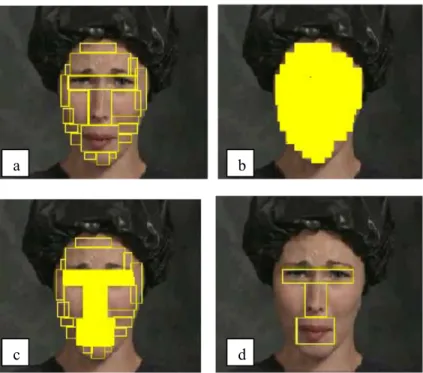
Figure 1. Stimulus regions compared in fixation time analyses: (a) all defined regions, ...37
(b) facial regions (shaded area) compared with non-facial regions (non-shaded),
(c) core regions (shared area) compared with periphery (non-shaded boxes on face),
and (d) three core regions compared (eyes, nose, mouth).
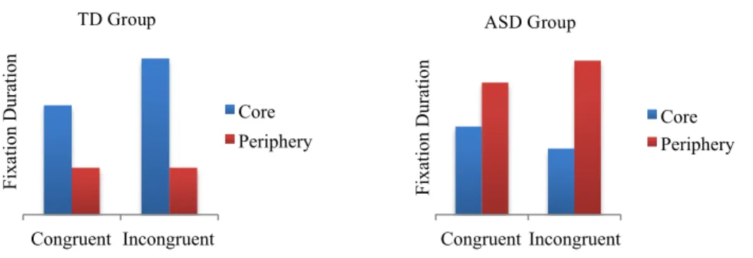
Figure 2. Illustration of expected three-way interaction between group, congruence, ...39
and region for the core versus periphery analysis.
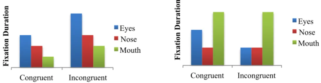
Figure 3. Illustration of expected three-way interaction between group, congruence, ...40
and region for the analysis between eyes, nose, and mouth.
Figure 4. Schematic of ASL 6000 set-up and eye-tracking data collection ...52
Figure 5. Total fixation duration for the face and non-face regions when viewing ...63
congruent movies.
Figure 6. Total fixation duration for the core and peripheral face regions when viewing ...64
congruent movies.
Figure 7. Total fixation duration for the eye, nose, and mouth regions when viewing ...65
congruent movies.
Figure 8. Total fixation duration for the face and non-face regions when viewing ...69
all movies.
Figure 9. Combined group data for total fixation duration to face and non-face regions ...69
when viewing congruent and incongruent movies.
Figure 10. Total fixation duration for the core and peripheral face regions when ...71
viewing all movies.
regions when viewing congruent and incongruent movies.
Figure 12. Total fixation duration for the eye, nose, and mouth regions when viewing ...74
congruent movies.
Figure 13. Total fixation duration for the eye, nose, and mouth regions when viewing ...74
congruent movies.
Figure 14. Number of first fixations devoted to the eye, nose, and mouth regions when ...76
viewing congruent movies.
Figure 15. Number of first fixations devoted to the eye, nose, and mouth regions when ...77
1 INTRODUCTION
Affective perception occurs quickly and requires the seemingly automatic integration of
multiple sources of information, including facial expression, semantic information, affective
prosody, affective memory, and physiological responses. The accurate perception of facial affect
is just one important component of affective perception, as facial expressions reveal a lot of
information about one’s state of mind, desires, and intentions. Researchers continue to explore
the manner in which humans derive affective information from the facial expressions of others,
but the process is not yet completely understood.In order to better understand the perception of
affective information in individuals with autism spectrum disorders (ASD) and their typically
developing (TD) peers, it is important to consider the underlying processes, including facial
processing strategies and the integration of facial expression and affective prosody, as well as
gaze behavior and the impact of biological motion on affect recognition. In accordance with this,
research on emotion perception will be reviewed, taking into consideration what is known about
the typical and atypical perception of visual and audio-visual information. Given that the
emotion perception literature is fairly narrow and primarily focuses on perception of visual
emotion cues, a broader perceptual literature provides a useful foundation from which
predictions can be made about the strategies utilized during audio-visual emotion processing in
ASD.
The data collected in this study provide information about the regions of the face (i.e.,
eyes, nose, mouth) that are sampled during a relatively more complex emotion perception task
than those typically used in the literature. The relative increase in complexity of the stimuli to be
used in the current study comes from the fact that stimuli provide both bimodal (auditory and
information about different visual scanning approaches to the perception of dynamic
audio-visual stimuli, and will increase understanding of emotion perception deficits in ASD. This
knowledge will inform treatment intervention strategies that aim to improve social skills in ASD.
Future directions will be discussed in the context of previous findings, as well as in terms of
clinical implications for such research.
1.1 Autism Spectrum Disorders (ASD)
Autism spectrum disorders (ASD), or pervasive developmental disorders, are
characterized by markedly abnormal or impaired development across three domains: social
interaction, communication, and restricted interests or stereotyped/repetitive behaviors
(American Psychiatric Association, 2000). Within the social domain, symptoms include limited
social-emotional reciprocity, impaired use of nonverbal behaviors (e.g., facial expression,
gestures, eye contact) to regulate social interactions, failure to develop age-appropriate peer
relationships, and deficits in joint attention. Symptoms within the communication domain
include language delay coupled with a failure to compensate with nonverbal communication,
difficulty with reciprocal conversation, limited imitative or pretend play, and stereotyped or
idiosyncratic use of language. The restricted, repetitive, and stereotyped patterns of behavior or
interests are observed as intense preoccupations, rigidity of nonfunctional routines, stereotyped
or repetitive motor movements, or preoccupations with parts of or sensory qualities of objects
(American Psychiatric Association, 2000).
Abnormal functioning or a delay in the development of six behaviors across the three
domains, with impairment in one of these areas (e.g., social interaction, social use of language,
imitative play) by the age of three years, meets criteria for a diagnosis of Autistic Disorder. A
behaviors and interests domains, in the absence of language delay and cognitive impairment.
Individuals who present with significant and pervasive impairments in one or more of these
areas, but at a level that does not meet criteria for another specific ASD, may be diagnosed with
Pervasive Developmental Disorder, Not Otherwise Specified (PDD-NOS; American Psychiatric
Association, 2000). In addition to these core symptoms, children with ASD may develop
behavioral changes or comorbid disorders, such as problems with sleep, appetite, mood, anxiety,
hyperactivity, irritability, anger regulation, and aggression, but these symptoms may be difficult
to recognize due to the broader psychosocial deficits associated with the diagnosis of ASD
(Ozonoff, Goodlin-Jones, & Solomon, 2005). Additional symptoms (e.g., guilt) that alert
professionals to other psychiatric disorders (e.g., depression, anxiety) may not present in
individuals with ASD (Ozonoff, Goodlin-Jones, & Solomon, 2005).
A recent epidemiological study conducted by the Centers for Disease Control and
Prevention estimates the current prevalence rate for all ASDs at 1/88, representing a 23%
increase in prevalence when compared to data collected in 2006 (Centers for Disease Control and
Prevention, 2012). With regard to subtypes of ASD, one recent epidemiological study reports
prevalence rates for Autistic Disorder at 22.0/10,000 and for AS around 10.5/10,000 (Saracino,
Noseworthy, Steiman, Reisinger, & Fombonne, 2010). With the apparent rise in rates of ASD
diagnosis, research that aims to explore early risk factors, identify methods for early detection,
and develop early intervention strategies is becoming more widely conducted. Dawson (2008)
describes a developmental model of such risk factors, risk processes, and outcomes in
individuals diagnosed with ASD, highlighting the neurophysiological changes occurring with
early experience and intervention and the potential for reduced symptomatology in ASD. With
improvement in symptoms of ASD (using cognitive, adaptive, language, and/or general
developmental measures) following early intervention (Howlin, 2005; Rogers & Vismara, 2008).
Other reviews indicate significant improvement and treatment efficacy with early
communication intervention (Brunner & Seung, 2009) and behavioral intervention (Howlin,
Magiati, & Charman, 2009).
Social deficits are viewed as a primary symptom that is characteristic of all diagnoses
within the autism spectrum (American Psychiatric Association, 2000). Individuals with ASD
exhibit significant weaknesses in social-emotional reciprocity beginning early in development,
with primary impairments in social orienting, joint attention, motor imitation, the processing of
facial information, and the recognition of emotion from both visual and auditory cues (Dawson
& Bernier, 2007; Loveland et al., 1997; Mundy, Sigman, Ungerer, & Sherman, 1986).
1.2 Development of emotion perception
Typical Development.Research shows that TD individuals are consistently able to
identify basic emotions that are recognized across cultures (e.g., Ekman & Friesen, 1975).
Researchers generally focus on six basic emotions when exploring the universality of emotion
perception in cross-cultural studies: happiness, sadness, surprise, fear, disgust, and anger (e.g.,
Bryant & Barrett, 2008; Ekman, 1992; Izard, 1971, 1992; Wang et al., 2006). Basic emotions
have been defined as those presumed to have innate neural substrates, a unique and universally
recognized facial expression, and a unique feeling state (Izard, 1972, 1992; Ortony & Turner,
1990; Panksepp, 1982). In addition to early arguments regarding the existence of emotion- or
system-specific biological correlates, evidence from recent neuroimaging and neurophysiological
studies supports the existence of discrete, biologically-based emotion response mechanisms. For
emotion experiences (e.g., mood induction, facial expression identification, perception of
arousing pictures) found a unique pattern of neural activation for each of the six basic emotions;
the most robust activation was found in the superior temporal gyrus for happiness, left medial
frontal gyrus for sadness, left inferior frontal gyrus for anger, left amygdala for fear, and right
insula and right inferior frontal gyrus for disgust. In addition, discrete patterns of autonomic
nervous system (ANS) responding have been found for each of the six basic emotions, as
indicated by electrodermal (i.e., skin-conductance, skin resistance, skin potential),
thermo-vascular (i.e., skin blood flow, skin temperature), and respiratory measures (Collet,
Vernet-Maury, Delhomme, & Dittmar, 1997; Kreibig, 2010).
The recognition of emotions requires understanding of nonverbal social cues, including
facial expression, prosodic information or tone of voice, and body posture of others. The
development of emotion perception skills during infancy and early childhood is observed in
terms of the child’s responsiveness to others’ emotions and evidence of social referencing
(Baldwin & Moses, 1996; Haviland & Lelwica, 1987). Research suggests that infants display a
sensitivity to social cues (e.g., Grossman et al., 2008) and are able to discriminate between
dynamic (i.e., in motion) displays of basic, prototypical emotions (i.e., happy, sad) by the age of
six months old (de Haan & Nelson, 1997). Maternal expression of happy, fearful, or neutral
emotions has been shown to influence an infant’s behavior as early as 12 to 18 months of age. In
situations designed to produce uncertainty in the child, maternal facial expression was found to
influence the distance to which the infant would explore (Klinnert, 1984). By the age of four
years, children are able to verbally label prototypical emotions (i.e., happy, sad, angry; Widen &
Russell, 2003). Although the ability to recognize most prototypical emotional expressions is
2003), there continues to be improvement in the recognition of less intense emotions and
increased sensitivity to more subtle displays of the basic emotions beyond adolescence (Thomas,
DeBellis, Graham, & Labar, 2007). Researchers have found that emotion recognition
proficiency, as defined by the speed of processing and the ability to detect subtle differences,
continues to develop throughout adolescence and peaks in adulthood (Rump, Giovannelli,
Minshew, & Strauss, 2009).
Atypical Development of Emotion Perception Abilities in Autism Spectrum Disorders.
Individuals with ASD encounter difficulties in basic visual processing of social situations (e.g.,
social use of eye gaze to interpret what others may be thinking, feeling, or communicating) and
even young infants with ASD respond less to others, with evidence for the reduced salience of
human faces (Klin, Jones, Schultz, & Volkmar, 2005; Osterling, Dawson, & Munson, 2002).
However, the results of studies exploring emotion recognition abilities are not as definitive as
those that highlight early deficits in social engagement. Some researchers assert that emotion
recognition is intact in ASD, whereas others claim that it is impaired (see Capps, Yirmiya, &
Sigman, 1992 and Celani, Battacchi, & Arcidiacono, 1999 for opposing findings). Equivocal
findings can broadly be attributed to differences in methodology, task demands, and age and
abilities of the participants being studied.
In general, individuals with ASD perform similarly to controls when they are presented
with unambiguous, prototypical emotions and allowed sufficient processing time before a
response is required (Capps, Yirmiya, & Sigman, 1992; Grossman, Klin, Carter, & Volkmar,
2000; Humphreys, Minshew, Leonard, & Behrmann, 2007; Ozonoff, Pennington, & Rogers,
1990). Weaknesses in emotion perception emerge when individuals with ASD are shown stimuli
Greimel et al., 2010; Humphreys et al., 2007; Law Smith et al., 2010; Pelphrey, et al., 2002;
Philip et al., 2010; Rump, Giavannelli, Minshew, & Strauss, 2009). In addition, children with
ASD, regardless of specific diagnosis on the autism spectrum, perform worse than controls on
emotion identification (Lindner & Rosen, 2006) and affect matching tasks (Boucher, Lewis, &
Collis, 2000). However, some researchers have failed to find these deficits in emotion
identification tasks with adolescents and adults with AS (e.g., Capps, Yirmiya, & Sigman, 1992;
O’Connor, 2007; Rutherford & Towns, 2008).
A recent study attempted to reconcile these inconsistent findings by assessing emotion
recognition abilities from a developmental perspective. Rump and colleagues (2009) found that
children diagnosed with ASD, ages five to seven years, performed more poorly on an emotion
recognition task than did their TD peers. When examining the developmental differences in the
same study, the authors found that older children (ages 8-12 years) and adolescents (ages 13-17
years) with ASD performed similarly to controls; however, adults (ages 18-53 years) with ASD
performed significantly worse than same-age controls. Results indicated that the adult controls
showed significant improvement in terms of proficiency (i.e., processing speed, ability to detect
subtle differences) over younger controls, but the ASD adults did not show the same advantage
over older children and adolescents with ASD. As such, the researchers postulated, based on the
data from this cross-sectional study, that the continued development of proficiency seen in
typical development does not occur in ASD (Rump et al., 2009).
1.3 Impact of Emotion Perception Deficits on Social Functioning in ASD
Basic emotion recognition through nonverbal cues has been shown to relate to performance
on brief measures of socio-cognitive abilities and social functioning and adjustment in
Hietanen, 2003). Inattention to important nonverbal social cues (e.g., faces, eyes) may have
deleterious consequences for social functioning because of the roles that these cues play in
signaling others’ intentions. As such, ongoing deficits in age-appropriate affective recognition
skills in children and adolescents with ASD can have adverse consequences that are additive and
persist into adulthood. Studies with TD children provide evidence that peer rejection and
victimization alters the way children process and respond to social cues by increasing sensitivity
to hostile cues and increasing aggressive responding (Dodge, Lansford, Burks, Bates, Pettit,
Fontaine, et al., 2003). For individuals with ASD, deficits in using nonverbal cues to make
inferences about others’ intentions and desires impact their social adjustment; some researchers
have found that children with ASD respond to peer rejection with negative behaviors (e.g.,
isolation, oblivion, emotionally-charged accounts relayed to family, behavioral acting out) in the
same way as typical children (Ames & Jarrold, 2007; Ochs et al., 2001).
Just as peer group factors (e.g., peer rejection) impact a child’s social and academic
adjustment, so do the resulting individual child factors (e.g., isolation, withdrawal; Ladd, 2006).
Peer group size and peer group acceptance contribute to socio-emotional adjustment, academic
competence, and self-concept in TD children (Vandell & Hembree, 1994). Despite reported
involvement in peer networks, children with ASD report lower centrality, acceptance,
companionship, and reciprocity (Chamberlain, Kasari, Rotheram-Fuller, 2007). Interestingly,
some have shown that children with ASD report more loneliness than do their TD peers, albeit
with a less complete understanding of the relationship between loneliness and friendship,
whereas other report no difference in reports of loneliness between children with ASD and their
TD peers (see Bauminger & Kasari, 2000 and Chamberlain, Kasari, & Rotheram-Fuller, 2007 for
the child’s peer group. Some researchers have shown that chronic peer rejection inhibits TD
children’s classroom participation (Ladd, Herald-Brown, Reiser, 1997). In addition, studies with
TD children have shown that peer acceptance and friendship predicts school perceptions,
avoidance, and performance (Ladd, 1990).
As a result of this pervasive deficit, a vast amount of research focuses on the social
abnormalities of individuals diagnosed with ASD. However, Brunner and Seung (2009) provide
a summary of recent studies and meta-analyses examining the efficacy of social skills
interventions for children with ASD and conclude that adequate efficacy studies for social skills
interventions (i.e., social skills training, social stories, and peer-mediated training) are as yet
unpublished. As programs are developed and evaluated, effective social skill intervention
strategies could benefit from more in depth exploration of specific deficits in emotion perception
and recognition. These specific emotion perception deficits will be explored in greater detail in
the following section.
1.4 Components of Emotion Perception
A poor understanding of atypical emotion perception by researchers and professionals,
specifically with regard to children and adolescents with ASD, can result in inadequate treatment
interventions. With complex and often disparate results from emotion perception studies in
individuals with ASD, as discussed previously, researchers may benefit from looking more
closely at the various components of emotion perception (e.g., audio-visual integration, face
perception) in order to better understand deficits that do exist. Results from studies in the broader
perceptual literature that examine these components more directly will inform hypotheses about
understanding of ways to improve the emotional and social functioning of children and
adolescents with ASD.
Audio-visual integration.In everyday social situations, individuals are required to
integrate information from both visual (i.e., facial expression) and auditory (i.e., tone of voice)
modalities. Evidence shows that the integration of facial and vocal information occurs early in
emotion perception, during the perceptual level of processing and not later during the response
selection stage (Hietanen, Leppanen, Illi, & Surakka, 2004; Pourtois, Debatisse, Desplan, & de
Gelder, 2002). When congruent visual and auditory cues are presented concurrently, this
integration occurs, as measured by event related brain potentials (ERPs), as early as 110 ms after
stimulus onset (Pourtois et al., 2000). In addition, congruent emotional information presented
through multiple modalities has been shown to modulate the amplitude size and latency of two
early ERP components (e.g., P200 and N300; Paulmann, Jessen, & Kotz, 2009). Specifically,
researchers found a gradual decrease in amplitude of these ERPs with the addition of each
modality (i.e., unimodal, bimodal, multimodal). Findings suggest that modulated amplitudes
reflect a reduced and speeded processing effort for bimodal (facial expression and prosody) as
compared to unimodal (facial expression) information.
Some researchers have found that one sensory modality has a greater influence on
perceptual outcome than others. When presented simultaneously with visual and auditory stimuli
and asked to respond by indicating whether they detected a visual stimulus, an auditory stimulus,
or both, participants made a significant number of errors. Specifically, participants were prone to
report visual only during trials in which both visual and auditory cues were present, termed the
visual dominance effect (Collignon et al., 2008; Hecht & Reiner, 2008). In another study, this
were semantically incongruent (Koppen, Alsius, & Spence, 2008). In this study, participants
made visual-only responses significantly more often than auditory-only responses on bimodal
trials, regardless of the congruence of auditory and visual syllables. Similar support has been
found for a visual dominance effect during emotion perception as well; a number of researchers
have demonstrated that typically developing individuals showed a bias toward facial affective
information when incongruent information was presented in another modality (e.g., tone of
voice, semantic information; de Gelder & Vroomen, 2000; de Gelder, Vroomen, & Bertelson,
1998; Massaro, 1998; Massaro & Egan, 1996). This influence held when participants were
instructed to attend to both modalities (Massaro & Egan, 1996) and when they were instructed to
attend to prosody (deGelder & Vroomen, 2000; Vroomen, Driver, & deGelder, 2001). However,
this influence did not hold when the face was presented upside-down. The processing of faces
was disrupted when the face was inverted (discussed in the following section) and this finding
highlighted the unique influence of facial expression on judgments of prosody (de Gelder,
Vroomen, & Bertelson, 1998). The literature is equivocal on whether prosody has the same
influence on perception of facial expression (see deGelder & Vroomen, 2000 and Massaro, 1998
for opposing findings).
Weaknesses in bimodal integration have been found in individuals diagnosed with ASD
for non-emotional (Gepner, deGelder, & deSchonen, 1996; Mongillo et al., 2008) and emotional
(Loveland et al., 1995) tasks. Children with ASD produced significantly fewer blends and
fusions on an audio-visual speech task than did matched controls (Gepner, deGelder, &
deSchonen, 1996). In a similar study, Mongillo and colleagues (2008) found that individuals
with ASD show less visual-speech influence on their responses during a task with mismatching
congruence between computer images and a simultaneously played auditory track (Loveland et
al., 1995) or matching photographs with tape recordings containing affective prosody (Boucher,
Lewis, & Collis, 2000). However, individuals with ASD are able to perform at levels comparable
to that of TD controls on audio-visual emotion recognition tasks when cued by semantic content
(Lindner & Rosen, 2006). Individuals with AS showed a bias toward visual-verbal information,
not facial expression, suggesting that people with AS rely on verbal mediation as a compensatory
strategy during complex emotion perception (Grossman et al., 2000).
In summary, studies of typically developing individuals show that visual information is
dominant during visual-auditory stimuli (Collignon et al., 2008; Hecht & Reiner, 2008; Koppen,
Alsius, & Spence, 2008) and visual affective information may have a greater influence on
prosodic information than vice versa (de Gelder & Vroomen, 2000; Massaro, 1998; Massaro &
Egan, 1996). However, given that this same dominance appears to be absent in individuals with
ASD, future research should continue to explore the differences in visual perception strategies
employed in ASD.
Face perception.Given that information from the visual modality appears to have a
greater influence on audio-visual integration, exploration of facial processing strategies is bound
to offer critical insight into typical and atypical bimodal emotion perception. Research indicates
that a global or configural representation of faces was related to a greater recognition of facial
emotion (Gross, 2005; Smith & Scott, 1998; White, 1999). A global or configural representation
refers to one in which the perceiver utilizes information about the position of facial features, or
configuration of the features, to a greater degree than information about the individual features of
the face. To identify a particular face or facial expression, typically developing individuals
mouth), utilizing a holistic processing approach, as opposed to relying on individual features,
which is associated with the processing of non-face objects (Pelphrey et al., 2002). Exploring
these various approaches to face processing (i.e., configural, featural, holistic) in greater detail is
important to furthering our knowledge of how and what information is extracted from the face
during emotion processing tasks.
There is a large body of literature that is dedicated to the exploration of face processing
strategies, particularly configural and featural strategies. Diamond and Carey (1986) are some of
the researchers at the forefront of this literature and have attempted to explain the inversion
effect, a manipulation frequently utilized in standard recognition memory paradigms. The
inversion effect is essentially a difference in performance on recognition tasks between upright
and inverted photographs of faces; the inversion effect occurs when faces that are presented
upside-down during encoding and recognition are more poorly recognized than faces presented
upright.
Diamond and Carey (1986) argued that the inversion effect emerges when two criteria are
met: (1) members of a class to be encoded share a configuration and can be distinguished from
one another based on second-order relational features and (2) perceivers are experts with a class
of stimuli. Second-order relational features, to be discussed in detail in the next section,
essentially refer to the spatial configuration of the individual features of a stimulus. With regard
to the criterion of expertise, Diamond and Carey (1986) found that the inversion effect emerges
when perceivers are as experienced with the class of stimuli presented for recognition as they are
with human faces. They found that the recognition of dogs is as sensitive to the inversion effect
as is the recognition of faces, provided that the perceivers are highly experienced with both dogs
theory proposed by Yin (1969, 1970), who found that memory recognition for human faces is
more vulnerable to stimulus inversion when compared to other classes of familiar objects (e.g.,
houses, airplanes, stick figures of people in motion) and argued that the inversion effect occurs
because neural specialization has evolved such that humans possess superior recognition
processing strategies for human faces. The first criterion proposed by Diamond and Carey
(1986), regarding stimulus-class characteristics, provides insight into the underlying processes of
facial and emotion processing and will be discussed in greater detail.
Distinguishing between first-order and second-order features is important to
understanding the differential effects of inversion on recognition. Rhodes (1988) defined
first-order features as the appearances of individual parts of the face, such as those labeled the eye,
nose, and mouth. Second-order features were those said to have configural properties, including
the spatial relations among first-order features, the position of first-order features, and
information about the shape of the face (Rhodes, 1988). Rhodes’ initial hypothesis of the
mechanisms of face representation utilized similar terminology as that proposed by Diamond and
Carey (1986), but the definitions appeared somewhat different. The distinction between the two
hypotheses became less clear in a later paper written by Rhodes, Brake, and Atkinson (1993). As
defined by Diamond and Carey (1986), first-order relational features refer to the relationship
between parts of a stimulus that are relatively unconstrained (i.e., the trees in a landscape). When
you have a class of stimuli that are homogenous in nature (i.e., human faces), the first-order
relational features are relatively constrained (i.e., eyes are located horizontally above the nose,
which is above the mouth; Diamond & Carey, 1986; Rhodes, Brake, & Atkinson, 1993). With
stimulus classes that possess fixed first-order relational features, another relationship becomes
of a stimulus (e.g., the distance between a specific individual’s eyes) are referred to as the
second-order relational features; this unique configuration is what is used to individuate faces
(Diamond & Carey, 1986; Rhodes, Brake, & Atkinson, 1993).
There is a growing body of literature focused on contrasting the configural versus featural
processing of objects, and in human faces in particular. A common assumption is that both
information about isolated features (i.e., featural properties) and the relations among them (i.e.,
configural properties) are used as a means of encoding upright faces. The configural information,
however, cannot be extracted from inverted faces and therefore results in less successful
recognition (Valentine, 1988). Diamond and Carey (1986) argued that the large inversion effect
that results from expertise and experience is due to individuals’ reliance on the configural
properties of stimuli (e.g., faces). Researchers have shown that the coding of second-order
relational features is more affected by inversion than the coding of first-order relations or of
isolated features (Rhodes, Brake, & Atkinson, 1993). Rhodes (1988) concluded that
second-order features are critical to face perception and thus it is, at least in part, configural by nature.
In addition to the second-order relations, the properties of a stimulus configuration
include the holistic, or gestalt, form. The overall structure or gestalt of human faces is of critical
importance when considering the superior recognition or processing of faces as compared to
other classes of objects and a number of theories have been proposed that attempt to explain how
this information is represented. In addition to the theories of relational or configural information,
others have proposed that gestalt form of the face is represented in parallel processing or through
interactive coding (Bradshaw & Wallace, 1971; Diamond & Carey, 1986; Rhodes, 1988;
Rhodes, Brake, & Atkinson, 1993; Sergent, 1984). Farah, Wilson, Drain, and Tanaka (1998)
recognition does not involve, or does so only to a lesser extent, the explicit representations of the
eyes, nose, and the mouth. They believe that faces are instead recognized as undifferentiated
wholes. In support of this theory, Farah and colleagues (1998) found that individuals perceived
upright faces in a holistic manner, significantly more so than inverted faces, houses, or words;
participants had significant difficulty differentiating and comparing individual parts of the
upright face independent of the whole face.
Others have found a similar whole-face advantage for upright faces. Homa, Haver, and
Schwartz (1976) found that individuals recognized features more accurately when presented in
the context of a natural face than if presented amongst scramble features. TD children recognized
eyes and mouths in the context of the whole face, with eyes being recognized more efficiently
than the mouth (Joseph & Tanaka, 2003). If these component features were isolated from the
whole context of the face, recognition was more disrupted for upright faces than inverted or
scrambled faces or of component features of houses (Tanaka & Farah, 1993). Gauthier and Tarr
(1997) also examined recognition along this isolation-configuration continuum with their novel
stimuli; parts were better recognized in the studied configuration than when presented in
isolation, indicating that they were encoded holistically, and extensively trained viewers (i.e.,
experts) were more sensitive to configuration change than were novice viewers.
Researchers that have compared the face processing strategies of TD children to adults
have found that configural processing strategies develop more slowly than feature-based
processing strategies, with a shift from analytic to holistic processing occurring with typical
development (Mondloch, Le Grand, & Maurer, 2002; Schwarzer, Huber, and Dummler, 2005).
On a task requiring participants to judge whether two faces were the same or different, TD
eyes and nose differed (i.e., configural changes). In contrast, children were almost as accurate as
adults in judgments of faces in which the external contour of the face changed and in judgments
about faces with differing eye and nose shapes (i.e., featural changes).
With regard to the difference in face perception between TD individuals and individuals
with ASD, individuals with ASD are slower and less accurate on face perception tasks (Schultz,
2005). In fact, deficits in the perception and recognition of complex social stimuli, such as faces,
has been shown to be a specific impairment in individuals with ASD; whereas individuals with
ASD are generally successful at recognizing nonsocial stimuli (i.e., simple objects, complex
block patterns), they show impairments in the recognition of faces (Bradshaw, J., Shic, F., &
Chawarska, K., 2011). In addition, individuals with ASD have consistently demonstrated a bias
for and heavy reliance on feature-level analyses and are less prone to use configural or holistic
information in the perception of faces, often evidenced by an abnormal or decreased inversion
effect (Behrmann et al., 2006; Davies, Bishop, Manstead, & Tantam, 1994; Hobson et al., 1988;
Joseph & Tanaka, 2003; Karatekin et al., 2007; Schultz, 2005). As previously discussed,
individuals with little experience with a class of objects, as opposed to those who have attained
expertise, have been shown to rely more on the featural aspects of the face. Behrmann and
colleagues (2006) found that adults with ASD performed more slowly on a face discrimination
task than controls, showed a strong preference for local components of stimuli, and that the
slowed processing of both faces and objects correlated with the preference for local processing.
This slowed processing of global features is generally consistent with others’ conclusion
that some individuals with autism are able to develop configural representations of faces; this
ability, however, appears to emerge in a qualitatively different way than TD individuals.
controls on a face inversion task under reduced memory demands and increased viewing times,
but utilized contextual information in a face composite tasks (i.e., visual-search tasks). Similarly,
Joseph and Tanaka (2003) found that children with ASD evidenced a typical whole-face
recognition advantage, albeit in a different manner than the advantage found in TD children. The
whole-face advantage only emerged for ASD children during trials in which feature-matching
relied on mouth-recognition, whereas TD children demonstrated the whole-face advantage for
eye-recognition and, to a lesser degree, mouth-recognition (Joseph & Tanaka, 2003).
1.5 Gaze behavior
Further exploration of physiological markers during emotion perception tasks can afford
researchers a better understanding of the emotion perception abilities of individuals with ASD.
Should group discrepancies in performance on emotion perception tasks occur because TD
individuals and individuals with ASD approach these tasks in fundamentally different ways,
direct measures of face processing and perception strategies can help to elucidate such
differences. Noton and Stark (1971) stated that the most direct and real-time method of assessing
processing strategies was to record visual scanpaths. The visual scanpath is defined as the pattern
of eye movements that occurs when an individual processes a complex stimulus. Walker-Smith,
Gale, and Findlay (1977) studied the eye movements of three participants to determine whether
typical scanpaths emerged during a facial recognition task. Results showed that participants will
normally adopt a regular and consistent scanning strategy when viewing faces for encoding and
during recognition. Research examining gaze behavior and eye movements during viewing of
faces regularly indicates a proportionally greater sampling of the internal region compared with
& Strauss, 1978; Macworth & Bruner, 1970; Manor et al., 1999; Mertens, Sigmeund, & Grusser,
1993; Stacey et al., 2005).
Records of eye movements have shown that foveal vision (i.e., central point of gaze) is
reserved for elements containing essential information to the observer. In the case of face
perception, most attention is paid to the eyes, nose, and mouth (Walker-Smith, Gale, & Findlay,
1977). Research with healthy individuals shows that participants first fixate on the eyes and
mouth when they are provided with no task instructions (Bar-Haim, 2006; Groner, Walder, &
Groner, 1984). Lansing and McConkie (1999) described the “Gaze Direction Assumption,” the
theory that observers direct their gaze toward those parts of the stimulus from which visual
information is being sought in order to carry out the task at hand. During emotion processing
tasks, typical children and adults have a preference towards fixating on the eyes, mouth, and
nose, regions argued to yield the greatest amount of information about the mental state of other
individuals (Pelphrey et al., 2002; Walker-Smith, Gale, & Findlay, 1977). In the case of emotion
perception, TD individuals looked significantly more towards the eye region than the mouth
region when processing simple emotions (e.g., happy, surprise, angry, afraid). When presented
with complex emotions (e.g., scheming, thoughtfulness, flirting), these individuals increased the
overall time spent looking at the core regions of the face (both eyes and mouth combined). The
researchers found a marginally significant effect of complexity, attributed to the weak statistical
power, in that TD individuals greatly increased time spent looking to the eyes, with reduced time
to the mouth region (Rutherford & Towns, 2008).
A number of researchers have shown that, in various psychiatric populations such as
those with ASD, social phobia, and schizophrenia, the failure to attend to the important
Gonsalvez, & Gordon, 2003, 2004; Manor et al., 1999; Pelphrey et al., 2002). In a study in which
participants were asked to identify the emotion portrayed by actors in static photographs,
Pelphrey and colleagues (2002) found that the visual scan-path of typical individuals resembled
an upside-down triangle between the eyes, the nose, and the mouth, whereas the visual scan-path
of individuals with ASD resembled a random pattern that focused on only one or two features
surrounding the face (i.e., an ear, a region of the hairline, or the chin). When individuals with
ASD did focus on a core feature, they selectively attended to the mouth (Pelphrey et al., 2002).
Other researchers have also found that, during emotion processing tasks, individuals with ASD
depend more on information from the mouth region than from the eye region (Hernandez, 2009;
Joseph & Tanaka, 2003; Spezio et al., 2007). Another group (Rutherford & Towns, 2008) found
that individuals with ASD did fixate on the eye region during the presentation (2500 ms) of
photographs displaying simple emotions at a rate similar to that of the group of TD individuals.
However, differences in fixation pattern emerged in this study during the presentation of more
complex emotions. As previously discussed, TD individuals increased fixations both eyes and
mouth combined when presented with complex emotions, with a trend toward specific increase
to the eye region. In contrast, the individuals with ASD decreased the time spent looking at the
eyes and mouth combined, with a trend toward decreased fixations to the eye region (Rutherford
& Towns, 2008).
Few researchers to date have utilized dynamic stimuli to track the visual-scanning
patterns of individuals with ASD. Fixation patterns during the passive viewing of dynamic
movies depicting emotionally-laden social interactions has been studied in toddlers, adolescents,
and young adults (Jones, Carr, & Klin, 2008; Klin, Jones, Schultz, Volkmar, & Cohen, 2002;
fixated less often on the eye region than did TD controls, with no group difference in the time
spent fixating on the mouth region (Norbury et al., 2009). In another study, adolescents and
young adults with ASD focused less on the eye region as compared to the TD controls and also
fixated more on the mouth, body, and object regions (Klin et al., 2002). These findings were
largely replicated in a group of toddlers with ASD, with a lesser percentage of fixations on the
eye and more on the mouth region (Jones et al., 2008). Bal and colleagues (2010) attempted to
examine visual strategies during an explicit emotion-labeling task using dynamic stimuli. When
viewing stimuli that digitally morphed from neutral to emotionally expressive over a period of
15-33 seconds, individuals with ASD demonstrated similar fixation duration to the eyes as did
TD individuals.
1.6 Static vs. Dynamic stimuli
Researchers have posited that dynamic emotion stimuli, as compared to static stimuli,
improve the perception of facial expressions by providing critical temporal information,
particularly in the case of subtle emotional expressions (Ambadar, Schooler, & Cohn, 2005; Hess
& Kleck, 1995; Lander & Chuang, 2005; LaBar, Crupain, Voyvodic, & McCarthy, 2003; Sato et
al., 2004). Neuroimaging studies have shown that the perception of dynamic facial stimuli
activates a more widespread and enhanced neural network when compared to the perception of
static stimuli (Arsalidou, Morris, & Taylor, 2001; Trautmann, Fehr, & Herrmann, 2009).
In addition, dynamic audio-visual stimuli more realistically simulate the emotion
decisions that are required during social interactions, both because they provide biological
motion and require rapid processing of changing facial configurations believed to signal unique
emotions (Aviezer et al., 2008; Kohler et al., 2004; Nusseck, Cunningham, Wallraven, &
indicates that a raised upper eye-lid and raised outer-eyebrows are characteristic and uniquely
qualifying features of fearful expressions (Kohler et al., 2004), the recognition of a happy facial
expression depends primarily on the motion of the mouth and its surrounding regions (i.e., upper
lip, cheeks; Kohler et al., 2004; Nusseck, Cunningham, Wallraven, & Bulthoff, 2008), and the
recognition of anger is facilitated by furrowed or lowered eyebrows, wrinkled nose, and a
depressed lower lip (Kohler et al., 2004). Research conducted with children with ASD shows
that they do not preferentially attend to biological motion, but instead attended to
phase-scrambled (i.e., motion trajectories of point-light stimuli played temporally out of sequence) and
biological motion (i.e., point-light display of walking human figure) for similar amounts of time
(Annaz et al., 2012). More importantly, the children with ASD in this study preferentially
attended to object motion (i.e., point-light display of a spinning top) over biological motion
stimuli.
The aforementioned evidence provides strong support for the importance of assessing
emotion perception abilities with bimodal, audio-visual (i.e., face, voice) and dynamic (i.e.,
movies) stimuli. However, previous studies have generally utilized static photographs when
attempting to determine which regions of the face provide the most information for individuals
under conditions requiring judgments of emotion (e.g., Dalton et al., 2005; Falkmer, Bjallmark,
Larsson, & Falkmer, 2011; Hernandez et al., 2009; Kirchner, Hatri, Heekeren, & Dziobek, 2011;
Pelphrey et al, 2002; Philip et al., 2010; Rutherford & Towns, 2008; Sawyer, Williamson, &
Young, 2012). In studies that have attempted to provide bimodal emotional information, printed
or audio-taped recordings of sentences with emotional content or spoken in various tones of
voice have most often been used with static photographs of faces (de Gelder & Vroomen, 2000;
Hietanen, Leppanen, Illi, & Surakka, 2004; Lindner & Rosen, 2006; Massaro, 1998; Massaro &
Egan, 1996; Pell, 2005), which still lacks the dynamic quality in the visual stimulus. Some
researchers (see previous section on gaze behavior in ASD) have examined passive viewing of
dynamic movies depicting emotionally laden scenes (Jones et al., 2008; Norbury et al., 2009),
explicit emotion labeling of dynamic (i.e., morphing) visual-only video clips (Bal et al., 2010),
and passive viewing an examiner’s face during an interactive conversation with that examiner
(Falkmer, Bjallmark, Larsson, & Falkmer, 2011). However, few studies to date have used sets of
brief, dynamic and audio-visual stimuli to explore the fixation patterns of individuals with ASD
during explicit emotion identification tasks.
In accordance with the reviewed literature, the accurate perception of emotions during
social interactions arguably requires the successful integration of several components (e.g., face
perception, audio-visual information, biological motion). Understanding each of these discrete
processes and their integration provides a framework for understanding the complex skill of
emotion perception and deficits found therein. Similarly, emotion perception and, more broadly,
social functioning involves a complex interplay of various neural regions. Therefore, the role of
each of these discrete neural regions in emotion perception, and the proposed connectivity
among these regions, is briefly discussed in the following sections.
1.7 Neural Correlates of Social Perception
A network of brain structures that has been described as central to social functioning
includes the superior temporal sulcus (STS), fusiform gyrus (FG), amygdala, and prefrontal
cortex (PFC; Zilbovicus et al., 2006). The presumed function of each of these regions will be
discussed briefly in terms of the available literature with TD individuals, followed by an
structure and function of these specific “social brain” regions may help to explain atypical
performances on emotion perception tasks by ASD individuals found in previous, and predicted
in the current, studies. Given that few papers have used dynamic and audio-visual stimuli to
study neural involvement in emotion perception, the broader neural literature is discussed here.
The STS plays a critical role in the perception of biological motion, including eye
movements and other changeable aspects of the face, the analysis of visual and social
information conveyed by gaze shifts, and in the identification of emotion (Haxby et al., 2000;
Itier & Batty, 2009; Pelphrey, Adolphs, & Morris, 2004). The STS region, along with the
superior temporal gyrus (STG), has also been implicated in the integration of visual and auditory
perceptual cues and in emotion perception (Allison, Puce, & McCarthy, 2000; Ethofer et al.,
2006; Robins, Hunyadi, & Schultz, 2009).
Researchers have identified a specific region of interest, known as the “fusiform face
area” (FFA), within the lateral aspect of the middle-FG that responds significantly more strongly
during the passive viewing of a human face than to other objects (Bukach, Gauthier, & Tarr,
2006; Gauthier et al., 1999; Kanwisher, McDermott, & Chun, 1997; Kanwisher & Moscovitch,
2000; Kanwisher & Yovel, 2006; Tarr & Gauthier, 2000; Tong et al. 2000). Specificity of this
region is for the perceptual identification of face (e.g., face recognition or discrimination);
although the FFA is engaged during emotion perception tasks, as indicated by greater activation
for emotional facial expressions as compared to neutral expressions, this region is not required
for the identification of emotion (Schultz, 2005; Vuilleumier, 2002, 2004). Whether the
increased activation in this region results from the unique specialization of the FFA for face
perception or from the extensive amount of experience we have with this class of stimuli (i.e.,
The amygdala has dense reciprocal connections with the ventral visual stream (e.g.,
“what pathway”), plays a critical role in the early stages of facial expression processing, and also
modulates the FFA (Schultz, 2005). More specifically, this subcortical structure is important for
face processing, emotion identification, perspective taking, social judgments, and detection of
threat (Adolphs, Baron-Cohen, & Tranel, 2002; Adolphs, Tranel, & Damasio, 1998; Carter &
Pelphrey, 2006; Grelotti et al., 2002; Whalen et al., 2004). Some of the most direct evidence for
the role of the amygdala in social cognition comes from functional imaging studies with patients
with amygdala lesions. Previous research has revealed profound impairments in emotion
recognition, particularly with recognition and processing of the intensity of fearful and other
negative emotions, and other social judgments, such as evaluating he trustworthiness and
approachability of others (for a review, see Pelphrey, Adolphs, & Morris, 2004). Additional
evidence suggests that amygdala activation may be mediated by the amount of attention paid to
the eye region (Dalton et al., 2005).
The PFC is a region that is highly connected with limbic structures, including the
amygdala and hippocampus (Price, 2006). The PFC, specifically the ventromedial PFC,
including the orbitofrontal cortex and the ventral part of the anterior cingulate cortex, has been
implicated in motivation, reward, and planning. In addition, increased activation in the medial
PFC has been found during tasks designed to elicit empathy, theory of mind, and emotion
processing (for a review, see Neuhaus, Beauchaine, & Bernier, 2010).
Some research has suggested that there may be a hierarchy of processing for
socially-relevant information, wherein the subcortical structures (i.e., amygdala, ventral striatum/ventral
visual stream) respond quickly and signal other regions to the emotional salience of stimuli and
social information (Adolphs, 2003; Pelphrey, Adolphs, & Morris, 2004; Schultz, 2005). It has
been suggested that the functions of these primary regions (i.e., amygdala, FG, and STS) work in
parallel and rapidly integrate information during typical social processing (Pelphrey, Adolphs, &
Morris, 2004).
Schultz (2005) suggests that an early, congenital abnormality in the amygdala could
decrease attention to faces that occurs very early in infancy. This would then carry over
throughout childhood and reduce the “expertise” for faces in the FFA, leading to the social
dysfunction in ASD. In a similar argument, Dziobek and colleagues (2010) argued that deficits in
social perception are precursors to the structural changes in and resulting abnormal activity of the
FG. Results of their study showed a cortical thickening of the FG that was negatively correlated
with emotion perception skills and a reduced covariation between amygdala volume and cortical
thickening. Given that typical development includes increasing covariance between these two
regions with maturation, the authors concluded that the reduced structural connectivity found in
ASD resulted from underlying pathology (Dziobek et al., 2010). Alternate hypothesized causes
of abnormal connectivity include, but are not limited to, pre- and postnatal neuronal and axonal
development (Geschwind & Levitt, 2007). Geschwind and Levitt (2007) describe a model of
ASD in which aberrant axonal development in higher-order brain regions (e.g., frontal lobe)
results in a “developmental disconnection syndrome”. The authors argue that, when considered
with findings from neuroimaging studies, recent genetic findings in support of this theory
provide a potential explanation for the developmental course and heterogeneity of ASD.
Research exploring dysfunctional connectivity in ASD is becoming increasingly
undertaken and is broadly motivated and supported by findings of dysfunction in individual
and the FG have been widely implicated in the social deficits of ASD. A number of researchers
have found both structural and functional abnormalities in the amygdala, including atypical
patterns of neuronal development, volume abnormalities, and reduced connectivity with other
regions (Conturo et al., 2008; Critchley et al., 2000; Schultz, 2005; Schumann & Amaral, 2000).
Smaller amygdala volume in adults with ASD has been linked to fewer fixations on the eye
region (Munson et al., 2006). Similarly, imaging studies have revealed both structural and
functional abnormalities within the FG and, more specifically, the FFA, with reduced activation
during emotion processing tasks and decreased connectivity with frontal regions, amygdala, and
thalamus, amongst others, when viewing faces (Kleinhans et al., 2008; Koshino et al., 2000;
Neuhaus, Beauchain, & Bernier, 2010; Pelphrey, Morris, McCarthy, & LaBar, 2007; Schultz et
al., 2000). It has been suggested that the decreased activation in the FFA is modulated by amount
of time spent fixating on the eyes (Dalton et al., 2005).
Abnormalities in the STS have also been strongly implicated ASD, including abnormal
grey matter volumes, cortical thinning, changes in cerebral blood flow (for a review, see
Zilbovicus, Meresse, Chabane, Brunelle, Samson, & Boddaert, 2006). It has been suggested that
ASD individuals’ early failure of joint attention and impaired ability to interpret the mental state
and intentions of others from eye gaze (e.g., Baron-Cohen et al., 1999) and emotional facial
expressions (Knutson, 1996) may result from abnormal STS functioning, given its importance in
social perception, eye-gaze processing, and perception of dynamic facial expressions (Allison,
Puce, & McCarthy, 2000; LaBar, Crupain, Voyvodic, & McCarthy, 2003; Pelphrey, Adolphs, &
Morris, 2004). Additional research shows reduced or altered activation in the PFC during social
ASD had an increased activation when matching facial and vocal affect despite a greater number
of errors.
Findings from neurodevelopmental studies indicate that the amygdala (e.g., Schumann et
al., 2004) and the FG (Aylward et al., 2005) continue to develop through late childhood and
adolescence. In his review, Schultz (2005) described a study conducted by his research group
that revealed an abnormal growth process of the FG that continues into adulthood for individuals
with ASD. Results indicated that there is bilateral enlargement of the FG above and beyond that
explained by whole-brain enlargement for a group of 15 and older adults only, but not in those
younger than 15 years (Schultz, 2005). Taken together, the previously discussed evidence for the
typical involvement in the amygdala-FG system in social and emotional perception, the
continued development of these regions into adolescence, and the increase in efficiency (e.g.,
speed, subtlety) of emotion detection into adulthood highlights the need to examine emotion
perception difference between young adults with ASD.
Researcher have more recently employed neuroimaging techniques to expand our
understanding of broader theories of social functioning in ASD, such as theories of reduced
social motivation and reward system dysfunction. Neuroimaging studies have found
hypoactivation across a network of brain regions involved in reward system for both monetary
and social rewards (i.e., mesocorticolimbic circuitry, midbrain, thalamus, amygdala, striatum,
ACC; Kohls et al., 2012). In addition, a recent review highlighted disruptions in the
orbitofrontal-striatal-amygdala circuitry in response to social stimuli or social judgments and in
the dysregulation of neurochemicals (e.g., oxytocin, glutamate) in social reward (Chevallier et
1.8 Preliminary Findings
A preliminary study was conducted in order to explore the visual scanning strategies of a
group of TD young adults during the perception of dynamic audio-visual emotion (DAVE)
stimuli to be used in the current study (McManus, Robins, Tone, & Washburn, Unpublished
Thesis). These stimuli were digitally altered to allow for the presentation of both congruent and
incongruent audio-visual affect and will be discussed in more detailed in the method section.
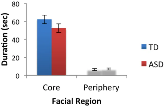
Fixation duration was consistently greater for the core versus peripheral regions and for the eye
region as compared to the other regions of interest. This preference for the eye region varied
somewhat by emotion, with a significant fixation time devoted to the nose in some cases (e.g.,
neutral; McManus et al., Unpublished Thesis).
Behavioral responses from this preliminary study also indicated that participants were
consistently able to identify angry, fearful, happy, and neutral expressions. Furthermore, the
increased task difficulty of the incongruent conditions was evidenced by the increase in response
times, wherein subjects showed a bias toward the visual affective information when asked to
determine the emotion portrayed (McManus et al., Unpublished Thesis). Other preliminary
studies have compared the behavioral performance (i.e., accuracy, modality bias, response time)
of individuals with ASD to the performance of TD individuals on emotion perception tasks
utilizing DAVE stimuli. Both TD and ASD groups made significantly more correct than
incorrect responses when responding to congruent movies. When presented with incongruent
movies, TD individuals showed a strong bias towards the facial expression, whereas individuals
with ASD did not demonstrate such bias, instead responding comparably toward facial
expression and prosody (McManus et al., 2008). With regard to response times, results have
incongruent movies. In addition, findings indicated that response times to provide an emotion
label for DAVE stimuli were significantly greater for individuals with ASD than TD individuals
(Banks et al., 2008).
1.9 The Current Study
Previous research suggests that individuals with ASD may be able to accurately identify,
at a level comparable to their TD peers, simple emotions presented as static visual stimuli and
under extended viewing conditions (Capps, Yirmiya, & Sigman, 1992; Grossman, Klin, Carter,
& Volkmar, 2000; Humphreys, Minshew, Leonard, & Behrmann, 2007; Ozonoff, Pennington, &
Rogers, 1990). However, research also suggests that individuals with ASD evidence weaknesses
in the integration of audio-visual integration (Boucher, Lewis, & Collis, 2000; Gepner, deGelder,
& deSchonen, 1996; Loveland et al., 1995; Mongillo et al., 2008), show no dominance effect for
visual information (Mongillo et al., 2008), show reduced salience of important affective
information provided by the eye region (Hernandez, 2009; Jones et al., 2008; Joseph and Tanaka,
2003; Klin et al., 2002; Norbury et al., 2009; Pelphrey et al., 2002; Rutherford & Towns, 2008),
and do not demonstrate the same level of proficiency as TD individuals on emotion perception
tasks, particularly as they reach adulthood (Humphreys et al., 2007; Pelphrey, et al., 2002;
Rump, Giavannelli, Minshew, & Strauss, 2009). Because the combined weaknesses in emotion
perception have lasting effects on peer and interpersonal relationships (Bonner, Hardy, Willard,
Anthony, Hood, & Gururangan, 2008; Leppanen & Hietanen, 2003), can potentially lead to
affective disorders (Vandell & Hembree, 1994), and impede academic performance (Ladd,
Herald-Brown, Reiser, 1997), it is imperative that researchers aim to explicate the factors