RESEARCH ARTICLE
Mesenchymal
Hox6
function is required for mouse pancreatic
endocrine cell differentiation
Brian M. Larsen1,2, Steven M. Hrycaj1, Micaleah Newman1, Ye Li3and Deneen M. Wellik1,2,3,*
ABSTRACT
Despite significant advances in our understanding of pancreatic endocrine cell development, the function of the pancreatic mesodermal niche in this process is poorly understood. Here we report a novel role for mouse Hox6 genes in pancreatic organogenesis. Hox6 genes are expressed exclusively in the mesoderm of the developing pancreas. Genetic loss of all three Hox6paralogs (Hoxa6,Hoxb6andHoxc6) leads to a dramatic loss of endoderm-derived endocrine cells, including insulin-secretingβ-cells, and to mild delays and disruptions in pancreatic branching and exocrine differentiation. Ngn3-expressing pan-endocrine progenitor cells are specified normally in Hox6 mutant pancreata, but fail to mature into hormone-producing cells. Reduced expression of Wnt5ais observed in mutant pancreatic mesenchyme, leading to subsequent loss of expression of the crucial Wnt inhibitors Sfrp3 andDkk1in endocrine progenitor cells. These results reveal a key role for Hox6 genes in establishing Wnt mesenchymal-epithelial crosstalk in pancreatic development.
KEY WORDS: Hox genes, Pancreatic development, Endocrine cells, Mesenchymal-epithelial crosstalk, Mouse, Wnt signaling
INTRODUCTION
Hox genes play a well-established role in axial and appendicular skeletal patterning, and knowledge of their importance in organogenesis is expanding. Hox genes have important roles in the development of organs that involve Hox expression along the anterior-posterior axis. Examples include theHox3paralogous group genes, which are crucial for thymus, thyroid and parathyroid development; Hox5genes in lung development;Hox9,Hox10andHox11genes in the reproductive tract; Hox10 and Hox11 genes in kidney development; and Hoxb13 for prostate development (Dolle et al., 1991; Benson et al., 1996; Gendron et al., 1997; Taylor et al., 1997; Manley and Capecchi, 1998; Podlasek et al., 1999; Economides and Capecchi, 2003; Schwab et al., 2006; Yallowitz et al., 2011; Boucherat et al., 2013; Raines et al., 2013; Hrycaj et al., 2015).
TheHox6paralogous group includes three genes:Hoxa6,Hoxb6 and Hoxc6.Paralogous Hox genes have been shown to exhibit a high degree of functional redundancy owing to sequence similarity and significant overlap in expression. Loss of a single gene within a paralogous group often results in little to no observable phenotype, but disruption of all members of a given paralogous group results in
dramatic patterning phenotypes (Davis and Capecchi, 1994; Horan et al., 1995; Fromental-Ramain et al., 1996a,b; Manley and Capecchi, 1998; Rossel and Capecchi, 1999; van den Akker et al., 2001; Wellik et al., 2002; Wellik and Capecchi, 2003; McIntyre et al., 2007; Yallowitz et al., 2009, 2011; Xu and Wellik, 2011; Xu et al., 2013). Single-mutant animals for each of theHox6 paralogs have undetectable or very mild defects, but collectively this group has been demonstrated to play important roles in patterning the rib cage and in determination of neuronal cell fate (Kostic and Capecchi, 1994; Rancourt et al., 1995; McIntyre et al., 2007; Mallo et al., 2010; Lacombe et al., 2013).Hox6genes are also expressed in the pancreatic mesoderm, suggesting a possible role for Hox6in pancreatic organogenesis.
In the mouse, the pancreas is specified at approximately embryonic day (E) 9.5 and expands as a dorsal and ventral bud from the endoderm-derived gut tube into the surrounding mesoderm (Wells and Melton, 2000; Gittes, 2009; Puri and Hebrok, 2010). At E11.5, these buds fuse to become the dorsal and ventral regions of the single pancreas. The pancreas has two main components: an exocrine and an endocrine component. The exocrine component is composed of digestive enzyme-secreting acinar cells and ductal cells. Ductal cells form a complex branching network that transports the digestive enzymes into the small intestine. The endocrine component is composed of five distinct types of endocrine cells, each of which secretes a single hormone: insulin, glucagon, somatostatin, ghrelin or pancreatic polypeptide. Endocrine cell differentiation is initiated in the ductal epithelium by the expression ofNgn3(Neurog3–Mouse Genome Informatics) in a subpopulation of the epithelial cells (Gu et al., 2002). These Ngn3+ cells subsequently delaminate from the ductal epithelium into the surrounding mesenchyme and differentiate further to the specific endocrine lineages. The bulk of endocrine cell differentiation occurs from E12.5 to E15.5, which is termed the secondary transition. Although all of the major functional components of the pancreas are derived from the endoderm, the surrounding mesodermally derived mesenchyme is crucial for the growth and development of these cell types (Golosow and Grobstein, 1962; Gittes et al., 1996; Miralles et al., 1998; Bhushan et al., 2001; Attali et al., 2007; Gittes, 2009; Puri and Hebrok, 2010).
Explant studies were first used to demonstrate the importance of the mesenchyme. When pancreatic epithelium was cultured in the absence of its surrounding mesenchyme, both endocrine and exocrine development arrested with defects in growth and differentiation. These defects were rescued by recombination with pancreatic mesenchyme (Golosow and Grobstein, 1962; Wessells and Cohen, 1967). A more recent study, in which the pancreatic mesenchyme was genetically ablated in vivo, showed a similar failure of all components of the pancreas to develop (Landsman et al., 2011).
Wnt signaling is crucial for multiple aspects of pancreatic development. A multitude of Wnt ligands, receptors, modifiers and
Received 9 June 2015; Accepted 30 September 2015
1
Department of Internal Medicine, Division of Molecular Medicine and Genetics, University of Michigan, Ann Arbor, MI 48109-2200, USA.2Cellular and Molecular Biology Program, University of Michigan, Ann Arbor, MI 48109-2200, USA.
3
Department of Cell and Developmental Biology, University of Michigan, Ann Arbor, MI 48109-2200, USA.
*Author for correspondence ([email protected])
DEVEL
O
inhibitors have reported expression in both the epithelium and the mesenchyme of the pancreas, with gene expression being highest early in development and declining with organ maturation (Heller et al., 2002). Wnt has largely been studied in the pancreas through manipulation of required canonical Wnt signaling components, such asβ-catenin. Others have shown specific roles for individual Wnt ligands and, taken together, there are significant roles for both Wnt/β-catenin signaling and the non-canonical Wnt planar cell polarity pathway for the proper development of both endocrine and exocrine pancreas (Heller et al., 2002; Kim et al., 2005; Murtaugh et al., 2005; Heiser et al., 2006; Attali et al., 2007; Wells et al., 2007; Jonckheere et al., 2008; Murtaugh, 2008; Baumgartner et al., 2014; Afelik et al., 2015).
Although Hox genes have been shown to be important for the development of many organs of endodermal and mesodermal origin, a role for Hox genes in development of the pancreas has not been reported. Herein, we report that mouseHox6genes function in pancreatic organogenesis. Hox6 genes are expressed only in the mesoderm of the developing pancreas and not in the endoderm. The Hox6 mutant pancreas buds normally and Ngn3+ endocrine progenitors are specified, but there is a >90% reduction of mature endocrine cells in the Hox6 mutant pancreas compared with controls. Loss of Hox6 function results in a decrease in Wnt5a expression in the pancreatic mesenchyme (although epithelial Wnt5aexpression is unperturbed). This leads to a subsequent loss of expression of two Wnt inhibitors,Sfrp3(Frzb–Mouse Genome Informatics) and Dkk1, in endocrine progenitor cells. The addition of recombinant Wnt5a protein to pancreatic explant cultures is sufficient to rescue endocrine cell differentiation in Hox6 mutant pancreata. Thus, regional mesodermal patterning factors are crucial for establishing the mesenchymal-epithelial crosstalk required for proper endocrine cell differentiation in pancreatic development, highlighting the potential importance of considering the mesodermal niche inex vivoβ-cell differentiation protocols.
RESULTS
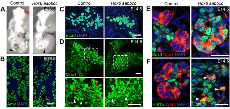
Pancreatic specification occurs normally inHox6mutants with mild defects in morphology and acinar differentiation Hox6 aabbcc(lower case letters represent null alleles) mice do not survive postnatally, but mutant embryos are indistinguishable in appearance (Fig. S1A) and mass (Fig. S1B) from littermate controls. Examination of internal organ defects reveals somewhat abnormal pancreatic morphology inHox6 mutants compared with controls (Fig. 1A). The ventral pancreas (black arrowheads) and trunk of the dorsal pancreas (yellow arrowheads) are more compact in Hox6 mutant pancreata compared with controls. Despite mildly perturbed pancreatic morphology, a normal expression pattern of acinar cell marker amylase (Amy) is observed in the mutant pancreas (Fig. 1B). Early stages of pancreatic initiation were examined by immunofluorescent staining for the pancreatic epithelial marker Pdx1. Quantification of immunofluorescent staining of the entire early pancreas (dorsal and ventral bud) showed that both pancreatic buds initiate normally and the volume of Pdx1-positive epithelium is unchanged between the control and Hox6 mutant at E10.5 (Fig. S2A,B). A small decrease in epithelial volume was observed at E11.5 (Fig. S2A,D). Minor decreases in both epithelial and mesenchymal proliferation were measured at E11.5 and E14.5 (Fig. S2C,G), but no overall differences in mesenchyme volume were measured (Fig. S2E), and there was no overall change in pancreatic mass at E18.5 regardless ofHox6genotype (Fig. S2H). There were no differences in apoptosis measured by cleaved caspase-3 staining (Fig. S2I).
[image:2.612.121.491.462.638.2]Examination of morphological defects at E14.5 by staining with cadherin 1 (Ecad) reveals typical loose, lobular branching epithelium in the control pancreas, but more compact clusters of epithelial cells with less branching in the mutant pancreas (Fig. 1C). Branch pattern was further analyzed at E14.5 using Muc1 antibody staining in whole pancreata (Villasenor et al., 2010). Hox6 null pancreata exhibit impaired branching and apparent defects in the remodeling of the early ductal plexus (Fig. 1D). Many thin, single
Fig. 1. Loss ofHox6function results in abnormal pancreatic morphology, but normal amylase expression.(A) E18.5 dissected pancreata from control and Hox6 aabbccmouse embryos.Hox6null pancreata show abnormal pancreatic morphology. Black arrowheads show compact ventral region of the pancreas in the mutant. Yellow arrowheads show narrower, more compact dorsal region with less lateral branching compared with controls. (B) Immunofluorescent staining for Amy (green) in sectioned E18.5 control andHox6 aabbccpancreata shows normal amylase patterns. (C) Immunofluorescent staining for Ecad (green) shows that the epithelium of theHox6null pancreas is more compact with less branching than the control pancreas at E14.5. (D) Whole-mount immunofluorescence for Muc1 (green) at E14.5 shows that the control has a well-defined multi-lumen plexus, whereas theHox6null pancreas is less developed. Higher-magnification images show both ductal (red arrowheads) and tip (white arrowheads) regions are readily visible in the control pancreas, but there are fewer ductal lumens apparent inHox6 mutant. (E) Immunofluorescent staining for Sox9 (green) and Cpa1 (red) shows no colocalization in control tip cells at E14.5, but many Cpa1/Sox9 double-positive cells in theHox6null pancreas. (F) Immunofluorescent staining for Hnf1b (green) and Cpa1 (red) shows no colocalization in control tip cells at E14.5, but Cpa1/ Hnf1b double-positive cells in theHox6null pancreas. Arrowheads indicate double-positive cells. Scale bars: 100μm in B,C; 50μm in D; 20μm in E; 15μm in F.
DEVEL
O
lumens have resolved throughout the control pancreas, whereas these lumens are less apparent in the mutant (Fig. 1D, red arrowheads). Bright, dense staining for Muc1, indicating acinar clusters, is found readily throughout both control and Hox6null pancreata (Fig. 1D, white arrowheads).
Before E13.5, the‘tip’acinar cells of the pancreas are multipotent progenitor cells and can produce exocrine, ductal and endocrine cells. All epithelial cells at these early stages expressSox9andHnf1b. After E13.5, asSox9andHnf1bare downregulated in tip cells, these cell types become unipotent and give rise only to acinar cells (Zhou et al., 2007; Kopp et al., 2011). In contrast to controls, Hox6 mutant pancreata demonstrate continued Sox9 and Hnf1b in Cpa1-positive tip cells at E14.5 (Fig. 1E,F). This has resolved by E16.5, consistent with the eventual differentiation and maturation of multipotent progenitor cells into mature exocrine acinar cells. This defect might be related to the perturbed morphology and branch pattern observed in mutants.
Endocrine differentiation is inhibited inHox6mutant pancreata
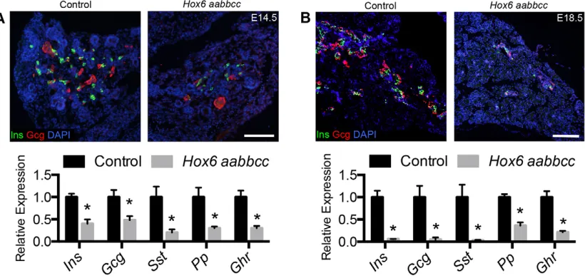
The mild defects in branching and in exocrine differentiation are in contrast to a dramatic reduction of all five mature endocrine cell hormones inHox6mutant pancreata (Fig. 2A,B). Antibody staining for insulin (Ins) and glucagon (Gcg) reveals a dramatic reduction of both endocrine protein and mRNA expression of all five endocrine hormones at E14.5 (Fig. 2A). This decrease is more pronounced by newborn stages, withInsandGcgmRNA expression reduced to less than 5% of control values (Fig. 2B).
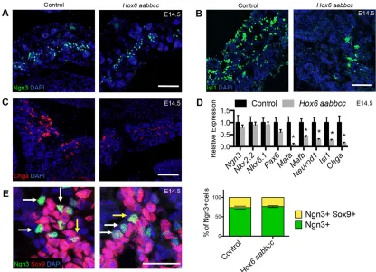
All five endocrine cell types derive from Ngn3+cells that arise from the ductal epithelium. During development of the pancreas, sporadic ductal cells expressNgn3, initiating a delamination process and allowing these cells to migrate into the surrounding mesenchyme, where they mature into the five types of hormone-producing cells. We investigated the cellular defects leading to the dramatic decrease of endocrine cells in the mutant by examining the initiation and differentiation of this cell type. There is no change in the amount of Ngn3+ immunofluorescent staining (Fig. 3A) or expression ofNgn3mRNA by qRT-PCR (Fig. 3D) in the mutant pancreas compared with controls. There are also no measured changes in expression of Nkx6-1 or Nkx2-2(Fig. 3D); however,
these genes are expressed more broadly in the epithelium (Sussel et al., 1998; Schaffer et al., 2010). There are significant reductions of pan-endocrine markers Chga and Isl1 protein and mRNA at E14.5 (Fig. 3B-D). The mRNA expression of endocrine lineage genes Mafa, Mafb and Neurod1 are also significantly reduced (Fig. 3D). Despite the apparent loss of endocrine differentiation, Ngn3 staining in controls and mutants is comparable, suggesting that Ngn3+progenitor cells do not accumulate inHox6mutants up to E18.5 (Fig. S3).
In the first phase of endocrine differentiation, sporadic Sox9+ ductal cells begin additionally to expressNgn3, allowing these cells to delaminate from the epithelium while concomitantly turning offSox9 expression (Gouzi et al., 2011; Shih et al., 2012). We examined this process by antibody staining control andHox6mutant pancreata for Ngn3 and Sox9. Immunostaining reveals that about 25% of Ngn3+ cells are also Sox9+in control pancreata at E14.5 (Fig. 3E, yellow arrows). This ratio is identical in theHox6mutant pancreas, providing evidence that endocrine progenitor cells are able to transition adequately from Sox9+/Ngn3+ductal cells into Sox9–/Ngn3+early endocrine cells (Fig. 3E). Ngn3+cells that have successfully migrated from the duct are observed throughout the control and in theHox6 mutant pancreas (Fig. 3E, white arrows), further supporting a differentiation defect after specification and delamination.
Hox6genes are expressed in the mesoderm of the pancreas during development
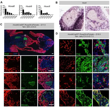
[image:3.612.103.513.499.694.2]All threeHox6genes are expressed in the embryonic pancreas, with expression highest early in development and persisting until E16.5 (Fig. 4A). Expression was below our limit of detection for all threeHox6genes at E18.5 (Fig. 4A).In situ hybridization (ISH) analyses of Hox6 mRNA reveal expression exclusively in the pancreatic mesoderm at E11.5 and E12.5 (Fig. 4B). Mesodermally restricted expression was confirmed using a previously reported Hoxb6-inducible Cre reporter line (Hoxb6CreERT; Rosa26-tdTomato; Nguyen et al., 2009; Madisen et al., 2010). Administration of tamoxifen at early stages (E9.5 and E10.5) reveals an anterior expression limit at the level of the budding dorsal and ventral pancreas, and also marks the majority of mesoderm posterior to this in the embryo (Fig. 4C).
Fig. 2.Hox6mutant pancreata demonstrate a drastic reduction of endocrine hormone expression.(A) Immunofluorescent staining for Ins (green), Gcg (red) and nuclei (blue) was performed at E14.5. qRT-PCR shows reduced expression of all five endocrine cell hormones at E14.5 in the mutant pancreas. (B) Immunofluorescence shows decreased staining for Ins (green) and Gcg (red) in the mutant pancreas at E18.5. There is a dramatic reduction of all five endocrine cell hormones at E18.5 in the mutant. Results are mean±s.e.m. *P<0.05 calculated by Student’st-test (n≥3). Scale bars: 100μm in A,B.
DEVEL
O
Expression from the ROSA locus in these mice is observed throughout the pancreatic mesenchyme, but is completely excluded from early Ins-positive endocrine cells, Pdx1-positive pancreatic endoderm and PECAM (Pecam1)-positive vasculature (Fig. 4C). As endocrine cells undergo epithelial-to-mesenchymal transition before differentiation, it was important to exclude possible initiation of Hox6 expression in endocrine cells during the secondary transition. To test this, tamoxifen was administered to pregnant dams at E12.5, E13.5 and E14.5 and embryos were examined at E15.5. We found no Cre activity in Ngn3+endocrine progenitors (Fig. S4A), with the pan-endocrine marker Chga (Fig. S4B) or with epithelium (Fig. S4C). Similar results were obtained with tamoxifen administration daily from E10.5 to E17.5. There was no overlap of tdTomato with Chga-positive endocrine cells, pancreatic epithelium, endothelial, neuronal or smooth muscle cells (Fig. 4D; Fig. S4D,E). We observed extensive and complete co-labeling of tdTomato and vimentin-positive mesenchyme (Fig. 4D), confirming that Hox6 expression is exclusive to the pancreatic mesoderm-derived mesenchyme.
Vasculature is normal in theHox6null pancreas
It has been reported previously that hypervascularization affects organogenesis of the pancreas by reducing branching and differentiation of epithelial cells, and signals from the vasculature are also important for the proper formation of endocrine cells of the pancreas (Lammert et al., 2001; Magenheim et al., 2011).
Immunofluorescence staining for PECAM at E14.5 (Fig. S5A) and E18.5 (Fig. S5B) reveals no difference in vasculature between the control and mutant pancreas. There are also no differences in the association of endothelial and insulin-positive endocrine cells between control andHox6null E18.5 pancreas (Fig. S5C).
Wnt signaling is disrupted in the mesoderm and
differentiating endocrine cells inHox6mutant pancreata Microarray analysis was performed on E12.5 control and Hox6 mutant pancreata to probe for the molecular mechanism(s) responsible for the Hox6 mutant phenotypes (http://www.ncbi. nlm.nih.gov/geo/query/acc.cgi?acc=GSE68390). ToppFun (https:// toppgene.cchmc.org/enrichment.jsp) analysis identified changes in members of the Wnt receptor-signaling pathway as significantly enriched in theHox6mutant pancreas, but no significant changes in other major signaling pathways.
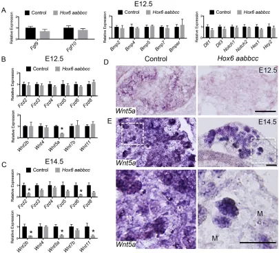
[image:4.612.97.513.58.359.2]To confirm our microarray results, we used qRT-PCR analysis to measure the expression levels of genes from major signaling pathways with reported or measured expression in the pancreas. No changes in the levels of Fgf, Bmp or Notch pathway members were measured in E12.5 mutant pancreata (Fig. 5A). There were also no differences in the measured expression of Wnt receptors between control and Hox6 mutant pancreata (Fig. 5B). There are no differences in expression ofWnt2b,Wnt4,Wnt7borWnt11, other Wnt ligands expressed in pancreas at E12.5, between mutant and control pancreata (Fig. 5B; Heller et al., 2002), and we were unable Fig. 3. Immature endocrine cell genes are decreased in the mutant, but there is no defect in endocrine progenitor delamination.(A) Immunofluorescent staining for Ngn3 (green) and nuclei (blue) shows no change between control andHox6null pancreata at E14.5. (B,C) Immunofluorescent staining shows a decrease in signal of both Isl1 (B) and Chga (C). (D) qRT-PCR analyses show no change in the expression ofNgn3,Nkx2-2,Nkx6-1orPax6, but there are significantly reduced levels of expression ofMafa,Mafb,Neurod1,Isl1andChgain theHox6mutant pancreas at E14.5.(E) Immunofluorescent staining for Sox9 (red) and Ngn3 (green) at E14.5 in control andHox6 aabbccpancreata. Quantification of Sox9+and Ngn3+cells shows that 25% of Ngn3+cells (white arrows) in both the control and theHox6null pancreas are also Sox9+(yellow arrows). Results are mean±s.e.m. *P<0.05 calculated by Student’st-test (n≥3). Scale bars: 100μm in A-C,E.
DEVEL
O
to detect expression of Wnt1 and Wnt8b, genes reported to be expressed in the mesenchyme later in development, in either the control or the Hox6 mutant pancreas at E12.5 (data not shown; Heller et al., 2002). However, we observed significantly reduced expression levels of mesodermally expressed Wnt ligand,Wnt5a, at both E12.5 and E14.5 (Fig. 5B,C).
Hox6genes are expressed solely in the mesoderm and therefore we examined mesodermally expressed Wnt genes as possible direct targets of Hox6. Wnt5a is expressed in both the pancreatic epithelium and mesenchyme (Heller et al., 2002). ISH analyses of Wnt5a revealed much weaker expression in the Hox6 mutant pancreatic mesenchyme at E12.5 compared with controls (Fig. 5D). At E14.5, reducedWnt5aexpression is even more apparent in the Hox6 mutant pancreatic mesenchyme compared with controls,
whereas expression in the epithelium appears identical (Fig. 5E). At E14.5 and beyond, we measured a significant reduction of more Wnt receptors and ligands as well, suggesting a secondary perturbation of Wnt signaling more globally in the Hox6mutant pancreas at later stages (Fig. 5C).Wnt2b,Wnt4,Wnt7bandWnt11 were also examined by ISH; however, no obvious changes in expression pattern were detected between the control and Hox6 mutant pancreas (Fig. S6A-D).
[image:5.612.95.519.57.468.2]We reasoned that this disruption in Wnt signaling in the mesoderm might lead directly to changes in the Ngn3-expressing endocrine precursors after delamination from the ductal epithelium. The expression of a number of Wnt inhibitors has been reported in the pancreatic epithelium and in endocrine cells throughout development, includingSfrp1-4,Dkk1-3andWif1(Heller et al., 2002; Gu et al., Fig. 4.Hox6expression is limited to the pancreatic mesenchyme.(A) qRT-PCR analysis shows expression ofHoxa6,Hoxb6andHoxc6in the pancreas throughout gestation, with expression of all three genes highest early in development. No expression above the limit of detection was measured for any of the three Hox6genes at E18.5. Values are relative to the colon, where there is a low level ofHox6expression. (B) ISH shows combinedHox6gene expression in the mesenchyme at E11.5 and E12.5. (C)Hoxb6CreERTandRosa26-tdTomatomice were crossed, and tamoxifen was administered to pregnant dams via intraperitoneal injection at E9.5 and E10.5 to examine reporter expression in the pancreas. Cre activity (red) was detected at an anterior limit in the lateral plate mesenchyme at the level of the forelimb; the anterior limit in the somatopleuric mesenchyme is at the level of the pancreatic buds. The dorsal pancreas (dp) and ventral pancreas (vp) exhibit strong td-tomato (red) signal throughout the mesenchyme. Epithelium is stained with Ecad (green). Cre activity is also excluded from Ins+cells, Pdx1+cells and PECAM+endothelium in the pancreas. (D) Daily tamoxifen administration to pregnant dams via intraperitoneal injection from E10.5 to E17.5 shows reporter
expression excluded from Chga+endocrine cells, Ecad+epithelial cells and PECAM+endothelial cells inHoxb6CreERT;Rosa26-tdTomatoembryos. Cre activity is detected throughout the pancreatic mesenchyme as co-labeled with vimentin (Vim). Scale bars: 100μm in B-D. Results are mean±s.e.m. (n≥3).
DEVEL
O
2004). Of the Wnt inhibitors examined in the pancreas, only two (Sfrp3andDkk1) showed significantly reduced expression levels in the Hox6 mutant at E12.5 (Fig. 6A). Using immunofluorescent staining with antibodies to Sfrp3 and Dkk1, we observe extensive overlap of Sfrp3 and Dkk1 with endocrine cells in wild-type pancreata (Fig. 6B). Overall, Hox6 mutant pancreata exhibited drastically reduced levels of Sfrp3 and Dkk1 (Fig. 6C,D). Close examination reveals colocalization of Sfrp3 and Dkk1 in a subset of Ngn3+endocrine progenitor cells in the control and none in theHox6 mutant pancreas (Fig. 6E,F). This reduction of Wnt inhibitors continues through later stages of pancreatic development (Fig. S7).
Wnt5a is the first mesodermally expressed Wnt ligand with disrupted expression in the Hox6 mutant pancreas. If the lack of endocrine cell differentiation in theHox6mutant pancreas stems from the lack of a sufficient amount of Wnt5a signaling from the mesoderm, we reasoned that it might be possible to rescue this phenotype with the addition of exogenous Wnt5a to theHox6mutant pancreata. To test this, we dissected control and Hox6 mutant pancreata at E12.5 and cultured them for 6 days in the presence or absence of exogenous Wnt5a (Fig. 7A). Without exogenous Wnt5a in the culture, we observed a significant reduction of mature endocrine cells in theHox6mutant pancreata compared with controls, similar to
thein vivophenotype (Fig. 7B,C). When recombinant Wnt5a protein was added to the culture media ofHox6mutant pancreata, the quantity of mature endocrine hormone staining was rescued to the same level as control pancreata (Fig. 7B,C).
DISCUSSION
Although the importance of the mesenchyme in pancreatic development was demonstrated decades ago, the molecular mechanisms involved in the crosstalk between the epithelium and the mesenchyme are poorly understood. Here we report a novel role forHox6genes in development of the pancreas as illustrated in the model in Fig. 8. Loss ofHox6function in the pancreatic mesoderm leads to mild defects in branching and delayed exocrine cell differentiation; however, the pancreas achieves its normal size by newborn stages with extensive acinar cell formation. Endocrine cells are specified normally, but immature endocrine cells do not mature, resulting in a dramatic reduction of all types of fully differentiated endocrine cells, including >90% decreases in insulin and glucagon expression.
[image:6.612.102.507.59.427.2]Specific components of the Wnt signaling pathway are disrupted by E12.5, whereas no changes in other crucial developmental signaling pathways, including Fgf, Bmp and Notch, are observed. Fig. 5. Expression ofWnt5aand Wnt inhibitorsSfrp3andDkk1is lost in theHox6mutant pancreas.(A) qRT-PCR analysis of genes from the Fgf, Bmp and Notch pathways at E12.5. (B,C) qRT-PCR analysis of Wnt receptor and ligand expression at E12.5 (B) and E14.5 (C). (D) ISH at E12.5 using probe forWnt5a shows staining throughout the pancreas and a significant reduction of signal in theHox6mutant pancreas. (E) At E14.5,Wnt5ais expressed in both the mesenchyme and epithelium of control pancreata.Wnt5aexpression in theHox6null pancreatic mesenchyme (M) is lost, but epithelial expression appears normal. Scale bars: 100μm in D,E. Results are mean±s.e.m. *P<0.05 calculated by Student’st-test (n≥3).
DEVEL
O
Mesenchymal ligand expression of Wnt5a is significantly downregulated (or absent), whereas epithelial expression of this ligand appears unperturbed.Wnt5ais expressed from E11 to the end
of gestation, with peak levels of expression at E12, similar to our measuredHox6gene expression profile (Heller et al., 2002). A study examining morpholino knockdown ofwnt5aandfz2 in zebrafish and global loss of function ofWnt5ain mice reported defects in islet formation in both organisms (Kim et al., 2005), consistent with loss ofWnt5aexpression in the mesoderm contributing to the endocrine phenotype in ourHox6mutant animals.
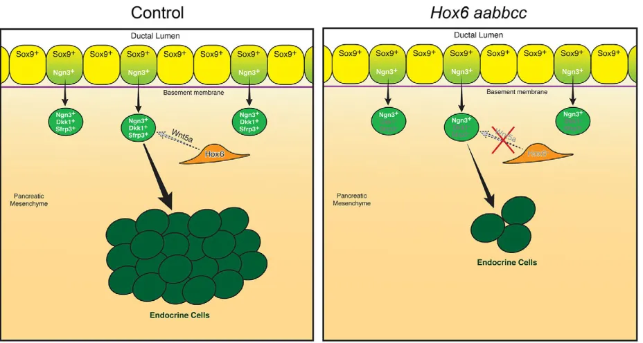
In addition to loss of Wnt5a signaling in the pancreatic mesoderm, which might result directly from loss of Hox6 function, there is a subsequent loss of expression of Wnt inhibitors Sfrp3 and Dkk1, specifically in delaminated Ngn3+ endocrine precursor cells as they are entering the mesoderm. As previous studies have demonstrated the importance of repressing Wnt signaling in developing endocrine cells (Pedersen and Heller, 2005), it is likely that loss of Wnt inhibitor induction in Ngn3+ progenitors leads directly to loss of further endocrine cell differentiation in Hox6 mutant pancreata. Pharmacological treatment ofHox6mutant pancreata with exogenous Wnt5a protein is sufficient to restore endocrine cell differentiation, and demonstrates that Wnt5a is a crucial mediator downstream ofHox6genes in the pancreatic mesenchyme during development. Collectively, our results suggest thatHox6genes are crucial for the establishment of the Wnt mesenchymal-epithelial crosstalk necessary for development of the pancreas and endocrine cell specification.
Hox genes are important for many aspects of development and organogenesis, but no disruption of Hox function has previously been implicated in the development of the pancreas. This work contributes to the growing understanding of pancreatic mesoderm signaling and the important roles that the mesoderm plays in the development of both the endocrine and the exocrine components of the pancreas. Here we show a direct link between Hox function and Wnt signaling; a theme that is reminiscent of a recent report of loss ofWnt2/2bexpression in the mesenchyme ofHox5triple-mutant lungs during development (Hrycaj et al., 2015). Another recent study examining Hoxd13 function in digit development reported thatHoxd13 promotes expression ofWnt5a in vitro(Kuss et al., 2014). All Hox proteins bind to the same (ATTA) binding sequence, and therefore it is plausible thatWnt5a is a direct target of Hox proteins during development. Future work will be required to establish possible direct regulation ofWnt5abyHox6.
This study adds to growing evidence that Hox function in the mesoderm of several organ systems plays region-specific roles associated with the establishment of proper Wnt signaling crosstalk during organogenesis (Kuss et al., 2014; Hrycaj et al., 2015). A more complete elucidation of mesodermal-endodermal crosstalk during development of the pancreas is crucial to the enhancement of ex vivo protocols for generating functional β-cells as a cellular therapy for the treatment of diabetes.
MATERIALS AND METHODS Generation of mouse mutants
Mice mutant for all three Hox6 paralogous genes were generated using standard genetic crosses (Kostic and Capecchi, 1994; Rancourt et al., 1995; Garcia-Gasca and Spyropoulos, 2000).Hoxb6CreERTmice were contributed
by Dr Susan Mackem (Nguyen et al., 2009).Rosa26-tdTomatomice were obtained from The Jackson Laboratory (Madisen et al., 2010). All experiments were performed following protocols approved by the University of Michigan’s Institutional Committee on the Use and Care of Animals.
Tamoxifen injections
[image:7.612.84.269.60.560.2]Tamoxifen and progesterone were dissolved in 100% ethanol and diluted in corn oil. Pregnant dams were given intraperitoneal injections with
Fig. 6. Expression of Wnt inhibitorsSfrp3andDkk1is lost in endocrine progenitors in theHox6mutant pancreas.(A) qRT-PCR analysis of Wnt inhibitors in control andHox6mutant pancreata at E12.5. (B) Immunofluorescent staining for Wnt inhibitors Sfrp3 (green) and Dkk1 (green) with endocrine markers Chga (red) and a combination of antibodies for Ins and Gcg (red) shows consistent overlap of staining in E13.5 mouse pancreas. (C,D) Immunofluorescent staining for Sfrp3 (C) and Dkk1 (D) in control andHox6 aabbccpancreata. There is a drastic reduction of signal for both Wnt inhibitors in the mutant pancreas at E12.5. (E,F) Sfrp3 (E; green), Dkk1 (F; green) and Ngn3 (E,F; red) in control andHox6mutant pancreata at E12.5. Signal for Sfrp3 and Dkk1 is absent in Ngn3+endocrine progenitor cells inHox6mutant pancreata. Scale bars: 100μm in C,D; 25μm in B,E,F. Results are mean±s.e.m. *P<0.05 calculated by Student’st-test (n≥3).
DEVEL
O
1.5 mg of tamoxifen and 0.75 mg of progesterone on the days noted in the Results.
In situhybridization
For section ISH, embryos were collected in PBS and fixed overnight in 4% PFA in PBS at 4°C. Embryos were then rinsed in PBS and immersed in 30%
sucrose at 4°C overnight before embedding into optimal cutting temperature (OCT) media. Frozen sections 20 µm in thickness were cut, and slides were stored at −80°C. Section ISH was performed as previously described (Mendelsohn et al., 1999; Di Giacomo et al., 2006). Detection ofHox6
[image:8.612.108.501.73.282.2]mRNA was done using probes generated against the 3′untranslated region of Hoxa6,Hoxb6and Hoxc6or against theNeorcassette as previously Fig. 7. Exogenous Wnt5a treatment rescues endocrine cell differentiation inHox6mutant pancreas.(A) Control andHox6mutant pancreata were dissected at E12.5 and cultured for 6 days in media with and without recombinant Wnt5a protein. On day 2 of culture, fresh media with or without Wnt5a was added to the explants. (B) Immunofluorescent staining for combination of Ins and Gcg primary antibodies on pancreatic explants collected after 6 days in culture. (C) Quantification of immunofluorescent staining using ImageJ software shows that untreatedHox6mutant pancreata have significantly less endocrine staining per total DAPI area than control pancreata.Hox6mutant pancreata treated with Wnt5a protein have the same amount of endocrine staining per DAPI area as controls.n=3 for each condition or genotype. Scale bar: 100μm in B. Results are mean±s.e.m.P-values were calculated by Student’st-test.
Fig. 8.Hox6regulates mesenchymalWnt5aexpression to promote endocrine cell differentiation.MesenchymalHox6promotes the expression ofWnt5ain the mesenchyme, which signals to endocrine precursor cells to initiate expression ofSfrp3andDkk1to allow differentiation to more mature, hormone-producing endocrine cells. WhenHox6function is disrupted, expression ofWnt5a,Sfrp3andDkk1is lost and differentiation of mature endocrine cells is inhibited in theHox6
mutant pancreas.
DEVEL
O
[image:8.612.76.540.445.693.2]described and with indistinguishable results from probes againstHox6gene mRNA (McIntyre et al., 2007).Wnt5acDNA was ligated into PCR4-TOPO vector and reverse transcribed with T3 RNA polymerase. The sequenced plasmid aligns to mouseWnt5a: GenBank gi 46909566/NM 009524.2 from bp 406 to 1440.Wnt2b,Wnt4,Wnt7bandWnt11ISH was performed with previously published riboprobes (Miller et al., 2012; Soofi et al., 2012; Ranghini and Dressler, 2015).
Immunofluorescent staining
Mouse embryos were collected as described above. Frozen sections 12 µm in thickness were cut, and slides were stored at−80°C. Slides were blocked for 1 h at room temperature in 0.1% or 0.5% Triton X-100 in PBS (PBS-T) with 1% donkey serum and treated with primary antibody overnight at 4°C. On day 2, slides were washed in PBS-T, incubated with secondary antibody for 2 h at room temperature, followed by a 10 min wash in PBS-T with DAPI (Sigma-Aldrich). Coverslips were added to the slides using Prolong Gold Antifade Reagent (Invitrogen). The primary and secondary antibodies and dilutions used are listed in Table S1. Slides were imaged using either an Olympus BX-51 or a Leica SP5X 2-Photon confocal microscope.
Whole-mount Muc1 staining
Tissue was fixed in 4% paraformaldehyde (PFA) for 3 h at 4°C, dehydrated in MeOH and stored at−20°C until stained. Fixed tissue was treated with Dent’s bleach (MeOH:DMSO:H2O2, 4:1:1) for 2 h at room temperature,
blocked with TNB (Perkin Elmer) and incubated overnight with anti-Muc1 antibody in TNB at 4°C. Pancreata were then washed with PBS and incubated with secondary antibody in TNB overnight at 4°C. Pancreata were imaged in BABB (benzyl alcohol:benzyl benzoate, 1:2) on a Leica SP5X Inverted 2-Photon FLIM Confocal microscope. Confocal z-stacks were reconstructed using ImageJ. Details of antibodies used and dilutions are listed in Table S1.
RNA isolation and quantitative RT-PCR
RNA was isolated from mouse pancreata with the Qiagen RNeasy Micro Kit. Quantitative RT-PCR (qRT-PCR) was carried out using Roche FastStart SYBR Green Master Mix and the Applied Biosystems StepOnePlus Real-time PCR system (Life Technologies). Relative expression values were calculated as 2−ΔΔCt, and values of controls were normalized to 1. Rn18s served as an internal control for normalization in all qRT-PCR experiments. All data are shown as the mean of at least three independent biological replicates; error bars represent s.e.m. Calculations andP-values (Student’s two-tailed, unpairedt-test) were generated in Microsoft Excel. Results were considered statistically significant atP<0.05. Graphs were generated using Prism 6 (GraphPad). Primer sequences are listed in Table S2.
Pancreatic explant cultures and rescue
Pancreata were dissected at E12.5 and cultured at the air-media interface on a Nucleopore Track-Etched Membrane (Whatman) in DMEM/F12 with
L-glutamine and 15 mM HEPES (Gibco) supplemented with
penicillin-streptomycin (Gibco). For rescue, the medium was supplemented with 500 ng/ml Recombinant Human/Mouse Wnt5a (R&D Systems) and refreshed with new media and Wnt5a protein on day 2 of culture. After 6 days in culture, pancreata were fixed at room temperature in 4% PFA for 3 h, immersed in 30% sucrose at 4°C overnight and frozen in OCT the next day. Pancreatic explants were cryosectioned at 12 µm and stained as described above for Ins/Gcg. ImageJ software was used to calculate the area of signal for DAPI and Ins/Gcg. Quantification is displayed as total Ins and Gcg signal/total DAPI signal for each explant.
Acknowledgements
We thank Dr Susan Mackem for contributingHoxb6CreERTmice. We would also like to thank Dr Ondine Cleaver for sharing the Muc1 whole-mount staining protocol and Drs D. Gumucio, Å. Kolterud and Kate Walton for theWnt5aISH probe. Dr Gregory Dressler provided riboprobes forWnt4andWnt11. Dr J. Spence provided several qRT-PCR primers. We thank Holly Fischer for model artwork. Bright Kim provided technical assistance on parts of this work. The PECAM antibody developed by Steven A. Bogen was obtained from the Developmental Studies Hybridoma Bank, created by the Eunice Kennedy Shriver National Institute of Child Health and Human Development of the National Institutes of Health and maintained at The University of Iowa, Department of Biology, Iowa City, IA, USA.
Competing interests
The authors declare no competing or financial interests.
Author contributions
B.M.L. performed and designed the majority of experiments and wrote the manuscript. S.M.H. contributed intellectual advice, performed experiments and edited the manuscript. M.N. and Y.L. performed experiments. D.M.W. conceived the project, contributed experimental and intellectual advice and edited the manuscript.
Funding
This work was supported by the National Institutes of Health [Cellular and Molecular Biology Training Grant T32GM007315 and the Training Program in Organogenesis T32HD007505 to B.M.L.]; The American Diabetes Association [ADA-7-13-BS-184]; a National Institute of Diabetes and Digestive and Kidney Diseases Michigan Diabetes Research and Training Center Pilot and Feasibility Award [P30DK020572]; the National Heart, Lung, and Blood Institute (NHLBI) [R01-HL119215]; a Ruth L. Kirschstein National Research Service Award (NSRA) training grant [5T32 HL 7749-20 to S.M.H.]; and a National Center for Research Resources grant number [UL1RR024986 to S.M.H.]. Deposited in PMC for release after 12 months.
Supplementary information
Supplementary information available online at
http://dev.biologists.org/lookup/suppl/doi:10.1242/dev.126888/-/DC1
References
Afelik, S., Pool, B., Schmerr, M., Penton, C. and Jensen, J.(2015). Wnt7b is
required for epithelial progenitor growth and operates during epithelial-to-mesenchymal signaling in pancreatic development.Dev. Biol.399, 204-217.
Attali, M., Stetsyuk, V., Basmaciogullari, A., Aiello, V., Zanta-Boussif, M. A.,
Duvillie, B. and Scharfmann, R.(2007). Control of beta-cell differentiation by the
pancreatic mesenchyme.Diabetes56, 1248-1258.
Baumgartner, B. K., Cash, G., Hansen, H., Ostler, S. and Murtaugh, L. C.(2014).
Distinct requirements for beta-catenin in pancreatic epithelial growth and patterning.Dev. Biol.391, 89-98.
Benson, G. V., Lim, H., Paria, B. C., Satokata, I., Dey, S. K. and Maas, R. L.
(1996). Mechanisms of reduced fertility in Hoxa-10 mutant mice: uterine homeosis and loss of maternal Hoxa-10 expression.Development122, 2687-2696.
Bhushan, A., Itoh, N., Kato, S., Thiery, J. P., Czernichow, P., Bellusci, S. and
Scharfmann, R. (2001). Fgf10 is essential for maintaining the proliferative
capacity of epithelial progenitor cells during early pancreatic organogenesis. Development128, 5109-5117.
Boucherat, O., Montaron, S., Berube-Simard, F.-A., Aubin, J., Philippidou, P.,
Wellik, D. M., Dasen, J. S. and Jeannotte, L. (2013). Partial functional
redundancy between Hoxa5 and Hoxb5 paralog genes during lung morphogenesis.Am. J. Physiol. Lung Cell Mol. Physiol.304, L817-L830.
Davis, A. P. and Capecchi, M. R.(1994). Axial homeosis and appendicular
skeleton defects in mice with a targeted disruption of hoxd-11.Development120, 2187-2198.
Di Giacomo, G., Koss, M., Capellini, T. D., Brendolan, A., Pöpperl, H. and
Selleri, L. (2006). Spatio-temporal expression of Pbx3 during mouse
organogenesis.Gene Expr. Patterns6, 747-757.
Dolle, P., Izpisua-Belmonte, J. C., Brown, J. M., Tickle, C. and Duboule, D.
(1991). HOX-4 genes and the morphogenesis of mammalian genitalia.Genes Dev.5, 1767-1776.
Economides, K. D. and Capecchi, M. R.(2003). Hoxb13 is required for normal
differentiation and secretory function of the ventral prostate.Development130, 2061-2069.
Fromental-Ramain, C., Warot, X., Messadecq, N., LeMeur, M., Dolle, P. and
Chambon, P.(1996a). Hoxa-13 and Hoxd-13 play a crucial role in the patterning
of the limb autopod.Development122, 2997-3011.
Fromental-Ramain, C., Warot, X., Lakkaraju, S., Favier, B., Haack, H., Birling,
C., Dierich, A., Doll e, P. and Chambon, P.(1996b). Specific and redundant
functions of the paralogous Hoxa-9 and Hoxd-9 genes in forelimb and axial skeleton patterning.Development122, 461-472.
Garcia-Gasca, A. and Spyropoulos, D. D. (2000). Differential mammary
morphogenesis along the anteroposterior axis in Hoxc6 gene targeted mice. Dev. Dyn.219, 261-276.
Gendron, R. L., Paradis, H., Hsieh-Li, H. M., Lee, D. W., Potter, S. S. and Markoff, E.(1997). Abnormal uterine stromal and glandular function associated with maternal reproductive defects in Hoxa-11 null mice.Biol. Reprod.56, 1097-1105.
Gittes, G. K.(2009). Developmental biology of the pancreas: a comprehensive
review.Dev. Biol.326, 4-35.
Gittes, G. K., Galante, P. E., Hanahan, D., Rutter, W. J. and Debase, H. T.(1996).
Lineage-specific morphogenesis in the developing pancreas: role of mesenchymal factors.Development122, 439-447.
Golosow, N. and Grobstein, C. (1962). Epitheliomesenchymal interaction in
pancreatic morphogenesis.Dev. Biol.4, 242-255.
DEVEL
O
Gouzi, M., Kim, Y. H., Katsumoto, K., Johansson, K. and Grapin-Botton, A.
(2011). Neurogenin3 initiates stepwise delamination of differentiating endocrine cells during pancreas development.Dev. Dyn.240, 589-604.
Gu, G., Dubauskaite, J. and Melton, D. A. (2002). Direct evidence for the
pancreatic lineage: NGN3+ cells are islet progenitors and are distinct from duct progenitors.Development129, 2447-2457.
Gu, G., Wells, J. M., Dombkowski, D., Preffer, F., Aronow, B. and Melton, D. A.
(2004). Global expression analysis of gene regulatory pathways during endocrine pancreatic development.Development131, 165-179.
Heiser, P. W., Lau, J., Taketo, M. M., Herrera, P. L. and Hebrok, M.(2006).
Stabilization of beta-catenin impacts pancreas growth. Development 133, 2023-2032.
Heller, R. S., Dichmann, D. S., Jensen, J., Miller, C., Wong, G., Madsen, O. D.
and Serup, P. (2002). Expression patterns of Wnts, Frizzleds, sFRPs, and
misexpression in transgenic mice suggesting a role for Wnts in pancreas and foregut pattern formation.Dev. Dyn.225, 260-270.
Horan, G. S., Ramirez-Solis, R., Featherstone, M. S., Wolgemuth, D. J., Bradley,
A. and Behringer, R. R.(1995). Compound mutants for the paralogous hoxa-4,
hoxb-4, and hoxd-4 genes show more complete homeotic transformations and a dose-dependent increase in the number of vertebrae transformed.Genes Dev.9, 1667-1677.
Hrycaj, S. M., Dye, B. R., Baker, N. C., Larsen, B. M., Burke, A. C., Spence, J. R.
and Wellik, D. M.(2015). Hox5 Genes Regulate the Wnt2/2b-Bmp4-Signaling
Axis during Lung Development.Cell Rep.12, 903-912.
Jonckheere, N., Mayes, E., Shih, H.-P., Li, B., Lioubinski, O., Dai, X. and Sander, M. (2008). Analysis of mPygo2 mutant mice suggests a requirement for mesenchymal Wnt signaling in pancreatic growth and differentiation.Dev. Biol.
318, 224-235.
Kim, H. J., Schleiffarth, J. R., Jessurun, J., Sumanas, S., Petryk, A., Lin, S. and
Ekker, S. C.(2005). Wnt5 signaling in vertebrate pancreas development.BMC
Biol.3, 23.
Kopp, J. L., Dubois, C. L., Schaffer, A. E., Hao, E., Shih, H. P., Seymour, P. A.,
Ma, J. and Sander, M.(2011). Sox9+ ductal cells are multipotent progenitors
throughout development but do not produce new endocrine cells in the normal or injured adult pancreas.Development138, 653-665.
Kostic, D. and Capecchi, M. R.(1994). Targeted disruptions of the murine Hoxa-4
and Hoxa-6 genes result in homeotic transformations of components of the vertebral column.Mech. Dev.46, 231-247.
Kuss, P., Kraft, K., Stumm, J., Ibrahim, D., Vallecillo-Garcia, P., Mundlos, S. and
Stricker, S.(2014). Regulation of cell polarity in the cartilage growth plate and
perichondrium of metacarpal elements by HOXD13 and WNT5A.Dev. Biol.385, 83-93.
Lacombe, J., Hanley, O., Jung, H., Philippidou, P., Surmeli, G., Grinstein, J. and
Dasen, J. S.(2013). Genetic and functional modularity of Hox activities in the
specification of limb-innervating motor neurons.PLoS Genet.9, e1003184.
Lammert, E., Cleaver, O. and Melton, D. (2001). Induction of pancreatic
differentiation by signals from blood vessels.Science294, 564-567.
Landsman, L., Nijagal, A., Whitchurch, T. J., VanderLaan, R. L., Zimmer, W. E.,
MacKenzie, T. C. and Hebrok, M.(2011). Pancreatic mesenchyme regulates
epithelial organogenesis throughout development.PLoS Biol.9, e1001143.
Madisen, L., Zwingman, T. A., Sunkin, S. M., Oh, S. W., Zariwala, H. A., Gu, H.,
Ng, L. L., Palmiter, R. D., Hawrylycz, M. J., Jones, A. R. et al.(2010). A robust
and high-throughput Cre reporting and characterization system for the whole mouse brain.Nat. Neurosci.13, 133-140.
Magenheim, J., Ilovich, O., Lazarus, A., Klochendler, A., Ziv, O., Werman, R.,
Hija, A., Cleaver, O., Mishani, E., Keshet, E. et al.(2011). Blood vessels restrain
pancreas branching, differentiation and growth.Development138, 4743-4752.
Mallo, M., Wellik, D. M. and Deschamps, J.(2010). Hox genes and regional
patterning of the vertebrate body plan.Dev. Biol.344, 7-15.
Manley, N. R. and Capecchi, M. R.(1998). HoxGroup 3 paralogs regulate the
development and migration of the thymus, thyroid, and parathyroid glands.Dev. Biol.195, 1-15.
McIntyre, D. C., Rakshit, S., Yallowitz, A. R., Loken, L., Jeannotte, L., Capecchi,
M. R. and Wellik, D. M. (2007). Hox patterning of the vertebrate rib cage.
Development134, 2981-2989.
Mendelsohn, C., Batourina, E., Fung, S., Gilbert, T. and Dodd, J.(1999). Stromal
cells mediate retinoid-dependent functions essential for renal development. Development126, 1139-1148.
Miller, M. F., Cohen, E. D., Baggs, J. E., Lu, M. M., Hogenesch, J. B. and
Morrisey, E. E.(2012). Wnt ligands signal in a cooperative manner to promote
foregut organogenesis.Proc. Natl. Acad. Sci. USA109, 15348-15353.
Miralles, F., Czernichow, P. and Scharfmann, R.(1998). Follistatin regulates the
relative proportions of endocrine versus exocrine tissue during pancreatic development.Development125, 1017-1024.
Murtaugh, L. C.(2008). The what, where, when and how of Wnt/beta-catenin
signaling in pancreas development.Organogenesis4, 81-86.
Murtaugh, L. C., Law, A. C., Dor, Y. and Melton, D. A.(2005). Beta-catenin is
essential for pancreatic acinar but not islet development.Development 132, 4663-4674.
Nguyen, M.-T., Zhu, J., Nakamura, E., Bao, X. and Mackem, S. (2009).
Tamoxifen-dependent, inducible Hoxb6CreERT recombinase function in lateral plate and limb mesoderm, CNS isthmic organizer, posterior trunk neural crest, hindgut, and tailbud.Dev. Dyn.238, 467-474.
Pedersen, A. H. and Heller, R. S.(2005). A possible role for the canonical Wnt
pathway in endocrine cell development in chicks. Biochem. Biophys. Res. Commun.333, 961-968.
Podlasek, C. A., Seo, R. M., Clemens, J. Q., Ma, L., Maas, R. L. and Bushman, W.
(1999). Hoxa-10 deficient male mice exhibit abnormal development of the accessory sex organs.Dev. Dyn.214, 1-12.
Puri, S. and Hebrok, M.(2010). Cellular plasticity within the pancreas—lessons
learned from development.Dev. Cell18, 342-356.
Raines, A. M., Adam, M., Magella, B., Meyer, S. E., Grimes, H. L., Dey, S. K. and
Potter, S. S.(2013). Recombineering-based dissection of flanking and paralogous
Hox gene functions in mouse reproductive tracts.Development140, 2942-2952.
Rancourt, D. E., Tsuzuki, T. and Capecchi, M. R.(1995). Genetic interaction
between hoxb-5 and hoxb-6 is revealed by nonallelic noncomplementation. Genes Dev.9, 108-122.
Ranghini, E. J. and Dressler, G. R.(2015). Evidence for intermediate mesoderm
and kidney progenitor cell specification by Pax2 and PTIP dependent mechanisms.Dev. Biol.399, 296-305.
Rossel, M. and Capecchi, M. R.(1999). Mice mutant for both Hoxa1 and Hoxb1
show extensive remodeling of the hindbrain and defects in craniofacial development.Development126, 5027-5040.
Schaffer, A. E., Freude, K. K., Nelson, S. B. and Sander, M.(2010). Nkx6
transcription factors and Ptf1a function as antagonistic lineage determinants in multipotent pancreatic progenitors.Dev. Cell18, 1022-1029.
Schwab, K., Hartman, H. A., Liang, H.-C., Aronow, B. J., Patterson, L. T. and
Potter, S. S.(2006). Comprehensive microarray analysis of Hoxa11/Hoxd11
mutant kidney development.Dev. Biol.293, 540-554.
Shih, H. P., Kopp, J. L., Sandhu, M., Dubois, C. L., Seymour, P. A.,
Grapin-Botton, A. and Sander, M.(2012). A Notch-dependent molecular circuitry initiates
pancreatic endocrine and ductal cell differentiation.Development139, 2488-2499.
Soofi, A., Levitan, I. and Dressler, G. R.(2012). Two novel EGFP insertion alleles
reveal unique aspects of Pax2 function in embryonic and adult kidneys.Dev. Biol.
365, 241-250.
Sussel, L., Kalamaras, J., Hartigan-O’Connor, D. J., Meneses, J. J., Pedersen,
R. A., Rubenstein, J. L. and German, M. S. (1998). Mice lacking the
homeodomain transcription factor Nkx2.2 have diabetes due to arrested differentiation of pancreatic beta cells.Development125, 2213-2221.
Taylor, H. S., Vanden Heuvel, G. B. and Igarashi, P.(1997). A conserved Hox axis
in the mouse and human female reproductive system: late establishment and persistent adult expression of the Hoxa cluster genes. Biol. Reprod. 57, 1338-1345.
van den Akker, E., Fromental-Ramain, C., de Graaff, W., Le Mouellic, H., Brulet,
P., Chambon, P. and Deschamps, J.(2001). Axial skeletal patterning in mice
lacking all paralogous group 8 Hox genes.Development128, 1911-1921.
Villasenor, A., Chong, D. C., Henkemeyer, M. and Cleaver, O.(2010). Epithelial
dynamics of pancreatic branching morphogenesis.Development137, 4295-4305.
Wellik, D. M. and Capecchi, M. R.(2003). Hox10 and Hox11 genes are required to
globally pattern the mammalian skeleton.Science301, 363-367.
Wellik, D. M., Hawkes, P. J. and Capecchi, M. R.(2002). Hox11 paralogous genes
are essential for metanephric kidney induction.Genes Dev.16, 1423-1432.
Wells, J. M. and Melton, D. A.(2000). Early mouse endoderm is patterned by
soluble factors from adjacent germ layers.Development127, 1563-1572.
Wells, J. M., Esni, F., Boivin, G. P., Aronow, B. J., Stuart, W., Combs, C.,
Sklenka, A., Leach, S. D. and Lowy, A. M.(2007). Wnt/beta-catenin signaling is
required for development of the exocrine pancreas.BMC Dev. Biol.7, 4.
Wessells, N. K. and Cohen, J. H. (1967). Early pancreas organogenesis:
morphogenesis, tissue interactions, and mass effects.Dev. Biol.15, 237-270.
Xu, B. and Wellik, D. M.(2011). Axial Hox9 activity establishes the posterior field in
the developing forelimb.Proc. Natl. Acad. Sci. USA108, 4888-4891.
Xu, B., Hrycaj, S. M., McIntyre, D. C., Baker, N. C., Takeuchi, J. K., Jeannotte, L.,
Gaber, Z. B., Novitch, B. G. and Wellik, D. M.(2013). Hox5 interacts with Plzf to
restrict Shh expression in the developing forelimb.Proc. Natl. Acad. Sci. USA110, 19438-19443.
Yallowitz, A. R., Gong, K.-Q., Swinehart, I. T., Nelson, L. T. and Wellik, D. M.
(2009). Non-homeodomain regions of Hox proteins mediate activation versus repression of Six2 via a single enhancer site in vivo.Dev. Biol.335, 156-165.
Yallowitz, A. R., Hrycaj, S. M., Short, K. M., Smyth, I. M. and Wellik, D. M.(2011).
Hox10 genes function in kidney development in the differentiation and integration of the cortical stroma.PLoS ONE6, e23410.
Zhou, Q., Law, A. C., Rajagopal, J., Anderson, W. J., Gray, P. A. and Melton,
D. A.(2007). A multipotent progenitor domain guides pancreatic organogenesis.
Dev. Cell13, 103-114.