Chemistry Theses Department of Chemistry
8-8-2017
Atomistic Insights into Binding Pocket Dynamics
and Regulation in the Interleukin-2 T-Cell Kinase
SH2 Domain
Mohamed Momin [email protected]
Follow this and additional works at:https://scholarworks.gsu.edu/chemistry_theses
This Thesis is brought to you for free and open access by the Department of Chemistry at ScholarWorks @ Georgia State University. It has been accepted for inclusion in Chemistry Theses by an authorized administrator of ScholarWorks @ Georgia State University. For more information, please [email protected].
Recommended Citation
Momin, Mohamed, "Atomistic Insights into Binding Pocket Dynamics and Regulation in the Interleukin-2 T-Cell Kinase SH2 Domain." Thesis, Georgia State University, 2017.
THE INTERLEUKIN-2 T-CELL KINASE SH2 DOMAIN
By
MOHAMED MOMIN
Under the Direction of Donald Hamelberg, PhD
ABSTRACT
Although the regulation of proteins functions by allosteric interactions has been
identified in many subcellular processes, long-range conformational changes in proteins are also
known to be induced by molecular switches. A molecular switch based on the cis-trans
isomerization of a peptidyl-prolyl bond is capable of inducing a conformational change directly
to the protein backbone, which is then propagated throughout the system. However, these
switches are elusive and difficult to identify due to their intrinsic dynamics in the biomolecules
where they are found. Herein, we explore the conformational dynamics and free energy
landscape of the SH2 domain of Interleukin-2-inducible T-Cell Kinase (ITK) to fully understand
the conformational coupling between the distal cis-trans molecular switch, and its
phosphotyrosine binding pocket. Using multiple microsecond-long all-atom molecular dynamics
phosphotyrosine binding pocket, in agreement with previous NMR studies. While the cis state is
localized to a single free energy basin and less dynamic, the trans state samples two distinct
conformations of its binding pocket – one that recognizes the phosphotyrosine motif, and another
that is similar the cis state. These results provide an atomic-level description of a less-well
understood allosteric regulation by a peptidyl-prolyl cis-trans molecular switch that could aid in
the understanding of normal and aberrant sub-cellular process and the identification of these
elusive molecular switches in other proteins.
INDEX WORDS: Molecular Dynamics, Molecular Switch, Substrate Recognition, Proline
THE INTERLEUKIN-2 T-CELL KINASE SH2 DOMAIN
By
MOHAMED MOMIN
A Thesis Submitted in Partial Fulfillment of the Requirements for the Degree of
Master of Science
in the College of Arts and Sciences
Georgia State University
Copyright by Mohamed Faizan Momin
THE INTERLEUKIN-2 T-CELL KINASE SH2 DOMAIN
By
MOHAMED MOMIN
Committee Chair: Donald Hamelberg
Committee: Donald Hamelberg
Markus Germann
Maged Henary
Electronic Version Approved:
Office of Graduate Studies
College of Arts and Sciences
Georgia State University
DEDICATION
My family members who have been patient with me throughout my lifetime and helping
me pursue greater heights in life overall. To my dearest Kaniz, who has helped me achieve this
ACKNOWLEDGEMENTS
First and foremost, thanks go to Dr. Hamelberg for giving me a position in the lab to
pursue my research. Computational chemistry is not something that I had initially thought of as a
method of research that I would’ve liked to study when I was an undergraduate. It was only after
I began researching topics upon acceptance into the MS Chemistry program at GSU that I was
hooked on the idea of doing biophysical research for its broad applications. Growing up, I loved
technology, so I had the skills on the hardware side of building a computer; choosing this field
was a no brainer, and the software came naturally to me. Dr. Hamelberg’s approach in explaining
the intricacies of computational and theoretical chemistry were easily understood. The seamless
transition between using a variety of examples to break down even the greatest of exponentially
complex concepts helped me become a better person not only in the research aspect, but also in
life.
The second person I would like to acknowledge is Arghya Barman, a former
post-doctoral fellow in our lab. His insight into a variety of topics, including software applications
and coding helped me learn how to apply my skills on a larger scale outside of my research. I
learned to simplify my code, and make it non-specific to a task such that others may benefit.
And lastly, I would like to send my thanks to my family and the many friends and
colleagues that have helped support me throughout these years in graduate school. To my friends
in the lab that have pushed me in striving to do quality research, even though my desk quickly
came to be known as the most unproductive corner of the entire lab, and also for being a pillar of
support in times of difficulty.
This research was supported by the National Science Foundation (MCB-1517617), and
TABLE OF CONTENTS
ACKNOWLEDGEMENTS ... v
LIST OF FIGURES ... viii
1 INTRODUCTION ... 1
1.1 Molecular Switch Regulating Substrate Recognition and Binding ... 1
1.2 Interleukin-2-Tyrosine Kinase Src Homology 2 (ITK SH2) ... 5
2 Molecular Dynamics ... 8
2.1 Algorithms That Govern MD ... 9
2.2 Overcoming the limitations of MD ... 11
2.2 Current State of MD ... 12
3 Experimental Procedure ... 14
3.1 Umbrella Sampling ... 15
3.2 Calculating Binding Energy using MM-PBSA ... 17
4 Results & discussion ... 18
4.1 Coupled conformational dynamics between distal regions and the cis-trans switch ... 20
4.2 Significant rearrangement of the network of residue-residue contacts as a result of the distal cis-trans switch ... 26
4.3 Direct modulation of the binding pocket dynamics and binding affinity by the proline switch ... 30
LIST OF FIGURES
Figure 1.1 A known intrinsic molecular switch. ... 2
Figure 1.2 Structure of Cyclophilin A19 and ITK36 ... 5
Figure 1.3 ITK SH2 domain with a phosphorylated tyrosine motif (PTR) bound and the
peptidyl-prolyl cis-trans isomerization Asn286-Pro287. ... 6
Figure 3.1 Free energy landscape determination via umbrella sampling windows ... 16
Figure 4.1 The cis-trans isomerization of the ω imide bond in ITK SH2. ... 20
Figure 4.2 PC plot of the intermediates taken along the positive ω bond isomerization and visual
overlay of trans and cis PC1. ... 23
Figure 4.3 Projection of the dominant Principal Component (PC1) onto the backbone of the SH2
domain of ITK when the Asn286-Pro287 peptidyl prolyl bond is in trans (A; red), and the
dominant cis (B; blue) conformations... 24
Figure 4.4 Dynamic contact statistics and Principal Component Analysis of the residue-residue
contact dynamics of the cis and trans state of the SH2 domain. ... 25
Figure 4.5 Residue-residue contact statistics from the trans state to the cis state, at intermediate
points of 135°, 90°, and 45° of the Asn286-Pro287 peptidyl-prolyl bond overlaid on top of
structure from PDB 1LUK10 ... 29
Figure 4.6 Probability distribution of the dominant Principal Component of the phosphotyrosine
Active Site Residues. Structure overlaid with PDB 1LUK10. ... 30
1 INTRODUCTION
1.1 Molecular Switch Regulating Substrate Recognition and Binding
Macromolecules are crucial components of biological processes that regulate many
complex and dynamic factors in cells. A type of macromolecule called proteins are key for cell
signaling, and therefore have their activities highly regulated.1-4 Proteins have a wide variety of
functions and corresponding conformations that are delimited by both allosteric and active site
interactions with cofactors. Although Allostery is the predominant form of regulation in many
proteins, intracellular signaling within proteins is thought to be sourced by molecular switches.2
Modulation of biomolecular functions via dynamical or conformational changes at distal
sites occur as a result of transient molecular interactions, which is central to many subcellular
biological processes.3, 4 Key components of catalysis are regulated by enzymes and binding of an
allosteric effector results in alterations of enzyme conformations, which can either increase or
decrease binding affinity and/or enzymatic activity at distal sites.4-8 Shifts in enzymatic activity
as a result of conformational changes demonstrate how allosteric regulation behaves in enzymes.
Fluctuations at the allosteric site lead to different permutations of dynamic contacts being broken
and formed, which in turn affect the energy landscape. This allows for a complete transformation
of enzymatic activity- in some cases, resulting in different substrate binding affinities. These
allosteric pathways vary from protein to protein, and understanding these processes on the
atomic level could help in the design and development of new classes of allosteric drugs.9
Although the regulation of biomolecular function by allosteric ligands has been identified in
many subcellular processes, molecular switches are thought to be one of the principal causes of
In contrast to the ligand binding model, where the ligand interacts with an allosteric site
to modulates activity, molecular switches are often intrinsic to the system. Molecular switches
are molecules that can undergo reversible changes between two or more states, often influenced
by changes in pH, temperature, light, electrical current, or the presence of a ligand. Due to their
broad regulatory spectrum, molecular switches have gained increasing popularity in drug
delivery systems.1 Molecular switches that can cause multiple reversible conformational changes
are significantly more difficult to locate; many macromolecules can be controlled without
affecting their conformation. Molecular switches are considered something of a “holy grail” in
explaining how modifying an intrinsic portion of the system can generate changes in
[image:13.612.108.521.346.452.2]conformational dynamics, which then adjusts activity.3-5
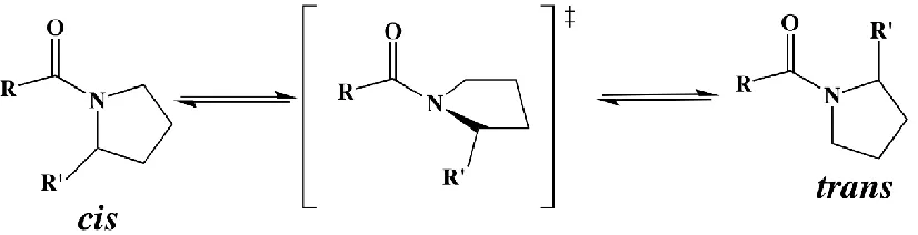
Figure 1.1 A known intrinsic molecular switch.
Figure 1.1 Proline switch is modulated through the cis-trans isomerization which results in many conformational changes to a protein backbone and contact dynamics.11
A known molecular switch is the peptidyl prolyl cis-trans isomerization.
Thermodynamically, the peptide amide bonds involving all amino acids favor the trans
conformation in linear and folded proteins due to its lower free energy.12, 13 Unlike other amino
acid sequences, linear peptide bonds preceding proline residues have a smaller free energy
difference of < 1 kcal/mol between the cis and trans conformations,14-17 consequently increasing
the cis conformation population. Peptidyl-prolyl bonds in folded proteins can be found in either
heterogeneous conformations marked by certain peptidyl-prolyl cis and trans states in both
solution and crystal environments.18, 19 The isomerization from cis to trans in peptidyl-prolyl
bonds can be propagated through the backbone to the entire protein, discretely shifting the
conformational ensemble of the protein. This switch, also referred to as a proline switch, is
known to direct conformational changes in several systems by mediating a variety of functions in
well-studied pathways.11, 20-26 While the isomerization of the peptidyl-prolyl bond is inherently
slow due to a free energy barrier of ~20 kcal/mol,14, 16, 27 the process can be sped up by a family
of enzymes known as peptidyl-prolyl cis-trans isomerases (PPIases).28 PPIases catalyze prolyl
cis-trans isomerization, typically the rate-limiting step in protein folding,29 from hundreds of
seconds to the more biologically relevant millisecond timescale. Recent studies have also shown
that PPIases are instrumental in controlling important intracellular signaling events.30 A majority
of PPIases, including Cyclophilin A and its homologues, and the phosphorylated dependent
PPIase Pin1, are found to be upregulated in all forms of cancer.31-33 Therefore, understanding the
molecular and atomistic basis of peptidyl-prolyl switches and their regulatory mechanisms could
provide insight into aberrant cellular processes, and provide new avenues for therapeutic design.
Cyclophilin A (CypA) is a peptiylpropyl isomerase that is located in the cytosol,
characterized by a beta barrel structure with two alpha helices and a beta-sheet. CypA is capable
of forming a complex with cyclosporine, which is capable of inhibiting the
calcium/calmodulin-dependent phosphate calcineurin.34 Inhibition of calcineurin is thought to aid in suppressing
organ rejection in transplants, achieved by halting the production of interleukin 2 and the
pro-inflammatory molecule TNF alpha. The side chains of CypA play a key role in protein catalysis.
Mutant CypA have been used to study different conformations and analyze catalytic activity.5
human diseases. CypA also play a key role in protein catalysis, with mutant CypA used to study
different conformations and analyze catalytic activity.5 Numerous biological pathways involve
CypA, including protein folding, cell signaling, and immune-modulation.32 CypA is vital in
regulating the immune system; this occurs through partner Src Homology proteins known as
Interleukin enzymes. These proteins regulate the cellular signaling pathways that lead to the
manufacturing of T-Cells in the human body. One such enzyme that has sparked interest in the
scientific community is Interleukin-2-Tyrosine Kinase Src Homology 2 (ITK SH2).
Interleukin-2 Tyrosine Kinase Src Homology 2 has been shown to exhibit a distal
intrinsic molecular switch in the form of a proline residue that undergoes cis-trans isomerization,
and is regulated by CypA.21 This isomerization modifies the conformational landscape of ITK
SH2, granting the protein the ability to bind to two different ligands determined by the state of
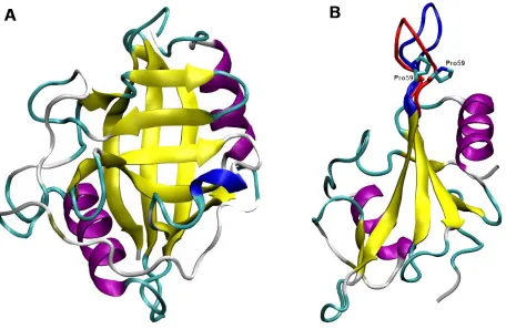
Figure 1.2 Structure of Cyclophilin A19 and SH2 domain of ITK36
Figure 1.2. (A) Structure of Cyclophilin A (B) ITK with both cis-transPro287
conformations in the loop highlighted36 The blue loop represents the cisPro287 conformation in
ITK while the red represents the transPro287 conformation.
1.2 Interleukin-2-Tyrosine Kinase Src Homology 2 (ITK SH2)
ITK SH2 is a nonreceptor tyrosine kinase protein that participates in an intracellular
signaling cascade that leads to the activation of T cells. Nonreceptor tyrosine kinases (nRTK) are
cytoplasmic enzymes which catalyze the transfer of a phosphate group to a tyrosine residue in
proteins from a nucleoside triphosphate donor.37 ITK is part of the Tec family which contains the
conserved SH3 and SH2 catalytic domains. Through previously reported NMR studies, ITK SH2
shows conformational heterogeneity by populating two distinct, low energy conformations in
solution, determined by the cis-trans conformation of Asn286-Pro287 peptidyl-prolyl bond.37
ITK SH2 has previously shown to have two distinct binding events that occur due to the two
conformation in ITK SH2 shows preference binding to ITK SH3, with weak binding shown to
occur when Asn286-Pro287 bond is in the trans conformation. Asn286-Pro287 bond in the cis
state upregulates the activation of T-cells.37 The trans Asn286-Pro287 bond conformation shows
recognition binding to phosphotyrosine-containing peptides. ITK SH2 has generated increasing
interest due to recent studies of HIV protease which binds to CypA. The binding of HIV to CypA
prevents CypA from isomerizing the Asn286-Pro287 peptidyl-prolyl bond in ITK SH2.36 The
stop in catalysis of the imide bond disrupts the ability of ITK SH2 in binding ITK SH3, which in
[image:17.612.69.541.300.622.2]turn prevents T-cell activation.
Figure 1.3 ITK SH2 domain with a phosphorylated tyrosine motif (PTR) bound and the peptidyl-prolyl cis-trans isomerization Asn286-Pro287.
between Asn286 and Pro287 in ITK SH2. The ω imide bond Asn286-Pro287 has shown to have heterogeneity between cis and trans conformation. The cis-trans isomerization is regulated by the peptidyl propyl isomerase enzyme Cyclophilin A. The atoms that account for the dihedral angle are labeled.
This study focuses on the ITK SH2 enzyme, which has a proline that undergoes the
cis-trans isomerization due to the distal switch that controls binding affinities for two different
substrates, one of which leads to the activation of T-cells. The binding of ITK SH2 to the ITK
SH3 domain has previously shown to be favored when Asn286-Pro287 of ITK SH2 is in the cis
state while the phosphotyrosine containing peptide is known to bind to the ITK SH2 when the
Asn286-Pro287 is in the trans state. The energy landscape that allows this conformational
change, in addition to the key residues that are affected by this change are not known. Using
computational studies combined with contact statistics and principal component analysis, it is
shown how the imide bond between Asn286-Pro287 undergoing cis-trans isomerization (Figure
1.4) can change the conformational landscape of ITK SH2. Through molecular dynamics (MD),
this study has defined the energy landscape and identified key contacts that are formed, shared,
2 MOLECULAR DYNAMICS
Enzymes have many dynamical properties that occur on a timescale many times faster than
what experimental conditions are able to measure. To study those effects, a statistical mechanics
approach known as molecular dynamics (MD) is utilized. This methodology allows us to study
the enzyme on a microscale using an ensemble to describe the physical properties of a system on
a macroscopic scale. MD has evolved from its initial application in studying the dynamics of
liquids, to simulating entire macromolecules and ligand binding events.38 In recent years the field
of computational chemistry has grown exponentially with the advancement of computing
resources available, offering an unparalleled tool for studying enzyme dynamics or calculating
the energy landscape of a system.
MD has branched into two subcategories- one known as quantum molecular dynamic
(QMD), and a classical approach using Newtonian physics. The mechanics behind the quantum
approach is to consider the contributions of the outer valence shelf electrons while calculating
the nucleus and inner electrons through classical integration. QMD refines the calculations on a
much more atomic level by considering the electrostatic interactions that occur in the system
from the valence shell electrons. The approach does have its drawbacks, namely in its resource
demands. As system size grows, the computational power needed increases exponentially, and
the approach has difficulty in dealing with transition metals.39 However classical MD ignores the
valence shell electrons for a more coarse grained approach, essentially considering the system in
a more simplified aspect by treating the atoms and bonds as a ball and stick model.40
MD simulations require an initial set of coordinates, usually provided in the form of an
accurate three-dimensional representation of the system. X-ray crystallography or nuclear
information of this nature.34 This in turn has given rise to structures determined by a mixture of
experimental and theoretical work.38 With the exponential increase in computing power, MD is
now able to provide a better understanding of key atomistic detailing in a complex system that
are subtle enough to change entire energy landscapes.
2.1 Algorithms that Govern MD
Every atom has a potential energy associated with it that needs to be taken into account to
predict the individual movements that then govern the overall system. MD simulations take into
those potentials by using a set of predetermined parameters to defines how the atoms are held
together using interatomic forces.38 Taking into account the potential energy of all the interacting
atoms (
𝑟
𝑁) as a function of position, and using that to take the gradient (
∇) of the function withrespect to displacement in the coordinates of the atoms will result finding the force acting upon
individual atoms.41
𝐹
𝑖= −∇
𝑟𝑖𝑈(𝑟
1, ⋯ , 𝑟
𝑁) = − (
𝜕𝑈 𝜕𝑥𝑖,
𝜕𝑈 𝜕𝑦𝑖
,
𝜕𝑈
𝜕𝑧𝑖
)
(1)
The entire movement of atoms in a MD simulations is governed through these different
potentials that act upon each atom in a given system.42
𝑈(𝑟
1, ⋯ , 𝑟
𝑁) = ∑
𝑎𝑖2
(𝑙
𝑖− 𝑙
𝑖0)
2𝑏𝑜𝑛𝑑𝑠
+ ∑
𝑏𝑖
2
(𝜃
𝑖− 𝜃
𝑖0)
2𝑎𝑛𝑔𝑙𝑒𝑠
+ ∑
𝐶𝑖
2 𝑡𝑜𝑟𝑠𝑖𝑜𝑛𝑠
[1 +
cos (𝑛𝑤
𝑖− 𝛾
𝑖] + ∑
4
𝜀𝑖𝑗[(
𝜎𝑖𝑗𝑟𝑖𝑗
)
12− (
𝜎𝑖𝑗 𝑟𝑖𝑗)
6
] +
𝑎𝑡𝑜𝑚𝑠 𝑝𝑎𝑖𝑟𝑠
∑
𝑘
𝑞𝑖𝑞𝑗
𝑟𝑖𝑗
𝑎𝑡𝑜𝑚 𝑝𝑎𝑖𝑟𝑠
(2)
Equation 2 considers all the potentials of a single atom by adding the potentials of bond
length, bond valence angle, bond dihedral angle, bonded van der Waals (vdw), and
non-bonded electrostatic potentials. Breaking down the equation further show the first two terms
from their equilibrium potential values of (li0) and (𝜃𝑖0). Harmonic force constants that prevent
modeling chemical changes are represented by terms ai and bi. The dihedral movement is the
rotational angle changes that happen around a chemical bond. These terms are represented by n
and ci which denotes periodicity and heights of rotational barriers respectively. Interatomic
forces that consider the attraction and repulsion terms of vdw forces are taken from the
Lennard-Jones 12-6 potential equation. The final terms represent the electrostatic potential by defining the
partial charges of two atoms in relation to the distance between two atoms. The terms qi and qj
represent the partial charges of the two atoms i and j respectively, the term to describe relative
distance is rij. According to Newton’s second law of motion, the force of a single atom is equal
to the mass of that atom times the change in acceleration, as shown in equation 3.43
𝐹
𝑖= 𝑚
𝑖𝑑2𝑟𝑖(𝑡)𝑑𝑡2
(3)
Taking the force vector on each individual atom in a system, an algorithm is used to
generate the movement in the simulation. The most popular algorithm in use today is the Verlet
algorithm (equation 4), hailed for its simplicity and stability. The Verlet algorithm describes the
movement of a set of atoms in a system by position and velocity vectors.38
𝑟
𝑖(𝑡 + Δ𝑡) ≅ 2𝑟
𝑖(𝑡) − 𝑟
𝑖(𝑡 − Δ𝑡) +
𝐹𝑖(𝑡)𝑚𝑖
Δ𝑡
2
(4)
Considering the previously established concepts, the sum of multiple potentials can be
used to generate the net force as applied on a single atom. Combining the Verlet algorithm with
Newton’s second law of motion provides change in position of a single atom over time.
Movement on the atomic scale for just a typical bond length between atoms happens on the
femtosecond scale. Thus, to measure the interactions the time scale needs to be sub-femtosecond
localization of electrons surrounding the atoms. Though less accurate, a femtosecond time step is
more efficient and detailed enough to thoroughly study a system of interest. However, by
continuously increasing the magnitude of time at the femtosecond scale the margin of error
increases, hence, a time step of more than 5 femtoseconds would not be a feasible approach for
the application of an MD simulation.43
2.2 Overcoming the limitations of MD
The positions and velocities of each atom in the system coupled with the summation of
all the potentials and forces must be continuously calculated at each step. The continuous process
has led to the development of algorithms that can continuously do so without error, having to
account for long range electrostatic effects and explicit assessment of atoms in a given system.38
In addition, the water model widely used in computational simulations (TIP3P) does not
accurately establish explicit electrostatic and conformational changes of physical water.
However, the TIP3P water model decreases the computational demand by more than three-fold.44
Furthermore, accelerated molecular dynamics (aMD), replica exchange molecular dynamics
(REMD), and high temperature simulations are also well established methods towards enhancing
sampling.45 The solution to the long range electrostatic interaction came in way of a summation
method called particle-mesh Ewald, which enabled a discrete cut off range that is applied to each
atom. As a result, a shorter cutoff would result in increased speeds but less precision, while a
longer cutoff increases computational time substantially.46
The primary aim of MD is to achieve convergence of a system, defined as sampling the
entire free energy landscape accessible by the system. Rigorous sampling is required to achieve
this state of convergence, and long timescales are needed to ensure the free energy landscape is
timestep is both the limiting factor, and timesteps that are too large result in inaccuracies. Even
with rigorous sampling, true convergence for an unknown energy landscape can only be
achieved when folding and unfolding are witnessed in a simulation. Since it is established that
most biological processes occur at the micro and millisecond timescale, complete understanding
of a system can only be achieved with exceedingly long simulations. For small systems (< 25K
atoms) with biological processes that occur on the sub microsecond timescale, it is possible to
run for ample enough time to achieve the microsecond scale. For larger systems, other methods
and algorithms have to be further developed and refined for more accurate sampling results.
A number of limitations in MD occur in relation to how an atom is defined in the system.
The classical approach considers the atoms and their corresponding electrons, as a single unified
ball, with bonds being represented as springs.43 Therefore, the approach cannot account for
proton tunneling, electron movements in acid-base chemistry, or even redox reactions. While
many of the descriptors for atoms are based on experimental data, this is reliant on the sensitivity
of the instruments used to collect the data. True achievement of an extremely refined and
accurate energy landscape would require QMD simulations on the entire system. There are also
limitations when parameters do not exist due to a lack of experimental data available and/or
mathematical limitations in defining the empirical basis for that parameter.
2.2 Current State of MD
The current state of MD has shown substantial progress after the achievement of running
on GPUs for calculations. As computational power has grown, it has given rise to the ability to
simulate complex systems that are larger in size for longer periods of time. The AMBER
software suite allows the calculations to be offloaded onto the GPU, and only passing back to the
update to GPUs. A small 110 residue protein, e.g. ITK, can be simulated for upwards of
150ns/day on a single Titan X GPU. Allowing for parallel processing and offloading calculations
onto two Titan X GPU does not scale as well, but the general speed is upwards of 240ns/day for
the same protein. This gives the ability to run on a routine microsecond scale and achieve
3 EXPERIMENTAL PROCEDURE
The three-dimensional structure of ITK SH2 and ITK SH2 with PTR substrate were taken
from an average NMR structure with Protein Data Bank (PDB) Identification (ID) no. 1LUK and
2ETZ respectively. Amber14 simulation package48 was used to carry out all simulations with
modified force field parameters of Cornell et al. (1995)47 by Perez et al. (2007)49, a re-optimized
dihedral parameter50 to both systems, in addition a phosphate parameter for the 2ETZ system
was applied.51,52 Each system was solvated using a 10 Å TIP3P explicit water model in an
octahedron box which added roughly 6000 TIP3P water molecules. The system was neutralized
by adding counter ions to negate the charge.
The systems were minimized for a total of 2000 steps and equilibrated while holding the
enzyme with a harmonic constraint force of 200, 100, and 5 kcal/mol/Å2. All simulations were
carried out at a constant temperature of 300 K and a constant pressure of 1 bar. A 2
femtoseconds time step was used to solve Newton’s equation of motion. To calculate the
electrostatic interactions, the particle mesh Ewald (PME) summation method was used with a 9
Å cutoff for long range non-bonded interactions.53
To sample the isomerization process, the umbrella sampling method was used to put a
harmonic restraint on the ω imide bond angle. The ω imide bond was rotated every five degrees
from -180° to 180°. Per the umbrella sampling method each angle rotation is considered a
window, this gave a total of 73 windows to sample the isomerization from both the negative and
positive rotational angle of the ω imide bond. Each window was simulated for 400 ns; the first
100 ns was part of the equilibration period. All windows were then combined via the Weighted
Histogram Analysis Method (WHAM) 54 to generate the potential mean force plot. Intermediates
the positive ω rotational angle had a slightly lower free energy barrier than the negative ω
rotational angle, the positive angle intermediates at 0°, 45°, 90°, 135°, and 180° was then
simulated for 4.0µs, in which case all trajectories were combined to create a principal component
(PC) plot. Using atom mask to select only the portion of ITK that does not involve the CD loop
in which the proline switch is located, the analysis shows the effect of the isomerization process
on the remaining portion of ITK. Further detailing of Umbrella Sampling is discussed in section
3.1. Contact statistics were generated by taking all heavy atoms into account that were within a
4.5 Å radius of another heavy atom, this is to set precedence of bonding interactions.8
The 4.0µs trajectory for the cis and trans conformational space was combined to create a
contacts trajectory PC plot. The contacts trajectory takes every frame into account and records
the contacts made and broken along the simulated time frame. All heavy atoms are considered
and if distances are less than 4.5 Å, they are contacts either made or, if greater than 4.5 Å, broken
or not there. ITK with PTR was simulated through the umbrella sampling method to sample for
the transition state (TS) and cis state of the Asn286-Pro287 for 400ns; the first 100 ns was
equilibration. This was to show the effect of isomerization on binding energies for the PTR
substrate, MMPBSA analysis was used to calculate the binding energies on ran on the final
300ns simulation. The PB portion allows us to measure more accurately by considering the
dielectric potential of the enzyme–substrate complex. This was done in a vacuum to eliminate
the electrostatic interactions of the explicit water. The surrounding was treated in a solvated
implicit surrounding.
3.1 Umbrella Sampling
Free energy calculation difference calculation is essential in computational chemistry and
experiments. The challenging task of calculating free-energy differences has led to a new way of
MD. Umbrella sampling is a biased MD simulation method that allows the user to control the
reaction coordinate, provided the reaction scheme is known. The methodology is used in
combination with multiple states that are simulated along the reaction pathway.55 This is
achieved through bias potentials that force the reaction to occur from one state to another. Each
step in the reaction pathway is considered to be a window, where each window is now simulated
while being held in accordance to that potential window.56,57
Mapping the entire reaction coordinate will determine the free energy pathway by
calculating the energetic differences between each window (Figure 3.1). An oversimplification
of this is shown a propyl imide bond that has a known reaction coordinate. This is simply taking
the ω bond between the X residue and proline and applying simple harmonic bias potentials.
The combination of windows can be analyzed through a method called the weighted histogram
analysis method (WHAM) which allows the recalculation of the bias potential and show the
potential mean force graph.58-59 The easiest case to describe this process is looking at the cis
-trans isomerization process of proline. The reaction pathway is long known to go from cis to
trans and vice versa.
[image:27.612.191.415.517.696.2]3.2 Calculating Binding Energy using MM-PBSA
Proteins are natively found in aqueous solutions or in membrane bound regions of a cell
and are always in an aqueous medium. The solvent plays a crucial role in ligand binding and
protein-protein association. This proves to be a challenging task in MD since the solvent effects
the intramolecular and intermolecular interactions. To overcome this challenge, the current
methodology allows the calculations of free energies by representing the molecular simulation in
atomic models that represent the explicit biomolecule and the environment surrounded by the
system. Water models have been published to describe successfully the aqueous environment in
molecular simulations.44
The explicit inclusion of water leads to a considerable increase in calculation time and
simulation system size. The computation of solvation requires an extensive sampling of the
solvent degrees’ freedom. The less costly approach is to treat the solvent as implicit and
incorporate additional potential mean force terms. The expression then depends on atomic
coordinates of the solute and the solvent degrees of freedom have been integrated out. This
results in the system having the same number of degrees of freedom for the solute as explicit
4 RESULTS & DISCUSSION
There have been substantial improvements made in finding heterogeneous cis-trans
proline through HSQC 15N NMR experiments. The key atomistic interactions of the
isomerization process however, is not evident through the experimental techniques. Through
current MD standards it is not known if these interactions can be captured, due to two key
limitations. The first limitation to MD simulations is the millisecond timescale to sample the
cis-trans isomerization process. The other is the limitation to MD is the force field used and its
capacity to capture accurately the elusive interactions. There have been substantial improvements
to the force field parameters to reproduce experimental observations. Current force field
parameters show that weak and elusive interactions can be captured using simulation methods
and there have been tremendous efforts to push the timescale limitations beyond the
microsecond. However, to sample the cis-trans peptidyl-prolyl bond remains a challenge using
regular MD. To sample the isomerization process here, the umbrella sampling method has been
used, this method helps explore the limits of sampling in MD. To study the interactions, the
recent molecular mechanics force field, ff14SB that comes with the AMBER 14 was used to run
all simulations. The force field has improved through consecutive adjustments to the preceding
AMBER force fields. A re-optimized parameter for the peptide bond dihedral angle was used as
well on all simulations. Along with the improved force field, the re-optimized parameters for
peptide bond dihedral angle, and the umbrella sampling method, the isomerization process was
sampled.
We have carried out extensive molecular dynamics simulations for a total of over 60 µs
in explicit water and ions to investigate an allosteric coupling between cis-trans isomerization of
domain. The free energy profile along the peptidyl-prolyl ω-bond angle between Asn286 and
Pro287 suggests that isomerization could predominantly occur in the positive angle direction
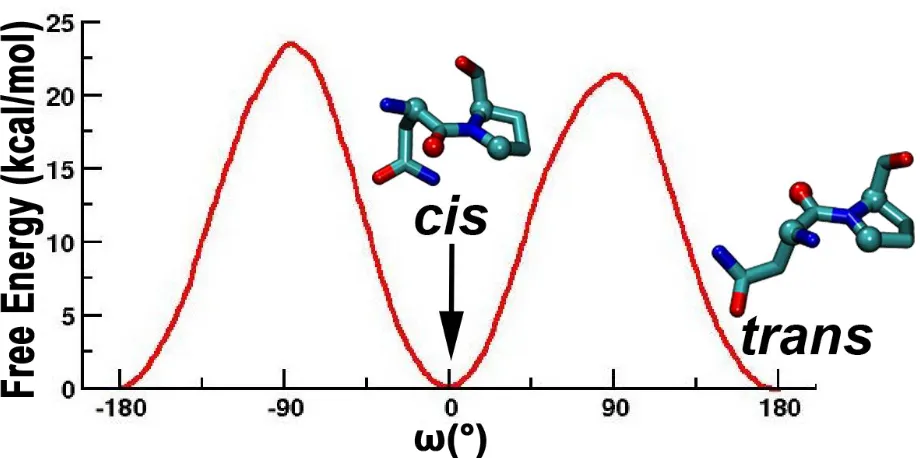
(Figure 4.1). The free energy barrier (~20 kcal/mol) along the positive direction is lower than
that (~24 kcal/mol) along with negative direction (Figure 4.1). The magnitude of the barrier is in
excellent agreement with experimentally measured barriers for uncatalyzed peptidyl-prolyl cis
-trans isomerization.14, 27 The peptidyl-prolyl bond angle would, therefore, isomerize mainly
clockwise from trans to cis, if looking along the backbone from Asn286 to Pro287, and
anticlockwise from cis to trans, since isomerization along the positive angle direction would be
more than 800 times faster than that in the other direction. Interestingly, Cyclophilin A, one of
the enzymes that catalyze peptidyl-prolyl cis-trans isomerization, has been shown to catalyze
peptidyl-prolyl bond angle in the positive direction, stabilizing the transition state of the
ω-bond angle around +90o.61 In general, the trans conformation of a peptidyl-prolyl bond in a linear
peptide is significantly more populated than the cis conformation. In a linear peptide containing
Asn-Pro, the cis population was shown to be ~11.6%, and about ~4.4% of Asn-Pro bonds in a set
of non-redundant proteins in the Protein Data Bank were shown to be in the cis conformation.27
The free energy profile however suggests that the trans state is almost isoenergetic to the cis
state of the SH2 domain, in excellent agreement with experiments. The trans and cis populations
were measured to be approximately 60% and 40%, respectively, in solution,21, 62, 63 suggesting
that the trans conformation of the Asn286 - Pro287 peptidyl prolyl bond in the folded native SH2
domain is similar in free energy to the cis conformation. The fold of the SH2 domain has
therefore further stabilized the cis conformation of the Asn286-Pro287 peptidyl prolyl bond to
Combining the umbrella sampling results via WHAM reveals the potential mean force
plot that shows the energy barriers of both the positive and negative ω rotational bond angle. The
results show that the cis state has the same relatively free energy as the trans state of the proline
and the positive ω rotational bond angle having a slightly less free energy barrier height than the
negative ω rotational bond angle as shown in Figure 4.1. The results from the WHAM conclude
that the positive ω rotational bond angle is more favorable in the isomerization process. Using
the WHAM data, intermediate points of 0°, 45°, 90°, 135°, and 180° ω imide bond angle were
simulated further for 4.0µs each. This allowed greater sampling to be added to the WHAM and
[image:31.612.76.536.320.549.2]to see the differences in conformational spaces along the isomerization process.
Figure 4.1 The cis-trans isomerization of the ω imide bond in ITK SH2.
The rotation bond from -180° to 80° shows that the free energy for the positive ω rotational bond angle is lower. The cis and trans ω imide bond between and Asn286-Pro287 is shown at their respective angles.
4.1 Coupled conformational dynamics between distal regions and the cis-trans switch
It was shown using NMR spectroscopy that the dynamics of the SH2 domain is coupled
However, a more detailed atomistic description of the conformational changes upon
isomerization is lacking. We carried out microsecond-long simulations (4 μs each) when the
Asn286-Pro287 prolyl bond is trans (~180o), cis (~0o), and in three intermediate states between
trans and cis (45°, 90°, 135°). The positive direction is chosen for the intermediate points
because the barrier in the positive direction is lower than that in the negative direction (Figure
4.1). We carried out Principal Component Analysis (PCA) on the Cartesian coordinates of the
backbone of the SH2 domain of the five states - excluding the CD loop, which contains the
Asn286-Pro287 motif, to only capture the effect of cis-trans isomerization on distal regions. The
dynamics of all of the states were projected on the top two eigenvectors representing the
dominant motions in the SH2 domain as shown in Figure 4.2. Interestingly, the slowest mode
(PC1) captures most of the changes observed (Figure 4.2). The results suggest that when the CD
loop is in the trans state, the rest of the SH2 domain visits two dominant conformational spaces
(Figure 4.2A). On the other hand, the backbone of the SH2 domain is predominantly localized in
one dominant conformational free energy well when the CD loop is in the cis state (Figure 4.2E).
As the CD loop goes from trans to cis, the backbone of the rest of the SH2 domain becomes
more localized in predominantly one free energy well (Figure 4.2B, C, D). The PCA results of
the backbone clearly show distinct differences in the conformational dynamics of the two
isomeric states of the CD loop of the SH2 domain, in agreement with previous NMR
experiments.63
The PCA results show that the cis state of proline, Figure 4.2e, results in one dominant
conformational space with a smaller secondary state that is less populated. The conformational
space results in two states that are connected, showing that the smaller secondary state is
transition angle 90°. In both cases, there is one dominant state with a secondary state that is
energetically less favorable. This agrees with experimental results that allow the ITK SH2
enzyme when the proline switch is in the trans state to bind to the ITK SH3. This is not shown to
be true in regards to the cis state of the proline experimentally to show binding to the
phosphotyrosine containing peptide substrate.
The PC plot shows that in all cases the separation of different dominant states occurs
along the PC 1. The separation that occurs in the PC plot is shown in Figure 4.2a-e along with
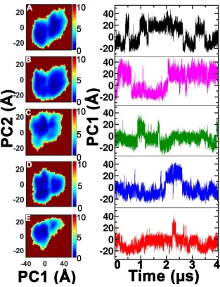
Figure 4.2 PC plot of the intermediates taken along the positive ω bond isomerization and visual overlay of trans and cis PC1.
Figure 4.2 Free energy landscape in kcal/mol of the conformational space explored by the backbone of (A) trans state, (B) 135° of the Asn286-Pro287 peptidyl-prolyl bond, (C) close to the transition at 90° of the Asn286-Pro287 peptidyl-prolyl bond (D) 45° of the Asn286-Pro287 peptidyl-prolyl bond, and (E) the cis state. On the left is the plot of the top two dominant
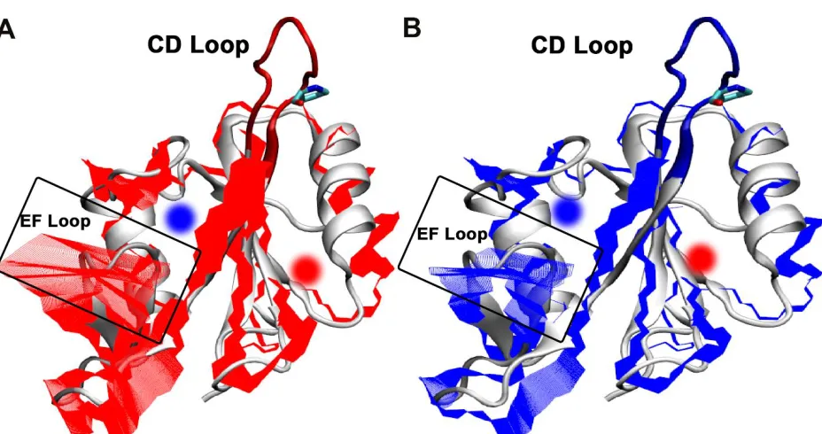
Figure 4.3 Projection of the dominant Principal Component (PC1) onto the backbone of the SH2 domain of ITK when the Asn286-Pro287 peptidyl prolyl bond is in trans (A; red), and the dominant cis (B; blue) conformations
The CD loop, containing the proline switch, is not considered in the analysis of the PCA. The EF loop (blue) shows the major conformational change. The dominant motions of SH2 within the proline switch in the trans state are distinctly spanned across that of the cis state. The blue circle highlights the pY+3 position of the phosphotyrosine binding pocket, and the red circle highlights the phosphotyrosine, pY, binding pocket. Structure is overlaid with 1LUK pdb10.
Using VMD, the major contributions of PC 1 is shown in overlay with ITK SH2 enzyme,
Figure 4.3. The visual aspect shows conformational change in the cis is limited in movements,
which could be the contributor to having a binding specificity to the ITK SH3 domain. The trans
conformation results in the enzyme having broader movement along the EF loop. The EF loop
has a wide range of motion in the trans conformation of proline but is restricted when the proline
is in cis conformation. This is shown in NMR experiments that the EF loop plays a vital role in
pY+3 substrate catalysis. The EF loop stabilizes the pY+3 region of the substrate and thus needs
plot did not consider the CD loop which contains the proline switch to see how the
conformational landscape changes as the proline undergoes isomerization.
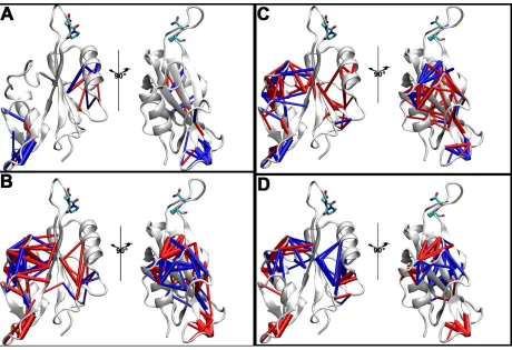
Figure 4.4 Dynamic contact statistics and Principal Component Analysis of the residue-residue contact dynamics of the cis and trans state of the SH2 domain.
The width of the cylinders between the residues is proportional to the magnitude of the change in probabilities. (C) Projection of the contacts dynamics for trans (black) and cis (red) states of the SH2 domain using the top two Principal Components of the contact trajectory.
4.2 Significant rearrangement of the network of residue-residue contacts as a result of
the distal cis-trans switch
According to the results in Figures 4.2 and 4.3, the backbone conformations of the trans
and cis states of the SH2 domain overlap. The backbone conformations of the cis state are a
subset of that of the trans state. While the trans state fluctuates in the nanosecond timescale
between two dominant states with a relatively low barrier separating the two (Figure 4.2A), the
cis state is mainly localized in one of the conformational basins (left basin of Figure 4.2E). It is
therefore easy to conclude that the overall conformational dynamics of the cis state is sampled by
the trans state from the results presented in Figures 4.2 and 4.3. Is this conclusion however
consistent with respect to the internal dynamics of the SH2 domain?
In answering this question, we analyze the dynamics of the residue–residue contacts of the
SH2 domain as previously described.8, 64 Specifically, we consider a residue-residue contact
between two non-adjacent residues to be formed if any two inter-residue heavy atoms are within
4.5 Å. In general, a small percentage of residue-residue contacts is dynamically forming and
breaking and is referred to as the “dynamic contacts.” The majority of the residue-residue
contacts are never formed, and another small percentage is always formed and mainly involved
in maintaining the secondary structure of the protein. A residue-residue contact is considered part
of the dynamic contacts if it is formed more than 10% and less than 90% of the total simulation
time. Figure 4.4A shows the difference of the probabilities of residue-residue contact formation
contacts are more formed and breaking as the trans state goes between the two backbone free
energy wells? Figure 4.4A shows that a subset of contacts, primarily involving the dynamics of
the EF loop, is more formed (blue) and less formed (red) as the backbone of the trans state move
from the right well to the left well, and vice versa. The width of the cylinders is proportional to
the magnitude of the difference in probabilities. Figure 4.2B shows the difference of the
probabilities between the left well of the trans state and the left dominant well of the cis state in
Figure 4.2A, E, that is what residue-residue contacts are more formed and breaking as the trans
state now moves to the cis state? Figure 4B shows that a predominantly different set of contacts -
now primarily involving the BG loop - is more formed (blue) and less formed (red) as the
Asn286-Pro287 peptidyl-prolyl bond switches form trans to cis. Interestingly, even though the
backbone conformations of the trans and cis states are similar (Figure 4.2A,E), the internal
residue-residue contact dynamics are quite different. The backbone dynamics of the trans state
can sample that of the cis state, but the internal residue-residue contacts are not in place until the
Asn286-Pro287 peptidyl-prolyl bond has fully switched over to the cis state. The results suggest
that the rearrangement of the contacts in the cis state somewhat locks the backbone
conformations in the left well, occluding the binding pocket for the pY+3 position of the
phosphotyrosine motif and opening the binding pocket of the phosphotyrosine, pY, to disfavor
favorable interactions with phosphate group. The results suggest that the phosphotyrosine motif
(PTR) could bind strongly to the right well of trans state (Figure 4.2A), and conversion from the
trans state to the cis state starts by rearrangements of the contacts involving the EF loop followed
by rearrangements of the contacts involving the BG loop, occluding the binding pocket for the
pY+3 position of the phosphotyrosine motif. Figure 4.5 shows the gradual rearrangements of the
to cis (0o) every 45o of the left free energy well in Figure 4.2, primarily rearranging the contacts
associated with the BG loop and the binding pocket of the phosphotyrosine residue, pY.
The contact trajectories of the dynamic contacts of the trans and cis states were further
analyzed using Principal Component Analysis. A dynamic contact was assigned a ‘1’ when
formed and a ‘0’ when not formed. A contact trajectory is therefore made up of binary
representations of the dynamic contacts at each time point of the simulation, reducing the
dimensionality of the simulations. Figure 4.4C shows the PCA of the contract trajectories of the
trans state and the cis state, projected on the top two Principal Components (PCs). The results in
Figure 4.4C show that there is a common set of contacts due to the overlap, but contact dynamics
between the trans and cis state are quite different, unlike the comparison between the backbone
dynamics (Figure 4.2). Changes in the contact dynamics are expected to tune the conformations
of the active site as the Asn286-Pro287 peptidyl-prolyl bond switches from trans to cis. Our
results complement existing experimental results to further explain the possible mechanism of
allosteric regulation by the Asn286-Pro287 proline switch in the SH2 domain of ITK, suggesting
that the contact dynamics play an enormous role in shaping the binding pocket. The results
highlight the multidimensional nature of biomolecular dynamics, such that the similarity in the
backbone dynamics of difference states of a protein does not always mean that the internal
dynamics or conformational states are similar. Therefore, caution should be taken when
Figure 4.5 Residue-residue contact statistics from the trans state to the cis state, at
intermediate points of 135°, 90°, and 45° of the Asn286-Pro287 peptidyl-prolyl bond overlaid on top of structure from PDB 1LUK10
Figure 4.5 Changes in the residue-residue contact probabilities above a magnitude of 25% going from the (A) left free energy well of the trans state to the left free energy well of the 135o intermediate state in Figure 4.2A, B, (B) left free energy well of the 135o state to the left free energy well of the 90o intermediate state in Figure 4.2B, C, (C) left free energy well of the 90o state to the left free energy well of the 45o intermediate state in Figure 4.2C, D, (D) left free energy well of the 45o state to the left free energy well of the, cis state in Figure 2D, E,
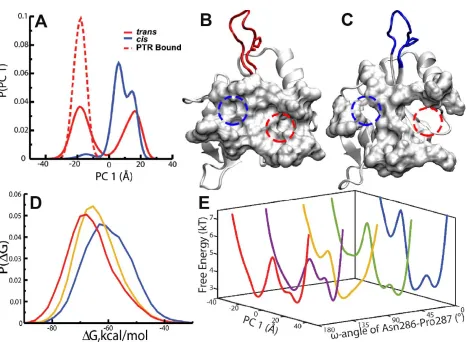
Figure 4.6 Probability distribution of the dominant Principal Component of the phosphotyrosine Active Site Residues. Structure overlaid with PDB 1LUK10.
Figure 4.5 (A) Free cis, free trans, and trans bound SH2 domain. (B) The binding pocket of the phosphotyrosine, pY, highlighted using a red dotted circle and the binding pocket of the pY+3 position of the phosphotyrosine motif highlighted using a blue dotted circle in the trans and (C) cis states of the SH2 domain. The entire binding pocket is represented using surf. (D) Probability distributions of MMPBSA binding free energies of PTR when the Asn286-Pro287 peptidyl-prolyl bond is in trans (red), the 90o (yellow), and the cis (blue) conformations. (E) The
potential mean force (PMF) profiles of the domain principal component of the binding site residues as the Asn286-Pro287 peptidyl-prolyl bond goes from trans to cis.
4.3 Direct modulation of the binding pocket dynamics and binding affinity by the proline
switch
We have shown that isomerization of the Asn286-Pro287 peptidyl-prolyl bond in the CD
[image:41.612.74.541.90.432.2]dynamics and conformational states are specific to the isomeric state of the Asn286-Pro287
peptidyl-prolyl bond. However, it is not exactly clear how the isomerization process directly
affects the binding pocket of the phosphotyrosine motif. We carried out an additional molecular
dynamics simulation of the phosphotyrosine motif (PTR) bound to the SH2 domain for 2.2 μs. In
analyzing the trajectory, we define the binding pocket of the SH2 domain by selecting the
residues surrounding the phosphotyrosine motif as shown in Figure 1.3A. Any residue within 5
Å of PTR was assumed to be part of the binding pocket. We carried out Principal Component
Analysis on the heavy atoms of the binding pocket residues of the trans bound, free trans state,
free cis states, and the free intermediate 135°, 90°, and 45° states of the Asn286-Pro287
peptidyl-prolyl bond. Figure 4.6A shows the probability distributions of the dominant principal
component (PC1) of the motions of the binding pocket of the trans bound, free trans and free cis
states. The active site of the free trans state has two almost equally distributed microstates, while
the active site of the cis form has one dominant conformational state. The binding pocket of the
trans bound SH2 domain has one dominant population resembling one of the microstate of the
free trans state, suggesting conformation selection65-68 and selective binding of the
phosphotyrosine motif to only one of the two possible conformations of the free trans state.
Experimentally, it has been shown using NMR experiments that PTR binds to the trans form of
the SH2 domain with a binding affinity of 4.0 mM-1, almost four times stronger than the binding
afinity to the cis form of the SH2 domain.69 The results suggest that the other conformational
state of the binding pocket of the trans state, which is similar to the binding pocket of the cis
state, would interact much weaker with PTR. According to the results in Figures 4.5 and 4.6, the
binding pocket of the pY+3 position of PTR is occluded by the inward motions of the BG and
completely disrupted in the cis state of the SH2 domain (Figure 4.6B, C). The rearrangement of
the BG and EF loops due to isomerization of the Asn286-Pro287 peptidyl-prolyl bond from trans
to cis has been suggested by NMR experiments to preorganize the SH2 domain to bind to the
SH3 domain.54 The cis form of the SH2 domain of ITK binds the SH3 domain approximately 3.5
times more stronger than the trans form, with an association constant of 2.6 mM-1 and 0.8 mM-1,
respectively.69
It is interesting to see that, as the Asn286-Pro287 peptidyl-prolyl bond isomerizes from trans
(180°) to cis (0°), the binding pocket changes accordingly. Figure 4.6E shows the free energy
profile along the dominant principal component (PC1) of the binding pocket for the trans, cis
and intermediate states of the Asn286-Pro287 peptidyl-prolyl bond. In the trans state of the SH2
domain, the binding pocket could adopt two possible conformations with a free energy barrier of
about 2 kcal/mol separating the two states. As the Asn286-Pro287 peptidyl-prolyl bond
isomerizes from trans to cis, the populations of the free energy wells of the binding pocket that is
pre-organized to recognize the phosphotyrosine motif decreases and the free energy barrier from
the dominant free energy well increases. The results suggest that the binding pocket is confined
to one state in the cis state and restricted to a single free energy basin, while that of the trans
state can sample two conformations of the binding pocket in the nanosecond time scale, one of
which is more optimized to recognize the phosphotyrosine motif.
We further test the hypothesis that the binding pocket of the trans state of the SH2 domain is
more suitable to bind the phosphotyrosine motif by estimating the binding affinity of PTR using
MMPBSA.70 The Asn286-Pro287 peptidyl-prolyl bond of the trans bound SH2 was then changed
energies were calculated for all of the three states using all of the snapshots using MMPBSA.
The results clearly suggest that PTR binds more tightly to the trans state of the SH2 domain, as
shown in Figure 4.6D. As the Asn286-Pro287 peptidyl-prolyl bond isomerizes to the cis form,
the binding affinity gets weaker (Figure 4.6D) and possibly facilitating the dissociation of PTR.
The results suggest that the shape of the binding pocket in the cis state could be partly formed in
the trans state, possibly allowing for the phosphorylated tyrosine motif (PTR) to easily dissociate
in the trans state as the Asn286-Pro287 peptidyl-prolyl bond is isomerized to the cis state. The
millisecond to second time scale of isomerization of the peptidyl-prolyl bond is much slower
than that of the nanosecond conformational rearrangement of the binding pocket, therefore the
conformational states could quickly and easily redistributed during the isomerization process.
5 CONCLUSION
Many advances have been made in MD to sample a wide range of enzyme dynamics to
explain the biological and physiological phenomena associated with them. There are however
just as many limitations that do not allow certain aspects to be measured computationally, such
as unrestricted isomerization process from cis to trans. However, there are many aspects that can
be measured computationally that make up for the loss in methods.
In this study, we carried out multiple microsecond-long molecular dynamics simulations
to provide an atomic level description of the coupling between a distal cis-trans molecular switch
and the phosphotyrosine binding pocket of the ITK SH2 domain. The results suggest that the
trans and cis states of the SH2 are almost isoenergetic, in agreement with previous NMR results.
Interestingly, the phosphotyrosine binding pocket of the trans state samples two distinct
conformation of the cis state somewhat resembles that of one of the conformations of the trans
state. Even though the backbone dynamics of the cis state is a subset of the trans state, the
residue-residue contact dynamics are quite different. Isomerization of the Asn286-Pro287
peptidyl-prolyl bond changes the backbone dynamics, which in turn rearranges the
residue-residue contacts and dynamics distal to the isomerization of the molecular switch. The
phosphotyrosine motif binds to the trans state with a lower free energy than that of the cis state.
The binding pocket conformation of the trans state that recognizes the phosphotyrosine motif is
one of two conformations of the binding site of the free trans state, suggesting conformational
selection. The other conformation of the free trans state is cis-like. However, the residue-residue
contacts of the transcis-like conformation of the binding pocket are only partially similar to that
of the cis state. The remaining residue-residue contacts are only formed in the cis state when the
Asn286-Pro287 peptidyl-prolyl bond is fully transitioned to the cis isomer and locks the cis state
into a single free energy well. Rearrangements of the contacts to form favorable residue-residue
contacts are partly responsible for the localized cis state. These results provide atomic level
description of the allosteric regulation by a cis-trans molecular switch that could have
implications for fully understanding subcellular normal and aberrant processes and identifying
similar elusive molecular switches.
REFERENCES
1. Molecular machines and motors. Springer: 2001.
2. Cooper, G. M., The Cell - A Molecular Approach 2nd Edition. Sunderland (MA): Sinauer Associates: 2000.
3. Gunasekaran, K.; Ma, B. Y.; Nussinov, R., Is allostery an intrinsic property of all dynamic proteins? Proteins-Structure Function and Bioinformatics 2004,57 (3), 433-443. 4. Tsai, C. J.; Nussinov, R., A Unified View of "How Allostery Works". Plos
Computational Biology 2014,10 (2).
5. Nussinov, R.; Tsai, C. J.; Ma, B. Y., The Underappreciated Role of Allostery in the Cellular Network. Annual Review of Biophysics, Vol 42 2013,42, 169-189.
6. Nussinov, R.; Tsai, C. J.; Csermely, P., Allo-network drugs: harnessing allostery in cellular networks. Trends in Pharmacological Sciences 2011,32 (12), 686-693.
7. Kern, D.; Zuiderweg, E. R., The role of dynamics in allosteric regulation. Current opinion in structural biology 2003,13 (6), 748-757.
8. Doshi, U.; Holliday, M. J.; Eisenmesser, E. Z.; Hamelberg, D., Dynamical network of residue–residue contacts reveals coupled allosteric effects in recognition, catalysis, and mutation. Proceedings of the National Academy of Sciences 2016,113 (17), 4735-4740.
9. Nussinov, R.; Tsai, C. J., Allostery in disease and in drug discovery. Cell 2013,153 (2), 293-305.
10. Blüthgen, N.; Herzel, H., How robust are switches in intracellular signaling cascades? Journal of theoretical biology 2003,225 (3), 293-300.
11. Andreotti, A. H., Native State Proline Isomerization: An Intrinsic Molecular Switch. Biochemistry 2003,42 (32), 9515-9524.
12. Weiss, M. S.; Jabs, A.; Hilgenfeld, R., Peptide bonds revisited. Nat Struct Biol 1998,5 (8), 676.
13. Fischer, G., Chemical aspects of peptide bond isomerisation. Chemical Society Reviews
2000,29 (2), 119-127.
14. Grathwohl, C.; Wüthrich, K., Nmr studies of the rates of proline cis–trans isomerization in oligopeptides. Biopolymers 1981,20 (12), 2623-2633.
15. Pal, D.; Chakrabarti, P., Cis peptide bonds in proteins: residues involved, their conformations, interactions and locations. J Mol Biol 1999,294 (1), 271-88.
16. Dugave, C.; Demange, L., Cis-trans isomerization of organic molecules and biomolecules: implications and applications. Chem. Rev. 2003,103 (7), 2475-532.
17. Neale, C.; Pomes, R.; Garcia, A. E., Peptide Bond Isomerization in High-Temperature Simulations. J. Chem. Theory Comput. 2016,12 (4), 1989-99.
18. Svensson, L. A.; Thulin, E.; Forsen, S., Proline cis-trans isomers in calbindin D9k observed by X-ray crystallography. J. Mol. Biol. 1992,223 (3), 601-6.
19. Howard, B. R.; Vajdos, F. F.; Li, S.; Sundquist, W. I.; Hill, C. P., Structural insights into the catalytic mechanism of cyclophilin A. Nat Struct Mol Biol 2003,10 (6), 475-481.
20. Sansom, M. S.; Weinstein, H., Hinges, swivels and switches: the role of prolines in signalling via transmembrane α-helices. Trends in Pharmacological Sciences 2000,21 (11), 445-451.