http://dx.doi.org/10.4236/aim.2014.42015
In Vitro Evaluation of Ozone Activity on Recent Clinically
Isolated Bacterial Strains
P. Tordiglione1*, F. S. M. Araimo Morselli1, I. Scarpa1, G. Puggioni2, C. Mancini2, G. Rosa1, A. Giordano2
1
Department of Anaesthesiology and Critical Care Medicine, Sapienza University of Rome, Rome, Italy 2
Department of Public Health and Microbiology, Sapienza University of Rome, Rome, Italy Email: *[email protected]
Received November 22, 2013; revised December 22, 2013; accepted December 29, 2013
Copyright © 2014 P. Tordiglione et al. This is an open access article distributed under the Creative Commons Attribution License, which permits unrestricted use, distribution, and reproduction in any medium, provided the original work is properly cited. In accor-dance of the Creative Commons Attribution License all Copyrights © 2014 are reserved for SCIRP and the owner of the intellectual property P. Tordiglione et al. All Copyright © 2014 are guarded by law and by SCIRP as a guardian.
ABSTRACT
This study aims to evaluate the cozone bactericidal activity in different suspension media (saline, broth and whole blood) at different exposure times. Methicillin-resistant Staphylococcus aureus, Enterococcus faecalis, ESBL- positive Escherichia coli, MDR Pseudomonas aeruginosa were suspended in different media. We used a bacterial concentration of 0.2 MF for all experiments, as this concentration is consistent with the results of septic shock blood experiments. We performed ozone insufflations in a “sealed environment”. The total number of in-suffla- tions for each experiment ranged from one to four. The gas concentration was maintained at 80 mcg/ml. We con- firmed the bactericidal activity of ozone on saline for all the bacterial strains. Experiments in broth re-vealed no changes in the bacterial growth. Ozone is primarily bactericidal against E. coli and bacteriostatic on P. aerugi- nosa, S. aureus and E. faecalis on whole blood. This study confirms the bactericidal efficacy of topical ozone ap- plications and supports the need for further evaluations of the therapeutic potential of major ozone autohemo- therapy. The results in E. coli promote further investigations of ozone activity on other Enterobacte-riaceae and its potential use in the treatment of urinary infections. In general, these results suggest that ozone-therapy might be an alternative therapy to overcome antibiotic resistance.
KEYWORDS
Ozone; Bactericidal Activity; in Vitro; Media; Therapeutic Treatment
1. Introduction
The microbicidal activity of ozone has been demonstrat- ed since the late 1800. The first municipal water purifica- tion plant dates back to 1906. On June 26, 2001, the US Food and Drug Administration (FDA) formally approved the use of ozone (gaseous phase and ozonized water) as an antimicrobial agent for the treatment, storage and pre- servation of food products [1].
Ozone is the most powerful oxidizing agent, showing ten times the effectiveness of chlorine, and it’s currently used to potabilize water [2-4], disinfect swimming pool water [5] and decontaminate bioclean rooms [6].
Ozone bactericidal activity seems to be primarily result-
ing from direct oxidative damage and the effects of ozone have been tested on different bacterial strains, including
E. coli, Salmonella sp., S. aureus and Bacillus subtilis [7- 18].
Using electron microscopy analysis, Thanomsub et al.
showed the destruction of the bacterial membrane with consequent cell lysis [19]. The treatment of Bacillus sub- tilis spores with various oxidizing agents (including ozone) damages the inner membrane, making the spores more sensitive to subsequent thermal and osmotic stresses, with the increased rapid penetration of methylamine with- in the core [20]. The literature also confirms the synergic action of ozone with antibiotic therapy in vivo [21-26].
The in vitro ozone bactericidal activity is compromis- ed when applied to blood and blood derivatives [27].
107
The aim of this study is to evaluate the ozone bacteri- cidal activity on different bacterial strains and media under different circumstances.
2. Materials and Methods
2.1. Microorganisms and Media
These experiments were divided into three phases ac- cording to the suspension media used: saline, nutrient broth (BHI) and whole blood.
We focused our study on the four multi-resistant bac- terial strains frequently found in nosocomial sepsis [28- 33]. The following bacterial strains were isolated from clinical samples obtained from patients at the Neurosur- gery Intensive Care Unit, suspended in glycerol and fro- zen at −80˚C:
• Methicillin-resistant Staph. aureus;
• Ent. faecalis;
• ESBL-positive E. coli;
• MDR Ps. aeruginosa.
The preparation of the bacterial strains was performed using the following steps:
1) Thawing of the bacterial strain for testing;
2) Seeding culture medium Trypticase Soy Agar (TSA);
3) Incubation at 37˚C for 18 - 24 hours;
4) Suspension of the bacterial strains in saline and di- lution until reaching 0.2 MF (McFarland);
5) Using a drop (0.02 ml) of the bacterial suspension; 6) Inoculating into 2.5 ml of the selected medium.
2.2. Ozone Generation
A Medica-srl machine, model E80, was used to generate medical ozone (Ozonline International, Medica S.r.l. Via Sante Vincenzi, 48 - 40138 Bologna
Italy) from oxygen and electricity. The machine converts medical oxygen into a mixture of O3 (0.05%) and O2
(99.95%) through an electrochemical process.
The Medica-srl machine is equipped with a photome- ter, calibrated according to the classic iodometric titra- tion of ozone, and a voltage system which regulates the concentration within a range from 5 to 80 µg/ml.
In all the experiments, we used an ozone concentration of 80 µg/ml, corresponding to the maximum concentra- tion within the therapeutic range recommended in vivo
[34-38].
The volume of the O2/O3 gas mixture used for each
insufflation was 6 ml, corresponding to 480 µg of ozone.
2.3. Treatment of the Bacterial Strains with Ozone
2.3.1. Suspension Medium: Saline
The four bacterial strains were suspended in saline and
diluted until reaching 0.2 MF in a final volume of 2.5 ml for each suspension. The obtained bacterial suspensions were sown onto TSA culture medium (Bio-Mérieux Italia) as (T0). We standardized the inoculum using a calibrated
loop of 10 µl and seeding in four quadrants.
We placed a 21-gauge needle (Troge/Hamburg), pre- mounted with a three-way cock (Axel S.r.l. connector), on the tubes containing the bacterial suspensions (BD 7-ml Vacutainer Red tube, Belliver Industrial Estate, Ply- mouth), thereby avoiding any leakage of the gas during the insufflation (“insufflation in closed tube”).
Because of excessive pressure, the insufflation could open the tubes. To avoid this problem, we aspirated the air from the tubes using a 60-ml syringe (Luer Lock Om- nifix/B. Braun) for a total of 360 ml, creating a vacuum. We proceeded with the insufflation of 6 ml of O2/O3
mixture.
This procedure was applied in all experiments. The tubes were subsequently shaken in a monodirec-tional oscillator for 40 minutes.
The experiment was terminated with a second seeding (T1).
2.3.2. Suspension Medium: Brain Heart Infusion (BHI)
The bacterial strains were suspended in saline, diluted until 0.2 MF and suspended in 2.5 ml of BHI (Bio-Mé- rieux Italia).
The bacterial suspensions were sown onto TSA culture medium (Bio-Mérieux Italia) as (T0).
We proceeded with the insufflation of 6 ml of O2/O3
mixture.
The tubes were subsequently shaken in a monodirec- tional oscillator for 40 minutes.
The experiment was terminated with a second seeding (T1).
2.3.3. Suspension Medium: Whole Blood
We performed the same procedure on medium containing the fresh whole blood of healthy donors. The blood was collected using a 21-gauge butterfly needle (Pic Indolor, Mirage, Artsana S.p.a. Grandate, CO, Italy) and BD Va- cutainer Light Blue tubes containing citrate (Belliver In- dustrial Estate, Plymouth).
We performed seven different experiments:
1) We inoculated the bacterial suspensions in 2.5 ml of whole blood; the obtained bacterial suspensions were sub- sequently sown onto TSA culture media (Bio-Mérieux Italia) as (T0).
The experiment continued, according to the following steps:
• Insufflation of the O2/O3 mixture; • Mechanical agitation for 5 minutes;
• Mechanical agitation for 20 minutes;
• Third seeding (T2);
• Mechanical agitation for 20 minutes; and
• Fourth seeding (T3).
Ozone: 480 µg × 1 = 480 µg.
Total time of mechanical agitation: 45’.
2) We inoculated the bacterial suspensions in 2.5 ml of whole blood; the obtained bacterial suspensions were subsequently sown onto TSA culture media (Bio-Mé- rieux Italia) as (T0).
The experiment continued according to the following steps:
• Insufflation of the O2/O3 mixture; • Mechanical agitation for 5 minutes;
• Second seeding (T1);
• Second insufflation of the O2/O3 mixture; • Mechanical agitation for 40 minutes; and
• Third seeding (T2).
Ozone: 480 µg × 2 = 960 µg.
Total time of mechanical agitation: 45’.
3) We inoculated the bacterial suspensions in 2.5 ml of whole blood, and the obtained bacterial suspensions were subsequently sown onto TSA culture media (Bio-Méri- eux Italia) as (T0).
The experiment continued according to the following steps:
• Insufflation of the O2/O3 mixture; • Mechanical agitation for 5 minutes;
• Second seeding (T1);
• Second insufflation of the O2/O3 mixture; • Mechanical agitation for 20 minutes;
• Third insufflation of the O2/O3 mixture; • Mechanical agitation for 20 minutes; and
• Third seeding (T2).
Ozone: 480 µg × 3 = 1440 µg.
Total time of mechanical agitation: 45’.
4) We inoculated the bacterial suspensions in 2.5 ml of whole blood, and the obtained bacterial suspensions were subsequently sown onto TSA culture media (Bio-Méri- eux Italia) as (T0).
The experiment continued according to the following steps:
• Insufflation of the O2/O3 mixture; • Mechanical agitation for 5 minutes;
• Second seeding (T1);
• Second insufflation of the O2/O3 mixture; • Mechanical agitation for 20 minutes;
• Third seeding (T2);
• Mechanical agitation for 20 minutes;
• Fourth seeding (T3);
• Mechanical agitation for 20 minutes; and
• Fifth seeding (T4).
Ozone: 480 µg × 1 = 480 µg.
Total time of mechanical agitation: 65’.
5) We inoculated the bacterial suspensions in 2.5 ml of whole blood, and the obtained bacterial suspensions were subsequently sown onto TSA culture media (Bio-Méri- eux Italia) as (T0).
The experiment continued according to the following steps:
• Insufflation of the O2/O3 mixture; • Mechanical agitation for 5 minutes;
• Second seeding (T1);
• Mechanical agitation for per 20 minutes;
• Third seeding (T2);
• Second insufflation of the O2/O3 minutes; • Mechanical agitation for 20 minutes;
• Fourth seeding (T3);
• Third insufflation of the O2/O3 mixture; • Mechanical agitation for 20 minutes;
• Fourth insufflation of the O2/O3 mixture; • Mechanical agitation for 20 minutes; and
• Fifth seeding (T4).
Ozone: 480 µg × 4 = 1920 µg.
Total time of mechanical agitation: 85’.
6) We inoculated the bacterial suspensions in 2.5 ml of whole blood, and the obtained bacterial suspensions were subsequently sown onto TSA culture media (Bio-Méri- eux Italia) as (T0).
The experiment continued according to the following steps:
• Insufflation of the O2/O3 mixture; • Mechanical agitation for 5 minutes;
• Second seeding (T1);
• Mechanical agitation for 40 minutes;
• Third seeding (T2);
• Second insufflation of the O2/O3 mixture, consistent
with the resumption of the bacterial growth;
• Mechanical agitation for 20 minutes; and
• Fourth seeding (T3).
Ozone: 480 µg × 2 = 960 µg.
Total time of mechanical agitation: 65’.
7) We inoculated the bacterial suspensions in 2.5 ml of whole blood, and the obtained bacterial suspensions were subsequently sown onto TSA culture media (Bio-Méri- eux Italia) as (T0).
The experiment continued according to the following steps:
• Insufflation of the O2/O3 mixture; • Mechanical agitation for 5 minutes;
• Second seeding (T1);
• Second insufflation of the O2/O3 mixture; • Mechanical agitation for 5 minutes;
• Third insufflation of the O2/O3 mixture; • Mechanical agitation for 5 minutes;
109
• Third seeding (T2);
• Mechanical agitation for 20 minutes;
• Fourth seeding (T3);
• Mechanical agitation for 40 minutes; and
• Fifth seeding (T4).
Ozone: 480 µg × 4 = 1920 µg.
Total time of mechanical agitation: 80’.
Before and after each experiment on whole blood, we performed a complete blood count.
All experiments, regardless of the suspension medium, were consistent with a control bacterial growth curve ob- tained without ozone.
2.4. Statistical Analysis
The results were expressed as the means ± SD of three independent measurements for each experiment. The statistical evaluations were performed using the statistic- al software SPSS ver. 10. Significance was defined as a
P value < 0.05.
3. Results
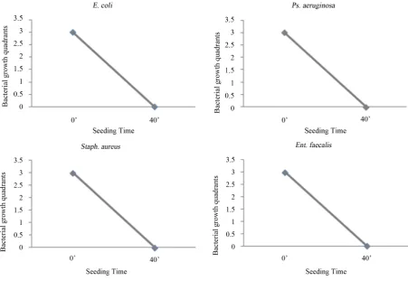
3.1. Suspension Medium: Saline
After contact with the O2/O3 mixture, we observed a
total reduction of the bacterial load and the absence of
growth for all four bacterial strains under examination (Figure 1).
3.2. Suspension Medium: Whole Blood
We performed seven experiments, adding ozone to the bacterial suspensions to verify the total dosage and inter- vals between insufflations.
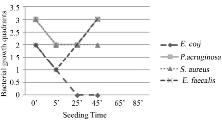
Experiment I -Ozone: 480 µg × 1; total time of me- chanical agitation: 45’.
Staph. aureus, Ps. aeruginosa,Ent. faecalis: reduction of bacterial growth at T1 (5’) and bacterial resumption at
T3 (45’), except for Staph. aureus which showed bacte-
riostatic activity.
E. coli: reduction of bacterial growth at T1 (5’) and to-
tal absence of colonies at T2 and T3. Bactericidal activity
(Figure 2).
ExperimentII - Ozone: 480 µg × 2; total time of me- chanical agitation: 45’.
Staph. aureus, Ps. aeruginosa, Ent. faecalis: Bacteri- ostatic activity.
E. coli: reduction of the bacterial growth at T1 (5’) and
total absence of colonies at T2. Bactericidal activity
(Figure 3).
ExperimentIII - Ozone: 480 µg × 3; total time of me- chanical agitation: 45’.
[image:4.595.74.526.403.714.2]Figure 2. Experiment I on whole blood: seeding T0, O3, me- chanical agitation for 5 minutes, seeding T1, mechanical agi- tation for 20 minutes, seeding T2, mechanical agitation for 20 minutes, seeding T3. T0 (0’); O3 (0’); T1 (5’); T2 (25’); T3 (45’). Ozone: 480 µg × 1. Total time of mechanical agitation: 45’.
Figure 3. Experiment II on whole blood: seeding T0, O3, me- chanical agitation for 5 minutes, seeding T1, O3, mechanical agitation for 40 minutes, seeding T2. T0 (0’); O3 (0’); T1 O3 (5’); T2 (45’). Ozone: 480 µg × 2. Total time of mechani- cal agitation: 45’.
Staph. aureus and Ent. faecalis remained in the two quadrants in each seeding, while Ps. aeruginosa showed a growth reduction at T1 (5’) and also remained in one quadrant at T2 (45’). Bacteriostatic activity.
E. coli: reduction of bacterial growth at T1 (5’) and to-
tal absence of colonies at T2 (45’). Bactericidal activity
(Figure 4).
Experiment IV - Ozone: 480 µg × 1; total time of mechanical agitation: 65’.
Staph. aureus and Ps. aeruginosa: a reduction of bac- terial growth at T2 (25’) which extended to T3 (45’).
Bacteriostatic activity.
Ent. faecalis: although a higher susceptibility to ozone than the first two bacteria was observed, these bacteria showed an evident resumption of growth at T4 (65’).
E. coli: progressive reduction of the bacterial growth from T0 to T3 (45’). Bactericidal activity (Figure 5).
Experiment V - Ozone: 480 µg × 4; total time of me- chanical agitation: 85’.
Staph. aureus and Ent. faecalis: reduction of bacterial growth for both bacteria, respectively at T2 (25’) and T1
(5’) with evident resumption at T4 (85’).
P. aeruginosa and E. coli: showed a progressive re- duction in bacterial growth from T0 to T3 (45’) which did
not increase from T3 (45’) to T4 (85’). Bacteriostatic ac-
tivity(Figure 6).
Experiment VI - Ozone: 480 µg × 2; total time of mechanical agitation: 65’.
Staph. aureus, Ps. aeruginosa and Ent. faecalis: fur- ther reduction of bacterial growth for all bacteria, respec- tively at T2 (45’), T1 (5’) and T3 (65’), and progressive
reduction of the bacterial growth in subsequent seedings. Bactericidal activity.
E. coli: evident reduction of bacterial growth at T1 (5’)
and total absence of colonies at T2 (45’) and T3 (65’).
Bactericidal activity(Figure 7).
ExperimentVII - Ozone: 480 µg × 4; total time of mechanical agitation: 80’.
The results for Staph. aureus and Ps. aeruginosa were similar: after an evident reduction at T2, the bacterial
growth resumed.
Ent. faecalis: bacterial growth was observed in three quadrants at T0 and in two quadrants at T1 (5’); the
growth returned in three quadrants at T2 (20’) and re-
mained constant until the last seeding (T4: 80’). Bacte-
riostatic activity.
E. coli: the evident reduction of bacterial growth at T1
(5’) and total absence of colonies in subsequent seedings. Bactericidal activity (Figure 8).
Theblood counts showed no significant changes. The curves of the control bacterial growthshowed a progressive increase in the bacterial growth.
4. Discussion
The study was divided into three phases, according to the suspension medium used for the bacterial cultures (saline, BHI and whole blood), to understand the interaction be- tween the ozone, bacterium and the suspension medium. Indeed, the milieu in which microbes are present deter- mines the effectiveness and outcome of the ozone treat- ment [39].
As shown in previous studies [7,11,16], we confirmed the ozone bactericidal activity on all bacterial strains when suspended in saline. Interestingly enough, ozone loses bactericidal activity with BHI suspension medium.
The results are interesting and discordant when the suspension medium is whole blood. Burgassi et al. ob- served that fresh plasma (as a biological substance with antioxidant systems), compromises ozone bactericidal activity.
Moreover, these authors suggested the incompatibility of using whole blood because it coagulates in the pres- ence of a bacterial suspension [27].
111
[image:6.595.159.431.284.432.2]Figure 4. Experiment III on whole blood: seeding T0, O3, mechanical agitation for 5 minutes, seeding T1, O3, mechanical agi- tation for 20 minutes, O3, mechanical agitation for 20 minutes, seeding T2. T0 (0’); O3 (0’); T1 O3 (5’); O3 (25’); T2 (45’). Ozone: 480 µg × 3. Total time of mechanical agitation: 45’.
Figure 5. Experiment IV on whole blood: seeding T0, O3, mechanical agitation for 5 minutes, seeding T1, mechanical agitation for 20 minutes, seeding T2, mechanical agitation for 20 minutes, seeding T3, mechanical agitation for 20 minutes, seeding T4. T0 (0’); O3 (0’); T1 (5’); T2 (25’); T3 (45’); T4 (65’). Ozone: 480 µg × 1. Total time of mechanical agitation: 65’.
Figure 6. Experiment V on whole blood: seeding T0, O3, mechanical agitation for 5 minutes, seeding T1, mechanical agitation for 20 minutes, seeding T2, O3, mechanical agitation for 20 minutes, seeding T3, O3, mechanical agitation for 20 minutes, O3, mechanical agitation for 20 minutes, T4. T0 (0’); O3 (0’); T1 (5’); T2 O3 (25’); T3 O3 (45’); O3 (65’); T4 (85’). Ozone: 480 µg × 4. Total time of mechanical agitation: 85’.
Compared to the results reported in the literature, our study on whole blood shows that the single insufflation with an O2/O3 mixture is sufficient to achieve a bacteri-
[image:6.595.158.437.485.640.2]Figure 7. Experiment VI on whole blood: seeding T0, O3, mechanical agitation for 5 minutes, seeding T1, mechanical agitation for 40 minutes, seeding T2, O3, mechanical agitation 20 minutes, seeding T3. T0 (0’); O3 (0’); T1 (5’); T2 O3 (45’); T3 (65’). Ozone: 480 µg × 2. Total time of mechanical agitation: 65’.
Figure 8. Experiment VII on whole blood: seeding T0, O3, mechanical agitation for 5 minutes, seeding T1, O3, mechanical agitation for 5 minutes, O3, mechanical agitation for 5 minutes, O3, mechanical agitation for 5 minutes, seeding T2, mechani- cal agitation for 20 minutes, seeding T3, mechanical agitation for 40 minutes, seeding T4. T0 (0’); O3 (0’); T1 (5’); O3 (10’); O3 (15’); O3 (20’); T3 (40’); T4 (80’). Ozone: 480 µg × 4. Total time of mechanical agitation: 80’.
periment VI, where we performed a second insufflation at 45 minutes after the first. We observed that the bac- terial growth typically resumed after approximately 40 minutes from the first exposure to the ozone oxidative insult, suggesting that the best results were obtained in experiment VI.
Although additional ozone insufflations prolonged ba- ctericidal activity (Staph. aureus, Ent. faecalis, Ps. aeru- ginosa), the effects of these treatments were not signifi- cantly different from those obtained in the experiments with only two insufflations.
Based on the differential results obtained on whole blood between E. coli and Ps. aeruginosa, and the failure of the experiments on broth culture, suggested that the mechanism of ozone, contrary to the literature [19,20], could also have a metabolic basis. On the other hand, changes in the size and morphology of the colonies, ob- served in all the bacterial strains after exposure to ozone, confirm the oxidative mechanism.
The bactericidal activity on E. coli, contrasts with the
increased effectiveness of ozone against Gram-positive bacteria [19]. The primary target of ozone on E. coli is the sulfhydryl group in the bacterial membrane [40]. Thus, we confirmed the correlation between ozone bac- tericidal activity and membrane permeability, which is specific for each microorganism [39].
The results of our study on whole blood represents an important confirmation of the ozone bactericidal activity in the topical treatment of wounds, and the daily persis- tence of topical ozone preparations kills even the most resistant bacteria [25,41].
The in vitro results were not consistent with the ob- servations of AHT-O3in vivo, except for the portion of
blood directly exposed to ozone. In fact, these results are consistent with those of Burgassi et al. [27], showing that the AHT enriched with ozone, even at highest ozone con- centration (80 µg/ml), is not able to oxidize and destroy circulating bacteria in the blood.
[image:7.595.158.431.284.435.2]113
volves exclusively its direct action in vitro. While in vivo, AHT enriched with ozone induces a series of effects that might also be indirectly useful in the treatment of infec- tions, such as: immune modulation [42-48], antioxidant systems activation [49-52] and improvement of the mi-crocirculation [53-56].
To clarify the mechanism of ozone, it would be useful to examine its affects on other Enterobacteriaceae (sus- pension medium: whole blood), such as Serratia and Kle- bsiella, and Aspergillus. Indeed, Aspergillus produces large colonies whose variations in size or color might be an important index for metabolic alterations.
In conclusion, we confirmed the bactericidal efficacy of topical ozone preparations, according to the evidence of antibody-catalyzed ozone formation in bacterial kill- ing [57], and suggest the need for further evaluations of the therapeutic potential of AHT-O3.
5. Conclusion
Further in vitro investigation on whole blood might re- veal the synergic action between traditional antibiotics and ozone treatment, and might provide the basis for de- veloping successive studies in vivo in patients affected through multidrug resistant nosocomial infections.
Conflict of Interest
The authors declare that they have no conflict of interest, neither commercial nor financial, in any of the products described in this article.
REFERENCES
[1] Safe Practices for Food Processes, Chapter V, “Methods to Reduce/Eliminate Pathogens from Produce and Fresh- Cut Produce,” US Food and Drug Administration. www.fda.gov/food/foodscienceresearch/safepracticesforf oodprocesses/ucm091363.htm
[2] D. S. Boyce, O. J. Sproul and C. E. Buck, “The Effect of Bentonite Clay on Ozone Disinfection of Bacteria and Viruses in Water,” Water Research, Vol. 15, No. 6, 1981, pp. 759-767.
http://dx.doi.org/10.1016/0043-1354(81)90169-X
[3] W. H. Glaze, “Drinking Water Treatment with Ozone,”
Environmental Science & Technology, Vol. 21, No. 3, 1987, pp. 224-230.
http://dx.doi.org/10.1021/es00157a001
[4] A. Joss, H. Siegrist and T. A. Ternes, “Are We about to Upgrade Wastewater Treatment for Removing Organic Micropollutans?” Water Science and Technology, Vol. 57, No. 2, 2008, pp. 251-255.
http://dx.doi.org/10.2166/wst.2008.825
[5] T. Glauner, P. Waldmann, F. H. Frimmel and C. Zwiener, “Swimming Pool Water-Fractionation and Genotoxicolo- gical Characterization of Organic Constituents,” Water Research, Vol. 39, No. 18, 2005, pp. 4494-4502.
http://dx.doi.org/10.1016/j.watres.2005.09.005
[6] T. Masaoka, Y. Kubota, S. Namiuchi, T. Takubo, T. Ueda, H. Shibata, H. Nakamura, K. Yoshitake, H. Doi and T. Kamiki, “Ozone Decontamination of Bioclean Rooms,”
Applied and Environmental Microbiology, Vol. 43, No. 3, 1982, pp. 509-513.
[7] A. Baysan, R. A. Whiley and E. Lynch, “Antimicrobial Effect of a Novel Ozone-Generating Device on Micro-Or- ganisms Associated with Primary Root Carious Lesions
in Vitro,” Caries Research, Vol. 34, No. 6, 2000, pp. 498- 501. http://dx.doi.org/10.1159/000016630
[8] D. Bialoszewski, E. Bocian, B. Bukowska, M. Czajkow- ska, L. B. Sokół and S. Tyski, “Antimicrobial Activity of Ozonated Water,” Medical Science Monitor, Vol. 16, No. 9, 2010, pp. 71-75.
[9] A. Castillo, P. Galindo Moreno, G. Avila, M. Valderrama, J. Liébana and P. Baca, “In Vitro Reduction of mutans streptococci by Means of Ozone Gas Application,” Quin-tessence International, Vol. 39, No. 10, 2008, pp. 827- 831.
[10] T. G. Fragell, W. Dietz, P. Lingström, F. Steiniger and J. G. Norén, “Effect of Ozone Treatment on Different Cari- ogenic Microorganisms in Vitro,” Swedish Dental Jour- nal, Vol. 32, No. 3, 2008, pp. 139-147.
[11] R. S. Hems, K. Gulabivala, Y. L. Ng, D. Ready and D. A. Spratt, “An in Vitro Evaluation of the Ability of Ozone to Kill a Strain of Enterococcus faecalis,” International En- dodontic Journal, Vol. 38, No. 1, 2005, pp. 22-29. http://dx.doi.org/10.1111/j.1365-2591.2004.00891.x
[12] K. C. Huth, M. Quirling, S. Maier, K. Kamereck, M. Al- khayer, E. Paschos, U. Welsch, T. Miethke, K. Brand and R. Hickel, “Effectiveness of Ozone against Endodontopa- thogenic Microorganisms in a Root Canal Biofilm Model,”
International Endodontic Journal, Vol. 42, No. 1, pp. 3- 13. http://dx.doi.org/10.1111/j.1365-2591.2008.01460.x
[13] G. M. Knight, J. M. McIntyre and P. S. Zilm, “The Inabi- lity of Streptococcus mutans and Lactobacillus acidophi- lus to Form a Biofilm in Vitro on Dentine Pretreated with Ozone,” Australian Dental Journal, Vol. 53, No. 4, 2008, pp. 349-353.
http://dx.doi.org/10.1111/j.1834-7819.2008.00077.x
[14] I. R. Komanapalli and B. H. S. Lau, “Ozone-Induced Da- mage of Escherichia coli K-12,” Applied Microbiology and Biotechnology, Vol. 46, No. 5-6, 1996, pp. 610-614. http://dx.doi.org/10.1007/s002530050869
[15] P. Müller, B. Guggenheim and P. R. Schmidlin, “Efficacy of Gasiform Ozone and Photodynamic Therapy on a Mul- tispecies Oral Biofilm in Vitro,” European Journal of Oral Sciences, Vol. 115, No. 1, 2007, pp. 77-80.
http://dx.doi.org/10.1111/j.1600-0722.2007.00418.x
[16] M. Nagayoshi, T. Fukuizumi, C. Kitamura, J. Yano, M. Terashita and T. Nishihara, “Efficacy of Ozone on Sur- vival and Permeability of Oral Microorganisms,” Oral Microbiology and Immunology, Vol. 19, No. 4, 2004, pp. 240-246.
http://dx.doi.org/10.1111/j.1399-302X.2004.00146.x
Using Ozone,” Journal of Medical and Dental Sciences, Vol. 45, No. 2, 1998, pp. 135-139.
[18] R. Stoll, L. Venne, A. J. Momeni, R. Mutters and V. Sta- chniss, “The Disinfecting Effect of Ozonized Oxygen in an Infected Root Canal: An in Vitro Study,” Quintessence International, Vol. 39, No. 3, 2008, pp. 231-236.
[19] B. Thanomsub, V. Anupunpisit, S. Chanphetch, T. Watcha- rachaipong, R. Poonkhum and C. Srisukonth, “Effects of Ozone Treatment on Cell Growth and Ultrastructural Chang- es in Bacteria,” The Journal of General and Applied Mi- crobiology, Vol. 48, No. 4, 2002, pp. 193-199.
http://dx.doi.org/10.2323/jgam.48.193
[20] D. E. Cortezzo, K. Koziol Dube, B. Setlow and P. Setlow, “Treatment with Oxidizing Agents Damages the Inner Membrane of Spores of Bacillus subtilis and Sensitizes Spores to Subsequent Stress,” Journal of Applied Micro- biology, Vol. 97, No. 4, 2004, pp. 838-852.
http://dx.doi.org/10.1111/j.1365-2672.2004.02370.x
[21] M. Bette, R. M. Nusing, R. Mutters, Z. B. Zamora, S. Me- nendez and S. Schulz, “Efficiency of Tazobactam/Pipera- cillin in Lethal Peritonitis Is Enhanced after Preconditio- ning of Rats with O3/O2-Pneumoperitoneum,” Shock, Vol. 25, No. 1, 2006, pp. 23-29.
http://dx.doi.org/10.1097/01.shk.0000187983.56030.dd
[22] R. Polgnano, P. L. Vannucchi and R. Degli Innocenti, “Ef- ficacia Antisettica Dell’Acqua Ozonizzata su Germi Iso- lati da Lesioni Croniche Della Cute,” Proceedings Inter- national Medical Ozone Society (IMOS) Congress, Ossi- geno Ozono Terapia: Dall’Empirismo alla Metodologia Scientifica, Siena, 2000, pp. 49-50.
[23] Z. Z. Rodriguez, D. Guanche, R. G. Álvarez, F. H. Rosal- es, Y. Alonso and S. Schulz, “Preconditioning with Ozone/ Oxygen Mixture Induces Reversion of Some Indicators of Oxidative Stress and Prevents Organic Damage in Rats with Fecal Peritonitis,” Inflammation Research, Vol. 58, No. 7, 2009, pp. 371-375.
http://dx.doi.org/10.1007/s00011-009-0001-2
[24] S. Schulz, Z. Z. Rodriguez, R. Mutters, S. Menendez and M. Bette, “Repetitive Pneumoperitoneum with Ozonized Oxigen as a Preventive in Lethal Polymicrobial Sepsis in Rats,” European Surgical Research, Vol. 35, No. 1, 2003, pp. 26-34. http://dx.doi.org/10.1159/000067032
[25] H. Steinhart, S. Schulz and R. Mutter, “Evaluation of Ozonated Oxygen in an Experimental Animal Model of Osteomyelitis as a Further Treatment Option for Skull- Base Osteomyelitis,” European Archives of Oto-Rhino- Laryngology, Vol. 256, No. 3, 1999, pp. 153-157. http://dx.doi.org/10.1007/s004050050130
[26] Z. B. Zamora, A. B. Orlay, Y. L´opez, R. Delgado, R. González, S. Menéndez, F. Hernández and S. Schulz, “Ef- fects of Ozone Oxidative Preconditioning on TNF-α Re- lease and Antioxidant-Prooxidant Intracellular Balance in Mice during Endotoxic Shock,” Mediators of Inflamma- tion, Vol. 2005, No. 1, 2005, pp. 16-22.
http://dx.doi.org/10.1155/MI.2005.16
[27] S. Burgassi, I. Zanardi, V. Travagli, E. Montomoli and V. Bocci, “How Much Ozone Bactericidal Activity Is Com- promised by Plasma Components?” Journal of Applied Microbiology, Vol. 106, No. 5, 2009, pp. 1715-1721.
http://dx.doi.org/10.1111/j.1365-2672.2008.04141.x
[28] S. K. Fridkin, J. C. Hageman, M. Morrison, L. T. Sanza, K. Como Sabetti, J. A. Jernigan, K. Harriman, L. H. Harri- son for the Active Bacterial Core Surveillance Program of the Emerging Infection Program Network, “Methicillin- Resistant Staphylococcus aureus Disease in Three Com- munities,” New England Journal of Medicine, Vol. 352, No. 14, 2005, pp. 1436-1444.
http://dx.doi.org/10.1056/NEJMoa043252
[29] R. M. Klevens, M. A. Morrison, J. Nadle, S. Petit, K. Ger- shman, S. Ray, L. H. Harrison and R. Lynfield, “Active Bacterial Core Surveillance (ABCs) MRSA Investigators, “Invasive Methicillin-Resistant Staphylococcus aureus In- fections in the United States,” JAMA, Vol. 298, No. 15, 2007, pp. 1763-1771.
http://dx.doi.org/10.1001/jama.298.15.1763
[30] G. J. Moran, A. Krishnadasan, R. J. Gorwitz, G. E. Foshe- im, L. K. McDougal, R. B. Carey, D. A. Talan and Emer- gency ID Net Study Group, “Methicillin-Resistant S. au- reus Infections among Patients in the Emergency Depart- ment,” New England Journal of Medicine, Vol. 355, No. 7, 2006, pp. 666-674.
http://dx.doi.org/10.1056/NEJMoa055356
[31] K. J. Popovich, R. A. Weinstein and B. Hota, “Are Com- munity-Associated Methicillin-Resistant Staphylococcus aureus (MRSA) Strains Replacing Traditional Nosocomi- al MRSA Strains?” Clinical Infectious Diseases, Vol. 46, No. 6, 2008, pp. 787-794.
http://dx.doi.org/10.1086/528716
[32] D. A. Robinson, A. M. Kearns, A. Holmes, D. Morrison, H. Grundmann, G. Edwards, F. G. O’Brien and F. C. Te- nover, “Re-Emergence of Early Pandemic Staphylococcus aureus as a Community-Acquired Meticillin-Resistant Clone,” Lancet, Vol. 365, No. 9466, 2005, pp. 1256- 1258. http://dx.doi.org/10.1016/S0140-6736(05)74814-5
[33] J. Vallés and R. Ferrer, “Bloodstream Infections in the ICU,” Infectious Disease of North America, Vol. 23, No. 3, 2009, pp. 557-569.
http://dx.doi.org/10.1016/j.idc.2009.04.005
[34] V. Bocci and L. Paulesu, “Studies on the Biological Ef- fects of Ozone: 1. Induction of Interferon Gamma on Hu- man Leucocytes,” Haematologica, Vol. 75, No. 6, 1990, pp. 510-515.
[35] V. Bocci, “Ozone as Bioregulator: Pharmacology and To- xicology of Ozone Therapy Today,” Journal of Biological Regulators & Homeostatic Agents, Vol. 10, No. 2-3, 1996, pp. 31-53.
[36] V. Bocci, “Can Ozone Therapy Be Performed If the Bio- chemistry of the Process Cannot Be Controlled?” Arc-
hives of Medical Research, Vol. 38, No. 5, 2007, pp. 584- 585. http://dx.doi.org/10.1016/j.arcmed.2007.03.005
[37] N. Di Paolo, V. Bocci, G. Marosi, E. Borrelli, A. Bravi, A. Bruci, C. Aldinucci and L. Capotondo, “Extracorporeal Blood Oxygenation and Ozonization in Man. Preliminary Report,” The International Journal of Artificial Organs, Vol. 23, No. 2, 2000, pp. 131-141.
115
Ambulatory Autohaemotherapy,” Biotherapy, Vol. 7, No. 2, 1994, pp. 83-90.
http://dx.doi.org/10.1007/BF01877731
[39] I. R. Komanapalli and B. H. S. Lau, “Inactivation of Bac- teriophage k, Escherichia coli and Candida albicans by Ozone,” Applied Microbiology and Biotechnology, Vol. 49, No. 6, 1998, pp. 766-769.
http://dx.doi.org/10.1007/s002530051244
[40] I. R. Komanapalli, J. B. Mudd and B. H. Lau, “Effect of Ozone on Metabolic Activities of Escherichia coli K-12,”
Toxicology Letters, Vol. 90, No. 1, 1997, pp. 61-66. http://dx.doi.org/10.1016/S0378-4274(96)03830-1
[41] G. Valacchi, V. Fortino and V. Bocci, “The Dual Action of Ozone on the Skin,” British Journal of Dermatology, Vol. 153, No. 6, 2005, pp. 1096-1100.
http://dx.doi.org/10.1111/j.1365-2133.2005.06939.x
[42] V. Bocci, E. Luzzi, F. Corradeschi, L. Paulesu, R. Rossi, E. Cardaioli and P. Di Simplicio, “Studies on the Biolog- ical Effects of Ozone 4: Cytokine Production and Gluta- thione Levels in Human Erythrocytes,” Journal of Bio-
logical Regulators and Homeostatic Agents, Vol. 7, No. 4, 1993, pp. 133-138.
[43] V. Bocci, E. Luzzi, F. Corradeschi and S. Silvestri, “Stu- dies of the Biological Effects of Ozone 6: Production of Transforming Growth Factor b1 by Human Blood after Ozone Treatment,” Journal of Biological Regulators and Homeostatic Agents, Vol. 8, No. 4, 1994, pp. 108-112.
[44] W. Doroszkiewicz, I. Sikorska and S. Jankowski, “Stu- dies on the Influence of Ozone on Complement-Mediated Killing of Bacteria,” FEMS Immunology & Medical Mi-
crobiology, Vol. 9, No. 4, 1994, pp. 281-285. http://dx.doi.org/10.1111/j.1574-695X.1994.tb00363.x
[45] L. Paulesu, E. Luzzi and V. Bocci, “Studies on the Biolo- gical Effects of Ozone 2: Induction of Tumor Necrosis Factor on Human Leucocytes,” Lymphokine and Cytokine Research, Vol. 10, No. 5, 1991, pp. 409-412.
[46] E. Riva Sanseverino, “Aspetti Immunologici Dell’Ozono- terapia,” Rivista Italiana di Omotossicologia, pp. 19-24.
[47] P. D. Thomson, G. O. Till and D. J. Smith, “Modulation of IgM Antibody Formation by Lipid Peroxidation Prod- ucts from Burn Plasma,” JAMA Surgery, Vol. 126, No. 8, 1991, pp. 973-976.
http://dx.doi.org/10.1001/archsurg.1991.01410320055006
[48] V. Bocci, “A Reasonable Approach for the Treatment of HIV Infection in the Early Phase with Ozone Therapy (Autohaemotherapy). How Inflammatory Cytokines May Have a Therapeutic Role,” Mediators of Inflammation,
Vol. 3, No. 5, 1994, pp. 315-321.
http://dx.doi.org/10.1155/S0962935194000438
[49] M. E. Bégin, “Fatty Acids, Lipid Peroxidation and Dis- eases,” Proceedings of the Nutrition Society, Vol. 49, No. 2, 1990, pp. 261-267.
http://dx.doi.org/10.1079/PNS19900029
[50] V. Bocci, G. Valacchi, F. Corradeschi, C. Aldinucci, S. Silvestri, E. Paccagnini and R. Gerli, “Studies on the Bi- ological Effects of Ozone 7: Generation of Reactive Oxygen Species (ROS) after Exposure of Human Blood to Ozone,” Journal of Biological Regulators and Homeo-
static Agents, Vol. 12, No. 3, 1998, pp. 67-75.
[51] R. D. Buckley, J. D. Hackney, K. Clark and C. Posin, “Ozone and Human Blood,” Archives of Environmental Health, Vol. 30, No. 1, 1975, pp. 40-43.
http://dx.doi.org/10.1080/00039896.1975.10666631
[52] B. D. Goldstein and O. J. Balchum, “Effect of Ozone on Lipid Peroxidation in the Red Blood Cell,” Experimental Biology and Medicine, Vol. 126, No. 2, 1967, pp. 356- 358. http://dx.doi.org/10.3181/00379727-126-32444
[53] L. Coppola and G. Verrazzo, “Oxygen-Ozone Therapy and Haemorheological Parameters in Peripheral Chronic Arterial Disease,” Trombosi e Aterosclerosi, Vol. 3, 1992, pp. 83-89.
[54] C. Di Filippo, M. Luongo, R. Marfella, F. Ferraraccio, B. Lettieri, A. Capuano, F. Rossi and M. D’Amico, “Oxy- gen/Ozone Protects the Heart from Acute Myocardial In- farction through Local Increase of eNOS Activity and Endothelial Progenitor Cells Recruitment,” Naunyn-Schmi-
edeberg’s Archives of Pharmacology, Vol. 382, No. 3, 2010, pp. 287-291.
http://dx.doi.org/10.1007/s00210-010-0545-2
[55] B. A. Freeman and J. B. Mud, “Reaction of Ozone with Sulphydryls of Human Erythrocytes,” Archives of Bio-
chemistry and Biophysics, Vol. 208, No. 1, 1981, pp. 212- 220. http://dx.doi.org/10.1016/0003-9861(81)90142-9
[56] A. Van Der Vliet, C. A. O’Neil, J. P. Eiserich and C. E. Cross, “Oxydative Damage to Extracellular Fluids by Ozone and Possible Protective Effects of Thiols,” Archives of Bio-
chemistry and Biophysics, Vol. 321, No. 1, 1995, pp. 43- 50. http://dx.doi.org/10.1006/abbi.1995.1366
[57] P. Wentworth, J. E. McDunn, A. D. Wentworth, C. Ta- keuchi, J. Nieva, T. Jones and C. Bautista, “Evidence for Antibody-Catalyzed Ozone Formation in Bacterial Kill- ing and Inflammation,” Science, Vol. 298, No. 5601, 2002, pp. 2195-2199.