Integrins in invasive growth
Cord Brakebusch, … , Takao Sakai, Reinhard Fässler
J Clin Invest.
2002;
109(8)
:999-1006.
https://doi.org/10.1172/JCI15468
.
Interactions between tumor cells and the ECM strongly influence tumor development,
affecting cell proliferation and survival, as well as the ability to migrate beyond the original
location into other tissues to form metastases. Many of these interactions are mediated by
integrins, a ubiquitously expressed family of adhesion receptors. Integrins are essential for
cell attachment and control cell migration, cell cycle progression, and programmed cell
death, responses that they regulate in synergy with other signal transduction pathways. This
large group of transmembrane proteins is formed from 18
a
and 8
b
subunits, which
dimerize to yield at least 24 different integrin heterodimers, each with distinct ligand binding
and signaling properties. With their extracellular domain, integrins can bind to different ECM
molecules, such as collagens and laminins, or to cellular receptors, such as VCAM-1. Their
intracellular domains connect directly or indirectly to the actin cytoskeleton, thus linking the
cytoskeleton to the ECM. Integrins also serve as bidirectional signaling receptors, inducing
changes in protein activities or gene expression in response to ligand binding, while also
modulating adhesive affinity on the cell surface in response to changes in cellular
physiology. Here, we describe how integrins affect migration, proliferation, and survival of
both transformed and normal cells, and we discuss how they modulate invasive growth in
vivo. Throughout, we stress that many of these functions […]
Perspective
Find the latest version:
PERSPECTIVE SERIES
Paolo Comoglio, Series Editor
Invasive growth
Interactions between tumor cells and the ECM strong-ly influence tumor development, affecting cell prolif-eration and survival, as well as the ability to migrate beyond the original location into other tissues to form metastases. Many of these interactions are mediated by integrins, a ubiquitously expressed family of adhesion receptors. Integrins are essential for cell attachment and control cell migration, cell cycle progression, and programmed cell death, responses that they regulate in synergy with other signal transduction pathways.
This large group of transmembrane proteins is formed from 18 αand 8 βsubunits, which dimerize to yield at least 24 different integrin heterodimers, each with distinct ligand binding and signaling properties. With their extracellular domain, integrins can bind to different ECM molecules, such as collagens and laminins, or to cellular receptors, such as VCAM-1. Their intracellular domains connect directly or indi-rectly to the actin cytoskeleton, thus linking the cytoskeleton to the ECM. Integrins also serve as bidi-rectional signaling receptors, inducing changes in pro-tein activities or gene expression in response to ligand binding, while also modulating adhesive affinity on the cell surface in response to changes in cellular physiolo-gy. Here, we describe how integrins affect migration, proliferation, and survival of both transformed and normal cells, and we discuss how they modulate inva-sive growth in vivo. Throughout, we stress that many of these functions are restricted to particular cell types and may be altered upon transformation.
Integrin avidity and affinity changes in cell migration
Cell migration is essential not only for tissue infiltra-tion and the formainfiltra-tion of metastases, but also for non-pathological processes, such as angiogenesis and leukocyte extravasation. In order to migrate, a cell has to pass through a sequence of distinct processes. Migration is initiated by cell polarization and the for-mation of membrane protrusions at the leading edge. Integrins fix cellular protrusions to the ECM, interact with the actin cytoskeleton, and trigger the association of many different signaling molecules at the so-called
focal contacts. Thereafter, integrin signals stimulate cell contraction, which allows the movement of the cell body on the adhesive contacts. Finally, the rear of the cell detaches from the substratum by inactivation of the integrins and disassembly of the adhesion com-plexes. Integrins further facilitate cell movement through the tissue by activating ECM-degrading enzymes. Although these basic mechanisms are con-sidered to be similar among the various types of migra-tory cells, there are also clear distinctions, since, for example, fibroblasts are about 3–20 times more adhe-sive than nonactivated leukocytes, move 10–60 times more slowly, and have a different cytoskeletal organi-zation. These differences in the migration mechanism might explain at least some of the known cell-specific effects of integrins on cell migration.
Integrin-mediated cell attachment depends not just on the expression of these receptors in a given cell type, but also on their affinity for various ligands and on their lateral mobility within the plasma mem-brane, which allows the formation of high-avidity clusters. Integrins adopt low- and high-affinity con-formations, which can be distinguished by their binding to soluble ligands or by conformation-spe-cific antibodies. High-affinity binding, which is important for the firm attachment of the leading edge to the ECM, can result from the binding of reg-ulatory intracellular molecules to the cytoplasmic domains of the αand βsubunits. Overexpression in osteosarcoma cells or normal human fibroblasts of chimeric proteins consisting of the β1, β3, or β5 intracellular domains, fused to the extracellular and transmembrane part of the IL-2 receptor, signifi-cantly reduces the affinity of the endogenous β1 inte-grins (1). This effect is most likely caused by seques-tration of intracellular molecules that would otherwise bind the cytoplasmic domain of the inte-grin βsubunits and alter the integrins’ extracellular conformation. Such an effect, called transdominant inhibition, has also been reported for α2 integrin cytoplasmic domains (2). More than 20 proteins are currently known to bind intracellular domains of integrin subunits (3), but it is not known which of
Integrins in invasive growth
Cord Brakebusch,
1,2Daniel Bouvard,
1Fabio Stanchi,
1Takao Sakai,
1and Reinhard Fässler
1,21Lund University Hospital, Department of Experimental Pathology, Lund, Sweden 2Max Planck Institute for Biochemistry, Martinsried, Germany
Address correspondence to: Cord Brakebusch, Max Planck Institute for Biochemistry, Department of Molecular Medicine, Am Klopferspitz 18a, 82152 Martinsried, Germany. Phone: 49-89-8578-2466; Fax: 49-89-8578-3777; E-mail: [email protected].
them are involved in the modulation of integrin affinity. Potential candidates for such regulation are ICAP-1 and TAP-20, which decrease, and β3-endonexin, which increases integrin-mediated adhesion in cells overexpressing these molecules. Intracellular signaling mechanisms, for example phosphorylation of ICAP-1 by CaMKII, are suggest-ed to modulate the binding of these molecules to integrin and thereby the affinity state.
Cell attachment can be induced not only by affinity regulation, but also by clustering of integrins, which leads to increased adhesive avidity. Such clusters are present in focal adhesions, readily detectable cell-matrix contacts that have been extensively studied in cultured fibroblasts. Increased lateral mobility of inte-grins in the membrane might also be important for efficient ligand binding. In leukocytes, even prior to lig-and binding, integrins seem to be associated with the actin network, which constrains integrin mobility and ligand binding. Releasing this contact by proteolytic digestion of the integrin connections with the cytoskeleton results in a rapid increase in lateral mobil-ity of integrins, which increases the chance of ligand encounters and facilitates integrin aggregation into high-avidity clusters (4).
Signaling pathways regulating integrin affinity
Growth factor and chemokine signaling can also mod-ulate the affinity and avidity of integrins. In particular, upregulation of integrin activity by growth factors often depends on phosphatidylinositol 3-kinase (PI3-K) acti-vation. Thus, in mast cells, activation of PI3-K by FcεRI, c-kit, or PDGF-R increases the affinity of α5β1 integrin. In metastatic breast cancer cells, increased cell adhesion and migration upon stimulation of EGF-R or erbB3 are also dependent on PI3-K. Integrin avidity is upregulat-ed in carcinoma cells by treatment with HGF (see Danilkovitch-Miagkova and Zbar, this Perspective series, ref. 5), but not with EGF. This increased integrin avidity is dependent on PI3-K and promotes invasive growth of these cells, suggesting an important role of integrin avidity regulation in metastasis (6).
Signaling initiated by chemokine receptors can induce a rapid and transient upregulation of integrin affinity, which is important for the tight adhesion of leukocytes to the endothelium during their extravasa-tion into inflamed tissues. At least in lymphocytes, this activation is independent of PI3-K and involves activa-tion of RhoA (4). Remarkably, chemokines can inde-pendently regulate the affinity and avidity of different integrins within the same cell. In eosinophils, the chemokines RANTES and monocyte chemoattractant protein–3 induce a transient upregulation of α4β1 avidity and a long-lasting affinity increase of αMβ2 (Mac-1). Chemokines such as secondary lymphoid tis-sue cytokine, EBI1-ligand chemokine, and stroma cell-derived factor 1αstimulate integrin motility in lym-phocytes through the coordinated action of cytosolic proteases and PI3-K. In contrast, PMA-stimulated inte-grin mobility is dependent on proteases, but inde-pendent of PI3-K (4).
The Ras and Rho GTPases can also regulate integrin affinity, although their effects are cell type– and inte-grin-specific. In Chinese hamster ovary (CHO) cells, for example, H-Ras inhibits activation of β1 and β3 inte-grins, while in a pro-B cell line it increases αLβ2 (LFA-1) activity in a PI3-K–dependent manner (7). R-Ras pro-motes integrin activation in myeloid cells but has no effect in a lymphoid cell line (7). The integrin-specifici-ty of Ras effects is also nicely demonstrated in T47D cells, where R-Ras promotes migration mediated by α2β1 but not by α5β1 (2). Ras signaling can affect many signaling cascades, including the activation of Erk and PI3-K. The integrin activating functions of both H-Ras and R-Ras require PI3-K activation.
Recently, Rap1 was shown to activate integrins on lymphoid and endothelial cells by inducing a high-affinity conformation (7, 8). Overexpression of consti-tutively active Rac1 in lymphoid cells increases integrin affinity (7). In mammary epithelial cells, however, nei-ther Rac1 nor Cdc42 induces obvious changes in inte-grin avidity or affinity, although they promote inteinte-grin- integrin-mediated motility and invasiveness through PI3-K.
Several reports have demonstrated cross-talk between integrins, in which signaling by one integrin influences the affinity or avidity of others. For example, stimula-tion of αIIbβ3 signaling has been shown to downregu-late α2β1- and α5β1-mediated adhesion.
A few reports have also shown that direct interaction of secreted molecules with the extracellular domain of integrins can influence integrin affinity. Galectin-8, for example, a secreted galactoside-binding protein, binds β1 integrins and induces a conformational change that decreases their affinity for other ligands (9).
Detachment of the cell rear
like EGF promote cell migration by reducing cell adhe-sion and the number of focal adheadhe-sions through the same mechanism. Cleavage products of FAK or the focal adhesion–associated docking protein HEF-1 can induce cell rounding, which is important for cell division and might also be involved in migration (13).
All of the detachment mechanisms discussed above appear to be sensitive to Ca2+transients. Migrating
fibroblasts have increased Ca2+levels in the rear,
per-haps as a consequence of stretch-activated calcium channels opening in response to cell contraction at the trailing edge. In neutrophils, Ca2+-induced activation
of calcineurin disrupts interactions between αvβ3 and the cytoskeleton, allowing detachment of the rear. Ca2+
transients are also essential to release α5β1-mediated attachment of neutrophils to fibronectin (14). This latter mechanism, however, is independent of cal-cineurin and may involve modulation of integrin affinity or of cellular contractility. Ca2+influx may also
lead to the activation of µ-calpain, which is proposed to sever the connection between the integrin and the cytoskeleton proteolytically.
During rear detachment, unbound integrins accu-mulate at the trailing edge. Endocytosis of these detached integrins and their transport to the leading edge are thought to be needed to prevent integrin depletion at the front (14). Such endocytotic transport of green-fluorescent protein–tagged α5 integrin sub-units has indeed been observed in migrating fibroblasts (11). Integrin molecules observed in endocytotic vesi-cles are not attached to α-actinin or vinculin, as they are in the focal contacts prior to endocytosis. In mono-cyte chemoattractant protein–7 breast cancer cells, pro-tein kinase Cαparticipates in the recycling of the acti-vated pool of β1 integrins (15).
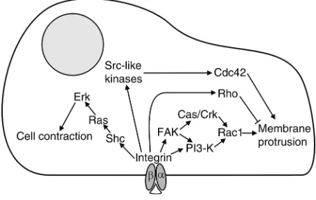
Integrin signaling in migration
Integrin signaling promotes cell migration by inducing changes in the cytoskeletal organization and by increas-ing cellular contractility (Figure 1). Activation of FAK plays a prominent role among the different integrin signaling pathways which affect migration. FAK is a non–receptor tyrosine kinase that is indirectly and per-haps also directly associated with integrins in focal con-tacts. Ligand binding to integrins leads to phosphory-lation of FAK on at least seven different tyrosines in vivo, allowing the interaction of FAK with Src, Grb2, and PI3-K, and also promoting the phosphorylation of associated proteins, such as Cas and paxillin. FAK therefore functions as an important adaptor molecule that recruits various other signaling molecules to focal contacts. FAK is also a target for tyrosine phosphory-lation induced by growth factor receptors. For this rea-son, FAK serves as an important integration point of growth factor and integrin signaling with respect to cell migration (16). Experiments in which FAK mutants are expressed in otherwise FAK-deficient fibroblasts have shown that FAK kinase activity and FAK autophos-phorylation of tyrosine 397 are required for integrin-stimulated cell migration, whereas FAK’s association with paxillin is dispensable.
FAK can promote cell movement by activating PI3-K and regulators of cytoskeletal dynamics like Rac1. Autophosphorylation of FAK on tyrosine 397 allows this molecule to bind and activate PI3-K, which can then influence integrin affinity and avidity, as dis-cussed above. In addition, PI3-K contributes to the activation of Rac1, which mediates membrane ruf-fling. In adherent but not in suspended cells, activat-ed Rac interacts with and activates p21-activatactivat-ed kinase (PAK), which can then stimulate migration by increasing the turnover of focal adhesions (17). Tyro-sine-phosphorylated FAK also promotes Rac activa-tion via an alternative pathway involving the adaptor proteins p130Casand Crk (18, 19).
Integrin signaling also stimulates Cdc42 and RhoA activity and facilitates the interaction of these Rho-family GTPases with their downstream targets. These GTPases cooperate and influence each other’s activity in a complex, cell-specific manner. In astrocytes, inte-grin-dependent stimulation of Cdc42 via Src-like kinas-es is crucial for cell polarity, protrusion formation, and migration (20). In CHO cells, α5β1-mediated activa-tion of Rac1 and Cdc42 is maximal already at interme-diate fibronectin levels, whereas Rho activity continues to increase with increasing fibronectin levels (21). Since Rac1 and Cdc42 promote, but RhoA inhibits, mem-brane protrusion, increased integrin-mediated Rho GTPase activation halts migration in these cells prefer-entially at high fibronectin concentrations.
[image:4.576.307.535.526.670.2]Recently, Chen et al. (22) found that the cytoplasmic tyrosine kinase Etk, which is highly expressed in migra-tory cells including endothelial cells and metastatic cell lines, is activated by integrin signaling. Integrin-medi-ated activation and autophosphorylation of FAK lead to interaction of FAK with the pleckstrin domain of Etk, and subsequent phosphorylation and activation of Etk. Etk may play an important role in migration, since changes in its expression directly correlate to changes in migration. Downstream targets of Etk mediating such a promigratory role are not yet known.
Figure 1
Integrin-triggered activation of Erk can contribute to migration via phosphorylation of MLCK, leading to increased phosphorylation of MLC and cell contraction (23). Erk activation furthermore leads to changes in gene expression, which have been suggested to promote migration. After integrin engagement or stimulation of v-Src, active Erk can also be targeted to newly forming focal adhesions, where it may phosphorylate focal adhe-sion proteins and influence cytoskeletal organization and migration (24). Erk can be activated via the interac-tion of FAK with Grb2. This pathway, however, appar-ently does not play a major role in integrin-mediated migration, since mutation of the Grb2-binding tyrosine 925 of FAK does not affect cell migration (18). Certain integrins can induce Erk activation independently of FAK via the Shc/Grb2/Sos/Ras cascade. α1β1, α5β1, and αvβ3 integrins activate this alternative pathway via an interaction of their αsubunit with the membrane-associated protein caveolin, which helps define a dis-tinctive membrane subdomain in which Shc becomes phosphorylated by the Src-like kinase Fyn (25).
Integrins and matrix metalloproteinases
Cell migration in vivo is often facilitated by a partial destruction of the surrounding ECM. Such degrada-tion is catalyzed by matrix metalloproteinases (MMPs), a family of more than 20 substrate-specific zinc-dependent endoproteases, most of them soluble, but some of them transmembrane proteins. MMP activity, which is controlled by regulated expression, by prote-olytic activation of inactive precursors (zymogens), and by the expression of a family of inhibitors, is crucial for tumor invasion, metastasis, and angiogenesis. Integrins can regulate the expression and activation of MMPs and can guide them to their targets by simultaneous binding of MMPs and ECM molecules.
Various integrin-induced signaling pathways are involved in the control of MMP expression. In collagen gels, for example, α1β1- and α2β1-dependent expres-sion of MMP-13 requires p38 activity and is inhibited by Erk. In other settings, overexpression of the integrin-linked kinase (ILK) can result in AP-1–dependent expression of MMP-9 and an invasive phenotype. Final-ly, stimulation of the α3β1-tetraspanin complex on mammary epithelial cells upregulates the expression of MMP-2 and increases their invasive potential. Some of these transcriptional effects might be mediated by the transcription factor Cas-interacting zinc finger protein (CIZ), which can shuttle between the focal adhesions and the nucleus (26). CIZ activates the expression of several MMPs, and its effect is enhanced in the presence of Cas, which mediates some of the effects of FAK as mentioned above. However, integrins can also reduce the expression of certain MMPs. Deletion of the mouse gene for α1 integrin, for example, leads to a marked increase of MMP-7 and MMP-9 in the serum (27). MMP activation by integrins can also occur indirectly, through the increased expression of zymogen-activat-ing proteases. Signalzymogen-activat-ing by β1 integrin on ovarian car-cinoma cells stimulates MMP-2 activity, perhaps by upregulating the activating protease MT1-MMP.
Yet another mechanism for modulating MMP func-tion involves the simultaneous binding of integrins to ECM molecules and to MMPs, thus bringing the pro-teases close to their target. Complex formation of inte-grins and MMPs is crucial for wound healing and angiogenesis, as has been shown in basal keratinocytes of injured skin, where α2β1 integrin not only induces MMP-1 but also forms a complex with pro–MMP-1 (28). Similarly, in endothelial cells, αvβ3 induces the production of MMP-2 and subsequently interacts with the newly synthesized MMP-2 to promote vascular invasion. Without binding to αvβ3, MMP-2 is not functional (29). Inhibition of the αvβ3–MMP-2 inter-action by small organic compounds or viral vector–encoded peptides might therefore allow for reduced metastasis without blocking the binding of other molecules to αvβ3.
Integrin-mediated proliferation
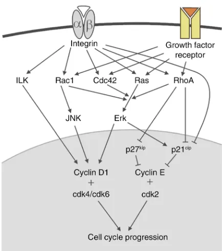
The cell cycle is controlled by cyclin-dependent kinas-es (cdks). Thkinas-ese proteins are constitutively exprkinas-essed, but their activity is regulated by binding to cyclins and by the action of cdk inhibitory proteins (CKIs). Pro-gression through the G1 phase is controlled by cdk4 and cdk6, which interact with cyclin D1, and by cdk2, which binds to cyclin E. The CKIs p21cip1and p27kip1
inactivate cdk2. Cdks are responsible for the phospho-rylation of Rb, which induces other cell cycle proteins including cyclin A. Association of cyclin A with cdk2 initiates then the G1/S transition.
Integrin signaling can regulate the G1/S transition in tight synergism with growth factors (Figure 2). Here again, however, different cell types require distinct inte-grins and growth factors for proliferation. In endothe-lial cells, for example, signaling of both bFGF and αvβ3 is required for cell cycle progression, whereas in fibrob-lasts bFGF can also synergize with β1 integrins (30). Integrin-mediated proliferation depends on the activa-tion of the Erk pathway, which controls expression of cyclin D1 and p21cip. Integrins can induce Erk via FAK
or via caveolin, Fyn, and Shc signaling, both converg-ing on the Ras/Erk cascade.
recognized by several jun/fos heterodimers, and a CRE site, to which heterodimers of jun/fos and certain ATF/CREB family members might bind.
A second integrin-dependent proliferation check-point exists at the level of the cdk inhibitors. Thus, transient, high-level Erk activation increases p21cip
expression, which inhibits cyclin E/cdk2. Integrin-dependent signals, but not Erk, downregulate p21cipin
mid- to late G1, allowing cell cycle progression. The molecular mechanisms by which integrin and growth factor receptor signaling pathways synergize to influence Erk activation are not yet clear, and several possible models should be considered. First, integrin signaling can influence the phosphorylation state of EGF and PDGF receptors as well as their association with signaling molecules such as SHP-2, Ras-GAP, IRS-1, and PI3-K. Interestingly, FAK activation is not required for the integrin-mediated increase of EGF-R phosphorylation. Alternatively, integrins may facilitate signaling of growth factor receptors via the Ras cas-cade. In some cells, for example, growth factor activa-tion of Raf or MEK, two serine/threonine kinases upstream of Erk, is dependent on cell adhesion. This effect might be due to the integrin-mediated activa-tion of PAK, which phosphorylates both Raf and MEK (32). Such cross-talk between integrin and growth fac-tor signaling can be essential for the transformation of cells. The malignant behavior of a breast cancer cell line in a three-dimensional culture is crucially depend-ent on both integrin and EGF-R signaling, which
together result in sustained Erk activation. Inhibition of integrins, EGF-R, or Erk leads to growth arrest and normal breast tissue morphogenesis. Interestingly, these effects are not seen in two-dimensional cell cul-tures, suggesting that the connections between sig-naling pathways depend on the spatial organization of the cytoskeleton.
Integrin-mediated cell cycle control has also been demonstrated in vivo. Thus, Faraldo et al. (33) have found that inhibition of integrin signaling in mice using a dominant negative integrin fusion protein reduces pro-liferation of mammary cells and attenuates Shc, Erk, and JNK activation without perturbing FAK activity.
Rho GTPases in integrin-mediated proliferation
In fibroblasts, but not in endothelial cells, the syner-gism between integrin and growth factor signaling is dependent on cell spreading, indicating an important role of the cytoskeleton. Rho GTPases might be key players in this regulation since they are activated by growth factors and, weakly, by integrins, and since they are pivotal for the organization of the cytoskele-ton. Rho GTPases are crucial for cell cycle progres-sion, as seen in the impairment of growth factor–induced production of cyclin D1 following treatment of adherent cells with the Rho inhibitor toxin A. RhoA downregulates the transcription of
p21cip and promotes degradation of the CDK
inhibitor p27kip1 by stimulation of cyclin E/cdk2
activity. Binding to fibronectin can induce cyclin D1 and suppress p21cipin a Rho-dependent manner (34).
Activation of RhoA and Cdc42 contributes to the integrin-mediated activation of Erk2. Activated Rac1 can induce cyclin D1 expression in serum-starved cells correlating with the activation of PAK-1, JNK, and NF-κB. Furthermore, in endothelial cells Rac1 can facilitate translation of cyclin D1 mRNA upon acti-vation by α5β1 integrin (35). Here, the integrin-induced Rac activation depends on Shc, FAK, and PI3-K signals, which converge to regulate the guanine nucleotide exchange factor Sos.
Other integrin-dependent proliferation pathways have also been documented. Integrin and growth fac-tor recepfac-tors both activate PI3-K, which downregu-lates p27kipand contributes to the induction of cyclin
[image:6.576.65.282.55.300.2]D1. These effects are at least partly mediated by the PDK-1–dependent stimulation of the p70 S6 kinase, a key regulator of cell growth that controls protein translation and induces cyclin D3 expression (36). Many human tumors carry mutations in proteins of the PI3-K signaling pathway, underlining the impor-tance of this mechanism in the prevention of malig-nant proliferation. In some cells, integrin-dependent activation of JNK also contributes to proliferation (37). JNK phosphorylates several members of the jun/fos family, causing increased transcriptional activ-ity and reduced degradation of AP-1 proteins, which are important for the induction of cyclin D1. Finally, integrins might induce cell cycle progression by ILK, which is proposed to induce cyclin D1 expression by activating the transcription factor LEF-1.
Figure 2
Integrins and apoptosis
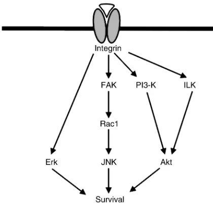
Apoptosis or programmed cell death regulates the lifespan of normal cells and eliminates cells after con-tact with certain toxic insults or following their detachment from the ECM. This last form of apopto-sis is also called “anoikis.” Reduced apoptoapopto-sis can con-tribute to tumorigenesis, since it helps tumor cells to escape natural elimination.
Apoptosis is initiated by either the signaling of death receptors or the release of cytochrome cfrom the mito-chondria, which then activates a cascade of intracellu-lar proteases of the caspase family. The first pathway is controlled by the expression of death receptors and soluble or inactive “decoy” receptors. Induction of pro-grammed cell death through the latter pathway is reg-ulated especially by the balance of pro- and antiapop-totic members of the Bcl-2 family. Although anoikis is initiated by cell detachment, increased death receptor signaling also contributes to cell death. Integrin sig-naling can modulate apoptotic induction, leading in most cases to increased, but sometimes also to decreased, survival. Integrins can promote survival by activating PI3-K, ILK, Erk, and JNK (Figure 3). Inte-grin-mediated activation of PI3-K can trigger several antiapoptotic mechanisms. Activated PI3-K produces PI(3,4,5)P3 and PI(3,4)P2, which promote the reloca-tion of Akt to the plasma membrane and stimulate its phosphorylation.
Akt blocks apoptosis by phosphorylating, and thus inactivating, a number of proapoptotic molecules, including the Bcl-2 family member Bad, caspase-9, and transcription factors of the forkhead family. Akt inhibits apoptosis via different pathways. It can also activate NF-κB, via phosphorylation of IκB, and thereby induce the expression of a set of survival fac-tors, such as osteoprotegerin in endothelial cells.
Finally, it prevents the release of cytochrome cfrom the mitochondria by an unknown mechanism. In CHO cells, integrin-mediated activation of PI3-K and Akt, and upregulation of antiapoptotic Bcl-2, are dependent on Shc and FAK-mediated Ras activation, but not on Erk (38).
Inhibition of β1 integrin function in the mammary gland in vivo using a dominant negative receptor leads to increased cell death and reduced phosphorylation of Akt and the Akt substrates Bad and the forkhead tran-scription factor FKHR. FAK phosphorylation is normal under these conditions (33), indicating that integrin-mediated activation of PI3-K in this tissue is inde-pendent of FAK, although it may be influenced by growth factor signaling, as has been described for mammary cells in culture. Some evidence suggests that the serine/threonine kinase ILK is activated after inte-grin binding and directly binds and phosphorylates Akt (39). In prostate carcinoma cells lacking the tumor suppressor PTEN, ILK is constitutively active and con-tributes to the survival of these cells.
Elicitation of Erk activity by integrins can prevent apoptosis, an effect that is limited to particular cell types and in some cases requires the additional activation of Rac1 by the FAK/Cas/Crk pathway (40). For example, in intestinal epithelial cells, α2β1-mediated Erk activation does not prevent apoptosis induced by serum depriva-tion (41). Integrin-mediated activadepriva-tion of JNK inhibits apoptosis of fibroblasts in the absence of growth factors (42). This effect is dependent on FAK, but independent of Akt and Erk. In the presence of growth factors, how-ever, integrins prevent apoptosis of fibroblasts by FAK-mediated activation of PI3-K and Akt.
Loss of these integrin-dependent survival signals after cell detachment can result in apoptosis. Detach-ment also leads to direct changes in the cytoskeleton and release of the proapoptotic molecule Bmf, which is normally bound to the cytoskeleton (43), an event that is considered to help initiate apoptosis, since it occurs before caspase activation. Furthermore, unligated inte-grin can transmit apoptosis-stimulating signals. Over-expression of unligated, but not of ligand-bound, α5β1 integrin can induce apoptosis in a variety of detached human cancer cells.
[image:7.576.65.275.59.260.2]As in migration and proliferation, the antiapoptotic functions of integrins depend on the context of other signaling pathways. Integrin signaling can enhance the survival effect of growth factors by facilitating down-stream signaling events, as has been shown for α6β1 on oligodendrocytes and for α5β1 on intestinal epithelial cells and on mammary epithelium (41). On the other hand, signaling by the growth factor receptor c-erbB2 in a mammary epithelial cell line reduces the affinity of α2β1 integrin, decreases integrin-mediated survival sig-nals, and results in apoptosis when the cells are grow-ing on collagen (44). The tumor suppressor p53 mod-ulates survival signals provided by α6β4 integrin in dramatic ways. α6β4 induces caspase-mediated inacti-vation of Akt and apoptosis in carcinoma cells express-ing wild-type p53, while it promotes Akt-dependent survival in the absence of p53 (45).
Figure 3
In endothelial cells, shear stress induces the upregula-tion of α5β1 integrin expression and inhibits pro-grammed death of these cells. Interestingly, abrogation of shear stress causes apoptosis mediated by an autocrine loop: It induces the secretion of throm-bospondin, which binds to αvβ3, which acts as a death receptor. In neutrophils as well, integrins can have a proapoptotic role, since attachment via β2 integrins ren-ders the cells susceptible to TNF-α–induced apoptosis.
Integrins and invasive growth in vivo
Many antibody and RGD peptide inhibition studies have suggested an important role for integrins in inva-sive growth during development. Analyses of mice lacking integrins, however, often fail to confirm such a role. For example, αvβ3 integrin has been shown to be essential for angiogenesis, although deletion of the mouse gene for the αv subunit does not impair angio-genesis. These discordant results might be explained by side effects of the antibody/peptide treatments or, alternatively, by compensatory mechanisms that are activated in the mutant mice, such as the upregulation of other, functionally similar molecules. Defects in cell migration have been described during the develop-ment of animals lacking integrins. Murine primordial germ cells lacking β1 integrin, for example, show impaired migration to the gonads. Interestingly, in
Drosophila, the PS1 integrin is required on tracheal cells of the visceral branch for normal migration of tracheal cells on the visceral endoderm; the interacting PS2 integrin is required on the visceral endoderm in the same migratory process (46).
A plethora of experimental evidence documents the importance of integrins in tumor progression, invasive growth, and metastasis. Using β1-null tumor cells, for instance, it has been shown that β1 integrin promotes but is not essential for metastasis. Mutations of the intracellular domain of the β1 integrin chain differen-tially affect cell adhesion, invasion, and metastasis, indicating that integrin signaling is important for inva-sion. Different observations suggest an important role of α6β4 integrin in tumorigenesis. Expression of α6β4 integrin in β4-deficient tumor cells increases their inva-siveness via a PI3-K–dependent pathway. Furthermore, different tumor types maintain or even increase their levels of α6β4 expression.
It is often difficult to demonstrate a positive or neg-ative correlation between tumor progression and inte-grin expression. This is due to the heterogeneity of tumors and to the fact that changes in the expression level of a single integrin subunit always have to be judged against the background of the expression levels of all other integrins, in addition to the activation sta-tus of growth factor and cytokine signal transduction pathways that modulate and synergize with integrin function. Although integrin-mediated binding events are essential for invasive growth, the migratory behav-ior of tumor cells is modulated by cytokines and growth factors to such a degree that the contribution of changes in integrin expression is often not obvious (47). As an example, α2β1 integrin expression seems to
promote invasive growth in pancreatic carcinomas and rhabdomyosarcomas but is strongly downregulated in bladder, breast, and colon cancer.
There is strong evidence that integrins are involved in the resistance of tumor cells to chemotherapy-induced apoptosis. Adhesion of small-cell lung cancer cells to ECM molecules protects them from chemotherapeu-tic, apoptosis-inducing agents (48). This effect is medi-ated by β1 integrins, which activate phosphotyrosine kinases in response to chemotherapy-induced DNA damage. In human myeloma cells, overexpression of α4β1 results in increased drug resistance. Finally, β1 integrin–mediated activation of PI3-K in breast cancer cells increases the resistance of tumor cells to apopto-sis-inducing drugs (49).
Integrins thus clearly offer attractive drug targets to fight tumor growth and metastasis and drug resist-ance, but the variability of their functions in different cell types is daunting. Much more will need to be learned about integrin function, cross-talk with other signaling pathways, and tumor-specific roles to ensure that such drugs will be effective and safe.
Acknowledgments
We thank Michael Dictor, Martin Pfaff, and Kristiina Vuori for critically reading the manuscript. We apolo-gize that, due to space limitations, we could not cite primary references for all the work mentioned and had to omit many interesting contributions to the field. D. Bouvard is supported by a Marie Curie fellowship.
1. Mastrangelo, A.M., Homan, S.M., Humphries, M.J., and LaFlamme, S.E. 1999. Amino acid motifs required for isolated βcytoplasmic domains to regulate ‘in trans’ β1 integrin conformation and function in cell attach-ment. J. Cell Sci. 112:217–229.
2. Keely, P.J., Rusyn, E.V., Cox, A.D., and Parise, L.V. 1999. R-Ras signals through specific integrin αcytoplasmic domains to promote migration and invasion of breast epithelial cells. J. Cell Biol. 145:1077–1088. 3. Liu, S., Calderwood, D.A., and Ginsberg, M.H. 2000. Integrin
cytoplas-mic domain-binding proteins. J. Cell Sci. 113:3563–3571.
4. Constantin, G., et al. 2000. Chemokines trigger immediate β2 integrin affinity and mobility changes: differential regulation and roles in lym-phocyte arrest under flow. Immunity.13:759–769.
5. Danilkovitch-Miagkova, A., and Zbar, B. 2002. Dysregulation of Met receptor tyrosine kinase activity in invasive tumors. J. Clin. Invest.
109:863–867. DOI:10.1172/JCI200215418.
6. Trusolino, L., et al. 2000. HGF/scatter factor selectively promotes cell invasion by increasing integrin avidity. FASEB J. 14:1629–1640. 7. Katagiri, K., et al. 2000. Rap1 is a potent activation signal for leukocyte
function-associated antigen 1 distinct from protein kinase C and phos-phatidylinositol-3-OH kinase. Mol. Cell. Biol. 20:1956–1969.
8. Reedquist, K.A., et al. 2000. The small GTPase, Rap1, mediates CD31-induced integrin adhesion. J. Cell Biol. 148:1151–1158.
9. Hadari, Y.R., et al. 2000. Galectin-8 binding to integrins inhibits cell adhesion and induces apoptosis. J. Cell Sci. 113:2385–2397.
10. Worthylake, R.A., Lemoine, S., Watson, J.M., and Burridge, K. 2001. RhoA is required for monocyte tail retraction during transendothelial migration. J. Cell Biol. 154:147–160.
11. Laukaitis, C.M., Webb, D.J., Donais, K., and Horwitz, A.F. 2001. Differ-ential dynamics of α5 integrin, paxillin, and α-actinin during formation and disassembly of adhesions in migrating cells. J. Cell Biol.
153:1427–1440.
12. Carragher, N.O., Fincham, V.J., Riley, D., and Frame, M.C. 2001. Cleav-age of focal adhesion kinase by different proteases during SRC-regulat-ed transformation and apoptosis. Distinct roles for calpain and caspas-es. J. Biol. Chem. 276:4270–4275.
13. O’Neill, G.M., and Golemis, E.A. 2001. Proteolysis of the docking pro-tein HEF1 and implications for focal adhesion dynamics. Mol. Cell. Biol.
21:5094–5108.
15. Ng, T., et al. 1999. PKCαregulates β1 integrin-dependent cell motility through association and control of integrin traffic. EMBO J.
18:3909–3923.
16. Sieg, D.J., et al. 2000. FAK integrates growth-factor and integrin signals to promote cell migration. Nat. Cell Biol. 2:249–256.
17. Kiosses, W.B., et al. 1999. A role for p21-activated kinase in endothelial cell migration. J. Cell Biol. 147:831–844.
18. Cary, L.A., et al. 1998. Identification of p130Cas as a mediator of focal adhesion kinase-promoted cell migration. J. Cell Biol. 140:211–221. 19. Klemke, R.L., et al. 1998. CAS/Crk coupling serves as a “molecular
switch” for induction of cell migration. J. Cell Biol. 140:961–972. 20. Etienne-Manneville, S., and Hall, A. 2001. Integrin-mediated activation
of Cdc42 controls cell polarity in migrating astrocytes through PKCζ. Cell.106:489–498.
21. Cox, E.A., Sastry, S.K., and Huttenlocher, A. 2001. Integrin-mediated adhesion regulates cell polarity and membrane protrusion through the Rho family of GTPases. Mol. Biol. Cell. 12:265–277.
22. Chen, R., et al. 2001. Regulation of the PH-domain-containing tyrosine kinase Etk by focal adhesion kinase through the FERM domain. Nat. Cell Biol.3:439–444.
23. Klemke, R.L., et al. 1997. Regulation of cell motility by mitogen-activat-ed protein kinase. J. Cell Biol. 137:481–492.
24. Fincham, V.J., James, M., Frame, M.C., and Winder, S.J. 2000. Active ERK/MAP kinase is targeted to newly forming cell-matrix adhesions by integrin engagement and v-Src. EMBO J. 19:2911–2923.
25. Wary, K.K., Mariotti, A., Zurzolo, C., and Giancotti, F.G. 1998. A requirement for caveolin-1 and associated kinase Fyn in integrin signal-ing and anchorage-dependent cell growth. Cell.94:625–634. 26. Nakamoto, T., et al. 2000. CIZ, a zinc finger protein that interacts with
p130(cas) and activates the expression of matrix metalloproteinases. Mol. Cell. Biol. 20:1649–1658.
27. Pozzi, A., et al. 2000. Elevated matrix metalloprotease and angiostatin levels in integrin α1 knockout mice cause reduced tumor vasculariza-tion. Proc. Natl. Acad. Sci. USA.97:2202–2207.
28. Dumin, J.A., et al. 2001. Pro-collagenase-1 (matrix metalloproteinase-1) binds the α2β1 integrin upon release from keratinocytes migrating on type I collagen. J. Biol. Chem. 276:29368–29374.
29. Silletti, S., et al. 2001. Disruption of matrix metalloproteinase 2 binding to integrin αvβ3 by an organic molecule inhibits angiogenesis and tumor growth in vivo. Proc. Natl. Acad. Sci. USA.98:119–124.
30. Roovers, K., and Assoian, R.K. 2000. Integrating the MAP kinase signal into the G1 phase cell cycle machinery. Bioessays.22:818–826. 31. Cook, S.J., Aziz, N., and McMahon, M. 1999. The repertoire of fos and
jun proteins expressed during the G1 phase of the cell cycle is deter-mined by the duration of mitogen-activated protein kinase activation. Mol. Cell. Biol. 19:330–341.
32. Howe, A.K., and Juliano, R.L. 2000. Regulation of anchorage-dependent signal transduction by protein kinase A and p21-activated kinase. Nat. Cell Biol. 2:593–600.
33. Faraldo, M.M., Deugnier, M.A., Thiery, J.P., and Glukhova, M.A. 2001. Growth defects induced by perturbation of β1-integrin function in the
mammary gland epithelium result from a lack of MAPK activation via the Shc and Akt pathways. EMBO Rep. 2:431–437.
34. Danen, E.H., Sonneveld, P., Sonnenberg, A., and Yamada, K.M. 2000. Dual stimulation of Ras/mitogen-activated protein kinase and RhoA by cell adhesion to fibronectin supports growth factor-stimulated cell cycle progression. J. Cell Biol. 151:1413–1422.
35. Mettouchi, A., et al. 2001. Integrin-specific activation of Rac controls progression through the G(1) phase of the cell cycle. Mol. Cell.8:115–127. 36. Feng, L.X., Ravindranath, N., and Dym, M. 2000. Stem cell factor/c-kit up-regulates cyclin D3 and promotes cell cycle progression via the phos-phoinositide 3-kinase/p70 S6 kinase pathway in spermatogonia. J. Biol. Chem. 275:25572–25576.
37. Oktay, M., et al. 1998. Integrin-mediated activation of focal adhesion kinase is required for signaling to Jun NH2-terminal kinase and pro-gression through the G1 phase of the cell cycle. J. Cell Biol.
145:1461–1469.
38. Matter, M.L., and Ruoslahti, E. 2001. A signaling pathway from the alpha5beta1 and αvβ3 integrins that elevates bcl-2 transcription. J. Biol. Chem. 276:27757–27763.
39. Persad, S., et al. 2001. Regulation of protein kinase B/Akt-serine 473 phosphorylation by integrin-linked kinase: critical roles for kinase activ-ity and amino acids arginine 211 and serine 343. J. Biol. Chem.
276:27462–27469.
40. Cho, S.Y., and Klemke, R.L. 2000. Extracellular-regulated kinase activa-tion and CAS/Crk coupling regulate cell migraactiva-tion and suppress apop-tosis during invasion of the extracellular matrix. J. Cell Biol. 149:223–236. 41. Lee, J.W., and Juliano, R.L. 2000. α5β1 integrin protects intestinal epithe-lial cells from apoptosis through a phosphatidylinositol 3-kinase and protein kinase B-dependent pathway. Mol. Biol. Cell. 11:1973–1987. 42. Almeida, E.A., et al. 2000. Matrix survival signaling: from fibronectin via
focal adhesion kinase to c-Jun NH(2)-terminal kinase. J. Cell Biol.
149:741–754.
43. Puthalakath, H., et al. 2001. Bmf: a proapoptotic BH3-only protein reg-ulated by interaction with the myosin V actin motor complex, activated by anoikis. Science.293:1829–1832.
44. Baeckstrom, D., Lu, P.J., and Taylor-Papadimitriou, J. 2000. Activation of the α2β1 integrin prevents c-erbB2-induced scattering and apoptosis of human mammary epithelial cells in collagen. Oncogene.19:4592–4603. 45. Bachelder, R.E., et al. 1999. p53 inhibits α6β4 integrin survival signaling by promoting the caspase 3-dependent cleavage of AKT/PKB. J. Cell Biol.
147:1063–1072.
46. Boube, M., Martin-Bermudo, M.D., Brown, N.H., and Casanova, J. 2001. Specific tracheal migration is mediated by complementary expression of cell surface proteins. Genes Dev. 15:1554–1562.
47. Kassis, J., Lauffenburger, D.A., Turner, T., and Wells, A. 2001. Tumor invasion as dysregulated cell motility. Semin. Cancer Biol. 11:105–117. 48. Sethi, T., et al. 1999. Extracellular matrix proteins protect small cell lung cancer cells against apoptosis: a mechanism for small cell lung cancer growth and drug resistance in vivo. Nat. Med. 5:662–668.