Preferential localization of systemically
administered radiolabeled interleukin 1alpha in
experimental inflammation in mice by binding to
the type II receptor.
C J van der Laken, … , F H Corstens, J W van der Meer
J Clin Invest.
1997;
100(12)
:2970-2976.
https://doi.org/10.1172/JCI119850
.
Previously, we have shown that systemically administered radiolabeled interleukin 1alpha
(IL-1alpha) accumulates preferentially in inflammatory foci in mice. Since inflammation is
characterized by influx of leukocytes, which represent IL-1 receptor (IL-1R) positive cells,
radiolabeled IL-1 may specifically localize in inflammation by binding to its receptors on
infiltrated leukocytes. This hypothesis was tested in a series of studies in mice with acute
focal inflammations. Evidence for specific IL-1-IL-1R interaction in induced inflammation
was found: microscopic autoradiography revealed that 125I-IL-1alpha localized at the site of
inflammatory cells with time; 125I-myoglobin, a similar-sized protein with no known
interactions in vivo, was not retained in the inflammation. Furthermore, the uptake
125I-IL-1alpha in inflammatory tissue was significantly lower in neutropenic mice than in
immunocompetent mice (0.05+/-0.004 vs. 0.65+/-0.06% ID/g at 48 h after injection, P <
0.0007). Moreover, the uptake of 125I-IL-1alpha at the inflammatory site could be blocked
with the anti-IL-1R type II antibody 4E2. At 48 h after injection, the uptake with and without
blocking the type II IL-1R was 0.13+/-0.01 and 0. 65+/-0.05% ID/g, respectively (P < 0.0001).
These in vivo studies provide evidence that systemically administered radiolabeled
IL-1alpha localizes in inflammatory tissue by specific receptor binding, predominantly by
binding to the type II IL-1R.
Research Article
Find the latest version:
J. Clin. Invest.
© The American Society for Clinical Investigation, Inc. 0021-9738/97/12/2970/07 $2.00
Volume 100, Number 12, December 1997, 2970–2976 http://www.jci.org
Preferential Localization of Systemically Administered Radiolabeled Interleukin 1
a
in Experimental Inflammation in Mice by Binding to the Type II Receptor
Conny J. van der Laken,* Otto C. Boerman,* Wim J.G. Oyen,* Marjo T.P. van de Ven,* Richard Chizzonite,§ Frans H.M. Corstens,* and Jos W.M. van der Meer‡
*Department of Nuclear Medicine and ‡Department of Internal Medicine, University Hospital Nijmegen, 6500 HB Nijmegen,
The Netherlands; and §Department of Inflammation/Autoimmune Diseases, Hoffmann-LaRoche Inc., Nutley, New Jersey 07110-1199
Abstract
Previously, we have shown that systemically administered radiolabeled interleukin 1a (IL-1a) accumulates preferen-tially in inflammatory foci in mice. Since inflammation is characterized by influx of leukocytes, which represent IL-1 receptor (IL-1R) positive cells, radiolabeled IL-1 may spe-cifically localize in inflammation by binding to its receptors on infiltrated leukocytes. This hypothesis was tested in a se-ries of studies in mice with acute focal inflammations. Evi-dence for specific IL-1–IL-1R interaction in induced inflam-mation was found: microscopic autoradiography revealed that 125I-IL-1a localized at the site of inflammatory cells with time; 125I-myoglobin, a similar-sized protein with no known interactions in vivo, was not retained in the inflam-mation. Furthermore, the uptake 125I-IL-1a in inflamma-tory tissue was significantly lower in neutropenic mice than in immunocompetent mice (0.0560.004 vs. 0.6560.06% ID/g at 48 h after injection, P , 0.0007). Moreover, the uptake of 125I-IL-1a at the inflammatory site could be blocked with the anti–IL-1R type II antibody 4E2. At 48 h after injection, the uptake with and without blocking the type II IL-1R was 0.1360.01 and 0.6560.05% ID/g, respectively (P , 0.0001). These in vivo studies provide evidence that systemically ad-ministered radiolabeled IL-1a localizes in inflammatory tis-sue by specific receptor binding, predominantly by binding to the type II IL-1R. (J. Clin. Invest. 1997. 100:2970–2976.) Key words: leukocytic infiltration • mice • microscopic auto-radiography • neutropenia • receptor blockade
Introduction
IL-1 is a 17-kD protein, produced by monocytes as a response to endotoxin. Two forms of IL-1, IL-1a and IL-1b, have been discovered, binding to two types of receptors on a wide variety of cells. The type I IL-1 receptor (IL-1R), a 80-kD glycopro-tein, is found on T cells, fibroblasts, hepatocytes, and
endothe-lial cells, whereas B cells, macrophages, monocytes, and neu-trophils express the 68-kD type II IL-1R (1). IL-1 binds to both receptors with affinity in the picomolar range (2).
Since infection and inflammation are characterized by in-flux of predominantly IL-1R positive leukocytes, systemically administered IL-1 may preferentially localize in inflammatory tissue by means of specific receptor binding. Recently, local-ization of radiolabeled IL-1a in Staphylococcus aureus–induced infections in mice was demonstrated (3). It cleared rapidly from noninflamed tissues.
Receptor binding of IL-1 to murine leukocytes has been well characterized both in vitro and ex vivo (2, 4, 5). However, in vivo in mice, receptor binding has not been demonstrated directly but only indirectly via inhibition of IL-1–induced bio-logic activity by blockade of type I IL-1Rs (6–10). In vivo bind-ing to type II IL-1Rs could not be shown due to lack of effect of type II IL-1R blockade on biologic activity (10). The present paper describes a series of studies conducted to vali-date the hypothesis of specific receptor binding of IL-1a in inflammation in mice. Binding of systemically administered IL-1a to type I as well as type II IL-1Rs was investigated by antibody-blocking studies. Our studies demonstrate localiza-tion of IL-1a in inflammatory tissue by specific binding to type II IL-1Rs on locally present infiltrated leukocytes.
Methods
Mice.Female Swiss mice weighing 20–25 g (Harlan Netherlands B.V., Zeist, The Netherlands) were kept in cages (5–15 mice per cage) and fed standard laboratory chow and water ad libitum.
Reagents. Human recombinant IL-1a (specific activity of 3 3 108
U/mg) was kindly provided by Dr. P. Lomedico (Hoffman-La Roche, Nutley, NJ). Myoglobin was purchased from Sigma Chemical Co. (St. Louis, MO). The anti–IL-1R type I antibody (35F5) and the anti–IL-1R type II antibody (4E2) (both monoclonal rat IgG) were prepared at Hoffmann-LaRoche.
Radioiodination. IL-1a and IL-1b were radiolabeled using the io-dogen method (11). In brief, for microscopic autoradiography studies, 10 mg IL-1a (0.68 mg/ml) in 10 ml 0.5 M and 80 ml 50 mM phosphate buffer, pH 7.2, and 111 MBq Na125I (Amersham International,
Amer-sham, UK; specific activity of 570 GBq/mg) were added to glass tubes, precoated with 25 mg of 1,3,4,6-tetrachloro-3a,6a -diphenylglu-couril (Pierce, Rockford, IL). For all other studies, 3 mg IL-1a (0.68 mg/ml) or 3 mg IL-1b (1 mg/ml) was labeled with 15–20 MBq Na125I.
The reaction was allowed to proceed for 10 min at room temperature, after which the reaction mixture was eluted with 0.5% BSA in PBS on a Sephadex column (PD-10; Pharmacia, Uppsala, Sweden) to sep-arate labeled IL-1a and IL-1b from free 125I. The void fractions were
pooled and sterilized through a 0.2-mm filter.
Analogously, 10 mg myoglobin (1 mg/ml), a protein with a molec-ular mass (18 kD) similar to IL-1a and IL-1b without any known in-teractions in vivo, was labeled with Na125I.
The labeling efficiency of IL-1a, IL-1b, and myoglobin was be-tween 50 and 80%. For the microscopic autoradiography studies, Address correspondence to Conny J. van der Laken, M.D.,
Depart-ment of Nuclear Medicine, University Hospital Nijmegen, P.O. Box 9101, 6500 HB Nijmegen, The Netherlands. Phone: 31-24-3613813; FAX: 31-24-3618942; E-mail: [email protected]
both IL-1a and myoglobin were labeled with Na125I at a specific
activ-ity of 8–12 MBq/mg. The specific activity used in all other studies was 3–4 MBq/mg.
The radiochemical purity of the radiolabeled proteins was deter-mined by instant thin-layer chromatography (ITLC) on Gelman ITLC-SG strips (Gelman Laboratories, Ann Arbor, MI) with 0.1 M citrate, pH 5.0, as the solvent. The radiochemical purity of all radiola-beled proteins was . 96% after removal of unbound iodine.
Typically, the receptor binding fraction and the dissociation con-stant of radioiodinated IL-1 preparations were 70–95% and 5 3 10211
mol/liter, respectively, as determined on the murine cell line EL-4-6.1, a variant subline of EL-4 thymoma cells (12), a kind gift of Dr. H.R. MacDonald (Ludwig Institute for Cancer Research, Epalinges, Swit-zerland) (3).
Mouse model of inflammation. Focal inflammations in the left calf muscles of ether anesthetized mice were induced either with 2 3 107 colony forming units of S. aureus in 0.05 ml 50–50% suspension of
autologous blood and normal saline or with 0.05 ml 7.5% zymosan in sterile saline. In some experiments, before the induction of focal in-flammation (day 0), neutropenia (, 109 white blood cells/liter) was
induced in mice by repeated subcutaneous injections of cyclophos-phamide: 150 mg/kg body wt in 200 ml saline on day 24 and 100 mg/ kg on day 21. 24 h after the induction of inflammation, when swelling of the muscle was apparent, mice were injected with 125I-IL-1a, 125
I-IL-1b, free 125I, or with the nonspecific control agent 125I-myoglobin
(microscopic autoradiography) in the tail vein.
Microscopic autoradiography. At 2, 6, and 24 h after intravenous injection of 0.2 ml 2 mg 24 MBq 125I-IL-1a or 2 mg 24 MBq 125
I-myo-globin (24 h after injection only), groups of three mice were killed under ether anesthesia by cervical dislocation. S. aureus–induced inflammations and contralateral noninflamed calf muscles were dissected. After removal, excised tissues were immediately fixed in 4% buffered formalin and embedded in paraffin. Four sections of 4 mm were cut of each excised tissue and mounted on glass slides by con-ventional procedures. The sections were deparaffinized with xylene and hydrated by passage through serial dilutions of ethanol. Subse-quently, slides were dipped in hypercoat LM1 photographic emulsion (Amersham) in the dark room. The slides were exposed in light-tight boxes for 3–4 wk at 48C. After exposure, the emulsions were devel-oped with Kodak D-19 developer (4 min), washed in water (20 s), and fixed in 24% thiosulfate (wt/vol, 4 min). The slides were slightly post-stained with hematoxylin. Parallel series of slides post-stained with hema-toxylin and eosin without photographic emulsion were included for better appreciation of morphology.
The autoradiographs were quantitated by automated image
anal-ysis (13). 10 high power fields per inflammation section, i.e., 5 fields in the area of the cellular infiltration and 5 fields in the area of the unaf-fected muscle, were selected. Furthermore, five fields per contralat-eral muscle were selected. The number of grains per field was deter-mined. The mean number of grains per field was calculated for the different areas in the tissue.
Ex vivo tissue biodistribution. Mice were killed under ether anes-thesia by cervical dislocation at 5 min, 1, 2, 6, 12, 24, and 48 h after in-jection of 125I-IL-1a, 125I-IL-1b, or free 125I. Blood samples, inflamed
left calf muscle, right calf muscle, thymus, lungs, spleen, liver, and kidneys were collected. The dissected tissues were weighed and counted in the gamma counter. To correct for radioactive decay, in-jection standards were counted simultaneously. The measured radio-activity in tissues and samples was expressed as percentage of in-jected radioactivity dose per gram of tissue (% ID/g).
Statistical analysis. All values are expressed as mean6SEM. Sta-tistical analysis was performed using the one-way ANOVA.
Results
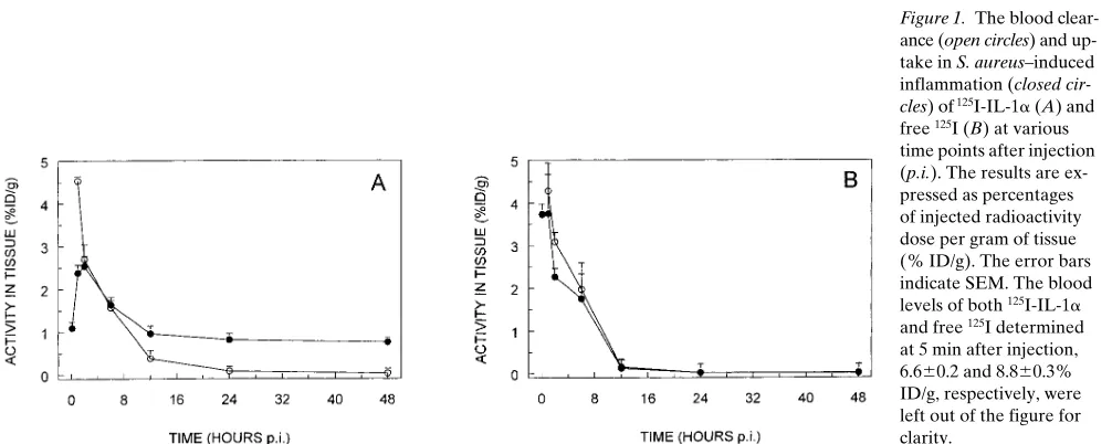
The uptake of 125I-IL-1a in inflammation in the course of time.
The blood clearance and uptake in inflammation of 125I-IL-1a
and free 125I was determined in mice with S. aureus–induced
in-flammation at various time points (n 5 5 for each preparation per time point) after intravenous injection of 0.4 MBq 120 ng of 125I-IL-1a or 0.4 MBq of free 125I. Despite rapid clearance
from the blood, 125I-IL-1a accumulated in the inflammation,
reaching maximum values of uptake within 2 h after injection (Fig. 1 A). 2 h after injection, the uptake of 125I-IL-1a in
in-flammation decreased. A significant level of 125I-IL-1a was
re-tained in the inflammatory tissue up to 48 h after injection (0.8% ID/g). In contrast, 125I-IL-1a levels in the blood
continu-ously decreased to a level , 0.05% ID/g at 48 h after injection. 12 h after injection, the uptake of 125I-IL-1a in inflammation
was significantly higher than the blood levels (P , 0.005). The retention of activity in inflammation with time was due to up-take of 125I-IL-1a and not of free 125I:free 125I rapidly cleared
from the inflammation in a similar fashion as from the blood (Fig. 1 B). To be able to study the specific receptor-binding mechanism of 125I-IL-1 in inflammation, the following studies
[image:3.612.57.560.536.737.2]focused on the time span of 12–48 h after injection because of high background activity levels at early time points.
Figure 1. The blood
clear-ance (open circles) and up-take in S. aureus–induced inflammation (closed
cir-cles) of 125I-IL-1a (A) and
free 125I (B) at various
time points after injection (p.i.). The results are ex-pressed as percentages of injected radioactivity dose per gram of tissue (% ID/g). The error bars indicate SEM. The blood levels of both 125I-IL-1a
and free 125I determined
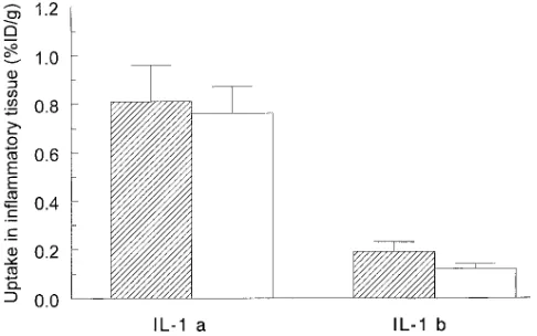
Comparison of uptake of 125I-IL-1a and 125I-IL-1b in in-flammation. The biodistribution of both 125I-IL-1a and 125I-IL-1b
was determined in mice with S. aureus–induced inflammations at various time points (n 5 5 for each radiolabeled preparation per time point) after intravenous injection of 0.4 MBq 120 ng of the radiolabeled preparation. The uptake of 125I-IL-1a in
in-flammation was higher than of 125I-IL-1b (Fig. 2). At 48 h after
injection, 0.7660.11% ID/g of 125I-IL-1a was still found in the
inflammation, while levels of 125I-IL-1b decreased to 0.156
0.01% ID/g at 48 h after injection. From 12 h after injection onwards the uptake of 125I-IL-1a at the site of inflammation
was significantly higher than of 125I-IL-1b (P , 0.05). Due to
the significantly higher uptake of 125I-IL-1a in inflammation as
compared with 125I-IL-1b, further studies exploring the
recep-tor-binding mechanism at the inflammatory site were per-formed with IL-1a.
Microscopic autoradiography. Microscopic
autoradiogra-phy was applied to study whether systemically administered radiolabeled IL-1a localizes at the site of infiltrated leukocytes in induced inflammation in mice. As early as 2 h after injec-tion, uptake of 125I-IL-1a in the inflammation was observed.
At this time point, most of 125I-IL-1a, found in areas of cellular
infiltration, was localized around the blood vessels. The radio-label was also found in the unaffected muscular tissue. With time, 125I-IL-1a was retained within the cellular infiltration,
while the uptake in the unaffected muscle tissue decreased. After 24 h, nearly all 125I-IL-1a was clearly associated with the
site of inflammatory cells, as demonstrated in Fig. 3, A and B. In contrast, no specific retention of 125I-IL-1a was found in the
contralateral muscle (Fig. 3 C). The size-matched control pro-tein 125I-myoglobin did not localize at the site of the
inflamma-tory cells (Fig. 3 D).
The above observations were analyzed quantitatively. The results are shown in Fig. 4. As early as 2 h after injection, most
125I-IL-1a was found in the area of the cellular infiltration at
2 h after injection. The number of grains in the contralateral muscle and in areas of unaffected muscular tissue in the in-flammation decreased during the time course of the experi-ment. Thus, with time, 125I-IL-1a migrated from the unaffected
muscle tissue to the inflammatory cells within the inflamma-tion. After 24 h, nearly all grains in the infection were found in
the area of cellular infiltration. At all time points, the number of grains in the cellular infiltration was significantly higher than the number of grains in the adjacent unaffected muscle and the contralateral muscle (P , 0.0001). Furthermore, the uptake of 125I-IL-1a in the area of the cellular infiltration was
significantly higher than of 125I-myoglobin at 24 h after
injec-tion (P , 0.0001).
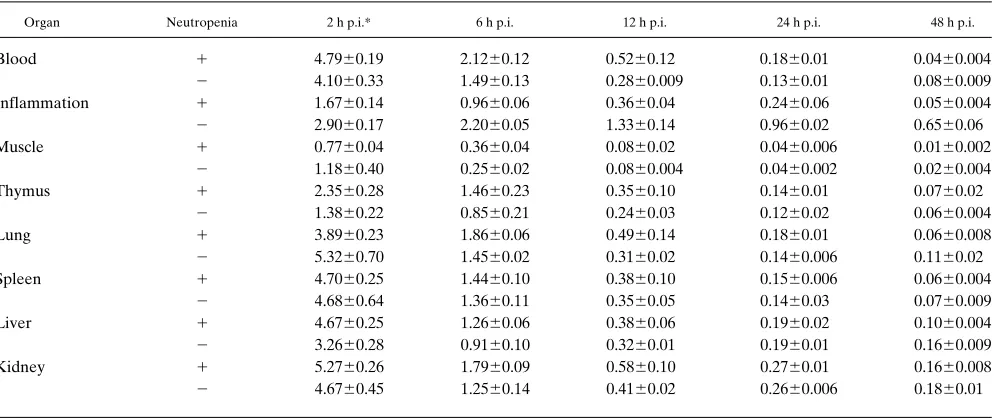
Comparison of biodistribution of 125I-IL-1a in immunocom-petent and neutropenic mice. To study whether the presence
of cellular infiltration is crucial to localization of radiolabeled IL-1a in inflammation, effects of neutropenia were investi-gated and compared with observations in immunocompetent mice. Zymosan, instead of S. aureus, was used for induction of inflammation in these studies to prevent mortality due to sep-sis in neutropenic mice. Histology demonstrated that the mas-sive infiltration of leukocytes, predominantly PMNs, as ob-served in immunocompetent mice was absent in neutropenic mice (data not shown). The biodistribution of 125I-IL-1a in
im-munocompetent (n 5 25) and neutropenic mice (n 5 25), de-termined at various times after intravenous injection of 0.4 MBq 120 ng of the radiolabeled preparation, was similar (Ta-ble I). 125I-IL-1a rapidly cleared from most organs. However,
one major difference was observed: 125I-IL-1a was retained at
the site of inflammation in immunocompetent mice, while vir-tually no retention of 125I-IL-1a in the inflammation was found
in neutropenic mice (Fig. 5). At all time points, the uptake of
125I-IL-1a at the site of inflammation was significantly higher
in immunocompetent mice than in neutropenic mice (P , 0.001). Maximum differences were obtained at 48 h after injec-tion; the uptake of 125I-IL-1a in inflammation was . 12 times
higher in immunocompetent mice compared with neutropenic mice, 0.6560.06 and 0.0560.004% ID/g, respectively.
Effect of in vivo blockade of IL-1Rs on localization of 125I-IL-1a in inflammation. To elucidate whether type I and/
or type II IL-1R is/are involved in entrapment of radioiodi-nated IL-1a in inflammation, groups of five mice with S.
au-reus–induced inflammations were injected intravenously with
200 mg anti–type I IL-1R antibody 35F5, 200 mg anti–type II IL-1R antibody 4E2, or a mixture of both antibodies (200 mg of each antibody) in 100 ml sterile saline at 6 h and 5 min be-fore intravenous injection of 0.4 MBq 120 ng 125I-IL-1a.
Con-trol mice (n 5 5) were injected intravenously with 100 ml ster-ile saline before injection of 125I-IL-1a. As depicted in Fig. 6,
mice with type I IL-1R blockade displayed significantly higher uptake of 125I-IL-1a in the inflammation than the control
group, i.e., 2.2660.12 vs. 0.6560.05% ID/g at 48 h after injec-tion (P , 0.0001). In contrast, the uptake of 125I-IL-1a at the
site of inflammation in mice with blockade of type II IL-1Rs (0.1360.009) was significantly lower than the value obtained in control mice (P , 0.0001). A similar low uptake of 0.146 0.009% ID/g in inflammation was found in mice with both type I and type II IL-1R blockade. The 125I-IL-1a levels in the blood
and all other organs were similar for all studied groups (data not shown).
Discussion
This study confirms the hypothesis that systemically adminis-tered radiolabeled IL-1a localizes in acute inflammatory foci by specific receptor binding. As demonstrated by microscopic autoradiography, IL-1a localized within the inflammation at the site of the infiltrated leukocytes, which were predomi-Figure 2. The uptake of 125I-IL-1a and 125I-IL-1b in S. aureus–
induced inflammation in mice at 24 (striped bars) and 48 h (open
bars) after injection. The results are expressed as percentages of
[image:4.612.57.300.60.212.2]nantly PMNs. The studies with neutropenic mice corroborated these findings, since low uptake of IL-1a in inflammation was found in the absence of cellular infiltration. Moreover, the IL-1 receptor blockade studies showed that the retention of IL-1a in inflammatory tissue was inhibited by blockade of type II IL-1Rs.
The binding of human recombinant IL-1 to murine leuko-cytes, predominantly PMNs, has also been described by Parker et al. (4). In their studies in mice, PMNs were removed from peritoneal exudate to test the binding of radiolabeled IL-1 to these cells ex vivo. Our results show binding of systemically administered radiolabeled IL-1 to murine infiltrated leuko-cytes in acute inflammation in vivo. The receptor binding ca-pacity of IL-1 after radiolabeling was tested in vitro on murine thymoma cells. The affinity was as high as reported in earlier studies (2, 5). It was not possible to isolate leukocytes from the inflammatory tissue in mice to test the binding to these cells ex vivo. Therefore, microscopic autoradiography was used to vi-sualize the localization of 125I-IL-1a in the cellular infiltration
of inflammatory tissue. This method has also been used by oth-ers to identify IL-1 receptors in various noninflamed murine tissues (14, 15). To exclude nonspecific accumulation in
in-flammatory foci by locally increased vascular permeability (16–18), inflammatory tissue of mice injected with the control agent 125I-myoglobin was also subjected to microscopic
autora-diography, showing no localization in the cellular infiltration. This indicates that IL-1a is retained in inflammatory foci by specific binding to its receptors.
For most of our studies, S. aureus–induced focal infections were used. These infections are characterized by massive in-flammatory cell infiltration. Mice with a focal S. aureus infec-tion do not show general signs of illness. However, neutro-penic mice with such infections become severely ill and die within 1–3 d, most likely due to sepsis. To allow comparison of uptake of radiolabeled IL-1a in inflammation in neutropenic and immunocompetent mice, the sterile zymosan model was chosen, which in immunocompetent mice induces a massive focal infiltration of PMNs and monocytes (19, 20). Neutro-penic mice with zymosan-induced inflammations did not show any signs of illness. No cellular infiltration was found in the in-duced inflammations of these mice. Despite similar biodistri-bution in both groups of mice, the uptake of 125I-IL-1a at the
site of inflammation was significantly higher in immunocompe-tent mice, again stressing the fact that radiolabeled IL-1a accu-Figure 3. Microscopic autoradiography of inflammatory tissue of a mouse 24 h after injection with 125I-IL-1a, without photographic emulsion
[image:5.612.60.532.60.419.2]mulates in inflammatory tissue by means of binding to its re-ceptors on infiltrated leukocytes.
The IL-1R blockade studies showed that the binding of ra-diolabeled IL-1a to infiltrated leukocytes was mainly due to binding to type II IL-1Rs. The inflammation uptake of IL-1a was significantly lower in case of blockade of type II IL-1Rs. Surprisingly, increased uptake of radiolabeled IL-1a in the in-flammation was found when type I IL-1Rs were blocked. Blockade of type I IL-1Rs, expressed on a wide range of cells in the body, may have increased levels of free IL-1a, available for binding to type II IL-1Rs, expressed on infiltrated neutro-phils and monocytes in the inflammation. When both type I and type II IL-1Rs were blocked, inflammation uptake of IL-1a was as low as that found with type II IL-1R blockade only. This indicates that the increased uptake of IL-1a in
inflamma-tory tissue (obtained by blockade of type I IL-1Rs) could be completely inhibited by blockade of type II IL-1Rs. Therefore, aspecific increased uptake of IL-1a in inflammation in case of type I IL-1R blockade could be ruled out. Binding to soluble type II IL-1Rs, increased during various inflammatory condi-tions (21–23), could also be excluded since the used anti–type II antibody 4E2 also binds and neutralizes these receptors.
[image:6.612.57.296.59.217.2]Although blockade of binding of radiolabeled IL-1a to type II IL-1Rs with the antibody 4E2 has been demonstrated in vitro on cell lines (24), in vivo, blockade of type II IL-1Rs did not affect particular studied biologic effects of IL-1a (7, 10). Since it is now generally accepted that the type II IL-1R acts as a decoy receptor for IL-1 (25–28), no direct effects of type II IL-1R blockade on IL-1 induced biologic activity are to be expected. Indirectly, type II IL-1R blockade may enhance Figure 4. The quantitative analysis of microscopic autoradiography
of inflammation (infiltration, open bars, and adjacent muscle, heavily
striped bars) and contralateral muscle (striped bars) sections of mice
injected with either 125I-IL-1a or 125I-myoglobin (at 24 h after
injec-tion only), expressed as mean number of grains per field 6SEM.
Figure 5. The uptake of 125I-IL-1a in zymosan-induced inflammation
[image:6.612.317.552.60.208.2]in neutropenic and immunocompetent mice at 24 (striped bars) and 48 h (open bars) after injection. The results are expressed as percent-ages of injected radioactivity dose per gram of tissue (% ID/g). The error bars indicate SEM.
Table I. Biodistribution of 125I-IL-1a in Neutropenic and Immunocompetent Mice with Zymosan-induced Inflammation
Organ Neutropenia 2 h p.i.* 6 h p.i. 12 h p.i. 24 h p.i. 48 h p.i.
Blood 1 4.7960.19 2.1260.12 0.5260.12 0.1860.01 0.0460.004
2 4.1060.33 1.4960.13 0.2860.009 0.1360.01 0.0860.009 Inflammation 1 1.6760.14 0.9660.06 0.3660.04 0.2460.06 0.0560.004
2 2.9060.17 2.2060.05 1.3360.14 0.9660.02 0.6560.06
Muscle 1 0.7760.04 0.3660.04 0.0860.02 0.0460.006 0.0160.002
2 1.1860.40 0.2560.02 0.0860.004 0.0460.002 0.0260.004 Thymus 1 2.3560.28 1.4660.23 0.3560.10 0.1460.01 0.0760.02
2 1.3860.22 0.8560.21 0.2460.03 0.1260.02 0.0660.004 Lung 1 3.8960.23 1.8660.06 0.4960.14 0.1860.01 0.0660.008
2 5.3260.70 1.4560.02 0.3160.02 0.1460.006 0.1160.02 Spleen 1 4.7060.25 1.4460.10 0.3860.10 0.1560.006 0.0660.004
2 4.6860.64 1.3660.11 0.3560.05 0.1460.03 0.0760.009 Liver 1 4.6760.25 1.2660.06 0.3860.06 0.1960.02 0.1060.004
2 3.2660.28 0.9160.10 0.3260.01 0.1960.01 0.1660.009 Kidney 1 5.2760.26 1.7960.09 0.5860.10 0.2760.01 0.1660.008
2 4.6760.45 1.2560.14 0.4160.02 0.2660.006 0.1860.01
[image:6.612.60.557.514.723.2]IL-1–induced biologic activity, as shown by others in vitro (25, 26), but this may be more difficult to demonstrate in vivo. The effects of type II IL-1R blockade on IL-1–induced biologic ac-tivity were not evaluated, but the inhibition of receptor bind-ing of radiolabeled IL-1a at the site of inflammation by type II IL-1R blockade could be shown in vivo.
The uptake of IL-1b in inflammation was lower than the uptake of IL-1a. Apparently, lower amounts of IL-1b bound to IL-1Rs in the inflammatory tissue. Since systemically ad-ministered IL-1 appears to mainly bind to type II IL-1Rs, these findings are in line with data obtained by Parker et al. (4). They also tested the binding of human recombinant IL-1a and IL-1b for the type II IL-1R on murine PMNs and found a two-to threefold lower affinity for IL-1b as compared with IL-1a. Opposite results have been reported for the binding of human recombinant IL-1a and IL-1b to the human type II IL-1R (29, 30), indicating that species differences play a role in receptor-binding characteristics of IL-1a and IL-1b.
Although the total amount of radiolabeled IL-1a in the in-flammation decreased with time, retention was still found at 48 h after injection. Most likely, as demonstrated by the micro-scopic autoradiography results, IL-1a accumulates in inflam-matory tissue shortly after injection and migrates toward the binding sites on infiltrated leukocytes during the next 24 –48 h. Some IL-1a molecules will bind easily accessible receptors early after injection and others reach binding sites several hours thereafter. After 24 h, radiolabeled IL-1a, retained in the inflammation, could almost only be found in areas of cellu-lar infiltration. In the meantime, most of the unbound and/or degraded IL-1a is cleared from the tissue, resulting in a de-creasing total amount of radiolabeled IL-1a in the inflamma-tion. Despite the latter, the uptake in areas of cellular infiltra-tion even increased with time, which could be explained by the fact that these areas form the minority of the total inflamma-tion. Since IL-1a seemed to bind mainly to type II IL-1Rs in the models of inflammation studied, these observations may be conflicting with data suggesting poor internalization of
re-ceptor-bound IL-1 (31) and a fast turnover of the receptor (2 h) (32). However, these characteristics were observed in vitro us-ing the Raji human B lymphoma cell line. In vivo, IL-1 bindus-ing to type II receptors on activated leukocytes in inflammatory tissue may be different.
This study supports first indications that systemically ad-ministered radiolabeled IL-1a localizes in inflammation by specific receptor binding. The type II IL-1R appeared to be the predominant binding site in acute inflammation models. Actual binding of IL-1a to type II IL-1R in inflammatory tis-sue could be demonstrated. Future studies will focus on the in vivo behavior of IL-1a and the correlation with IL-1R expres-sion in various other inflammation models.
Acknowledgments
The authors thank C. Diepenbroek and L. Schalkwijk (University of Nijmegen, Department of Pathology) for technical assistance in the microscopic autoradiography studies, P. Mast for determination of the leukocyte counts to establish neutropenia (University of Nij-megen, Department of Hematology), and G. Grutters, H. Eijkholt, and Y. Brom (University of Nijmegen, Central Animal Laboratory) for technical assistance in all the studies with mice.
References
1. Dinarello, C.A. 1991. Interleukin-1 and interleukin-1 antagonism. Blood. 77:1627–1652.
2. Lowenthal, J.W., and H.R. MacDonald. 1986. Binding and internaliza-tion of interleukin 1 by T cells. J. Exp. Med. 164:1060–1074.
3. Van der Laken, C.J., O.C. Boerman, W.J.G. Oyen, M.T.P. van de Ven, R.A.M.J. Claessens, J.W.M. van der Meer, and F.H.M. Corstens. 1995. Specific targeting of infectious foci with radioiodinated human recombinant interleukin-1 in an experimental model. Eur. J. Nucl. Med. 22:1249–1255.
4. Parker, K.P., W.R. Benjamin, K.L. Kaffka, and P.L. Kilian. 1989. Pres-ence of IL-1 receptors on human and murine neutrophils: relevance to IL-1 me-diated effects in inflammation. J. Immunol. 142:537–542.
5. Chizzonite, R., T. Truitt, and P.L. Kilian. 1989. Two high-affinity inter-leukin-1 receptors represent separate gene products. Proc. Natl. Acad. Sci.
USA. 86:8029–8033.
6. McIntyre, K.W., G.J. Stepan, K.D. Kolinsky, W.R. Benjamin, J.M. Plocinsky, K.L. Kaffka, C.A. Campen, R.A. Chizzonite, and P.L. Kilian. 1991. Inhibition of interleukin 1 (IL-1) binding and bioactivity in vitro and modula-tion of acute inflammamodula-tion in vivo by IL-1 receptor antagonist and anti–IL-1 re-ceptor monoclonal antibody. J. Exp. Med. 173:931–939.
7. Hestdal, K., S.E. Jacobsen, F.W. Ruscetti, C.M. Dubois, D.L. Longo, R. Chizzonite, J.J. Oppenheim, and J.R. Keller. 1992. In vivo effect of interleukin-1 alpha on hematopoiesis: role of colony-stimulating factor receptor modulation.
Blood. 80:2486–2494.
8. Dubois, C.M., F.W. Ruscetti, J.R. Keller, J.J. Oppenheim, K. Hestdal, R. Chizzonite, and R. Neta. 1991. In vivo interleukin-1 (IL-1) administration indi-rectly promotes type II IL-1 receptor expression on hematopoietic bone mar-row cells: novel mechanism for the hematopoietic effects of IL-1. Blood. 78: 2841–2847.
9. Neta, R., S.N. Vogel, J.M. Plocinsky, N.S. Tare, W. Benjamin, R. Chizzo-nite, and M. Pilcher. 1990. In vivo modulation with anti-interleukin-1 (IL-1) re-ceptor (p80) antibody 35F5 of the response to IL-1. The relationship of radio-protection, colony-stimulating factor, and IL-6. Blood. 76:57–62.
10. Oldenburg, H.S., J.H. Pruitt, D.D. Lazarus, M.A. Rogy, R. Chizzonite, S.F. Lowry, and L.L. Moldawer. 1995. Interleukin-1 binding to its type I, but not to type II receptor, modulates the in vivo acute phase response. Cytokine. 7: 510–516.
11. Fraker, P.J., and J.C. Speck. 1978. Protein and cell membrane iodination with a sparingly soluble chloramide 1,3,4,6-tetrachloro-3a,6a -diphenyl-glu-couril. Biochem. Biophys. Res. Commun. 80:849–857.
12. Zubler, R.H., F. Erard, R.K. Lees, M. van Laer, C. Mingari, L. Moretta, and H.R. MacDonald. 1985. Mutant EL-4 thymoma cells polyclonally activate murine and human B cells via direct cell interaction. J. Immunol. 134:3662.
13. Tamimi, Y., H.G. van der Poel, M.M. Denyn, R. Umbas, H.F. Karthaus, F.M. Debruyne, and J.A. Schalken. 1993. Increased expression of high mobility group protein I(Y) in high-grade prostatic cancer determined by in situ hybrid-ization. Cancer Res. 53:5512–5516.
[image:7.612.58.295.59.217.2]14. Takao, T., W.M. Mitchell, D.E. Tracey, and E.B. De Souza. 1990. Iden-tification of interleukin-1 receptors in mouse testis. Endocrinology. 127:251– 258.
Figure 6. The uptake of 125I-IL-1a in inflammation at 48 h after
injec-tion in mice in which IL-1Rs were blocked with anti–IL-1R type I an-tibodies, anti–IL-1R type II anan-tibodies, or both IL-1R type I and type II antibodies before injection of 125I-IL-1a. Control mice were
in-jected with saline before injection of 125I-IL-1a. The results are
15. Takao, T., W.M. Mitchell, and E.B. De Souza. 1991. Interleukin-1 re-ceptors in mouse kidney: identification, localization, and modulation by li-popolysaccharide treatment. Endocrinology. 128:2618–2624.
16. Lavender, J.P., J. Lowe, J.R. Barker, J.I. Burn, and M.A. Chaudri. 1971. Gallium-67 citrate scanning in neoplastic and inflammatory lesions. Br. J.
Ra-diol. 44:361–366.
17. McAfee, J.G., G. Gagne, G. Subramanian, and R.F. Schneider. 1991. The localization of indium-111-leucocytes, gallium-67, polyclonal IgG and other radioactive agents in acute focal inflammatory lesions. J. Nucl. Med. 32: 2126–2131.
18. Oyen, W.J.G., R.A.M.J. Claessens, J.W.M. van der Meer, and F.H.M. Corstens. 1992. Biodistribution and kinetics of radiolabeled proteins in rats with focal infection. J. Nucl. Med. 33:388–393.
19. Lefkowith, J.B. 1988. Essential fatty acid deficiency inhibits the in vivo generation of leukotriene B4 and suppresses levels of resident and elicited leu-kocytes in acute inflammation. J. Immunol. 140:228–233.
20. Dawson, J., A.D. Sedgewick, J.C. Edwards, and P. Lees. 1991. A com-parative study of the cellular, exudative and histological responses to carra-geenan, dextran and zymosan in the mouse. Int. J. Tissue React. 13:171–185.
21. Symons, J.A., J.A. Eastgate, and G.W. Duff. 1990. A soluble binding protein specific for interleukin-1 beta is produced by activated mononuclear cells. Cytokine. 2:190–198.
22. Giri, J.G., J. Wells, S.K. Dower, C.E. McCall, R.N. Guzman, J. Slack, T.A. Bird, K. Shaneback, K.H. Grabstein, J.E. Sims, and M.R. Alderson. 1994. Elevated levels of shed type II IL-1 receptor in sepsis. J. Immunol. 153:5802– 5809.
23. Arend, W.P., M. Malyak, M.F. Smith, T.D. Whisenand, J.L. Slack, J.E. Sims, J.G. Giri, and S.K. Dower. 1994. Binding of IL-1a, IL-1b, and IL-1 recep-tor antagonist by soluble IL-1 receprecep-tors and levels of soluble IL-1 receprecep-tors in synovial fluids. J. Immunol. 153:4766–4774.
24. Hestdal, K., F.W. Ruscetti, R. Chizzonite, M. Ortiz, J.M. Gooya, D.L.
Longo, and J.R. Keller. 1994. Interleukin-1 (IL-1) directly and indirectly pro-motes hematopoietic cell growth through type I IL-1 receptor. Blood. 1:125– 132.
25. Colotta, F., F. Re, M. Muzio, R. Bertini, N. Polentarutti, M. Sironi, J.G. Giri, S.K. Dower, J.E. Sims, and A. Mantovini. 1993. Interleukin-1 type II re-ceptor: a decoy target for IL-1 that is regulated by IL-4. Science. 261:472–475.
26. Sims, J.E., M.A. Gayle, J.L. Slack, M.R. Alderson, T.A. Bird, J.G. Giri, F. Colotta, F. Re, A. Mantovani, K. Shanebeck, K.H. Grabstein, and S.K. Dower. 1993. Interleukin-1 signaling occurs exclusively via the type I receptor.
Proc. Natl. Acad. Sci. USA. 90:6155–6159.
27. Colotta, F., S.K. Dower, J.E. Sims, and A. Mantovini. 1994. The type II “decoy” receptor: a novel regulatory pathway for interleukin-1. Immunol.
To-day. 15:562–566.
28. Re, F., M. Sironi, and M. Muzio. 1996. Inhibition of interleukin 1 re-sponsiveness by type II receptor gene transfer: a surface “receptor” with anti– interleukin 1 function. J. Exp. Med. 183:1841–1850.
29. Scapigliati, G., P. Ghiari, M. Bartalini, A. Tagliabue, and D. Boraschi. 1989. Differential binding of IL-1a and IL-1b to receptors on B and T cells.
FEBS Lett. 243:394–398.
30. Re, F., M. Muzio, M. De Rossi, N. Polentarutti, J.G. Giri, A. Mantovani, and F. Colotta. 1994. The type II “receptor” as a decoy target for interleukin 1 in polymorphonuclear leukocytes: characterization of induction by dexametha-sone and ligand binding properties of the released decoy receptor. J. Exp. Med. 179:739–743.
31. Horuk, R., J.J. Huang, M. Covington, and R.C. Newton. 1987. A bio-chemical and kinetic analysis of the interleukin-1 receptor. Evidence for differ-ences in molecular properties of IL-1 receptors. J. Biol. Chem. 262:16275– 16278.